the cultivation problem - ilslila.ilsl.br/pdfs/v33n3pt2a20.pdf · the cultivation problem ......
TRANSCRIPT
1 1''''1 RN \IIO \l \1. )01 Jt\'" 0" l , I, " I{O,y Volllme :tL NUlIlhl.'r ~ Pr;"I,.ri;1/ U.,", " .
The Cultivation Problem Leads from the Psittacine and Rickettsial Groups
Chairman : R. S. Weiser
Dr. Weiser. Until the leprosy organism can he cuhi vatcd we are forced to consider it an obligate parasite in the strictest sensc. By that I mean an organ ism that demands the li ving host for its reproduction, and, more particularly perhaps, an intracellular invironment. Inasmuch as the organisms of leprosy are commonly seen in cells, particularly phagocytic cells of the macrophage series, I think we must consider that these organisms demand this intracellular environment for their reproduction. Until we can fulfill all of Koch's postulates, we must also consider the possibility, however remote, that this organism is not actually the etiologic agent of the disease. Of course other alternative possibilities come to mind, e.g. , that the organism is simply
along for the ride, and some other microbe is causing the disease. I do not believe this myself for one moment. Still another possibility is that the form of the organism that we observe, i.e., acid-fast rods, is not the form in which the etiologic agent operates; it is possible that soft forms may be involved. With these possibilities in mind, however remote, I believe it is highly appropriate at this time that we hear from Dr. James W. Moulder, who is Professor of Microbiology at the University of Chicago, chairman of the department, and one of the contributors to the famous textbook of Jordan and Burrows on bacteriology. We will now hear from Dr. Moulder on the "Metabolic capabilities and deficienCies of rickettsiae and the psittacosis group."
Metabolic Capabilities and Deficiencies of Rickettsiae
and the Psittacosis Group
James W. Moulder, Ph .D.l
The causative agents of leprosy, the typhus fevers, psittacosis, and trachoma have in common, for the moment at leas t, the distinction of never having been cultivated in artificial media. In their natural hosts they multiply only intracellularly - this is known for certain for the rickettsiae and the psittacosis agents and is generally as-
1 Professor of Mi crobiology, Dept. of MicrobiOlogy, University of Chicago, Chicago, Illin ois 60637.
494
sumed for the leprosy bacillus. Such obligate intracellular parasites are unusual not on ly in that they cannot multiply outside of host cells, but also because they do multiply inside other cells. This is a property they share with another exclusive group, the facultative intercellular parasites, such as Brucella abortus and Mycobacterium t llberculosi~. Most free-living organisms can no mor~ grow inside a cell than the obligate intracellular parasites can
.'3.'3, .'3 (Pt. 2) M o/llde'/': Rickettsiae & Psittacosis Gmllps 495
grow outside one. So, in examining the metaboli c capabilitics and deficiencies (11 these organisms, we are consciously or unconsciously asking two important questions:
1. Why cannot they grow outside of other cells ?
2. Why do they grow so well inside other cells?
The answer to the first ques tion almost certainly li es in the accumulation of mutations that reduce biosynthetic competence, mutations that would be lethal in any envi ronment other than the rich intracellular one. Even when we do not know just which biosynthetic capabilities have disappeared, we can make good guesses as to their general nature and the mechanism of their loss. \lVe are encouraged in our search for metabolic lesions by the hope that the long evolutionary adaptation of microorganisms to intracellular existence may have engendered some unusual and interesting nutritional deficiencies or metabolic inadequacies.
The answer to the second question is not nearly so obvious. Intracellular parasites have evolved special mechanisms that enable them to grow and multiply inside suitable host cells. Such mechanisms could be positive-the acquisition of new metabolic patterns that allow a more efficient exploitation of the intracellular environment;-or they could be negative-the appearance of defense mechanisms that enable preexisting metabolic patterns to function in the face of hostile host influences. But whatever they are, we have no real idea at all as to what their molecular basis might be.
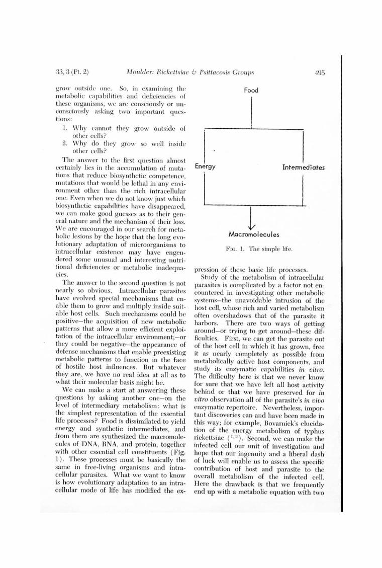
We can make a start at answering these questions by asking another one-on the level of intermediary metabolism: what is the simplest representation of the essential life processes? Food is dissimilated to yield energy and synthetic intermediates, and from them are synthesized the macromolecules of DNA, RNA, and protein, together with other essential cell constituents ( Fig. 1). These processes must be basically the same in free-living organisms and intracellular parasites. What we want to know is how evolutionary adaptation to an intracellular mode of life has modified the ex-
Food
Energy Intennediates
1 Macromolecules
Flc . 1. The simple life.
pression of these basic life processes. Study of the metabolism of intracellular
parasites is complicated by a factor not encountered in investigating other metabolic systems-the unavoidable intrusion of the host cell, whose rich and varied metabolism often overshadows that of the parasite it harbors. There are two ways of getting around-or trying to get around-these difficulties. First, we can get the parasite out of the host cell in which it has grown, free it as nearly completely as possible from metabolically active host components , and study its enzymatic capabilities in vitro. The difficulty here is that we never know for sure that we have left all host activity behind or that we have preserved for in vitro observation all of the parasite's in vivo enzymatic repertoire. Nevertheless, important discoveries can and have been made in this way; for example, Bovarnick's elucidation of the energy metabolism of typhus rickettsiae (1,2). Second, we can make the infected cell our unit of investigation and hope that our ingenuity and a liberal dash of luck will enable us to assess the specific contribution of host and parasite to the overall metabolism of the infected cell. H ere the drawback is that we frequ ently end up with a metabolic equation with two
49fl J ntenwtio/1al JOllrnal of Lepl'Osu ]96,5
unknowns an d an in deter minate solution, but again this procedure has proven itse lf an indisp cnsable mcans for learnin g ahout the mctabolism of intracellular parasi tes. It may well be tha t a goal almost as desirable as tha t of artificia l cultivation is the development of a cell culture system in w hich the intracellular growth of the leprosy bacillus can be studied under controlled and reproducible conditions,
This is enough generali ties; so I shall use the rest of my time to examine three specific tOPIcs : energy metabolism, synthesis of proteins, and synthesis of nucleic acids,
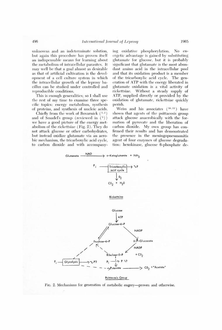
Chiefl y from the work of Bovarnick ( \,2) and of Smadel's grou p ( reviewed in ( ~)) we have a good p icture of the energy metabolism of the r ickettsiae ( F ig. 2 ) . They do not attack glucose or other carbohydra tes, but instead oxidize glutamate via an aerobi c mechanism, the tri carboxylic acid cycle, to carbon dioxide and with accompany-
ing oxidative phosphorylati on. No energet ic ad vantagc is gained hy suhstituting glutamate for g lucosc. hut it is probabl y significant tha t glutamate is the mos t abull dant amino acid in the intracellular pool and tha t its oxidation product is a member of the tricarboxylic acid cycle. The genera tion of ATP with the energy libera ted in glutamate oxidation is a vita l activity of rickettsiae. Without a steady supply of ATP, supplied directly or provided by the oxida tion of glutamate, ri ckettsiae quickl y perish ,
W eiss and his associates ( 10. II ) have shown tha t agents of th e psittacos is group attack glucose anaerobically w ith the formation of pyru va te and the liberation of carbon dioxide. My own group has confirm ed their results and has demonstrated the presence in the meningopneumonitis agent of four enzymes of glu cose degradation : hexokinase, glucose 6-phospha te de-
Glutamate NAD ----~). a-Ketoglutarate + NH3
Ricketts iae
Glucose
1 ATP
GI ucose-6-P
F~Lp ~-:'"'OOO'. // / 1", "'" ~ADP
/ Ribulose-5-P + CO2 I
Pi -rGl~I;SiS 1_ "7"\.,P? Pi '7 P \.?
~ CO2 + "Aceta te " - -;:>pyruvate ----7
Psittacos is Group
F IG. 2. Mechanisms for generation of metabolic engery-proven and otherwise.

33, 3 (Pt. 2) Moulder: Ricketts iae & Psittacosis Gro ll ])S 497
hydrogenase, 6-phosphogluc:onate dehydrogenase, and phospho hexose isomerase (( 6) and unpublished results ). These results provide an enzymatic basis for the observed liberation of carbon dioxide from the Cl position of glucose and suggest the presence of a pentose cycle. The series of reactions from hexose to pentose generate no energy. In fact W eiss (10) has shown that they require stoichiometric amounts of ATP and NADP for their occurrence. These reactions may, however, be important sources of intermediates: pentose for synthesis of nucl eic acids, NADPH for synthesis of fatty acids, and fructose-6-phosphate for syn thesis of ~lucosamine and muramic acid. The production of pyruvate from either pentose or hexose by as yet unknown mechanisms could well generate high-energy phosphate, but this remains to be proven experimentally.
Before leavin g energy metabolism, let us make a few comparisons between glutamate oxidation in the rickettsiae and glucose breakdown in the psittacosis group. Both proceed optimally in high K+, low Na + media, media which at least approximate the intracellular inorganic environment. NAD is the first electron acceptor in glutamate oxidation, and the ultimate acceptor is mol ecular oxygen. NADP is the first known electron acceptor in glucose breakdown. Clem'cut evidence of its reoxidation is lacking; although glutathione reductase may be involved. In any event, there is no evidence for aerobic oxidations in the psittacosis group. Glutamate oxidation generates ATP; glucose breakdown requires it. The one generalization we can make is that successful intracellular parasites may exhibit quite different metabolic patterns. Evolution in a common environment, the inside of a cell , does not necessarily lead to the same end result.
Turning now to the synthesis of proteins, I think we can be reasonably certain ' that the rickettsiae and the psittacosis group agents synthesize their own proteins in ribosomal systems typical or bac:leria. Both groups of organisms are sl1 sceptihle to chloramphenicol and the tctracyclin cs, antibiotics that inhibit the growth of ordinary
bacteria hy preventing rihosomal synthesis of protein , and Higashi (;1) has recently mention ed that his group has actually prepared ribosomes from the meningopneumonitis agent. In my laboratory, Schechter (8) has followed the uptake of labeled amino acids into the meningopneumonitis agent growing in L cells and has observed an early ·and long-continued synthesis of agent protein.
Bovarnick and her associates (~) and Bovarnick and Schneider ( I ) have obtained incorporation of amino acids into the protein of isolated rickettsia l suspensions (Table 1) . The requirements for in v itro amino acid incorporation are complex, even more complex than for maintenance of infectivity itself. There is a peculiar double requirement for A TP: for maximum incorporation it must he supplied exogcnously and also generated endogenously by oxidation of glutamate. The necessity that amino acids be present in order that one may be incorporated indicates that thc rickettsiae have no amino acid pools. Bovarnick and Schneider ( 1) have pointed out that the requirements for protein synthesis in whole rickettsial cells resemble more than anything else the requirements for protein synthesis in subcellular particulates, perhaps because both function insi.de of cells.
I believe we can be cqually certain that the rickettsiae and the psittacosis agents also synthesize their own RNA and DNA by the same general mechanisms used by other organisms. For example, Jones and Paretsky (4) have obtained from the agent of Q fever a cell -free system capable of synthesizing RNA.
TABLE I.-Requirements for incorporation of amino acids into protein of typhus rickettsiae maintained in vitro (1,2) .
Yolk sac protein
NAD Coenzyme A
i\ TP and othf'r rihof)llckoticle mono-, di-, and triphosphates
Glutathione Glutamin e All naturally oc
curring amino acids
ITigh,K + , low Na -t 19++
Mn ++
498 International Journal of Leprol>!J 1965
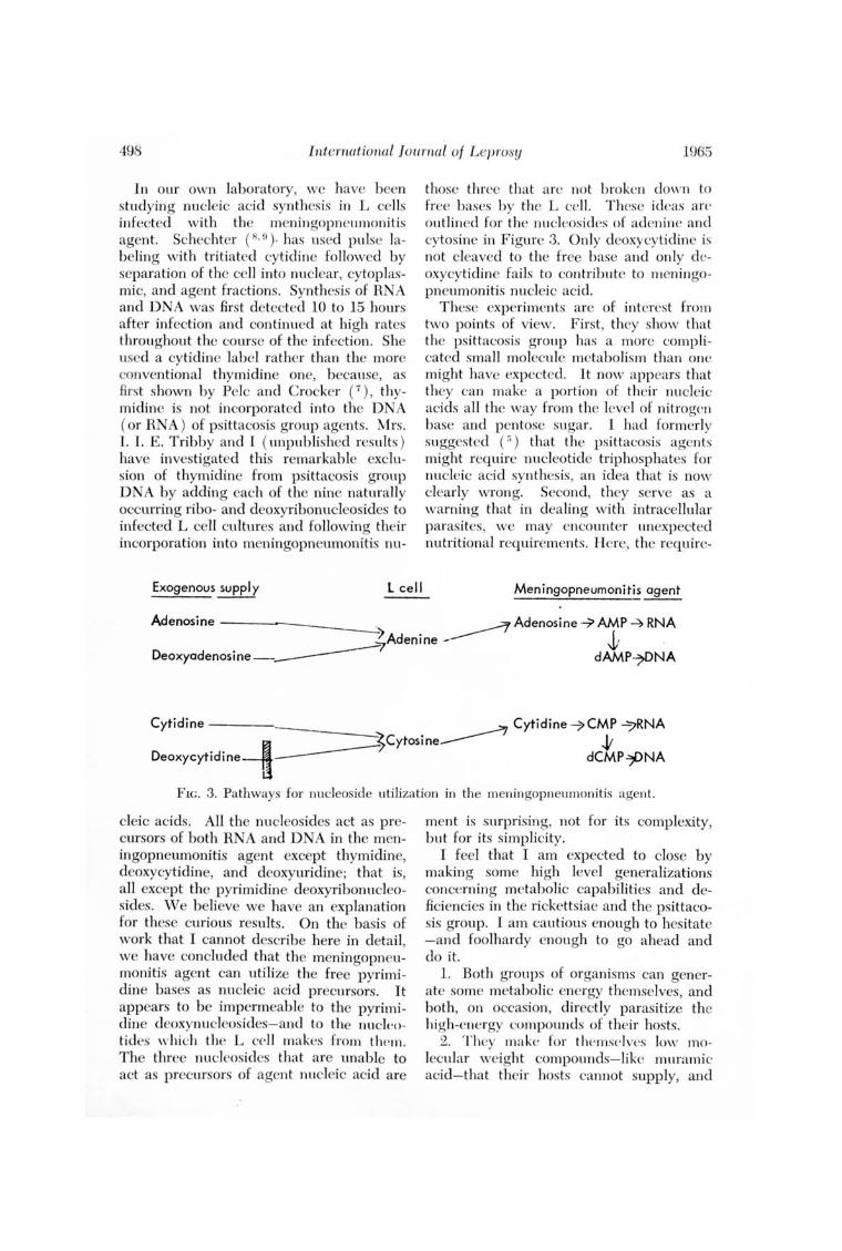
In our own laboratory, we have been studyin g nucleic acid synthesis in L cells infected with the menin gopneumonitis agent. Schechter (8 .0). has used pulse labeling with tritiated cytidin e followed by separation of the cell into nuclear, cytoplasmi c, and agent fractions. Synthesis of RNA and DNA was first detected 10 to 15 hours after infection and continued at hi gh rates throughout the course of the infection. She used a cytidine label rather than the more conventional thymidine one, because, as fi rst shown by Pelc and Crocker (7 ), thymidine is not incorporated into the DNA (or RNA ) of psittacosis group agents. Mrs. I. I. E. Tribby and I ( unpublished results ) have investigated this remarkable exclusion of thymidine from psittacosis group DNA by adding each of the nine naturally occurring ribo- and deoxyribonucleosides to infected L cell cultures and following their incorporation into meningopneumonitis nu-
Exogenous supply l cell
those three that arc not broken down ta free bases by the L cel l. These ideas art' outlined for the nucleasides of adenine and cytosine in Figure 3. Only deoxycytidine is not cleaved to the free base and only deoxycytidine fails to contribute to meningopneumonitis nu cleic acid.
These experiments are of interest from two points of view. First, they show that the psittacosis group has a more complicated small molecule metabolism than onc might have expectcd. It now appears that they can make a portion of their nucleic acids all the way fram the level of nitrogen base and pentase sugar. I had formerly suggested (r;) that the psittacosis agents might require nucleotide triphosphates far nucleic acid synthesis, an idea that is now clearly wrong. Second, they serve as a warning that in dealin g with intracellular paras ites, we may encounter unexpected nutritional req uirements. Here, the require-
Meningopneumonitis agent
Adenosine ~ ~ Adenosine --7> AMP ~ RNA ~Adenine ---- . J; .
Deoxyadenosine_~ dAMP7f)NA
Cytidine ~ Cytidine-7>CMP 7RNA --.n ~Cytosine______ . .,y DeOXycytidine----u~ dCMP~NA
FIG. 3. Pathways for nucleoside utilization in the meningopneumonitis agent.
cleic acids. All the nucleosides act as precursors of both RNA and D NA in the meningopneumonitis agent except thymidine, deoxycytidine, and deoxyuridine; that is, all except the pyrimidine deoxyribonucleosides. vVe believe we have an explanation for these curious results. On the basis of work that I cannot describe here in detail, we have concluded that the meningopneumonitis agent can utilize the free pyrimidin e bases as nucleic acid precursors. It appears to be impermeable to the pyrimidine deaxynucleas ides- and to the nuclt'utides which the L ce ll ma kes from them. The three nucleosides that are unable to act as precursors of agent nu cleic acid are
ment is surprising, not for its complexity, but for its simplicity.
I feel that I am expected to close by making some high level generalizations concerning metabolic capabilities and deficiencies in the rickettsiae and the psittacosis group. I am cautious enough to hesitate -and foolhardy enough to go ahead and do it.
1. Both groups of organisms can generate some metabolic energy themselves, and both, on occasion, directly parasitize the high-energy compounds of their hos ts.
2. They ma ke fo r themselves Jow molecul ar weight compounds-like mu rami c acid- that their hosts cannot supply, and

.'3.1, .3 (Pt. 2) M ouldel': Rickettsiae & Psittacosis G'I'O llPS 499
they depend on their hos ts for many others. T his dependency reflects in part adaptations to intrace llular life and is in part a legacy from their free- livin g ancestors, whatever they may have b een like.
3. They make their own lipids and carbohydrates, p roteins and nucleic acids by the same mechanisms as other cells.
4. They have abnormally small p ools of oxidizable substratcs, amino acids, nucleosides and nucleotides, and oxidation-reduction cofactors, and these compounds pass in and out of these organisms with more than ordinary facility. This makes them extremely vuln erabl e to irreversible damage when they are transferred from the rich intracellular environment to the poor extracellul ar one.
5. They are highly adapted b oth genotypi cally and phenotypically to intracellular growth . Removal from this environment gives ri se to lethal metaboli c dislocations.
6. Reproduction in v itro of the essential properties of the intracellular milieu will permit artificial cultivation . D elineation of these prop erties will take mu ch more hard work.
7. Finally, even after we have grown them in artificial media, we shall still b e left with the most fascinating question of all , the molecular nature of th eir successful adaptation to intracellul ar life. For in our zeal to grow these organisms in artificial media we mus t never forget that, however successful wc may b e, they are still going to grow intracellularly in their natural hosts, and that we are still goin g to have to explain the pathogenesis of their diseases in terms of the consequences of intracellular growth.
UEFEHENCES I. BOVARNICK, M. R. and SCHNEJDER, L. T h e
incorporat ion of g lycine- l -C" by typhus r ickettsiae. J. BioI. Chem. 235 ( 1960) 1727-1931.
2. BOVARNICK, M . R., SCHNElDER, L. and WALTER, H. The incorpora tion of labeled methionine by typhus rickettsiae. Biochim . Bioph ys . Acta 33 ( 1959) 415-422.
3. HIGASI-II, N. E lectron m icroscop ic stu d ies on th e mode of reprod ucti on of trachoma virus and psittacosis virus in cell cultures. Expel'. & Molecu lar Path. 4 (1965) 24-39.
4. JONES, F . JR. and PAHETSKY, D . Polyri bonu cleotide synthesis by Coxiella h111'11 etii. Bact. P roc. (1965) , p . 87.
5. MOULDER, T. W. T he Biochem is try of I nt racellular P arasi tism, C hicago, U niversi ty of Chicago Press, 1962.
6. MOULDER, J . W., GHISSO, D . L. and BnuBAKER, R. H. E nzymes of glucose catab olism in a member of the psittacosis group. J . Bact. 89 (1965) 810-812.
7. PELC, S. R. and CnocKER, T. T. D ifferences in th e utili zation of labelled precu rsors for the synthesis of D NA in cell nu cle i and psittacosis virus. Biochem . J . 78 (1961) 20-P.
8. SCHEcHTEn, E. M. Synthesis of p rote in and nu cle ic acid d urin g growth of the men ingopneumonitis agen t in L cells. Bact. Proc. (1965) p . 37.
9. SCHECHTER, E. M., Tm BBY, 1. 1. E. and MOULDEH, T. W. Nucle ic acid metabolism in L cells infected w ith, a member of th e p sittacos is group. Science 145 (1964) 819-821.
10. W EISS, E. Ad enosine triphospha te and other r equirements for the utiliza tion of g lucose b y agents of the psittacosis-trachoma group. T. Bact. 90 (1965) 243-2.53.
11. WEISS, E., MYEHs, 'vV. F., DHESSLEH, H. R. and CHUN-H oON, H . Glucose metabolism b y agen ts of the psittacosis- trachoma group . Virology 22 (1964) 551-562.
DISCUSSION
Dr. Weiser. Thank you, Dr. Moulder, for those very enlightening remarks. The concepts presented are indeed intriguing and if and when Dr. Moulder succeeds in cultiva tin g various other ri ckettsiae in an extracellular environment, the results may give us real hope of being able to cultivate M. leprae. Dr. Weiss, D eputy Director of the D epartment of Microbiology a t the
Naval Medical Research Institute in Bethesda, will open the discussion .
Dr. Weiss. Dr. Moulder analyzed two problems of intracellular parasitism : ( 1 ) loss in biochemical competence preventing growth of the microorganism in an extracellular environment, and (2) gain in biochemical competence permitting intracellular growth. Of the two problems the first
.500 Internationa l Jnr l1'lw l of Lep'I'Os!I 196.5
is the easier and the only one that I want to discuss. .I hope that other discussants will take on the sec<?ncl p rohkm.
In his prescntation, as well as in previous publi cati ons, Dr. Moulder has made a comprehensive effort to discover similarities among intracellular microorganisms and to draw guidelines for further investi gation. I shall act as the devil's advocate and emphasize the dissimilariti es.
The typhus ri ckcttsia can well be defin ed as the prototype intracellul ar bacterium. It has th e main fun ctions of other bacteria, but does nothing well. It produces adenosin e triphosphate (ATP ) th rough the oxidation of glutamate, but this ATP is not sufficient fo r synthesis. Preformed ATP must be added. Nico tinamide adenine dinucleotide (N AD ) is the first electron acceptor in glutamate oxidation, but under certain conditions this cofactor can be los t. For optimal stability and function NAD must be present in the mcdium. Amino acids can be incorporated into proteins, but the rickettsia has no amino acid pool to sustain the reaction, even brieRy, under conditions of nutritional deficiency. It has a cell wall , but this wall does not eliminate a requirement for osmotic protection. Nucleic acid synthesis has been studi ed in Coxiella bumetii (:!) but not in typhus rickettsiae. But even if the rickettsia can synthesize nucleic acids more effi ciently than other macromolecules, cultivation in a cell -free medium seems to be a long way off. It needs biochemical assistance over a very wide fron t. Some of the ass istance, undoubtedly, will be easy to provide, such as ATP or NAD, but we must not preclude the possibility that one of the requirements might be the timely inhibition or activation of enzymes, i.e., the mechanism that controls synthesis in other bacteria, as illustrated yesterday by Dr. Goldman. Such a need would be most difficult to satisfy.
This particular portra it of a parasite p robably applies only to a small group of microorgan isms. As Ormsbee and Peacock (G) have recently shown, the close relative of the typhus rickettsia, C. burnetii, has a somewhat different type of metabolism. Pyruvate, rather than glutamate, is the chief substrate of intact cells. Preliminary
experimcnts in our lahoratory with C. /Jllfl l(' lii ( Wciss a lld ()nnshc(" 1I11puhlisllt'u experi ments) ind ica ted that amino aeids arc incorporated as in the caSl' of tl w ri ckettsia, but the requirements are not so strin gent.
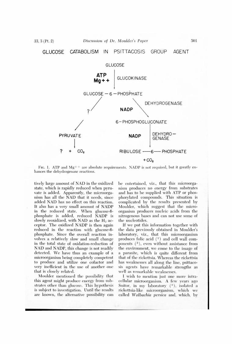
If we go still furth er and consider the psittacos is group of agents, we find a situation that is certainly different ( Fig. 1 ), as pointed out by Moulder. The mi croorganism utilizes glucose and has at leas t the three enzyme sys tems indicated in Figure 1, viz. , glucokin ase and the two dehydrogenases, plus the other enzymes described by Mouldcr. It has an absolute requirement for ATP and Mg + + and partial requirement for nicotinamide adenine dintlcleotide phosphate (NADP ). While the dehydrogenase reactions will take place without added NADP, they are markedly stimulated by it. \IVe have reason to believe that pyruvate is produced via a pathway other than pentose. CO~ is produ ced from the first carbon of pyruvate. The dissimilation of. glucose by this agent is very much like an ideal biochemical reaction : If there is no ATP, no CO~ is produced; if ATP is added, large amoi.lllts of CO~ evolve. This occurs despite the fact that we are dealing with whole cells. If we start with glucose-6-phosphate, A TP is not required. Pyruvate utili zation requires no added cofactors. ATP, NAD, diphosphothiamin , and, probably, coenzyme A, have no effect on the reaction . Surprisingly, lipoic acid has a slight enhancing effect.
A recent experiment by Kiesow and my-self (unpublished experiments ) illustrates some of the deficiences and capabilities of this agent. This experiment was carried out with the Britton Chance dual wavelength spectrophotometer. \Vith this instrument we can detect very small changes in the state of oxidation-reduction of the pyridine nucleotides of whole cells. If we add pyruvate to a suspension of microorganisms, the reduction of the pyridine nucleotides is rapid and marked . On the other hand, if we add glucose-6-phosphate, which yields approximately the same amount of CO~, the response is small and irregular. There is a very simple explanation for these observations: the microorganisms have a rela-
33, 3 (Pt. 2) DiscLlssion of Dr. Moulder's Paper 501
GLUCOSE CATABOLISM IN PSITTACOSIS GROUP AGENT
GLUCOSE
ATP Mg++ GLUCOK I NASE
GLUCOSE - 6 -PHOSPHATE
7/ /
PYRUVATE
1 ? + cO2
~ DEHYDROGENASE
NADP ~
6- PHOSPHOGLUCONATE
NADP DEHYDROGENASE
RIBULOSE ~6- PHOSPHATE
FIG. 1. ATP and Mg + + are absolute requirements . 1 ADP is not required, but it greatl y enhances the dehydrogenase reactions.
tively large amount of NAD in the oxidized state, which is rapidly reduced when pyruvate is added. Apparently, the microorganism has all the NAD that it needs, since added NAD has no effect on this reaction. It also has a very small amount of NADP in the reduced state. When glucose-6-phosphate is added, reduced NADP is slowly reoxidized, with NAD as the H~ acceptor. The oxidized NADP is then again reduced in the reaction with glucose-6-phosphate. Since the overall reaction involves a relatively slow and small change in the total state of oxidation-reduction of NAD and NADP, this change is not readily detected. We have thus an example of a microorganism being completely competent to produce and utilize one cofactor and very ineffici ent in the use of another ·one that is closely related .
Moulder mentioned the possib iliLy Lha t this agent might produce energy from substntes other thnn glucose. This hypothcsis is subject to investigation. Unti l thc results are known, the alternative possibility can
be entertained, viz., that this microorganism produces no energy from substrates and has to be supplied with ATP or phosphorylated compounds. This situation is complicated by the results presented by Moulder, which suggest that the microorganism produces nucleic acids from the nitrogenous bases and can not use some of the nucleotides.
If we put this information together with the data previously obtained in Moulder's laboratory, viz. , that this microorganism produces folic acid (1) and cell wall components (3), even without assistance from the environment, we come to the image of a parasite, which is quite different from that of the rickettsia. Whereas the rickettsia has weaknesses all along the line, psittacosis agents have remarkable strengths as well as rcmarkahlc wcakn esses.
1 wish to mention just one more inl'r:1-cellular microorganism. A fcw years ago Suitor, in my lahoratory ( R) , isolated a rickettsia-like microorgariism, which wc called W olbachia persica and, which, by
502 inte'l'1wtional ] otl1'luil of Leprosy 1965
the way, docs not r esemble a rickettsia. Suitor m a de very extensive attempts to grow thi s microorganism in cell-free m edi a and a ll these a ttempts h ave b een completely unsuccessful (7) . H owever , w hen we studied the carbohydra te m etaboli sm of this microorganism, it appeared to b e extremely active (n). An inves tigation of the lipid m etabolism carri ed out b y Neptune and m yself (4. 5) again yielded excellent results. A few a ttempts a t d emonstration of prote in synthesis indicated tha t glucose and other substrates w ere actively incorpora ted into prote in fractions ( 10 ). At this point the work was stopped , but it would have b een interesting to find ou t exactly why this microorganism did n ot grow in any of the m edia tested. It appears tha t the m etabolic defi ciency of this microorganism is a highly speci fic one and does not involve a broad m etab olic function.
When one emphasizes differences am on g microorganisms r a ther than similarities, one tends t o b e p essimisti c and, in effect, say tha t there is very little that we can learn from other systems. However, this is not necessarily so. We are confronted w ith a number of models of intracellular parasitism. W e must study them all and select the one that presents the greatest similarity to the intracellular microorganism we w ish to investigate or, even b etter , to construct our own model from w h atever information is avai lable. I hope tha t those of you w ho are trying to grow the agent of leprosy w ill not have to select the model of the rickettsia, w hich has a series of deficiencies. I hope, instead, tha t information now available will permit you to select a model of a microor ganism that has select deficiencies as well as excellent cap abilities . As Moulder has just pOinted out-and thi s seems to b e the theme of this conference-a requirem ent for intracellular growth may surprise you for its complexity or for its simplicity .
REFERENCES
1. COL6N, J. I. Enzymes for forma tion of citrovol'l1m fac tor in members of tJw psittacosis group of m icroorga nisms. J. Bact. 79 (1960 ) 741-746.
2. JONES, F. , In. and PAnETSKY, D. Polyribon ucleotide syn thesis in Coxiella 1J111'11 -ettii enzymes. Bact. Proc. (1965) 87.
3. MOULDER, J. W., NOVOSEL, D . L. and TRIBBY, I. C. Diaminopimelic acid decarboxylase of the agent of meningopneumonitis. J. Bact. 85 (1963) 701-706.
4. NEPTUNE, E. M. , JR., WEISS, E., DAVIES, J. A. and SUITOR, E. C. Lipid metabolism of the rickettsia-like microorganism V/o1-bachia persica. I. Incorporation of longchain fatty acids into phosphatides. J. Infect. Dis. 114 (1964) 39-64.
5. NEPTUNE, E. M., In ., ViTEtSS, E. and DAVIES, J. A. L ipid metabolism of th e rickettsia-like microorgan ism vVo1h(whia persica. II. Studies with labeled non lipid substrates. J. Infect. Dis. 114 (1964) 45-49.
6. OnMsBEE, R. A. and PEACOCK, M. C. Metabolic activity in Coxiella bll1'l1etii. J. Bact. 88 (1964 ) 1205-1210.
7. SUlTOR, E. c., TR. , Stud ies on the cell envelope of W n17Jachia 11ersica. J. Infect. Dis. 114 (1964) 125-134.
8. SUlTOR, E. c., J.R. , and WEISS, E. Isolation of a rickettsia-like microorganism (Wolbachia persica, n . sp.) from Argas persictls (Oken). J. Infect. Dis. 108 (1961 ) 95-106.
9. WEISS, E., MYERS, W. F. , SUITon, E. C. , In. and EPTUNE, E. M. , In. Respiration of a rickettsia-like microorganism , W nlhachia persica. J. Infect. Dis. 110 (1962 ) 155-164.
10. WEISS, E., NEPTUNE, E. M. , In. and D AVIES, J. A. Lipid metabolism of the rickettsia-like microorganism 'Volbachia persica. III. Comparison with other metabolic activities . J. Infect. Dis . 114 (1964) 50-54.
Dr. Weisel'. vVe are now ready for open discussion.
Dr. Dannenberg. I would like to com m ent on intracellula r parasitism in general. First of all the p sittacosis group seems to h ave properti es of b oth viruses and b acteri a , an d ye t M!lcobac[pl'ill1n 7eprae look~ like a full fl edged aciu-fast bac terium , :11-though it may a lso exist in L form s.
\ "'ould it b e possible, if on e considers the lysosom es of macrophages to piece parts of

33,3 (Pt. 2) Discllssion of Dr. MOlllder'!> Pa})(: r 503
this conferencc together? Mycobactcria, when ingested by macrophagcs, seem to rcside in phagocytic vacuoles, or phagosomcs. Zanvil, Cohn and Hirsch (1. Expe1'. Merl. 118 (1963) 1009), have shown that lysosomal enzymes are pomed into the vacuole surrounding such bacteria. As the bacterium divides, the phagosome may divide, and morc and more lysosomes may be produced to discharge their enzymcs into thc new vacuoles. vVhat is the environment in a phagosome? First of all, it is acid. And Dr. Hanks' group havc shown that mycobacteria prefer a slightly acid environmcnt. Second, it is full of diges tive enzymes, as protcinases, Jipases and nucleases. Hanks and Tepper have impli ed that the lipid coat around acid-fast bacteria tends to impede the absorption of nutrients by the bacteria. The thicker the coat, the more difficulty they have in growing. Could it be that the enzymes in the phagosome digest off this impeding coat and make the intracellular organism better able to absorb nutrients? This may be a reason why it grows in a phagosome. Finally, what are these nutrients? It may be that the digestive enzymes of lysosomes (nucleases, lipases, and proteinases) break down the major building blocks of protoplasm partially but not completely. The resulting peptides, lipid components and short chains of nucleotides may be growth factors for acid-fast bacteria . We have startcd to purify some of the lysosomal enzymes of macrophages, but will not complete the job for several years. Analogous enzymes, however, could perhaps be used to produce such gro"vth factors by partially breaking down tissue or bacteria elements. The protein, lipid or nucleic acid fragm ents which result might aid the clllture of the leprosy bacillus in the test tubc.
Dr. Emmons. It might be interesting to point out that in medical mycology we have intracellular parasites that grow luxuriantly and exuberantly il1 vitro without any special attcntion to tIlt' composition of the culture medium. The three species of Tlistoplasma alld Pellicillitlln lII{/l'll effei illllstrate this phenomenon . The fungi that WE' havc not succeeded in growing in v it'l'O
arc extracellular. This certainly presents a contrast to the concept that intracellular parasitism is associated invariabl y with diffi culty of in vit ro culture.
Dr. Segal. I would like to ask Dr. Moulder if the DNA content of these agents has been analyzed as an index of genetic competence, and, secondly, if the base ratios have been analyzed as an index of taxonomic position.
Dr. Moulder. I shall answer Dr. Segal's questions first, and then comment on something Dr. Dannenberg said. First, the DNA content. The DNA content, is about a 5th or a 10th of that of Esch l! richia coli . This would give quite a lot of codin g capacity. vVe have determined the guanine-cytosine ratios of the meningopneumonitis agent by physical methods and it comes out to be about 40 per cent; this is a little disappointing b ecause it is just like that of the host. At the 1965 American Society for Microbiology meetings in Atlantic City, Dr. Gerloff and other investigators from the Rocky Mountain Laboratory gave a very interesting account of some hybridization experiments with meningopneumonitis DNA. and the DNA of three other members of the psittacosis group. There was practically complete homology between meningopneumonitis DNA and each of the other three DNA's tes ted, a fact indicating, as all of us had thought, that the psittacosis group is a tru e evolutionary group. They obtained no hybridization between the DNA of meningopnemnonitis agent and that of rickettsiae, a number of other bacteria, L cells, etc.
Next, to comment on what Dr. Dannenberg said: First, I do not think you can nam e one property of the psittacosis O'J'OUIl
and rickettsiae that is virus-like. I do not believe there is any borderline agent that is really half way b etween bacteria and viruses. They are either all viruses or all bacteria . I b elieve the rickettsiae and psitticosis organisms are highly modified bacteria . Now the question of the phagocytic vacuole. I think it becomes very important because the recent fIlt e work of Armstrong and Heed in London and of Higashi in Kyoto, has finally resulted in good electron
504 International Journal of Leprosy 1965
micrographs showing all stages from the first absorption of a psittacosis group agent to the host cell membrane and onward. There is no doubt that it is taken in by a process strongly resembling phagocytosis. The invading psittacosis group cell appears in a little vacuole surrounded by a unit membrane, and as the inclusion grows the membrane grows and it seems that the whole life of the psittacosis agent in the infected cell occurs in a vacuole, which is scparated from the cytoplasm by a llnit membra'ne, Dr. D annenberg's discuss ion of the acidic properties of the phagocytic vacuole reminds me of something I had not thought of before. We have been struck by the fac t that the meningopneumonitis agent is quite stable down to pH 5 at vvhi ch pH it agglutinates, hut one can bring the pH
back to neutrality and lose no infec tivity a t all. It will be very interestin g to sec how far down the pH scale its acid res istance goes. vVe noticed also many years ago that the psittacosis group agents are resistant to enzymes of all kinds. Therefore, it is entirely conceivable that they could survive in a phagocytic vacuole into which the lysosomal enzymes wcre heing poured ,
Dr. Weiser. Our next speaker Dr, p . D 'Arcy Hart, was fo rmerly D irector of the Tuherculosis Research Unit of the Medical Research Council of Great Britain . He is now associated with Dr. Rees' laboratory at Mill I-lill. The topic of his discussion will be "Further analysis of the growth (elongation) phenomenon of M. lepmemurium in vit ro and relevant studies with M. lepme."
Cultivation of M. leprae including leads
from M. leprae7nuriul1t
Chairman : R. S. Weiser
Further Analysis of the Growth (Elongation) Phenomenon
of Mycobacterium, leprael1turium in vitro, and Relevant
Studies with Mycobacterium, leprae
P. D'Arcy Hart, F.R.C.P.!
Many bacilli elongate when in an un favorable environment, but on restoration of normal conditions cell division and multiplica tion can occur and the long forms become replaced. On the contrary, it is still doubtful whether M ycobacterium lepmemllrium has hecn ohserved to multiply in a cell -free mediu m; consequ entl y elonga tion, (,V(,11 tholl gh an irr('vl'rsihk process upp:1rl' lItly, rnay be cOlisiu t>n ·d- at Ipas t for t hl' mO llw llt - nn ad vallcc.
'\:<1 l io ll a l Ill st i lli te for M edio " R esearch , ~ I i l l Ilill , LOll doll :'>I.W.7 , E ll g la ll d.
Many of the fea tures of elongation of M. lepraemurillm in a cell -free medium have already been reported (~), and these will only be summarized here. The usual source of bacilli has been homogenized infected mouse liver ; the inocul ated liquid medium is incuhated for several weeks at 37°C and exa min ed by Ziehl- c('lsen stain , or the e It'el roll III i('ros('opc ', for Il-Ilg1 h aml lll-generation. Lcngthening is apparent hy 2 days, with doublin g II slla lly by 7-14 days; the ra te of increase gradually declines, a maximum of about 3-4 times the initial