the calcineurin β-like interacting protein kinase cipk25
TRANSCRIPT
Copyedited by: OUP
Journal of Experimental Botanydoi:10.1093/jxb/eraa004 Advance Access Publication 13 February 2020
© The Author(s) 2020. Published by Oxford University Press on behalf of the Society for Experimental Biology. This is an Open Access article distributed under the terms of the Creative Commons Attribution License (http://creativecommons.org/licenses/by/4.0/), which permits unrestricted reuse, distribution, and reproduction in any medium, provided the original work is properly cited.
RESEARCH PAPER
The calcineurin β-like interacting protein kinase CIPK25 regulates potassium homeostasis under low oxygen in Arabidopsis
Andrea Tagliani1,2,†, Anh Nguyet Tran1,*,†, Giacomo Novi1, Riccardo Di Mambro1,3, , Michele Pesenti4, Gian Attilio Sacchi4, Pierdomenico Perata1,2, and Chiara Pucciariello1,2,‡
1 PlantLab, Institute of Life Sciences, Scuola Superiore Sant’Anna, 56127 Pisa, Italy2 nanoPlant Center @NEST, Institute of Life Sciences, Scuola Superiore Sant’Anna, 56127 Pisa, Italy3 Department of Biology, University of Pisa, 56126 Pisa, Italy4 Department of Agricultural and Environmental Science, University of Milano, 20133 Milano, Italy
* Current address: Department of Plant Biotechnology, Cuu Long Delta Rice Research Institute, 9400 Can Tho, Vietnam† These authors contributed equally to the work‡ Correspondence: [email protected]
Received 28 June 2019; Editorial decision 2 January 2020; Accepted 12 February 2020
Editor: Hideki Takahashi, Michigan State University, USA
ABSTRACT
Hypoxic conditions often arise from waterlogging and flooding, affecting several aspects of plant metabolism, including the uptake of nutrients. We identified a member of the CALCINEURIN β-LIKE INTERACTING PROTEIN KINASE (CIPK) family in Arabidopsis, CIPK25, which is induced in the root endodermis under low-oxygen conditions. A cipk25 mutant exhibited higher sensitivity to anoxia in conditions of potassium limitation, suggesting that this kinase is involved in the regulation of potassium uptake. Interestingly, we found that CIPK25 interacts with AKT1, the major inward recti-fying potassium channel in Arabidopsis. Under anoxic conditions, cipk25 mutant seedlings were unable to maintain potassium concentrations at wild-type levels, suggesting that CIPK25 likely plays a role in modulating potassium homeostasis under low-oxygen conditions. In addition, cipk25 and akt1 mutants share similar developmental defects under waterlogging, further supporting an interplay between CIPK25 and AKT1.
Keywords: Anoxia, Arabidopsis, calcineurin β-like interacting protein kinase, CIPK25, hypoxia, potassium homeostasis.
Introduction
The intensification of flooding events is one of the conse-quences of climate change that is strongly affecting plant bio-diversity and crop productivity. In a flooded environment, the availability of external oxygen (O2) is reduced, because gas diffusion in water is lower than in aerobic conditions (Armstrong, 1979; Colmer, 2003). Low O2 availability for plants is not only a consequence of environmental stress but
also occurs during the development of specific organs and tissues, such as fruits, root vasculature, and seeds (Van Dongen and Licausi, 2015).
In plants, hypoxia is perceived by members of the group VII ETHYLENE RESPONSIVE FACTORS (ERF-VIIs), whose protein stability is regulated by PLANT CYSTEINE OXIDASE (PCO) enzymes in an O2-dependent manner
applyparastyle "fig//caption/p[1]" parastyle "FigCapt"
Dow
nloaded from https://academ
ic.oup.com/jxb/advance-article-abstract/doi/10.1093/jxb/eraa004/5735510 by guest on 08 April 2020
Copyedited by: OUP
Page 2 of 12 | Tagliani et al.
(Gibbs et al., 2011; Licausi et al., 2011; Weits et al., 2014; White et al., 2017). PCO enzymes destabilize ERF-VIIs through the O2-dependent oxidation of an N-terminal cyst-eine, targeting the ERF-VIIs for proteasomal degradation. This process is prevented under O2 limitation, allowing ERF-VIIs to act as transcriptional activators of genes in-volved in anaerobic metabolism.
Together with a direct O2 sensing mechanism, additional signaling pathways contribute to the plant’s adaptation to low O2 availability. These pathways rely on perturbations of cel-lular homeostasis due to changes in available sugars, energy status, cytosolic calcium (Ca2+), pH, reactive oxygen species, reactive nitrogen species, and possibly potassium (K+) levels (Bailey-Serres and Chang, 2005; van Dongen and Licausi, 2015; Shahzad et al., 2016; Pucciariello and Perata, 2017; Wang et al., 2017b).
Among the second messengers, Ca2+ is involved in the re-sponse to many stimuli related to plant development and en-vironmental cues (Dodd et al., 2010). Release of Ca2+ into the cytosol from internal stores or from the extracellular space occurs under various conditions, so that different ex-ternal stimuli are transduced by distinct spatio-temporal vari-ations in the frequency, amplitude, and location of Ca2+ waves (Kudla et al., 2018).
In line with the widespread signaling function of Ca2+, early reports suggested that O2 deprivation triggers a cytosolic Ca2+ flux in several plants, which indirectly regulates the expres-sion of anaerobic genes (Subbaiah et al., 1994; Sedbrook et al., 1996; Nie et al., 2006). Using rice protoplasts, the increased cytosolic Ca2+ concentrations observed under anoxia have been suggested to depend on both external and internal stores (Yemelyanov et al., 2011). More recently, a CALMODULIN-LIKE PROTEIN 38 (CML38) was found to be induced under low O2 and associated with cytosolic stress granules in a Ca2+-dependent manner (Lokdarshi et al., 2016).
Due to its ubiquitous role, the Ca2+-dependent network is multifaceted, and plants are equipped with a plethora of sensors able to transfer the message to downstream trans-ducers. A major family of Ca2+ sensors is the CALCINEURIN β-LIKE PROTEIN (CBL) family, which is unique to plants. CBLs modulate the activity of CBL-INTERACTING PROTEIN KINASE (CIPK) partners, which have a catalytic activity (Weinl and Kudla, 2009), thus acting as a signaling relay in which the sensor and the effector are two separate proteins (Kudla et al., 2018).
CIPKs belong to the subgroup of SUCROSE NON-FERMENTING 1 (SNF1) RELATED PROTEIN KINASE 3 (SnRK3) of plants, which is functionally similar to SNF1 in yeast and AMPK in mammals (Mao et al., 2016). CIPKs have a typical structural organization consisting of an N-terminal kinase catalytic domain and a C-terminal regulatory domain (Sanyal et al., 2015). The C-terminus contains the NAF/FISL motif, which is responsible for self-inhibition of the enzyme, and a protein phosphatase interaction domain. The Ca2+-dependent interaction of CBLs with the CIPK NAF/FISL motif activates the kinase, releasing it from autoinhibition (Chaves-Sanjuan et al., 2014). Additionally, the activity of
CIPKs is influenced by phosphorylation within the activation loop (Chaves-Sanjuan et al., 2014).
The CBL–CIPK complex transmits the Ca2+-dependent signal to downstream target proteins via phosphorylation (Sanyal et al., 2015). Each CBL can interact with multiple CIPKs and vice versa, providing a substantial level of versa-tility and flexibility in the Ca2+ signal transduction pathway (DeFalco et al., 2009).
Many physiological functions have been assigned to CBL–CIPK complexes, including the regulation of ion transport, the stress response, and plant development (for a review see Kudla et al., 2018). Some CBL–CIPK combinations—CBL1/9 and CIPK23 (Li et al., 2006; Xu et al., 2006; Cheong et al., 2007), CBL4 and CIPK6 (Held et al., 2011), and CBL3 and CIPK9 (Liu et al., 2013)—are involved in regulating K+ homeostasis in Arabidopsis roots and/or modulating the activity of plasma membrane channels (Wang et al., 2018).
K+ is the most abundant inorganic cation in plants, con-tributing up to 10% of their dry mass (Leigh and Wyn Jones, 1984) and having a high concentration (~100–200 mM) in-side the plant cytosol (Wyn Jones and Pollard, 1983). It is crucial in several processes, such as the maintenance of cell turgor and growth, the regulation of metabolism through direct interaction with enzymes, and the regulation of ionic balance in the cell (Sharma and Sharma, 2013). In this framework, the CBL1–CBL9/CIPK23 effector module is activated under K+ starvation and regulates the Shaker inward-rectifying K+ channel AKT1 through interaction and phosphorylation (Li et al., 2006; Xu et al., 2006; Lee et al., 2007). In addition, the CBL4/CIPK6 complex me-diates plasma membrane targeting as well as the activity of the highly selective and weak inward-rectifying K+ channel AKT2 (Held et al., 2011).
Under low O2 conditions, membrane depolarization occurs as a consequence of reduced proton pumping at the plasma membrane, due to a reduced ATP pool (Gout et al., 2001). This depolarization is likely transient, since the increased con-centration of H+ ions in the cytosol is counteracted by a rapid stimulation of depolarization-activated K+ efflux channels, which repolarizes the plasma membrane potential (Zeng et al., 2014, Cuin et al., 2018). This process may thus cause a latent K+ starvation. In fact, in plants exposed to low-O2 conditions, the K+ pool in the root is markedly reduced, and exogenous foliar or root applications of K+ alleviate the adverse effect on plants (for a review see Shabala et al., 2014). In line with this finding, Arabidopsis gork1-1 mutants lacking functional K+ efflux channels possess higher tolerance to hypoxia (Wang et al., 2017a).
The modification of K+ flux inside the cell may also indirectly alter the fermentative metabolism activated under O2 shortage. Shahzad et al. (2016) identified a MAPKKK, HYDRAULIC CONDUCTIVITY of ROOT 1 (HCR1), which contributes to RAP2.12 (ERF-VII) stabilization under hypoxia only when K+ is available. Moreover, K+ gradients may be exploited by Arabidopsis plants as a source of energy under low O2 condi-tions, since they stimulate loading of sucrose into the phloem sap (Gajdanowicz et al., 2011). This mechanism exploits the
Dow
nloaded from https://academ
ic.oup.com/jxb/advance-article-abstract/doi/10.1093/jxb/eraa004/5735510 by guest on 08 April 2020

Copyedited by: OUP
CIPK25 role under low oxygen | Page 3 of 12
differential operative status of the AKT2 K+ channel, which can partially replace the H+-ATPase when ATP is limited in availability (Dreyer et al., 2017). However, little is currently known about the regulation of K+ uptake after the onset of anoxia.
In this work, we identified a CIPK protein, named CIPK25, which is involved in the regulation of K+ homeostasis under O2 shortage. CIPK25 is transcriptionally induced by low O2, preferentially in the root endodermis, and directly interacts with the inward rectifying K+ channel AKT1. Misregulation of CIPK25 under O2 shortage results in a lower K+ content in Arabidopsis seedlings, suggesting that this protein plays a role in maintaining ion homeostasis in these conditions.
Materials and methods
Plant material and growth conditionsThe genotypes used were Arabidopsis thaliana ecotype Col-0 and Wassilewskija-2 (Ws-2), T-DNA insertion mutants cipk25-2 (Col-0 SALK_070911c, previously isolated also by Meena et al., 2019), cipk25-3 (Col-0 SALK_059092), cipk23-5 (Ragel et al., 2015), akt1-2 (Ws-2 NASC stock number: N3762; Nieves-Cordones et al., 2019), and akt1-1 (Hirsch et al., 1998). The genetic status of the cipk25-3 and cipk25-2 lines was experimentally verified using primers listed in Supplementary Table S1 at JXB online. Homozygous plants for the CIPK25 locus were isolated in the cipk25-2 and cipk25-3 T-DNA insertion lines.
In order to visualize the CIPK25 promoter activity using the GUS and GFP reporter genes, a 1 kb genomic fragment corresponding to the 5′ region upstream of the gene was cloned and recombined into the pKGWFS7 destination vector (Karimi et al., 2002). The CIPK25 pro-moter was analyzed with AGRIS AtcisDB (https://agris-knowledgebase.org/AtcisDB) and PlantPAN 2.0 (http://plantpan2.itps.ncku.edu.tw). To overexpress the gene, the full-length coding DNA sequence of CIPK25 and the CIPK25∆C version, lacking the C-terminal domain, were amplified and recombined into the pK7WG2 destination vector (Karimi et al. 2002). Transgenic plants were obtained using Agrobacterium-mediated transformation by the floral dip method (Clough and Bent, 1998). T1 seeds were screened on 0.9% agar plates containing the ap-propriate selective antibiotic. Resistant plants showing green cotyledons were screened until the T3 generation on selective medium.
To grow plants in pots, seeds were germinated in a moist soil mixture at 18–20 °C under a 12 h light photoperiod. The seeds were covered with plastic film for 1 week to maintain humidity. Seedlings were then transferred into new pots containing a growing mixture composed of soil, vermiculite, and fertilizer (ONE, Valagro). The plants were grown at 23 °C with a 12 h light photoperiod (120 μmol photons m–2 s–1) for 3 weeks.
To evaluate the plants’ submergence tolerance, plants were grown in pots for 3 weeks and then submerged in the dark for 72–96 h, starting the treatment at 20.00 h (the plants had been exposed to a 12 h light photoperiod with the light switched on at 08.00 h). The plants were then allowed to recover for 2 weeks.
For seedlings grown on six-well plates, a custom half-strength Murashige and Skoog (MS)-type medium, pH 5.7, was used, with the following recipe: 1.5 mM CaCl2, 0.75 mM MgSO4, 15 mM NH4NO3, 0.63 mM NH4H2PO4, 50 µM FeNaEDTA, 15 µM ZnSO4, 0.5 µM Na2MoO4, 50 µM MnSO4, 5 µM KI, 50 µM H3BO3, 0.05 µM CoCl2, and 0.5 µM CuSO4, with KCl added at 10, 2.5, or 0.1 mM. In Fig. 1A, MS (Duchefa Biochemie, product number M0221) was used. Seedlings were stratified in the dark at 4 °C for 2 days and then grown at 23 °C with a 12 h light photoperiod for 3 days before the dark-anoxia treat-ment. This treatment was applied in an enclosed anaerobic workstation (1 Person Hypoxic Glove Box, Coy Laboratory Products).
For gene expression analysis, seedlings were harvested directly after the treatment. For quantification of chlorophyll content and K+ content, seedlings were allowed to recover in the growth chamber for 1 additional week after the treatment before analysis.
For measurement of stalk height, plants were grown in pots for 3 weeks, then waterlogged for an additional 3 weeks before being meas-ured with a ruler. To overcome differences in the vegetative to flowering transition between Col-0 and Ws-2 wild-type plants, both neutral (12 h light/12 h dark) and long-day (16 h light/8 h dark) conditions were evaluated, since in long-day conditions Ws-2 showed the phenotype after 5 days of waterlogging.
Isolation and transfection of Arabidopsis protoplastsArabidopsis Col-0 mesophyll protoplasts were obtained from leaves of 3-week-old plants grown in a plastic pot filled with soil and peat (3:1) at 25/20 °C day/night under a 12 h light photoperiod, with photosyn-thetically active radiation of 100 µmol m–2s–1 provided by fluorescence lamps. Protoplasts were isolated as previously described (Yoo et al., 2007) and transformed with polyethylene glycol, using 5 µg of each plasmid. Protoplasts were incubated for 16 h at 25 °C in the dark and then im-mediately visualized.
RNA extraction and real-time PCR analysisTotal RNA was extracted as previously described (Kosmacz et al., 2015). Total RNA was reverse transcribed using the Maxima First Strand cDNA synthesis kit (Thermo Scientific). Real-time PCR reactions were car-ried out using SYBR® Green PCR Master Mix (Bio-Rad Laboratories, USA), using specifically designed primers (see Supplementary Table S1). The ∆∆Ct method was applied for relative quantification (Livak and Schmittgen, 2001).
Localization of GUS/GFP in plants and protoplastsProtoplasts transfected with a 35S:GFP:CIPK25 construct were observed with a Nikon Eclipse Ti-5 ViCo epi-fluorescence microscope (Nikon, Japan) using GFP and TRITC filters. The pAVA vector (von Arnim et al., 1998) was used as a 35S:GFP control. For bimolecular fluorescence com-plementation (BiFC) experiments, protoplasts were visualized with a ZEISS LSM880 Airyscan confocal microscope. Yellow fluorescent protein (YFP) fluorescence was excited at 488 nm and collected at between 520 and 560 nm. Chlorophyll autofluorescence was excited at 633 nm and collected at between 650 and 750 nm. Images were analyzed with ZEN 2010 software (Zeiss).
PromCIPK25-GUS/GFP seeds were plated on half-strength MS me-dium and seedlings were grown vertically in long-day conditions (16 h light/8 h dark) as previously described (Di Mambro and Sabatini, 2018). At 7 days after germination, seedlings were submerged by direct injec-tion of distilled water into the plate. Plants were submerged vertically up to the level of the hypocotyl/root junction. After 14 h of being sub-merged, water was drained from the plate and the plants were analyzed by confocal microscopy. For visualization of cells, seedlings were directly mounted on a slide in 10 µl of propidium iodide (PI) solution (10 μg ml–1 dissolved in water) to stain the cell wall; PI and GFP fluorescence were acquired using a 488 nm laser and a highly sensitive gallium ar-senide phosphide (GaAsP) spectral photodetector [EC Plan-Neofluar ×40/1.30 Oil differential interference contrast (DIC) objective, pinhole 90 µm]. DIC microscopic analysis followed GUS histochemical staining performed as described in Vitha et al. (1995).
Cloning and protein–protein interaction assaysCoding and regulatory CIPK25 sequences were amplified from Arabidopsis Col-0 genomic DNA template using Phusion High Fidelity DNA poly-merase (New England Biolabs, UK) following the manufacturer’s in-structions and using the primers listed in Supplementary Table S1. The PCR products were purified and cloned into the Gateway pENTR/D-TOPO vector (Life Technologies, USA). The resulting entry clones were
Dow
nloaded from https://academ
ic.oup.com/jxb/advance-article-abstract/doi/10.1093/jxb/eraa004/5735510 by guest on 08 April 2020
Copyedited by: OUP
Page 4 of 12 | Tagliani et al.
recombined into destination vectors using the LR Reaction Mix II (Life Technologies, USA). Each cloning product was verified by restriction-site mapping and sequencing. For the BiFC experiments, the CIPK25∆C sequence, consisting of the kinase domain, and the C-terminal cytosolic domain of AKT1 were cloned into the pDH51-GW-YFPC or pDH51-GW-YFPN vector (Zhong et al., 2008), respectively, and co-transfected into Arabidopsis mesophyll protoplasts. The tag orientation was defined in line with previous results (Xu et al., 2006). Corresponding empty vec-tors were used as negative controls. pAVA (35S:GFP) (von Arnim et al., 1998) was used as a positive control of transformation.
For yeast two-hybrid (Y2H) analysis, the CIPK25∆C sequence and the C-terminal cytosolic domain of AKT1 were cloned into the pDEST32 or pDEST22 vector (ThermoFisher), respectively. Co-transformation was performed in the MaV203 yeast strain following the Li/Ac protocol (ThermoFisher Proquest). Positive colonies for transform-ation were screened in SD-LT medium and interaction was screened in SD-LTUH+15 mM 3AT medium (Sigma-Aldrich). Empty vectors were used as negative controls. X-gal staining was performed on the same plates with filter paper soaked in Z-buffer (ThermoFisher) and left at 37 °C for 2–3 h. Homodimerization of HRU1 (Gonzali et al., 2015) was used as a positive control.
Total chlorophyll content analysisChlorophyll extraction was performed in the dark, using ethanol (96% v/v) as a solvent. The samples were incubated at 4 °C overnight. After centrifugation (11 200 g for 5 min at 4 °C), total chlorophyll content was measured spectrophotometrically using the formula of Lichtenthaler and Buschmann (2001).
Quantification of potassium contentTo quantify the K+ content, seedlings grown as described above were collected, dried in an oven at 60 °C for 10 days, and their dry weight was recorded. They were then mineralized in 65% HNO3 at 200 °C in an Anton Paar Multiwave 7000 microwave and finally analyzed in a Bruker AURORA ICP-MS mass spectrometer.
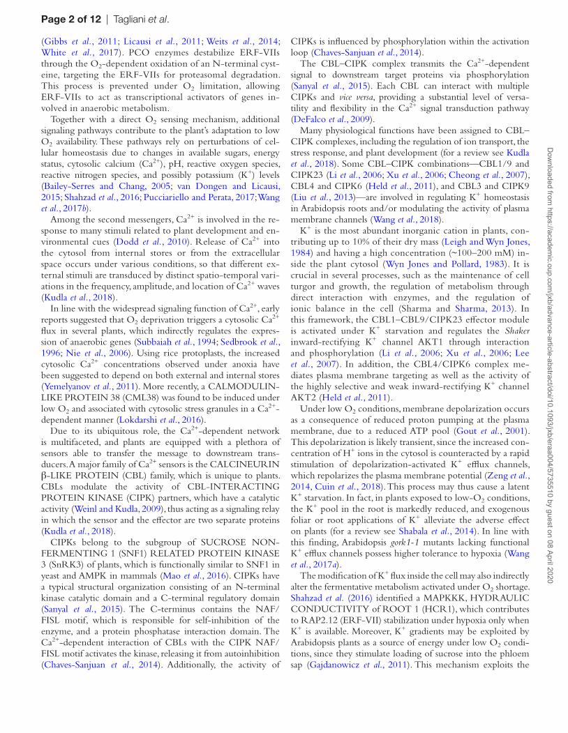
Fig. 1. (A) Effect of abiotic stresses on the expression of CIPK25. Four-day-old seedlings were treated with 4 h of anoxia in darkness, 100 mM NaCl, 100 mM mannitol, 5 mM H2O2, 4 °C (cold), and 37 °C (heat). The value of the untreated control (Ctrl) was arbitrarily set to 1. Each value represents the mean ±SE (n=4). Statistical significance (Ctrl versus treatment) was determined using Student’s t-test: *P<0.05. (B) Time-course of CIPK25 mRNA accumulation under anoxia in roots and shoots dissected from 3-week-old seedlings grown on vertical agar plates. Expression levels are shown as relative units, with the shoot value at time 0 arbitrarily set to 1. Each value represents the mean ±SE (n=3). Statistical significance (shoot versus root) was determined using Student’s t-test: *P<0.05. (C) Histochemical GUS staining of 10-day-old seedlings of the promCIPK25:GUS line under air and after 14 h of waterlogging (Sub). The experiment was performed in quadruplicate (n=15). (D) Confocal analysis of promCIPK25:GFP plants under air and after 14 h of waterlogging, showing longitudinal (left) and cross (right) sections along the z-axis. The experiment was performed in quadruplicate (n=10). (E) Confocal images of Arabidopsis protoplasts transiently transformed with 35S:GFP:CIPK25 and (as positive control) 35S:GFP in air. (F) CIPK25 mRNA accumulation in leaves of 3-week-old seedlings grown on vertical agar plates in Col-0 wild type and Δ13rap2.12 (overexpressing a stable version of RAP2.12). The value of the wild type was arbitrarily set to 1. Each value represents the mean ±SE (n=3). Statistical significance was determined using Student’s t-test: **P<0.01.
Dow
nloaded from https://academ
ic.oup.com/jxb/advance-article-abstract/doi/10.1093/jxb/eraa004/5735510 by guest on 08 April 2020
Copyedited by: OUP
CIPK25 role under low oxygen | Page 5 of 12
Results
CIPK25 is induced in Arabidopsis roots under oxygen shortage
Among the 26 CIPKs encoded by the Arabidopsis genome, CIPK25 (At5g25110) is transcriptionally induced under low-O2 conditions (Supplementary Fig. S1), pointing to a putative role for this kinase under this stress condition. Gene expression analysis in Arabidopsis seedlings ex-posed to different abiotic stresses identified CIPK25 as a salt- and anoxia-induced gene (Fig. 1A), preferentially in roots (Fig. 1B). CIPK25 expression was also detected in 10-day-old seedlings using promCIPK25:GUS (Fig. 1C) and promCIPK25:GFP (Fig. 1D) plants, revealing a prefer-ential induction under waterlogging, localized in the root endodermis. Observation of mesophyll protoplasts transi-ently transformed with the 35S:GFP:CIPK25 construct revealed that CIPK25 protein is preferentially localized in the cytosol (Fig. 1E). Bioinformatic inspection of the CIPK25 promoter identified the presence of several MYB, ABRE, and ARF binding motifs (Supplementary Fig. S2A). However, the hypoxia-responsive promoter element (HRPE), which is responsible for the regulation of core an-aerobic genes (Gasch et al. 2016), was absent. Interestingly, a GCC-box (position –138), a known target of AP2/ERF transcription factors (Yang et al., 2009; Lee et al., 2015), was
also found (Supplementary Fig. S2B). In line with this ob-servation, CIPK25 is expressed at a higher level in plants overexpressing a chimeric form of RAP2.12 lacking the first 13 N-terminal amino acids containing the destabilizing Cys2 (35S:∆13RAP2.12; Giuntoli et al., 2017) (Fig. 1F).
CIPK25 is involved in tolerance to hypoxia
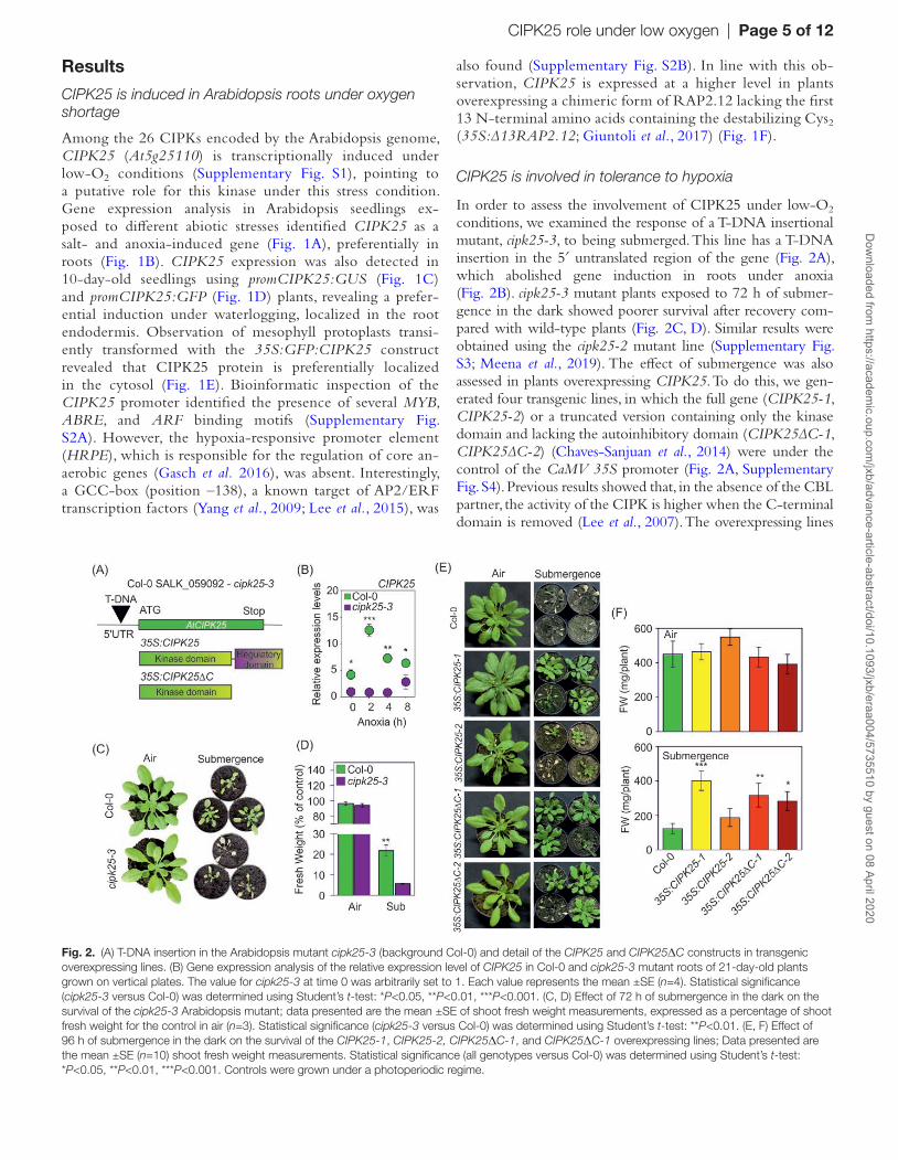
In order to assess the involvement of CIPK25 under low-O2 conditions, we examined the response of a T-DNA insertional mutant, cipk25-3, to being submerged. This line has a T-DNA insertion in the 5′ untranslated region of the gene (Fig. 2A), which abolished gene induction in roots under anoxia (Fig. 2B). cipk25-3 mutant plants exposed to 72 h of submer-gence in the dark showed poorer survival after recovery com-pared with wild-type plants (Fig. 2C, D). Similar results were obtained using the cipk25-2 mutant line (Supplementary Fig. S3; Meena et al., 2019). The effect of submergence was also assessed in plants overexpressing CIPK25. To do this, we gen-erated four transgenic lines, in which the full gene (CIPK25-1, CIPK25-2) or a truncated version containing only the kinase domain and lacking the autoinhibitory domain (CIPK25∆C-1, CIPK25∆C-2) (Chaves-Sanjuan et al., 2014) were under the control of the CaMV 35S promoter (Fig. 2A, Supplementary Fig. S4). Previous results showed that, in the absence of the CBL partner, the activity of the CIPK is higher when the C-terminal domain is removed (Lee et al., 2007). The overexpressing lines
Fig. 2. (A) T-DNA insertion in the Arabidopsis mutant cipk25-3 (background Col-0) and detail of the CIPK25 and CIPK25ΔC constructs in transgenic overexpressing lines. (B) Gene expression analysis of the relative expression level of CIPK25 in Col-0 and cipk25-3 mutant roots of 21-day-old plants grown on vertical plates. The value for cipk25-3 at time 0 was arbitrarily set to 1. Each value represents the mean ±SE (n=4). Statistical significance (cipk25-3 versus Col-0) was determined using Student’s t-test: *P<0.05, **P<0.01, ***P<0.001. (C, D) Effect of 72 h of submergence in the dark on the survival of the cipk25-3 Arabidopsis mutant; data presented are the mean ±SE of shoot fresh weight measurements, expressed as a percentage of shoot fresh weight for the control in air (n=3). Statistical significance (cipk25-3 versus Col-0) was determined using Student’s t-test: **P<0.01. (E, F) Effect of 96 h of submergence in the dark on the survival of the CIPK25-1, CIPK25-2, CIPK25ΔC-1, and CIPK25ΔC-1 overexpressing lines; Data presented are the mean ±SE (n=10) shoot fresh weight measurements. Statistical significance (all genotypes versus Col-0) was determined using Student’s t-test: *P<0.05, **P<0.01, ***P<0.001. Controls were grown under a photoperiodic regime.
Dow
nloaded from https://academ
ic.oup.com/jxb/advance-article-abstract/doi/10.1093/jxb/eraa004/5735510 by guest on 08 April 2020
Copyedited by: OUP
Page 6 of 12 | Tagliani et al.
(both full and truncated versions of CIPK25) showed a similar phenotype under aerobic conditions, comparable with previous observations (Meena et al., 2019). Overexpression of the two CIPK25∆C lines and CIPK25-1 conferred enhanced tolerance to submergence compared with the wild type after 96 h of submergence in darkness (Fig. 2E, F). When grown on vertical plates, the 35S:CIPK25-1 and 35S:CIPK25∆C transgenic lines showed longer roots, while the roots of cipk25-3 were signifi-cantly shorter than those of wild-type plants (Supplementary Fig. S5), as previously observed (Meena et al., 2019).
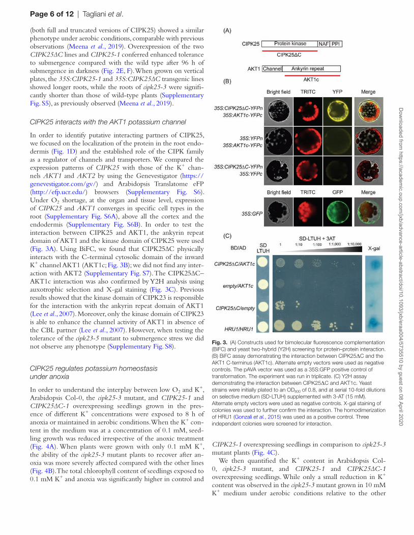
CIPK25 interacts with the AKT1 potassium channel
In order to identify putative interacting partners of CIPK25, we focused on the localization of the protein in the root endo-dermis (Fig. 1D) and the established role of the CIPK family as a regulator of channels and transporters. We compared the expression patterns of CIPK25 with those of the K+ chan-nels AKT1 and AKT2 by using the Genevestigator (https://genevestigator.com/gv/) and Arabidopsis Translatome eFP (http://efp.ucr.edu/) browsers (Supplementary Fig. S6). Under O2 shortage, at the organ and tissue level, expression of CIPK25 and AKT1 converges in specific cell types in the root (Supplementary Fig. S6A), above all the cortex and the endodermis (Supplementary Fig. S6B). In order to test the interaction between CIPK25 and AKT1, the ankyrin repeat domain of AKT1 and the kinase domain of CIPK25 were used (Fig. 3A). Using BiFC, we found that CIPK25∆C physically interacts with the C-terminal cytosolic domain of the inward K+ channel AKT1 (AKT1c; Fig. 3B); we did not find any inter-action with AKT2 (Supplementary Fig. S7). The CIPK25∆C–AKT1c interaction was also confirmed by Y2H analysis using auxotrophic selection and X-gal staining (Fig. 3C). Previous results showed that the kinase domain of CIPK23 is responsible for the interaction with the ankyrin repeat domain of AKT1 (Lee et al., 2007). Moreover, only the kinase domain of CIPK23 is able to enhance the channel activity of AKT1 in absence of the CBL partner (Lee et al., 2007). However, when testing the tolerance of the cipk23-5 mutant to submergence stress we did not observe any phenotype (Supplementary Fig. S8).
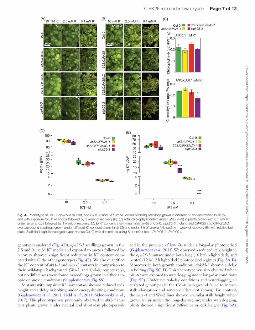
CIPK25 regulates potassium homeostasis under anoxia
In order to understand the interplay between low O2 and K+, Arabidopsis Col-0, the cipk25-3 mutant, and CIPK25-1 and CIPK25∆C-1 overexpressing seedlings grown in the pres-ence of different K+ concentrations were exposed to 8 h of anoxia or maintained in aerobic conditions. When the K+ con-tent in the medium was at a concentration of 0.1 mM, seed-ling growth was reduced irrespective of the anoxic treatment (Fig. 4A). When plants were grown with only 0.1 mM K+, the ability of the cipk25-3 mutant plants to recover after an-oxia was more severely affected compared with the other lines (Fig. 4B). The total chlorophyll content of seedlings exposed to 0.1 mM K+ and anoxia was significantly higher in control and
CIPK25-1 overexpressing seedlings in comparison to cipk25-3 mutant plants (Fig. 4C).
We then quantified the K+ content in Arabidopsis Col-0, cipk25-3 mutant, and CIPK25-1 and CIPK25∆C-1 overexpressing seedlings. While only a small reduction in K+ content was observed in the cipk25-3 mutant grown in 10 mM K+ medium under aerobic conditions relative to the other
Fig. 3. (A) Constructs used for bimolecular fluorescence complementation (BiFC) and yeast two-hybrid (Y2H) screening for protein–protein interaction. (B) BiFC assay demonstrating the interaction between CIPK25ΔC and the AKT1 C-terminus (AKT1c). Alternate empty vectors were used as negative controls. The pAVA vector was used as a 35S:GFP positive control of transformation. The experiment was run in triplicate. (C) Y2H assay demonstrating the interaction between CIPK25ΔC and AKT1c. Yeast strains were initially plated to an OD600 of 0.8, and at serial 10-fold dilutions on selective medium (SD-LTUH) supplemented with 3-AT (15 mM). Alternate empty vectors were used as negative controls. X-gal staining of colonies was used to further confirm the interaction. The homodimerization of HRU1 (Gonzali et al., 2015) was used as a positive control. Three independent colonies were screened for interaction.
Dow
nloaded from https://academ
ic.oup.com/jxb/advance-article-abstract/doi/10.1093/jxb/eraa004/5735510 by guest on 08 April 2020
Copyedited by: OUP
CIPK25 role under low oxygen | Page 7 of 12
genotypes analyzed (Fig. 4D), cipk25-3 seedlings grown in the 2.5 and 0.1 mM K+ media and exposed to anoxia followed by recovery showed a significant reduction in K+ content com-pared with all the other genotypes (Fig. 4E). We also quantified the K+ content of akt1-1 and akt1-2 mutants in comparison to their wild-type background (Ws-2 and Col-0, respectively), but no differences were found in seedlings grown in either aer-obic or anoxic conditions (Supplementary Fig. S9).
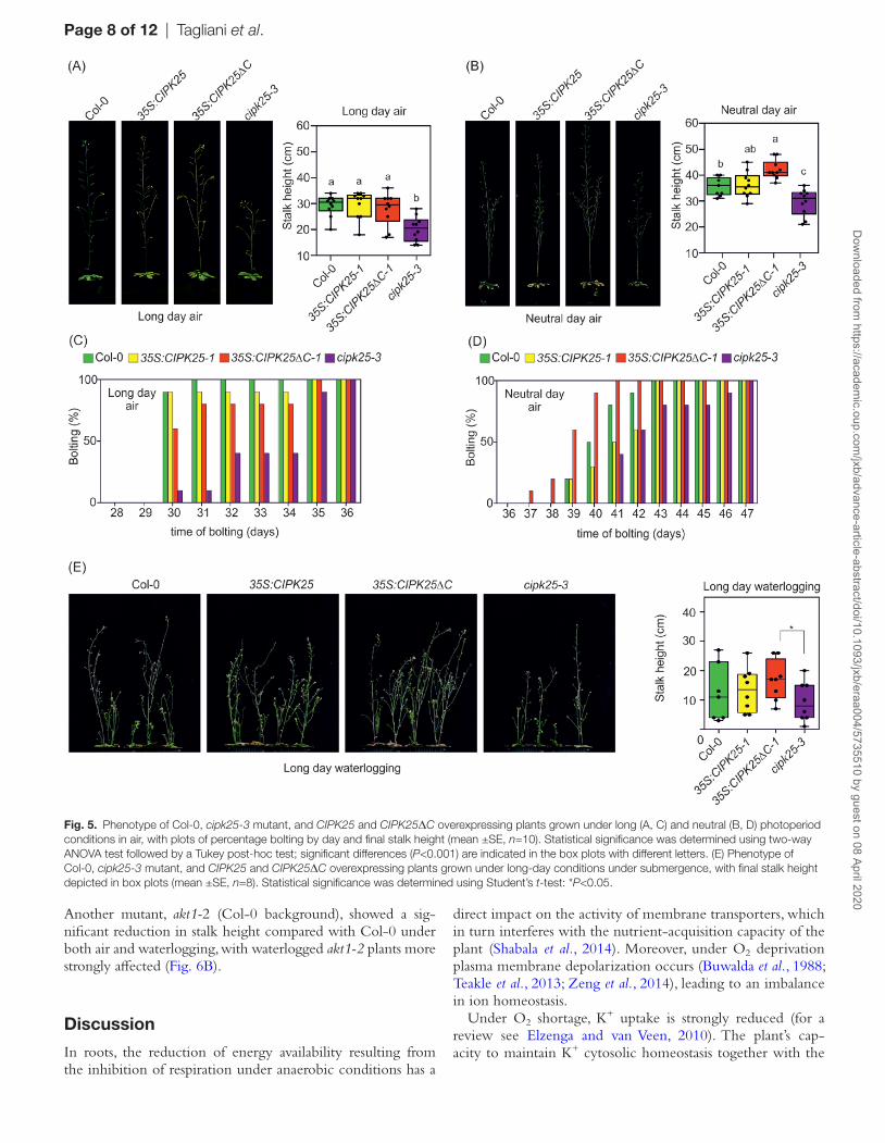
Mutants with impaired K+ homeostasis showed reduced stalk height and a delay in bolting under energy-limiting conditions (Gajdanowicz et al., 2011; Held et al., 2011; Sklodowski et al., 2017). This phenotype was previously observed in akt2-1 mu-tant plants grown under neutral and short-day photoperiods
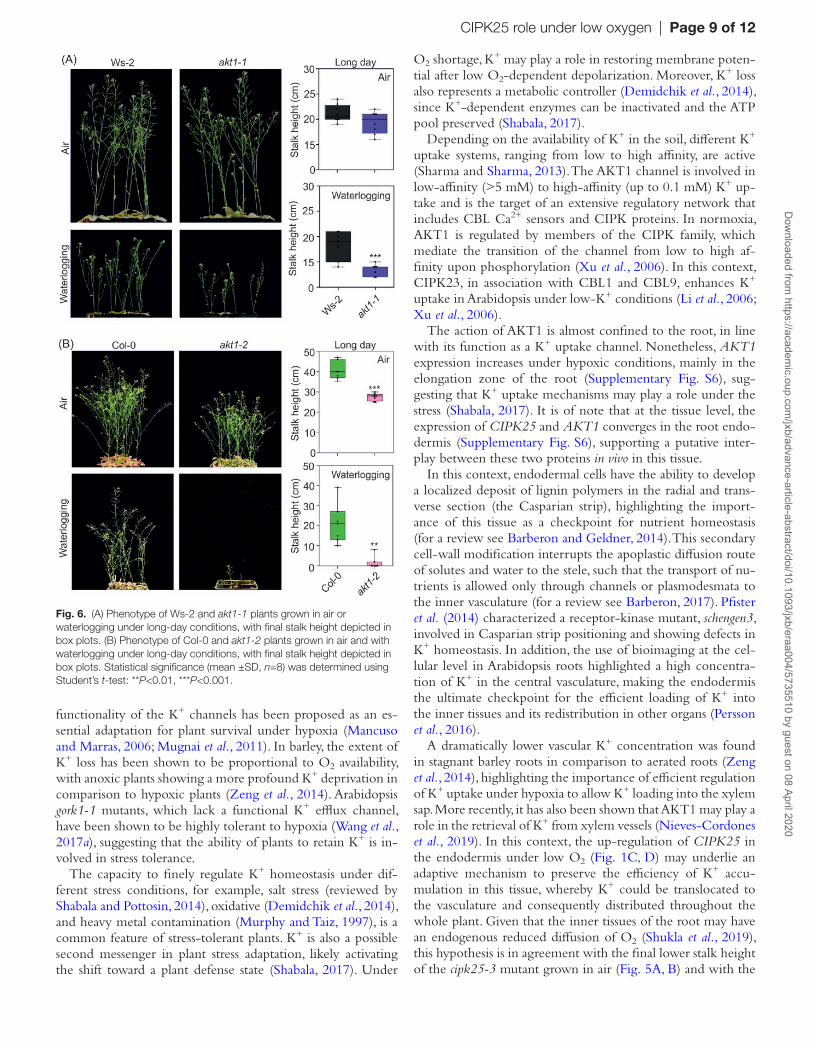
and in the presence of low O2 under a long-day photoperiod (Gajdanowicz et al., 2011). We observed a reduced stalk height in the cipk25-3 mutant under both long (16 h/8 h light/dark) and neutral (12 h/12 h light/dark) photoperiod regimes (Fig. 5A, B). Moreover, in both growth conditions, cipk25-3 showed a delay in bolting (Fig. 5C, D). This phenotype was also observed when plants were exposed to waterlogging under long-day conditions (Fig. 5E). Under neutral-day conditions and waterlogging, all analyzed genotypes in the Col-0 background failed to induce stalk elongation and senesced (data not shown). By contrast, the akt1-1 and Ws-2 lines showed a similar stalk height when grown in air under the long-day regime; under waterlogging, plants showed a significant difference in stalk height (Fig. 6A).
Fig. 4. Phenotype of Col-0, cipk25-3 mutant, and CIPK25 and CIPK25ΔC overexpressing seedlings grown in different K+ concentrations in air (A) and with exposure to 8 h of anoxia followed by 1 week of recovery (B). (C) Total chlorophyll content (mean ±SD, n=3) in plants grown with 0.1 mM K+ under air or anoxia followed by 1 week of recovery. (D, E) K+ concentration (mean ±SE, n=5) of Col-0, cipk25-3 mutant, and CIPK25 and CIPK25ΔC overexpressing seedlings grown under different K+ concentrations in air (D) and under 8 h of anoxia followed by 1 week of recovery (E), with relative box plots. Statistical significance (genotypes versus Col-0) was determined using Student’s t-test: *P<0.05, ***P<0.001.
Dow
nloaded from https://academ
ic.oup.com/jxb/advance-article-abstract/doi/10.1093/jxb/eraa004/5735510 by guest on 08 April 2020
Copyedited by: OUP
Page 8 of 12 | Tagliani et al.
Another mutant, akt1-2 (Col-0 background), showed a sig-nificant reduction in stalk height compared with Col-0 under both air and waterlogging, with waterlogged akt1-2 plants more strongly affected (Fig. 6B).
Discussion
In roots, the reduction of energy availability resulting from the inhibition of respiration under anaerobic conditions has a
direct impact on the activity of membrane transporters, which in turn interferes with the nutrient-acquisition capacity of the plant (Shabala et al., 2014). Moreover, under O2 deprivation plasma membrane depolarization occurs (Buwalda et al., 1988; Teakle et al., 2013; Zeng et al., 2014), leading to an imbalance in ion homeostasis.
Under O2 shortage, K+ uptake is strongly reduced (for a review see Elzenga and van Veen, 2010). The plant’s cap-acity to maintain K+ cytosolic homeostasis together with the
Fig. 5. Phenotype of Col-0, cipk25-3 mutant, and CIPK25 and CIPK25ΔC overexpressing plants grown under long (A, C) and neutral (B, D) photoperiod conditions in air, with plots of percentage bolting by day and final stalk height (mean ±SE, n=10). Statistical significance was determined using two-way ANOVA test followed by a Tukey post-hoc test; significant differences (P<0.001) are indicated in the box plots with different letters. (E) Phenotype of Col-0, cipk25-3 mutant, and CIPK25 and CIPK25ΔC overexpressing plants grown under long-day conditions under submergence, with final stalk height depicted in box plots (mean ±SE, n=8). Statistical significance was determined using Student’s t-test: *P<0.05.
Dow
nloaded from https://academ
ic.oup.com/jxb/advance-article-abstract/doi/10.1093/jxb/eraa004/5735510 by guest on 08 April 2020
Copyedited by: OUP
CIPK25 role under low oxygen | Page 9 of 12
functionality of the K+ channels has been proposed as an es-sential adaptation for plant survival under hypoxia (Mancuso and Marras, 2006; Mugnai et al., 2011). In barley, the extent of K+ loss has been shown to be proportional to O2 availability, with anoxic plants showing a more profound K+ deprivation in comparison to hypoxic plants (Zeng et al., 2014). Arabidopsis gork1-1 mutants, which lack a functional K+ efflux channel, have been shown to be highly tolerant to hypoxia (Wang et al., 2017a), suggesting that the ability of plants to retain K+ is in-volved in stress tolerance.
The capacity to finely regulate K+ homeostasis under dif-ferent stress conditions, for example, salt stress (reviewed by Shabala and Pottosin, 2014), oxidative (Demidchik et al., 2014), and heavy metal contamination (Murphy and Taiz, 1997), is a common feature of stress-tolerant plants. K+ is also a possible second messenger in plant stress adaptation, likely activating the shift toward a plant defense state (Shabala, 2017). Under
O2 shortage, K+ may play a role in restoring membrane poten-tial after low O2-dependent depolarization. Moreover, K+ loss also represents a metabolic controller (Demidchik et al., 2014), since K+-dependent enzymes can be inactivated and the ATP pool preserved (Shabala, 2017).
Depending on the availability of K+ in the soil, different K+ uptake systems, ranging from low to high affinity, are active (Sharma and Sharma, 2013). The AKT1 channel is involved in low-affinity (>5 mM) to high-affinity (up to 0.1 mM) K+ up-take and is the target of an extensive regulatory network that includes CBL Ca2+ sensors and CIPK proteins. In normoxia, AKT1 is regulated by members of the CIPK family, which mediate the transition of the channel from low to high af-finity upon phosphorylation (Xu et al., 2006). In this context, CIPK23, in association with CBL1 and CBL9, enhances K+ uptake in Arabidopsis under low-K+ conditions (Li et al., 2006; Xu et al., 2006).
The action of AKT1 is almost confined to the root, in line with its function as a K+ uptake channel. Nonetheless, AKT1 expression increases under hypoxic conditions, mainly in the elongation zone of the root (Supplementary Fig. S6), sug-gesting that K+ uptake mechanisms may play a role under the stress (Shabala, 2017). It is of note that at the tissue level, the expression of CIPK25 and AKT1 converges in the root endo-dermis (Supplementary Fig. S6), supporting a putative inter-play between these two proteins in vivo in this tissue.
In this context, endodermal cells have the ability to develop a localized deposit of lignin polymers in the radial and trans-verse section (the Casparian strip), highlighting the import-ance of this tissue as a checkpoint for nutrient homeostasis (for a review see Barberon and Geldner, 2014). This secondary cell-wall modification interrupts the apoplastic diffusion route of solutes and water to the stele, such that the transport of nu-trients is allowed only through channels or plasmodesmata to the inner vasculature (for a review see Barberon, 2017). Pfister et al. (2014) characterized a receptor-kinase mutant, schengen3, involved in Casparian strip positioning and showing defects in K+ homeostasis. In addition, the use of bioimaging at the cel-lular level in Arabidopsis roots highlighted a high concentra-tion of K+ in the central vasculature, making the endodermis the ultimate checkpoint for the efficient loading of K+ into the inner tissues and its redistribution in other organs (Persson et al., 2016).
A dramatically lower vascular K+ concentration was found in stagnant barley roots in comparison to aerated roots (Zeng et al., 2014), highlighting the importance of efficient regulation of K+ uptake under hypoxia to allow K+ loading into the xylem sap. More recently, it has also been shown that AKT1 may play a role in the retrieval of K+ from xylem vessels (Nieves-Cordones et al., 2019). In this context, the up-regulation of CIPK25 in the endodermis under low O2 (Fig. 1C, D) may underlie an adaptive mechanism to preserve the efficiency of K+ accu-mulation in this tissue, whereby K+ could be translocated to the vasculature and consequently distributed throughout the whole plant. Given that the inner tissues of the root may have an endogenous reduced diffusion of O2 (Shukla et al., 2019), this hypothesis is in agreement with the final lower stalk height of the cipk25-3 mutant grown in air (Fig. 5A, B) and with the
Fig. 6. (A) Phenotype of Ws-2 and akt1-1 plants grown in air or waterlogging under long-day conditions, with final stalk height depicted in box plots. (B) Phenotype of Col-0 and akt1-2 plants grown in air and with waterlogging under long-day conditions, with final stalk height depicted in box plots. Statistical significance (mean ±SD, n=8) was determined using Student’s t-test: **P<0.01, ***P<0.001.
Dow
nloaded from https://academ
ic.oup.com/jxb/advance-article-abstract/doi/10.1093/jxb/eraa004/5735510 by guest on 08 April 2020
Copyedited by: OUP
Page 10 of 12 | Tagliani et al.
lower K+ content found in cipk25-3 grown in air in the pres-ence of 10 mM K+ in the medium (Fig. 4D).
Among the CIPK family, we found that CIPK25 is posi-tively regulated at the transcriptional level by O2 deficiency in roots (Supplementary Fig. S1, Fig. 1A–D). CIPK25 is also up-regulated in the ate1-2 mutant in comparison to wild-type Col-0, in HRE1 and HRE2 overexpressing plants under low O2 (Supplementary Fig. S1), and in 35S:∆13RAP2.12 trans-genic plants (Fig. 1F). It might thus be possible that either CIPK25 is a target of RAP2.12 or that HRE1 and HRE2 are responsible for CIPK25 expression under low O2. In fact, a known target of AP2/ERF transcription factors, a GCC-box, is present in the CIPK25 promoter (Supplementary Fig. S2). Alternatively, CIPK25 expression might be indirectly affected by the O2 sensing machinery, downstream of RAP2.12.
Overall, the activation of a CBL–CIPK sensor relay com-plex, post-translationally regulated by Ca2+, suggests a mech-anism in which low O2 and the presence of Ca2+ spiking converge in protecting the plant from K+ leakage. We noticed that CIPK23, which is known to regulate AKT1 under K+ starvation (Li et al., 2006; Xu et al., 2006), seems not to be involved in adaptation to submergence, since the survival of the cipk23-5 mutant was similar to that of the Col-0 control (Supplementary Fig. S8). This suggests that the possible im-pairment in K+ homeostasis that occurs under anoxia may fail to activate the mechanism that in air enhances the capability for K+ uptake—that is, disruption in K+ homeostasis-activated Ca2+ spiking, activation of CBL1/9 by Ca2+, interaction of CBL1/9 with CIPK23, phosphorylation of AKT1 by CIPK23, and transition of AKT1 from low to high affinity (Li et al., 2006; Xu et al., 2006; Cheong et al., 2007; Lee et al., 2007). The result of low-O2-dependent induction of CIPK25 may compensate for this impairment by activating the K+ channel AKT1. Y2H and BiFC results (Fig. 3) identified the presence of an interaction between CIPK25 and AKT1, supporting this hypothesis. Recently, CIPK25 has been found to interact with CBL4 (Meena et al., 2019), which is also strongly expressed in Arabidopsis roots under hypoxia (eFP Translatome browser; data not shown), suggesting a post-translational activation mechanism mediated by Ca2+ under low O2. The presence of an early Ca2+ spike under low O2 has been recently con-firmed using a FRET-based biosensor (NES-YC3.6) (Wagner et al., 2019).
The activation of CIPK25 may be a prerequisite for AKT1 functioning under combined O2 shortage and low K+, as sug-gested by the strong reduction in K+ content in the cipk25-3 mutant under anoxia and low K+ content in the medium (Fig. 4E). Indeed, the reduction in cellular K+ concentration in the cipk25-3 mutant was observed exclusively in media with low K+ concentrations (2.5 and 0.1 mM K+) where almost only high-affinity K+ channels, such as AKT1, play a role.
AKT1 is probably not the only K+ uptake mechanism func-tioning under O2 shortage, since the akt1-1 and akt1-2 mutants did not show a strong reduction in K+ content relative to their respective wild type (Supplementary Fig. S9). The mechanism of K+ uptake under low O2 by CIPK25 likely includes some other transporters, which have not yet been identified.
Interestingly, cipk25 mutant lines have a reduced root length (Supplementary Fig. S5; Meena et al., 2019) and altered auxin transport, possibly due to misregulated PIN protein expression (Meena et al., 2019). Philippar et al. (2006) reported that the application of exogenous auxin transcriptionally regulates the Zea mays inward K+ channel ZMK1. Moreover, Arabidopsis AKT1 is involved in the sensing of external K+ concentration, with a subsequent regulation of PIN protein abundance and auxin redistribution in roots (Li et al., 2017). In fact, mutants for K+ efflux channels, which likely have a higher concentra-tion of K+ in the cytosol, show increased cell expansion, likely connected to auxin (Osakabe et al., 2013). It thus seems that in aerobic conditions an interplay between CIPK25, AKT1, and auxin might occur in Arabidopsis roots in order to regulate growth, a mechanism that deserves further investigation.
Our results show that CIPK25 plays a key role in maintaining K+ homeostasis under low-O2 conditions. This mechanism is transcriptionally regulated by low O2 and likely by Ca2+-dependent signaling at the post-translational level. In addition to AKT1, other targets of CIPK25 could be involved in leading to adaptive responses that modify K+ fluxes not only under en-vironmental low O2 but also under endogenous tissue-specific hypoxia.
Supplementary data
Supplementary data are available at JXB online.Table S1. List of primers.Fig. S1. Expression of CIPK25 under low O2 conditions and
different genetic backgrounds related to hypoxia.Fig. S2. CIPK25 promoter analysis through AGRIScisDB
platform and PlantPAN2.0.Fig. S3. Phenotype of cipk25-2 mutant under submergence
stress.Fig. S4. CIPK25 expression level in overexpressing plants. Fig. S5. Root length of seedlings grown on plates.Fig. S6. Comparison between CIPK25, AKT1 and AKT2
expression pattern under O2 shortage in various Arabidopsis tissues using Genevestigator software and eFp Translatome Browser.
Fig. S7. BiFC assay showing no interaction between CIPK25 and AKT2.
Fig. S8. Effect of submergence on the survival of cipk23-5 Arabidopsis mutants.
Fig. S9. Potassium cellular concentration of Ws-2 and akt1-1 and Col-0 and akt1-2 seedlings grown under different external K+ concentrations in air and anoxia.
Acknowledgements
This work was supported by the Scuola Superiore Sant’Anna. ANT was funded by a PhD fellowship in Agrobiodiversity. AT was funded by a PhD in Agrobiosciences. We thank Dr Manuel Nieves-Cordones for providing akt1-2 and cipk23-5 mutant seeds.
Dow
nloaded from https://academ
ic.oup.com/jxb/advance-article-abstract/doi/10.1093/jxb/eraa004/5735510 by guest on 08 April 2020
Copyedited by: OUP
CIPK25 role under low oxygen | Page 11 of 12
Author contributions
CP and PP conceived the study; AT, ANT, RDM, GN, and MP per-formed the experiments; CP and AT analyzed the data; CP, AT, and PP interpreted the data and wrote the manuscript; GN, RDM, and GAS contributed to revising the manuscript.
ReferencesArmstrong W. 1979. Aeration in higher plants. Advances in Botanical Research 7, 225–332.
Bailey-Serres J, Chang R. 2005. Sensing and signalling in response to oxygen deprivation in plants and other organisms. Annals of Botany 96, 507–518.
Barberon M. 2017. The endodermis as a checkpoint for nutrients. New Phytologist 213, 1604–1610.
Barberon M, Geldner N. 2014. Radial transport of nutrients: the plant root as a polarized epithelium. Plant Physiology 166, 528–537.
Buwalda F, Thomson CJ, Steigner W, Barrett-Lennard EG, Gibbs J, Greenway H. 1988. Hypoxia induces membrane depolarization and po-tassium loss from wheat roots but does not increase their permeability to sorbitol. Journal of Experimental Botany 39, 1169–1183.
Chaves-Sanjuan A, Sanchez-Barrena MJ, Gonzalez-Rubio JM, Moreno M, Ragel P, Jimenez M, Pardo JM, Martinez-Ripoll M, Quintero FJ, Albert A. 2014. Structural basis of the regulatory mechanism of the plant CIPK family of protein kinases controlling ion homeostasis and abiotic stress. Proceedings of the National Academy of Sciences, USA 111, E4532–E4541.
Cheong YH, Pandey GK, Grant JJ, Batistic O, Li L, Kim BG, Lee SC, Kudla J, Luan S. 2007. Two calcineurin B-like calcium sensors, interacting with protein kinase cipk23, regulate leaf transpiration and root potassium uptake in Arabidopsis. The Plant Journal 52, 223–239.
Clough SJ, Bent AF. 1998. Floral dip: a simplified method for Agrobacterium-mediated transformation of Arabidopsis thaliana. The Plant Journal 16, 735–743.
Colmer T. 2003. Long-distance transport of gases in plants: a perspec-tive on internal aeration and radial oxygen loss from roots. Plant, Cell & Environment 26, 17–36.
Cuin TA, Dreyer I, Michard E. 2018. The role of potassium channels in Arabidopsis thaliana long distance electrical signalling: AKT2 modu-lates tissue excitability while GORK shapes action potentials. International Journal of Molecular Sciences 21, 926.
DeFalco TA, Bender KW, Snedden WA. 2009. Breaking the code: Ca2+ sensors in plant signalling. The Biochemical Journal 425, 27–40.
Demidchik V. 2014. Mechanisms and physiological roles of K+ efflux from root cells. Plant Physiology 171, 696–707.
Di Mambro R, Sabatini S. 2018. Developmental analysis of Arabidopsis root meristem. Methods in Molecular Biology 1761, 33–45.
Dodd AN, Kudla J, Sanders D. 2010. The language of calcium signaling. Annual Review of Plant Biology 61, 593–620.
Dreyer I, Gomez-Porras JL, Riedelsberger J. 2017. The potassium bat-tery: a mobile energy source for transport processes in plant vascular tis-sues. New Phytologist 216, 1049–1053.
Elzenga JTM, van Veen H. 2010. Waterlogging and plant nutrient uptake. In: Mancuso S, Shabala S, eds, Waterlogging signalling and tolerance in plants. Heidelberg: Springer, 23–36.
Gajdanowicz P, Michard E, Sandmann M, et al. 2011. Potassium (K+) gradients serve as a mobile energy source in plant vascular tissues. Proceedings of the National Academy of Sciences, USA 108, 864–869.
Gasch P, Fundinger M, Müller JT, Lee T, Bailey-Serres J, Mustroph A. 2016. Redundant ERF-VII transcription factors bind to an evolutionarily conserved cis-motif to regulate hypoxia-responsive gene expression in Arabidopsis. The Plant Cell 28, 160–180.
Gibbs DJ, Lee SC, Isa NM, et al. 2011. Homeostatic response to hypoxia is regulated by the N-end rule pathway in plants. Nature 479, 415–418.
Giuntoli B, Shukla V, Maggiorelli F, Giorgi FM, Lombardi L, Perata P, Licausi F. 2017. Age-dependent regulation of ERF-VII transcription
factor activity in Arabidopsis thaliana. Plant, Cell & Environment 40, 2333–2346.
Gonzali S, Loreti E, Cardarelli F, Novi G, Parlanti S, Pucciariello C, Bassolino L, Banti V, Licausi F, Perata P. 2015. Universal stress protein HRU1 mediates ROS homeostasis under anoxia. Nature Plants 1, 15151.
Gout E, Boisson A, Aubert S, Douce R, Bligny R. 2001. Origin of the cytoplasmic pH changes during anaerobic stress in higher plant cells. Carbon-13 and phosphorous-31 nuclear magnetic resonance studies. Plant Physiology 125, 912–925.
Held K, Pascaud F, Eckert C, et al. 2011. Calcium-dependent modula-tion and plasma membrane targeting of the AKT2 potassium channel by the CBL4/CIPK6 calcium sensor/protein kinase complex. Cell Research 21, 1116–1130.
Hirsch RE, Lewis BD, Spalding EP, Sussman MR. 1998. A role for the AKT1 potassium channel in plant nutrition. Science 280, 918–921.
Karimi M, Inzé D, Depicker A. 2002. GATEWAYTM vectors for Agrobacterium-mediated plant transformation. Trends in Plant Science 7, 193–195.
Kosmacz M, Parlanti S, Schwarzländer M, Kragler F, Licausi F, Van Dongen JT. 2015. The stability and nuclear localization of the tran-scription factor RAP2.12 are dynamically regulated by oxygen concentra-tion. Plant, Cell & Environment 38, 1094–1103.
Kudla J, Becker D, Grill E, Hedrich R, Hippler M, Kummer U, Parniske M, Romeis T, Schumacher K. 2018. Advances and current challenges in calcium signaling. New Phytologist 218, 414–431.
Lee SC, Lan WZ, Kim BG, Li L, Cheong YH, Pandey GK, Lu G, Buchanan BB, Luan S. 2007. A protein phosphorylation/dephosphorylation network regulates a plant potassium channel. Proceedings of the National Academy of Sciences, USA 104, 15959–15964.
Lee SY, Hwang EY, Seok HY, Tarte VN, Jeong MS, Jang SB, Moon YH. 2015. Arabidopsis AtERF71/HRE2 functions as transcriptional activator via cis-acting GCC box or DRE/CRT element and is involved in root devel-opment through regulation of root cell expansion. Plant Cell Reports 34, 223–231.
Leigh RG, Wyn Jones RA. 1984. A hypothesis relating critical potassium concentrations for growth to the distribution and functions of this ion in the plant cell. New Phytologist 9, 1–13.
Li J, Wu WH, Wang Y. 2017. Potassium channel AKT1 is involved in the auxin-mediated root growth inhibition in Arabidopsis response to low K+ stress. Journal of Integrative Plant Biology 59, 895–909.
Li L, Kim BG, Cheong YH, Pandey GK, Luan S. 2006. A Ca2+ signaling pathway regulates a K+ channel for low-K response in Arabidopsis. Proceedings of the National Academy of Sciences, USA 103, 12625–12630.
Licausi F, Kosmacz M, Weits DA, Giuntoli B, Giorgi FM, Voesenek LA, Perata P, van Dongen JT. 2011. Oxygen sensing in plants is mediated by an N-end rule pathway for protein destabilization. Nature 479, 419–422.
Lichtenthaler HK, Buschmann C. 2001. Chlorophylls and caroten-oids: measurement and characterization by UV-VIS spectroscopy. Current Protocols in Food Analytical Chemistry 1, F4.3.1–F4.3.8.
Liu LL, Ren HM, Chen LQ, Wang Y, Wu WH. 2013. A protein kinase, Calcineurin B-like Protein-Interacting Protein Kinase9, interacts with calcium sensor Calcineurin B-Like Protein3 and regulates potassium homeostasis under low-potassium stress in Arabidopsis. Plant Physiology 161, 266–277.
Livak KJ, Schmittgen TD. 2001. Analysis of relative gene expression data using real-time quantitative PCR and the 2−ΔΔCt method. Methods 25, 402–408.
Lokdarshi A, Conner WC, McClintock C, Li T, Roberts DM. 2016. Arabidopsis CML38, a calcium sensor that localizes to ribonucleoprotein complexes under hypoxia stress. Plant Physiology 170, 1046–1059.
Mancuso S, Marras AM. 2006. Adaptative response of Vitis root to an-oxia. Plant & Cell Physiology 47, 401–409.
Mao J, Manik SMN, Shi S, Chao J, Jin Y, Wang Q, Liu H. 2016. Mechanisms and physiological roles of the CBL-CIPK networking system in Arabidopsis thaliana. Genes 7, 1–15.
Meena MK, Vishwakarma NK, Tripathi V, Chattopadhyay D. 2019. CBL-interacting protein kinase 25 contributes to root meristem develop-ment. Journal of Experimental Botany 70, 133–147.
Mugnai S, Marras AM, Mancuso S. 2011. Effect of hypoxic acclimation on anoxia tolerance in Vitis roots: response of metabolic activity and K+ fluxes. Plant & Cell Physiology 52, 1107–1116.
Dow
nloaded from https://academ
ic.oup.com/jxb/advance-article-abstract/doi/10.1093/jxb/eraa004/5735510 by guest on 08 April 2020
Copyedited by: OUP
Page 12 of 12 | Tagliani et al.
Murphy A, Taiz L. 1997. Correlation between potassium efflux and copper sensitivity in 10 Arabidopsis ecotypes. New Phytologist 136, 211–222.
Nie X, Durnin DC, Igamberdiev AU, Hill RD. 2006. Cytosolic calcium is involved in the regulation of barley hemoglobin gene expression. Planta 223, 542–549.
Nieves-Cordones M, Lara A, Ródenas R, Amo J, Rivero RM, Martínez V, Rubio F. 2019. Modulation of K+ translocation by AKT1 and AtHAK5 in Arabidopsis plants. Plant, Cell & Environment 42, 2357–2371.
Osakabe Y, Arinaga N, Umezawa T, et al. 2013. Osmotic stress re-sponses and plant growth controlled by potassium transporters in Arabidopsis. The Plant Cell 9, 25609–25624.
Persson DP, Chen A, Aarts MG, Salt DE, Schjoerring JK, Husted S. 2016. Multi-element bioimaging of Arabidopsis thaliana roots. Plant Physiology 172, 835–847.
Pfister A, Barberon M, Alassimone J, et al. 2014. A receptor-like kinase mutant with absent endodermal diffusion barrier displays selective nutrient homeostasis defects. eLife 3, e03115.
Philippar K, Büchsenschütz K, Edwards D, Löffler J, Lüthen H, Kranz E, Edwards KJ, Hedrich R. 2006. The auxin-induced K+ channel gene Zmk1 in maize functions in coleoptile growth and is required for em-bryo development. Plant Molecular Biology 61, 757–768.
Pucciariello C, Perata P. 2017. New insights into reactive oxygen spe-cies and nitric oxide signalling under low oxygen in plants. Plant, Cell & Environment 40, 473–482.
Ragel P, Ródenas R, García-Martín E, et al. 2015. The CBL-interacting protein kinase CIPK23 regulates HAK5-mediated high-affinity K+ uptake in Arabidopsis roots. Plant Physiology 169, 2863–2873.
Sanyal SK, Pandey A, Pandey GK. 2015. The CBL–CIPK signaling module in plants: a mechanistic perspective. Physiologia Plantarum 155, 89–108.
Sedbrook JC, Kronebusch PJ, Borisy GG, Trewavas AJ, Masson PH. 1996. Transgenic AEQUORIN reveals organ-specific cytosolic Ca2+ re-sponses to anoxia and Arabidopsis thaliana seedlings. Plant Physiology 111, 243–257.
Shabala S. 2017. Signalling by potassium: another second messenger to add to the list? Journal of Experimental Botany 68, 4003–4007.
Shabala S, Pottosin I. 2014. Regulation of potassium transport in plants under hostile conditions: implications for abiotic and biotic stress tolerance. Physiologia Plantarum 151, 257–279.
Shabala S, Shabala L, Barcelo J, Poschenrieder C. 2014. Membrane transporters mediating root signalling and adaptive responses to oxygen deprivation and soil flooding. Plant, Cell & Environment 37, 2216–2233.
Shahzad Z, Canut M, Tournaire-Roux C, Martinière A, Boursiac Y, Loudet O, Maurel C. 2016. A potassium-dependent oxygen sensing pathway regulates plant root hydraulics. Cell 167, 87–98.e14.
Sharma V, Sharma KM. 2013. Influence of accompanying anions on po-tassium retention and leaching in potato growing alluvial soils. Pedosphere 23, 464–471.
Shukla V, Lombardi L, Iacopino S, Pencik A, Novak O, Perata P, Giuntoli B, Licausi F. 2019. Endogenous hypoxia in lateral root primordia controls root architecture by antagonizing auxin signaling in Arabidopsis. Molecular Plant 12, 538–551.
Sklodowski K, Riedelsberger J, Raddatz N, Riadi G, Caballero J, Chérel I, Schulze W, Graf A, Dreyer I. 2017. The receptor-like pseudokinase MRH1 interacts with the voltage-gated potassium channel AKT2. Scientific Reports 7, 44611.
Subbaiah CC, Zhang J, Sachs MM. 1994. Involvement of intracellular calcium in anaerobic gene expression and survival of maize seedlings. Plant Physiology 105, 369–376.
Teakle NL, Bazihizina N, Shabala S, Colmer TD, Barrett-Lennard EG, Rodrigo-Moreno A, Lauchli AE. 2013. Differential tolerance to com-bined salinity and O2 deficiency in the halophytic grasses Puccinellia ciliata and Thinopyrum ponticum: the importance of K+ retention in roots. Environmental and Experimental Botany 87, 69–78.
van Dongen JT, Licausi F. 2015. Oxygen sensing and signaling. Annual Review of Plant Biology 66, 345–367.
Vitha S, Benes K, Phillips JP, Gartland KM. 1995. Histochemical GUS analysis. Methods in Molecular Biology 44, 185–193.
von Arnim AG, Deng XW, Stacey MG. 1998. Cloning vectors for the ex-pression of green fluorescent protein fusion proteins in transgenic plants. Gene 221, 35–43.
Wagner S, Steinbeck J, Fuchs P, et al. 2019. Multiparametric real‐time sensing of cytosolic physiology links hypoxia responses to mitochondrial electron transport. New Phytologist 224, 1668–1684.
Wang F, Chen ZH, Liu X, Colmer TD, Shabala L, Salih A, Zhou M, Shabala S. 2017a. Revealing the roles of GORK channels and NADPH oxidase in acclimation to hypoxia in Arabidopsis. Journal of Experimental Botany 68, 3191–3204.
Wang F, Chen ZH, Shabala S. 2017b. Hypoxia sensing in plants: on a quest for ion channels as putative oxygen sensors. Plant & Cell Physiology 58, 1126–1142.
Wang X, Hao L, Zhu B, Jiang Z. 2018. Plant calcium signaling in re-sponse to potassium deficiency. International Journal of Molecular Sciences 19, 1–16.
Weinl S, Kudla J. 2009. The CBL–CIPK Ca2+-decoding signaling network: function and perspectives. New Phytologist 184, 517–528.
Weits DA, Giuntoli B, Kosmacz M, Parlanti S, Hubberten HM, Riegler H, Hoefgen R, Perata P, van Dongen JT, Licausi F. 2014. Plant cysteine oxidases control the oxygen-dependent branch of the N-end-rule pathway. Nature Communications 5, 3425.
White MD, Klecker M, Hopkinson RJ, et al. 2017. Plant cysteine oxidases are dioxygenases that directly enable arginyl transferase-catalysed arginylation of N-end rule targets. Nature Communications 8, 14690.
Wyn Jones RG, Pollard A. 1983. Proteins, enzymes and inorganic ions. Encyclopedia of Plant Physiology 15B, 528–562.
Xu J, Li HD, Chen LQ, Wang Y, Liu LL, He L, Wu WH. 2006. A protein kinase, interacting with two calcineurin B-like proteins, regulates K+ trans-porter AKT1 in Arabidopsis. Cell 125, 1347–1360.
Yang S, Wang S, Liu X, Yu Y, Yue L, Wang X, Hao D. 2009. Four diver-gent Arabidopsis ethylene-responsive element-binding factor domains bind to a target DNA motif with a universal CG step core recognition and different flanking bases preference. The FEBS Journal 276, 7177–7186.
Yemelyanov VV, Shishova MF, Chirkova TV, Lindberg SM. 2011. Anoxia-induced elevation of cytosolic Ca2+ concentration depends on dif-ferent Ca2+ sources in rice and wheat protoplasts. Planta 234, 271–280.
Yoo SD, Cho YH, Sheen J. 2007. Arabidopsis mesophyll protoplasts: a versatile cell system for transient gene expression analysis. Nature Protocols 2, 1565–1572.
Zeng F, Konnerup D, Shabala L, Zhou M, Colmer TD, Zhang G, Shabala S. 2014. Linking oxygen availability with membrane potential maintenance and K+ retention of barley roots: implications for waterlogging stress tolerance. Plant, Cell & Environment 37, 2325–2338.
Zhong S, Lin Z, Fray RG, Grierson D. 2008. Improved plant transform-ation vectors for fluorescent protein tagging. Transgenic Research 17, 985–989.
Dow
nloaded from https://academ
ic.oup.com/jxb/advance-article-abstract/doi/10.1093/jxb/eraa004/5735510 by guest on 08 April 2020