talar fracture
TRANSCRIPT
SEMINAR ON
FRACTURE TALUS
CHAIRPERSON PRESENTED BY
Dr Rupa Kumar .C.S Dr Harsimran Sidhu
HOD and UNIT chief P.G in ORTHOPAEDICS
Date : 07/04/15
IntroductionThe complexity of anatomy , physiological role of talus
in functioning of lower extremity and variability of fracture pattern often complicates the outcomes of talus fracture
To gain full confidence in the treatment , orthopaedic surgeon should have thorough knowledge of osseous anatomy and vascular supply and modern method of fixation .
Surgeon should be prepared to deal with complications which often occur.
HISTORY
Roman Times- The heel bone of horse was used
as dice and was called Taxillus. This Word evolved
into Talus
Year 1500-Talus Fracture was first described by
Egyptians .
Year 1608 –Fabricius Von Hilden -Reported
fracture dislocation resulting in Talectomy.
HISTORYYear 1882- SHEPARD- Described fracture of
lateral tubercle in English literature.
Year 1919-Anderson – reported the first series of talar neck fractures in World War I pilots and coined the term Aviators Astragalus.
YEAR 1943- BLAIR described talectomy of the Talus body with tibial slide fusion to the head and neck of talus.
HISTORYYear 1970- Hawkins – presented
classification of neck of talus fracture based
on pattern of injury and disruption of blood
supply .
He determined the risk of osteonecrosis to talar dome
HISTORY YEAR 1978- Canale and Kelly Expanded the
HAWKINS classification system and introduced type 4 .
Canale – pioneered specific radiographic techniques for talus
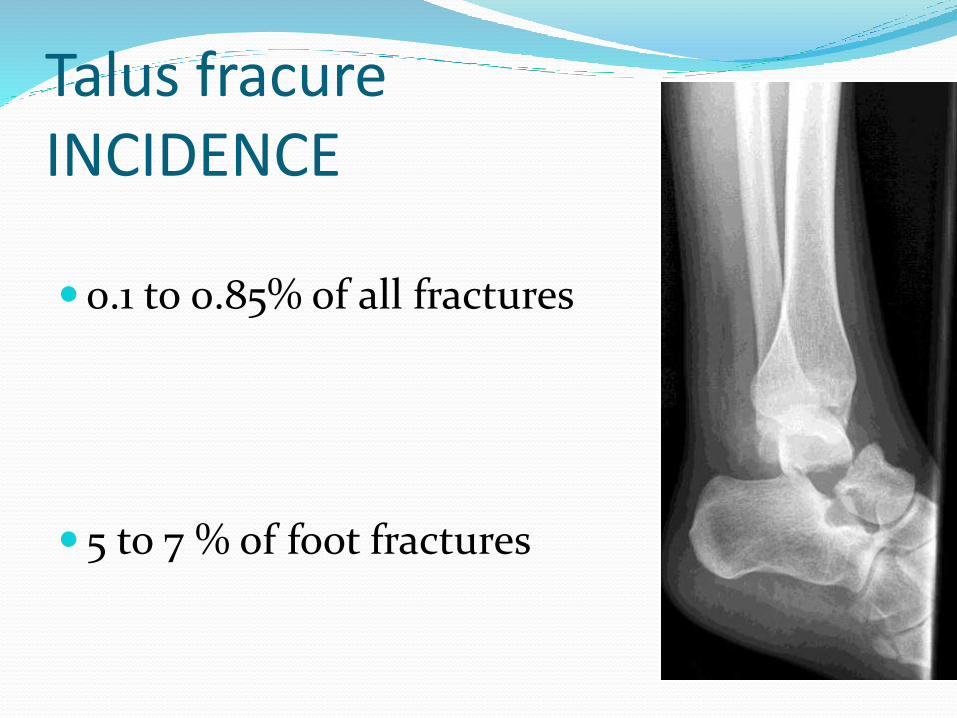
Talus fracureINCIDENCE
0.1 to 0.85% of all fractures
5 to 7 % of foot fractures
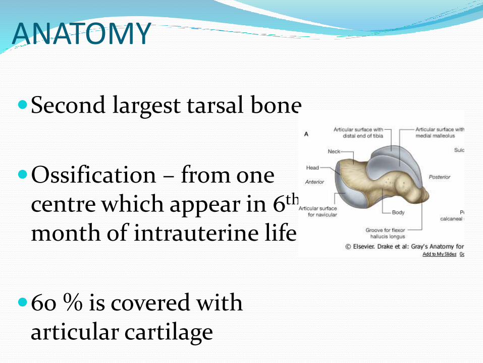
ANATOMY
Second largest tarsal bone.
Ossification – from one centre which appear in 6th
month of intrauterine life
60 % is covered with articular cartilage
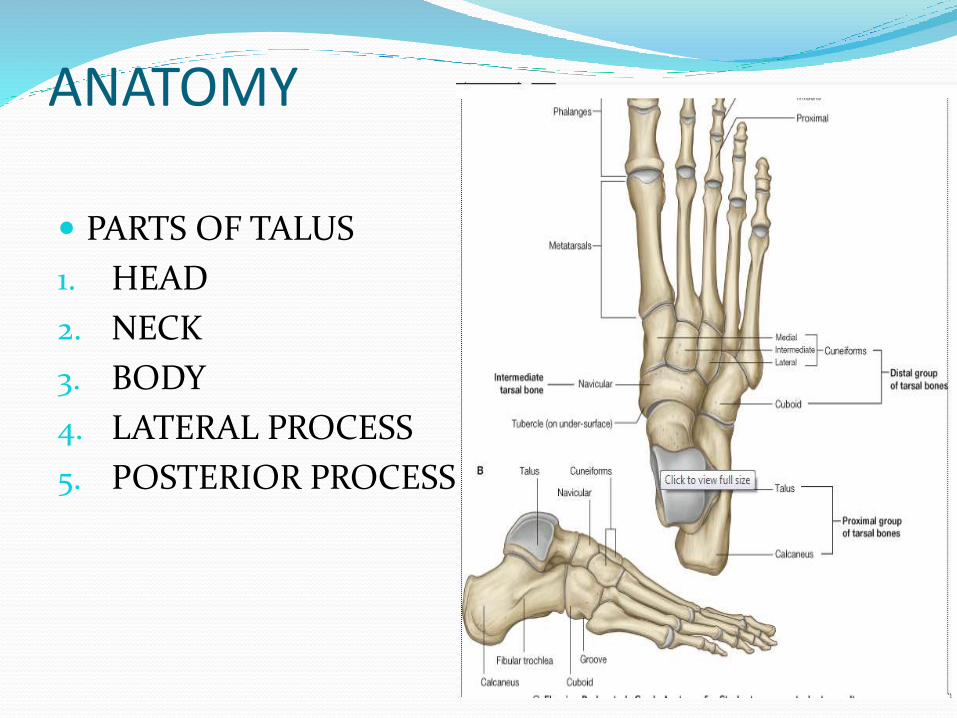
ANATOMY
PARTS OF TALUS
1. HEAD
2. NECK
3. BODY
4. LATERAL PROCESS
5. POSTERIOR PROCESS PROCESS
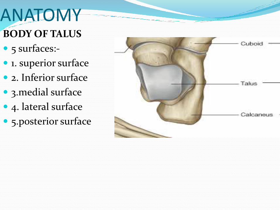
ANATOMYBODY OF TALUS
5 surfaces:-
1. superior surface
2. Inferior surface
3.medial surface
4. lateral surface
5.posterior surface
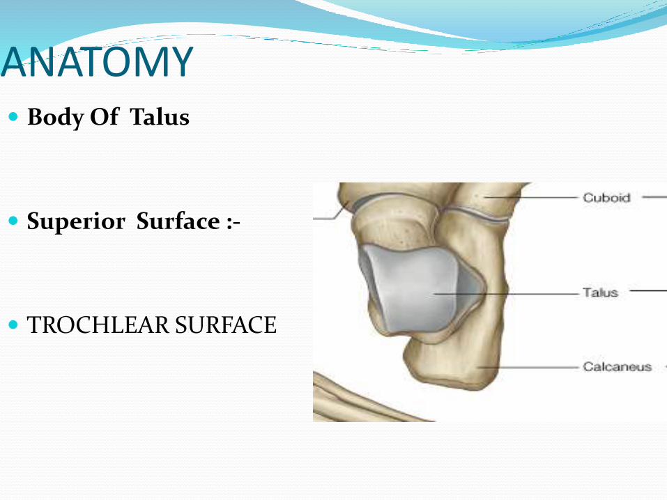
ANATOMY Body Of Talus
Superior Surface :-
TROCHLEAR SURFACE
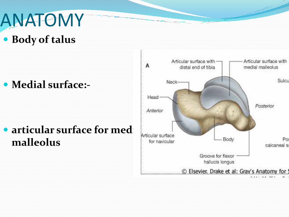
ANATOMY Body of talus
Medial surface:-
articular surface for medial malleolus
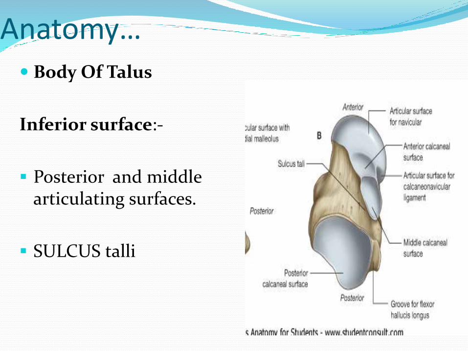
Anatomy… Body Of Talus
Inferior surface:-
Posterior and middle articulating surfaces.
SULCUS talli
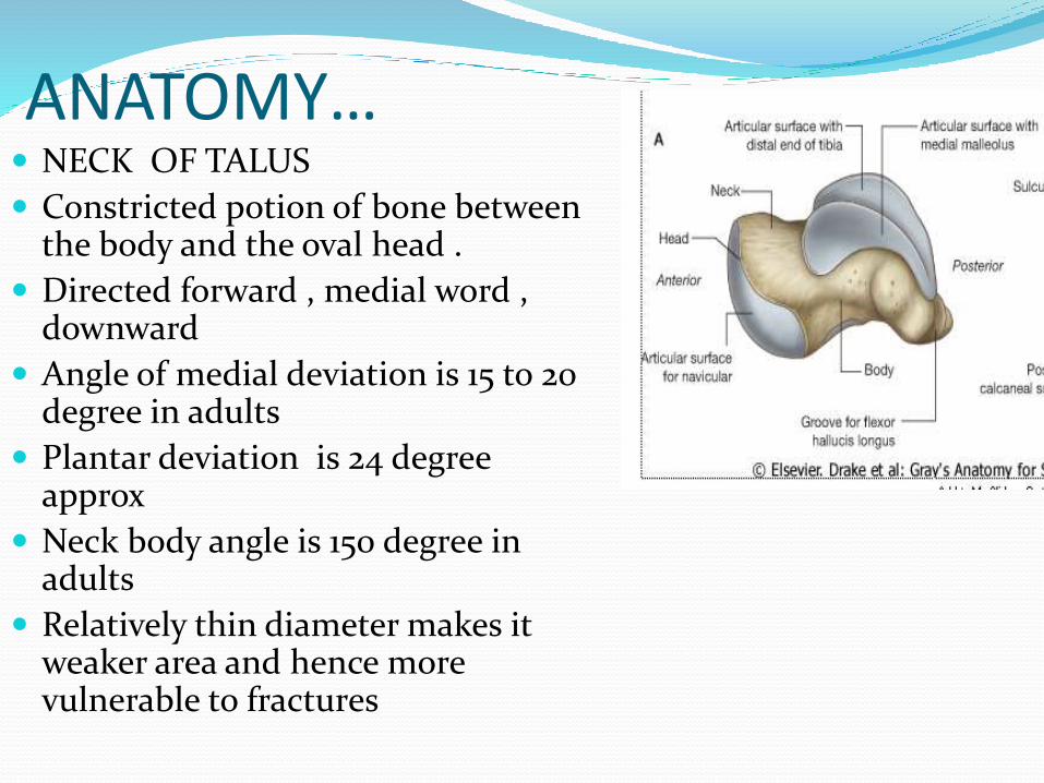
ANATOMY… NECK OF TALUS
Constricted potion of bone between the body and the oval head .
Directed forward , medial word , downward
Angle of medial deviation is 15 to 20 degree in adults
Plantar deviation is 24 degree approx
Neck body angle is 150 degree in adults
Relatively thin diameter makes it weaker area and hence more vulnerable to fractures
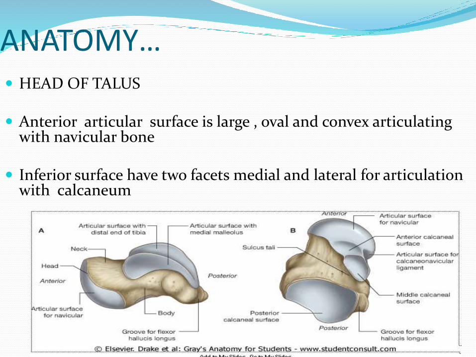
ANATOMY… HEAD OF TALUS
Anterior articular surface is large , oval and convex articulating with navicular bone
Inferior surface have two facets medial and lateral for articulation with calcaneum
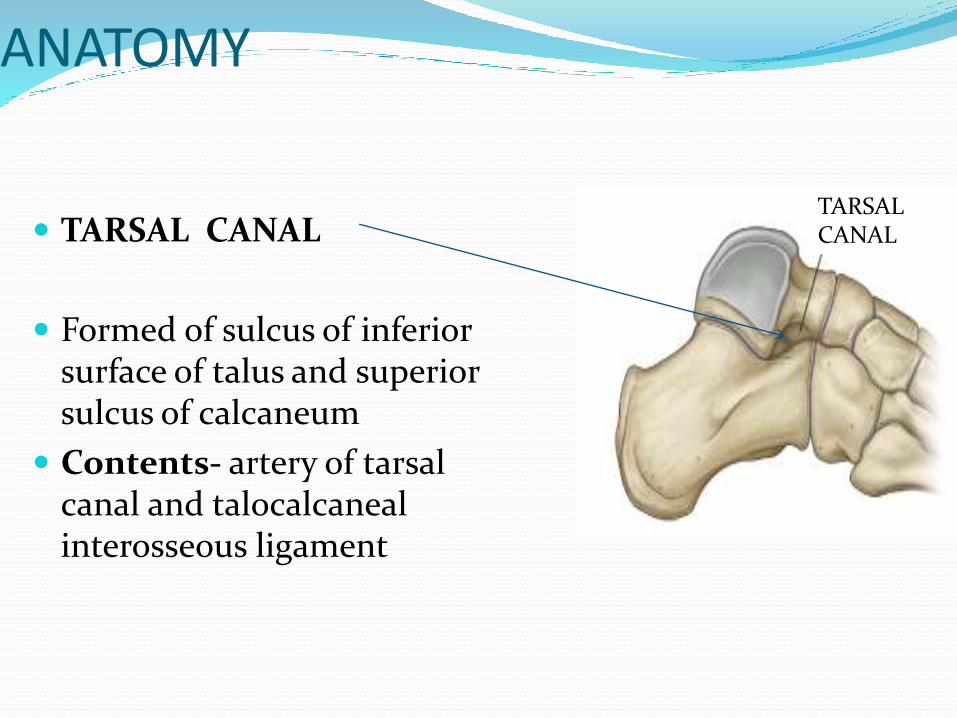
ANATOMY
TARSAL CANAL
Formed of sulcus of inferior surface of talus and superior sulcus of calcaneum
Contents- artery of tarsal canal and talocalcanealinterosseous ligament
TARSAL CANAL
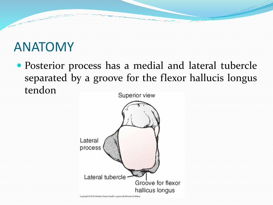
ANATOMY Posterior process has a medial and lateral tubercle
separated by a groove for the flexor hallucis longustendon
ANATOMY …
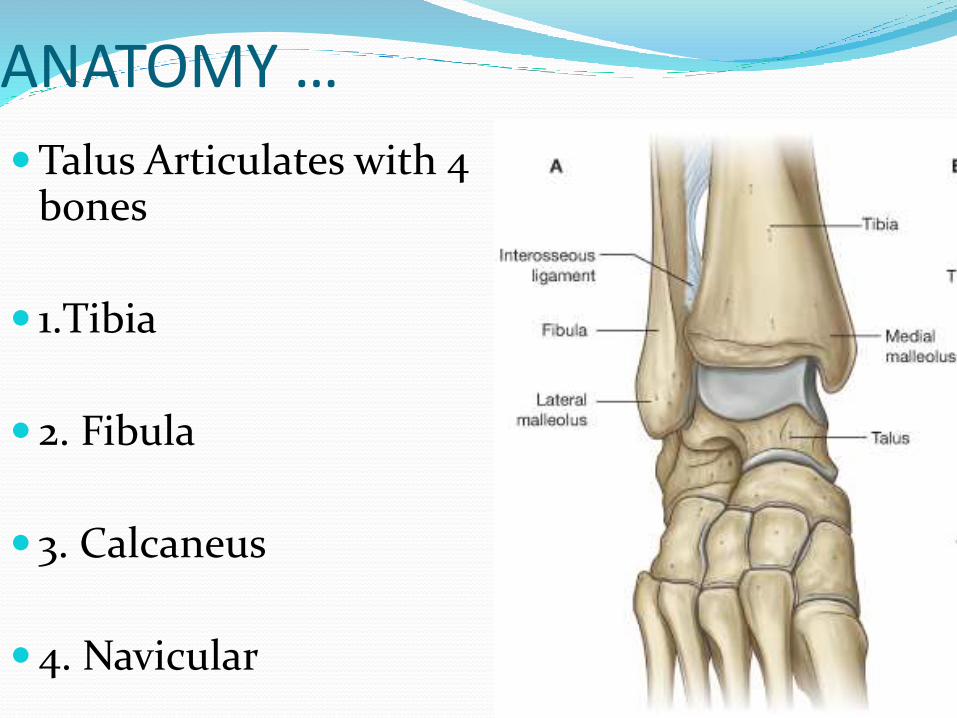
Talus Articulates with 4 bones
1.Tibia
2. Fibula
3. Calcaneus
4. Navicular
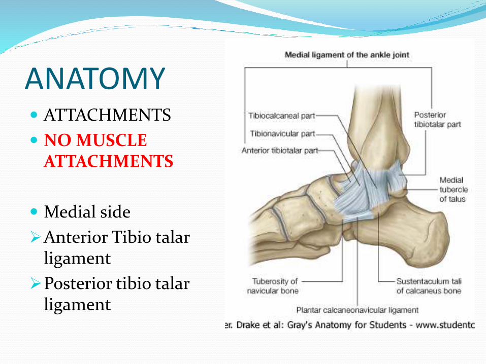
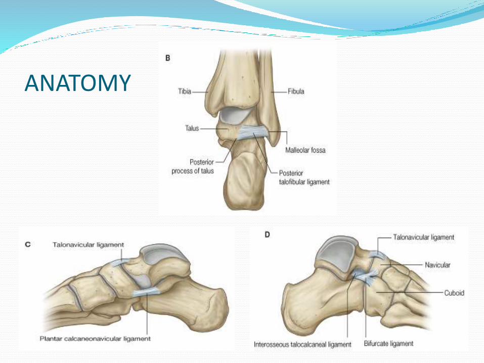
ANATOMY ATTACHMENTS
NO MUSCLE ATTACHMENTS
Medial side
Anterior Tibio talarligament
Posterior tibio talarligament
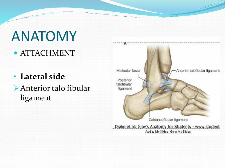
ANATOMY ATTACHMENT
• Lateral side
Anterior talo fibular ligament
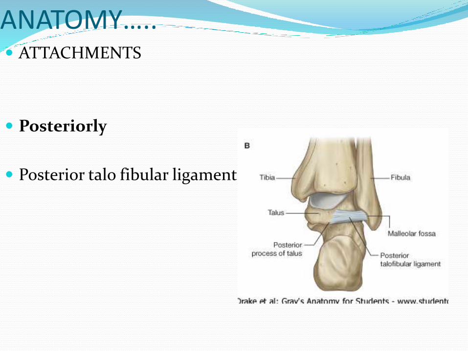
ANATOMY….. ATTACHMENTS
Posteriorly
Posterior talo fibular ligament
ANATOMY
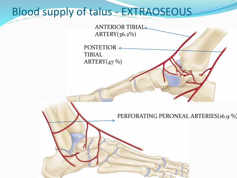
Blood supply of talus - EXTRAOSEOUSANTERIOR TIBIAL ARTERY(36.2%)
POSTETIOR TIBIAL ARTERY(47 %)
PERFORATING PERONEAL ARTERIES(16.9 %)
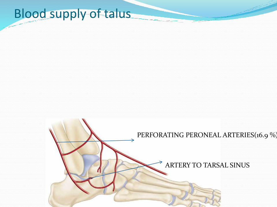
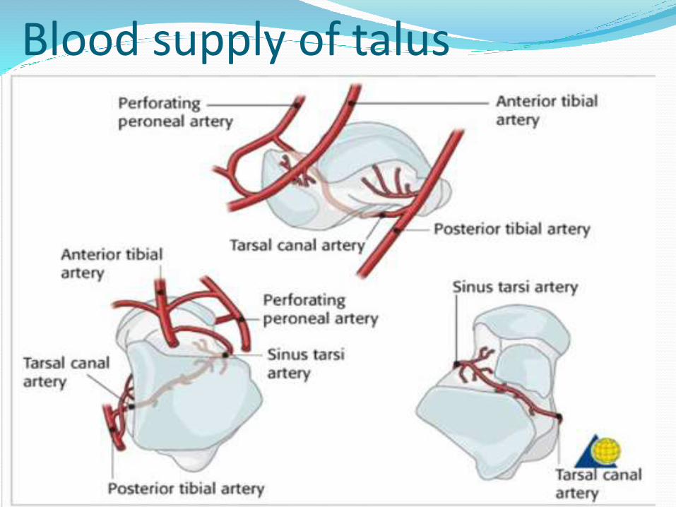
Blood supply of talus
PERFORATING PERONEAL ARTERIES(16.9 %)
ARTERY TO TARSAL SINUS
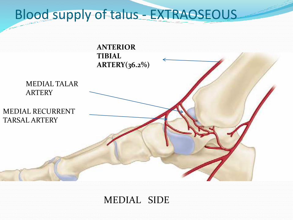
Blood supply of talus - EXTRAOSEOUS
ANTERIOR TIBIAL ARTERY(36.2%)
MEDIAL RECURRENT TARSAL ARTERY
MEDIAL TALAR ARTERY
MEDIAL SIDE
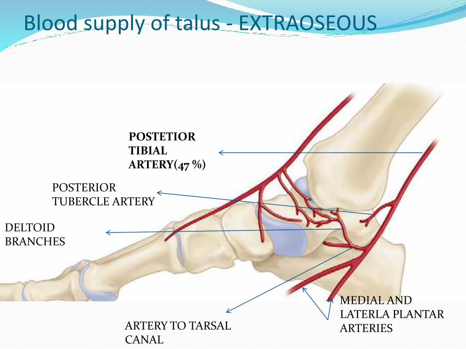
Blood supply of talus - EXTRAOSEOUS
POSTETIOR TIBIAL ARTERY(47 %)
POSTERIOR TUBERCLE ARTERY
ARTERY TO TARSAL CANAL
DELTOID BRANCHES
MEDIAL AND LATERLA PLANTAR ARTERIES
Blood supply of talus
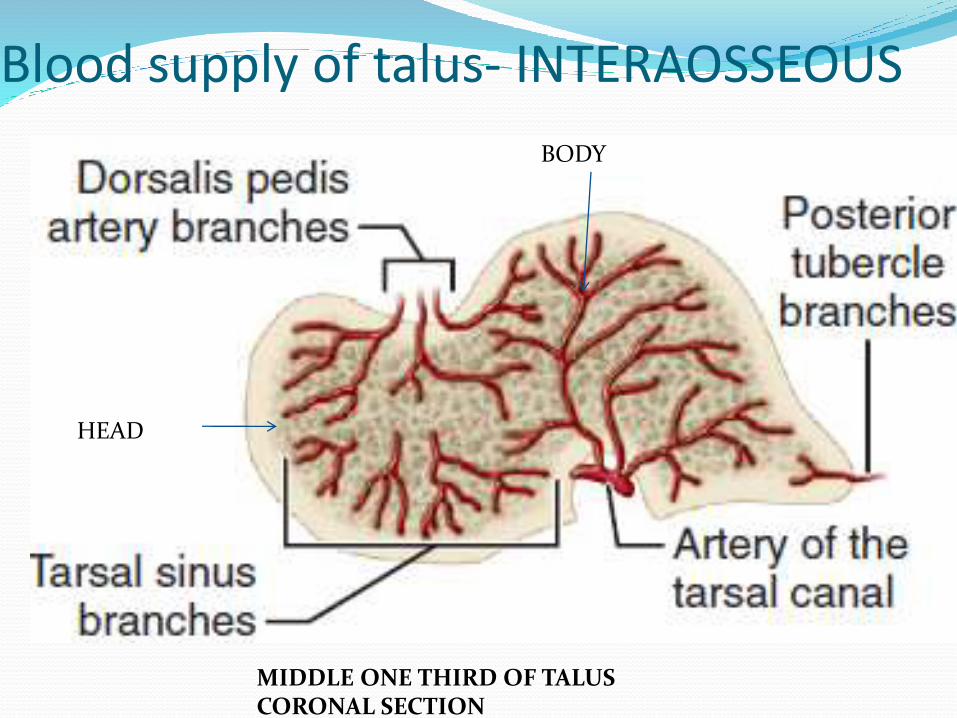
Blood supply of talus- INTERAOSSEOUS
HEAD
BODY
MIDDLE ONE THIRD OF TALUS CORONAL SECTION
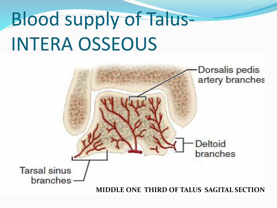
Blood supply of Talus-INTERA OSSEOUS
MIDDLE ONE THIRD OF TALUS SAGITAL SECTION
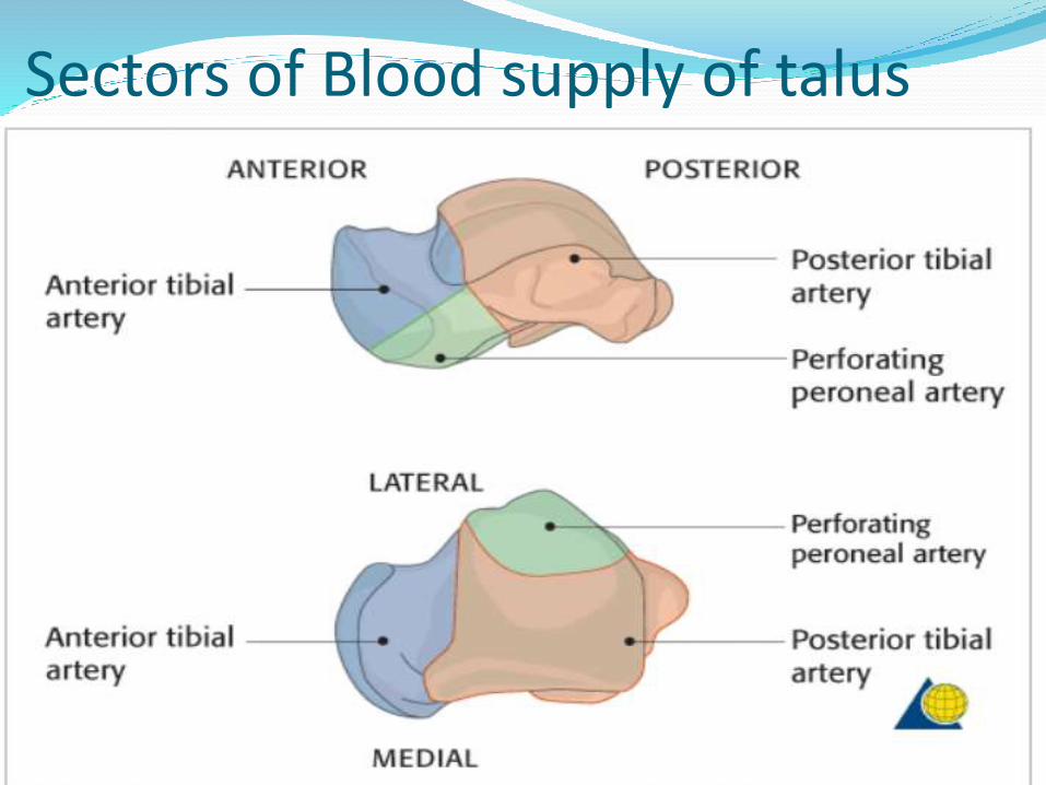
Sectors of Blood supply of talus

FRACTURE TALUSANATOMICAL CLASSIFICATION OF TALUS
FRACTURE :-
1. Talar neck fracture
2. Talar body fracture
3. Talar head fracture
4. Lateral process fracture
5. Posterior process fracture
CLINICAL PRESENTATION
Talus fractures frequently occur in a young, active, and mobile population
History of high velocity injury present
• Clinically :-
Intense pain , unable to move ankle,
Gross edema and echymosis usually present
When there is subluxation or dislocation the normal contours of ankle and hind foot are distorted
Open injury may occur if there is significant distortment

Diagnosis RADIOGRAPHIC EVALUATION
XRAYS
ANTEROPOSTERIOR VIEWS
ANKLE MORTISE VIEW
LATERAL VIEW
CANALE VIEW
RADIOGRAPHIC XRAYS
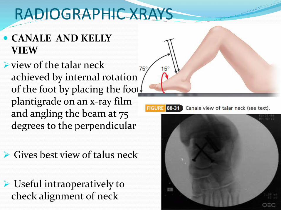
CANALE AND KELLY VIEW
view of the talar neck achieved by internal rotation of the foot by placing the foot plantigrade on an x-ray film and angling the beam at 75 degrees to the perpendicular
Gives best view of talus neck
Useful intraoperatively to check alignment of neck
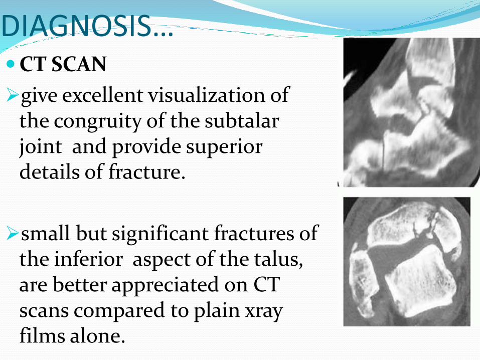
DIAGNOSIS…CT SCAN
give excellent visualization of the congruity of the subtalar joint and provide superior details of fracture.
small but significant fractures of the inferior aspect of the talus, are better appreciated on CT scans compared to plain xrayfilms alone.
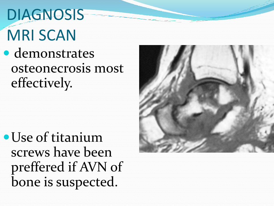
DIAGNOSISMRI SCAN demonstrates
osteonecrosis most effectively.
Use of titanium screws have been preffered if AVN of bone is suspected.
FRACTURE NECK OF TALUS
Constitue 30 % of talus fractures.
MECHANISM OF INJURY
Forced hyperdorsiflexion of the ankle and impingement of the taller neck on the distal anterior tibia .
Axial load to plantar foot causes talar neck fracture
HAWKIN CLASSIFICATION OF TALAR NECK FRACTURE
Hawkins 1970 - talar neck fractures into three type
Canale and Kelly added type IV
Based on displacement of body of talus.
Useful to perdict long term outcome and development of avn of talar body
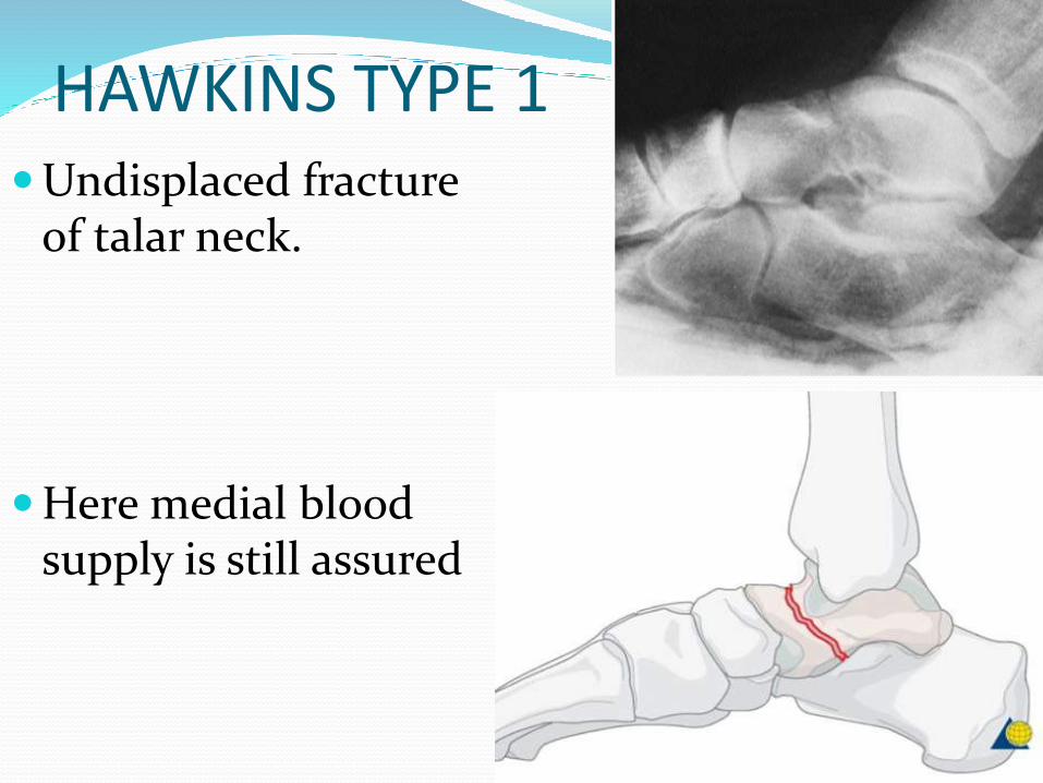
HAWKINS TYPE 1Undisplaced fracture
of talar neck.
Here medial blood supply is still assured
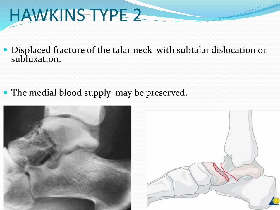
HAWKINS TYPE 2
Displaced fracture of the talar neck with subtalar dislocation or subluxation.
The medial blood supply may be preserved.
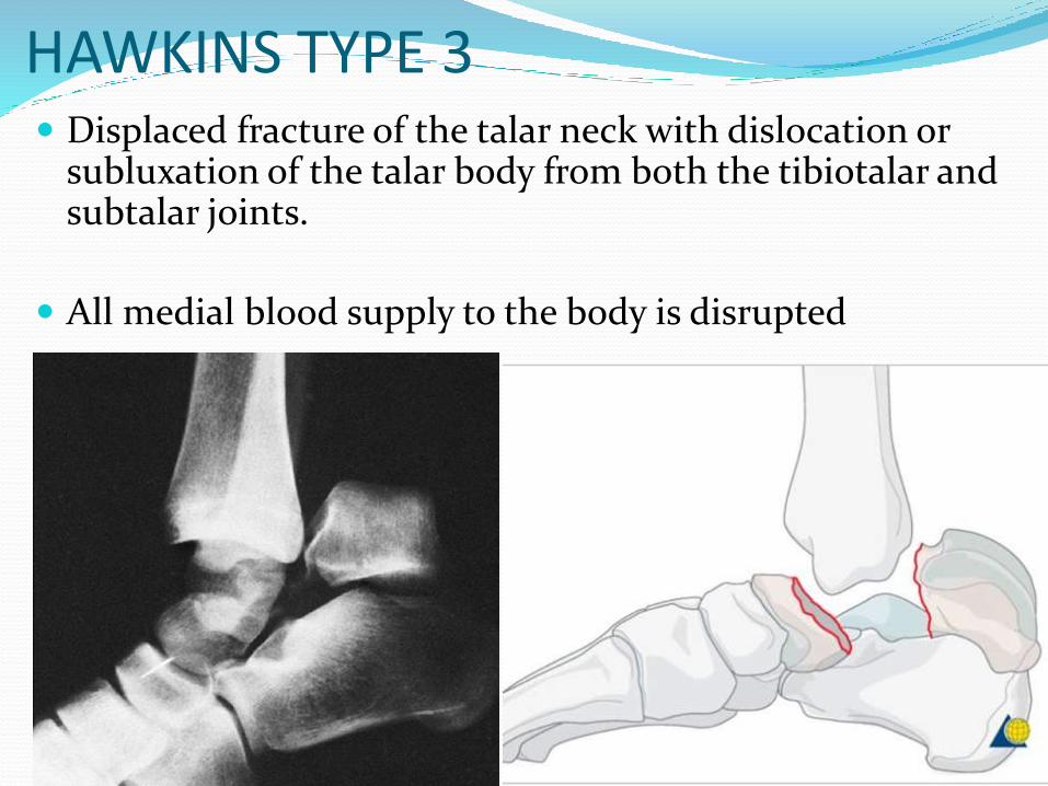
HAWKINS TYPE 3 Displaced fracture of the talar neck with dislocation or
subluxation of the talar body from both the tibiotalar and subtalar joints.
All medial blood supply to the body is disrupted
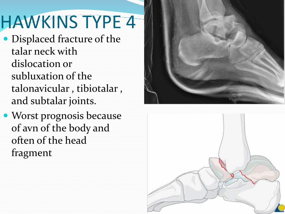
HAWKINS TYPE 4 Displaced fracture of the
talar neck with dislocation or subluxation of the talonavicular , tibiotalar , and subtalar joints.
Worst prognosis because of avn of the body and often of the head fragment

TREATMENT Goals of treatment:
1. Early anatomic reduction of the neck fracture
2. Reduction of dislocated joints
3. Stable fixation
4. Avoidance of complications
Treatment Options
Hawkins type 1 fractureNonoperative Management
Considered for fractures in which there is no displacement of the fracture line and no incongruity of the subtalar joint.
SHOULD BE CONFIRMED WITH CT SCAN IF DOUBTFULL
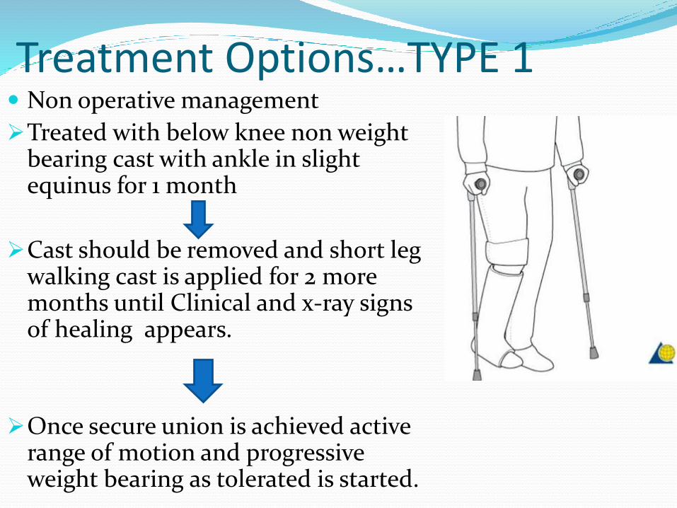
Treatment Options…TYPE 1 Non operative management
Treated with below knee non weight bearing cast with ankle in slight equinus for 1 month
Cast should be removed and short leg walking cast is applied for 2 more months until Clinical and x-ray signs of healing appears.
Once secure union is achieved active range of motion and progressive weight bearing as tolerated is started.
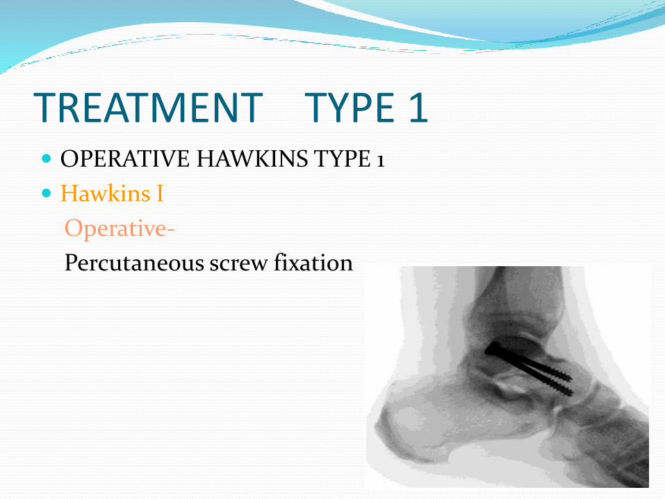
TREATMENT TYPE 1 OPERATIVE HAWKINS TYPE 1
Hawkins I
Operative-
Percutaneous screw fixation
TREATMENTHAWKINS TYPE 2
NON OPERATIVE
Achieving closed reduction is very difficult.
Should be only attempted if surgery is delayed.
TREATMENT HAWKINS TYPE 2 CLOSED REDUCTION
firstly, adequate analgesia and sedation
technique involves bringing the foot, including the talar head, to the residual talar body fragment
requires the talar body to be reduced within the ankle mortise
the knee is flexed and the foot is flexed plantar ward. This relaxes the gastrocsoleus complex and brings the talar head fragment into proper relation to the body
At that point, any varus or valgus malalignment can be corrected as well
reduction is achieved, excessive dorsiflexion will cause a redisplacementof the head fragment, and therefore radiographs to confirm reduction should be performed with the foot in a comfortable position of equinus.
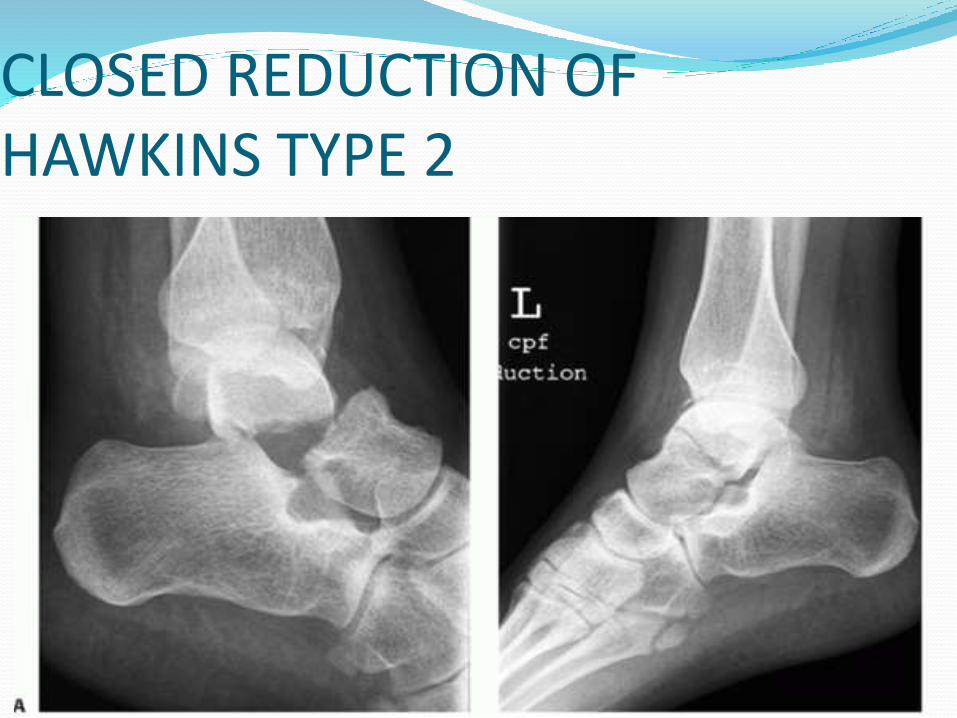
CLOSED REDUCTION OF HAWKINS TYPE 2
Treatment SURGICAL APPROACHES
Anterolateral approach
Anteromedial approach
Anteromedial approach combined with medial malleolar osteotomy
TREATMENTHAWKINS TYPE 3 AND 4
Most authors agree that group III and IV cannot be reduced and held by closed attempts
• Almost all require surgical stabilization.
Most patients require additional surgery for relief of complications resulting from the initial injury
SURGICAL OPTIONS
TYPE 3 & TYPE 4
SCREW FIXATIONANTERIOR TO POSTEROR
POSTERIOR TO ANTERIOR
• DIRECT PLATE FIXATION
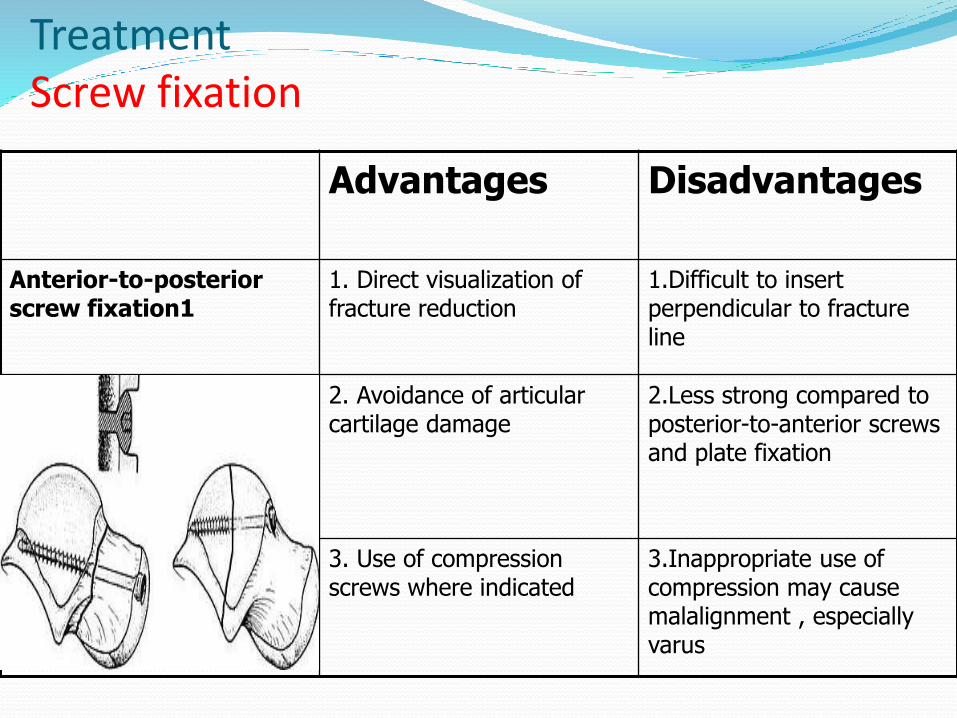
Treatment Screw fixation
Advantages Disadvantages
Anterior-to-posterior screw fixation1
1. Direct visualization of fracture reduction
1.Difficult to insert perpendicular to fracture line
2. Avoidance of articular cartilage damage
2.Less strong compared to posterior-to-anterior screws and plate fixation
3. Use of compression screws where indicated
3.Inappropriate use of compression may cause malalignment , especially varus
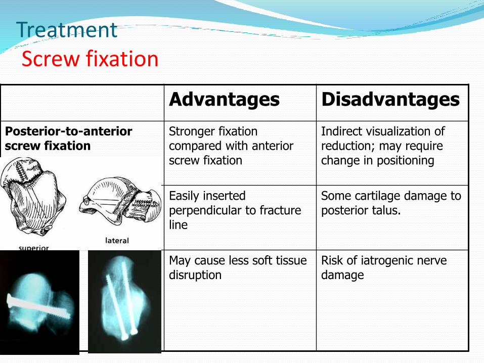
TreatmentScrew fixation
Advantages Disadvantages
Posterior-to-anterior screw fixation
Stronger fixation compared with anterior screw fixation
Indirect visualization of reduction; may require change in positioning
Easily inserted perpendicular to fracture line
Some cartilage damage to posterior talus.
May cause less soft tissue disruption
Risk of iatrogenic nerve damage
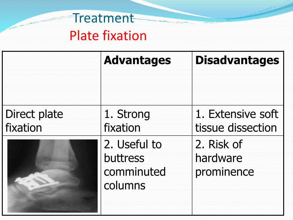
TreatmentPlate fixation
Advantages Disadvantages
Direct plate fixation
1. Strong fixation
1. Extensive soft tissue dissection
2. Useful to buttress comminuted columns
2. Risk of hardware prominence
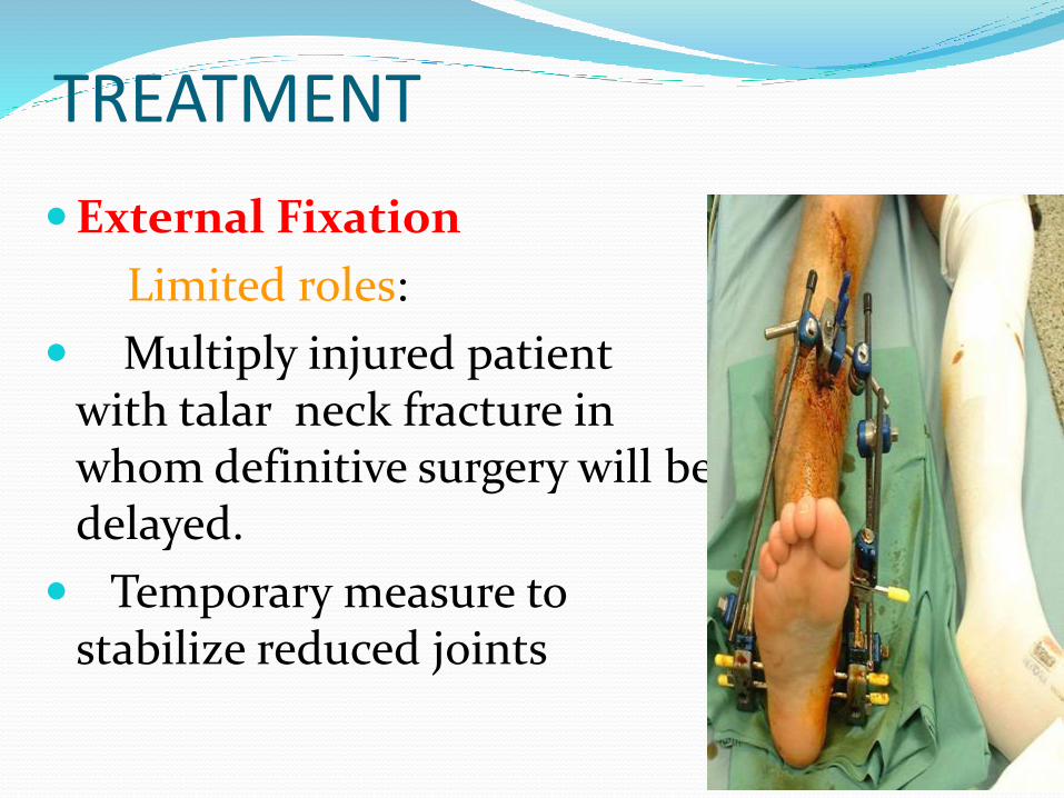
TREATMENT
External Fixation
Limited roles:
Multiply injured patient with talar neck fracture in whom definitive surgery will be delayed.
Temporary measure to stabilize reduced joints

Complications
AVN
Malunion
Nonunion
Arthritis
AVN OF TALUS
Most common complication of talar neck fracture.
Extent of involvement of talar body by osteonecrosis is directly related to degree of vascular disruption
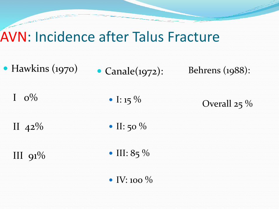
AVN: Incidence after Talus Fracture
Hawkins (1970)
I 0%
II 42%
III 91%
Canale(1972):
I: 15 %
II: 50 %
III: 85 %
IV: 100 %
Behrens (1988):
Overall 25 %
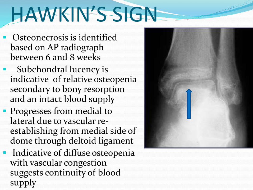
HAWKIN’S SIGN Osteonecrosis is identified
based on AP radiograph between 6 and 8 weeks
Subchondral lucency is indicative of relative osteopeniasecondary to bony resorptionand an intact blood supply
Progresses from medial to lateral due to vascular re-establishing from medial side of dome through deltoid ligament
Indicative of diffuse osteopeniawith vascular congestion suggests continuity of blood supply
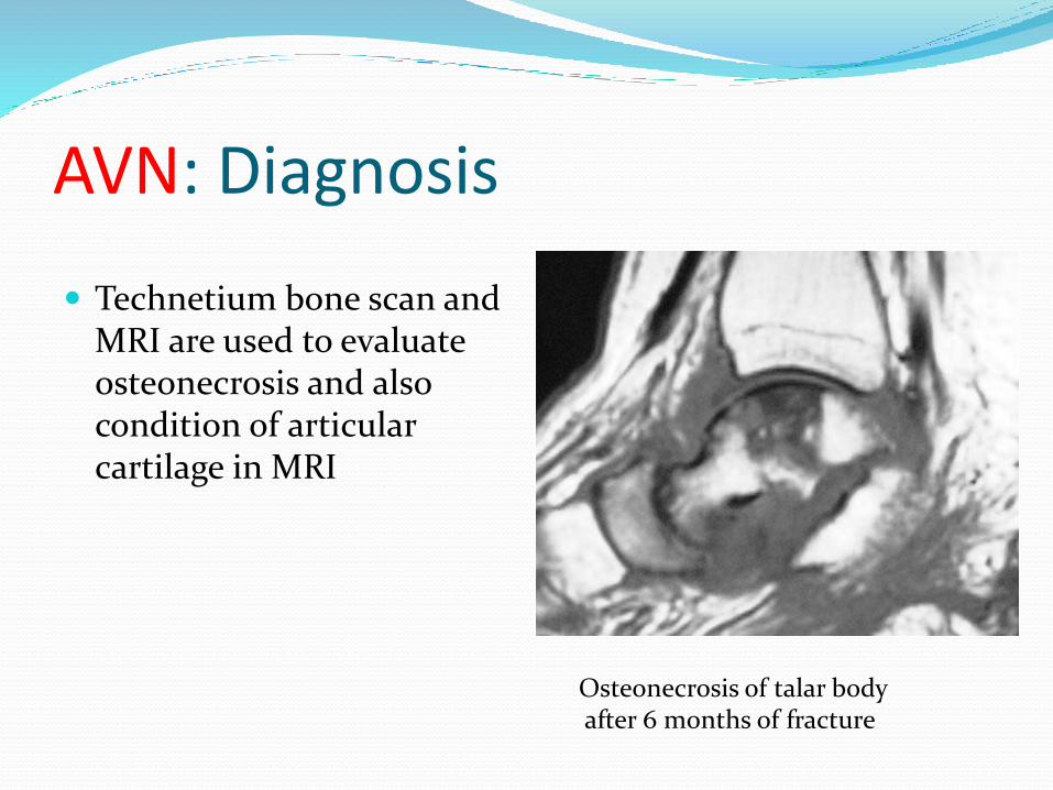
AVN: Diagnosis
Technetium bone scan and MRI are used to evaluate osteonecrosis and also condition of articularcartilage in MRI
Osteonecrosis of talar bodyafter 6 months of fracture
AVN Treatment Precollapse:
Modified Weight Bearing
Patela tendon brace cast
Compliance difficult
Efficacy unknown
Postcollapse:
Observation
Arthrodesis if symptomatic
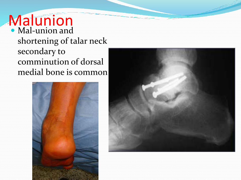
Mal-union and shortening of talar neck secondary to comminution of dorsal medial bone is common
Malunion
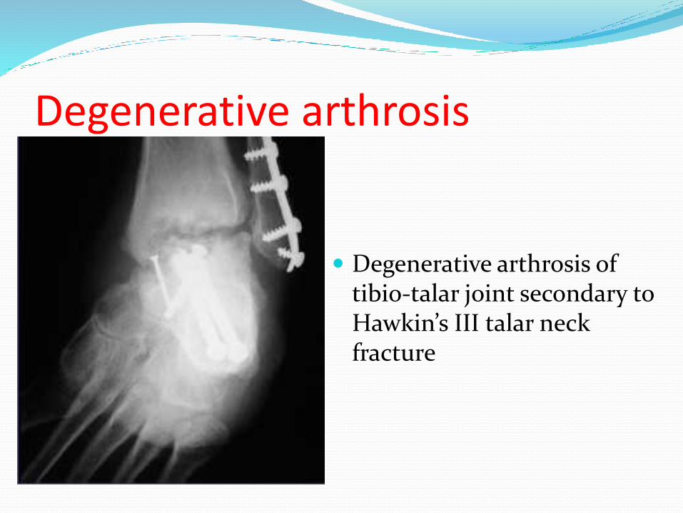
Degenerative arthrosis of tibio-talar joint secondary to Hawkin’s III talar neck fracture
Degenerative arthrosis
TALAR BODY FRACTURE
DEFINITION-fractures of the talar body are intra-articular injuries in which the articular surfaces of the tibiotalar and the subtalar joints are involved.
RADIOGRAPHIC –LATERAL XRAY VIEWS
fractures extending into or posterior to the lateral process of the talus are defined as talar body fractures, whereas fractures anterior to the lateral process are defined as talar neck fractures.
TALAR BODY FRACTURE MECHANISM OF INJURY
AXIAL COMPRESSION OF THE TALUS BETWEEN TIBIAL PLAFOND AND THE CALCANEUS
USUALLY SEEN IN MOTOR VEHICLE ACCIDENTS AND FALLS FROM HEIGHT
TALUS BODY FRACTURE
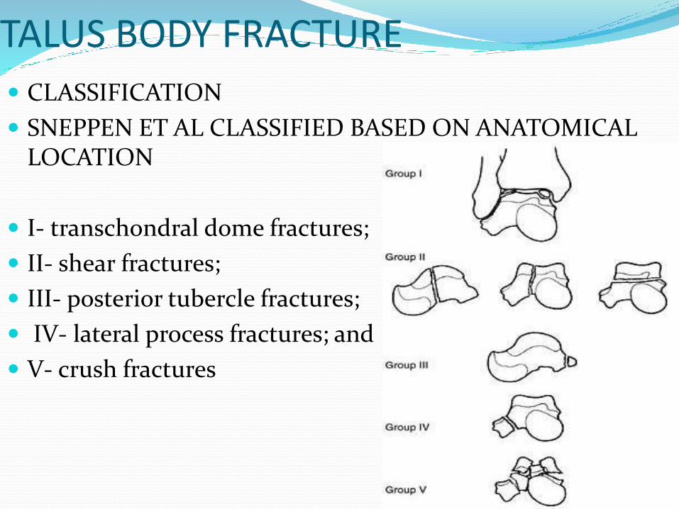
CLASSIFICATION
SNEPPEN ET AL CLASSIFIED BASED ON ANATOMICAL LOCATION
I- transchondral dome fractures;
II- shear fractures;
III- posterior tubercle fractures;
IV- lateral process fractures; and
V- crush fractures
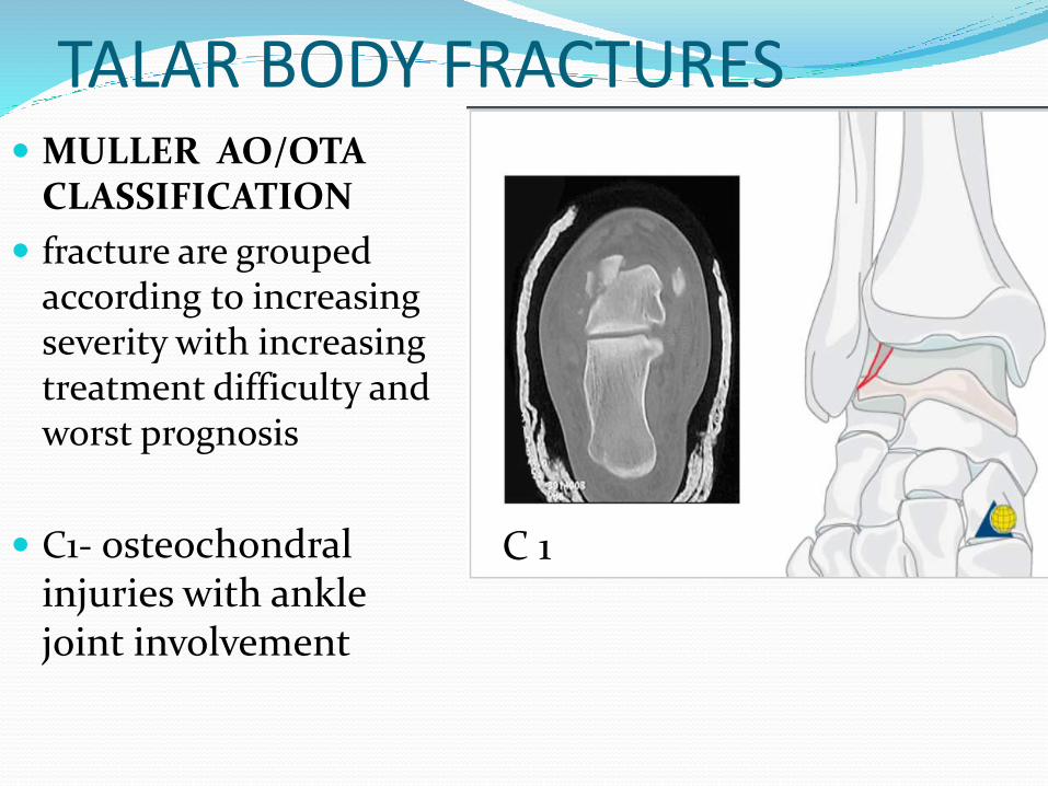
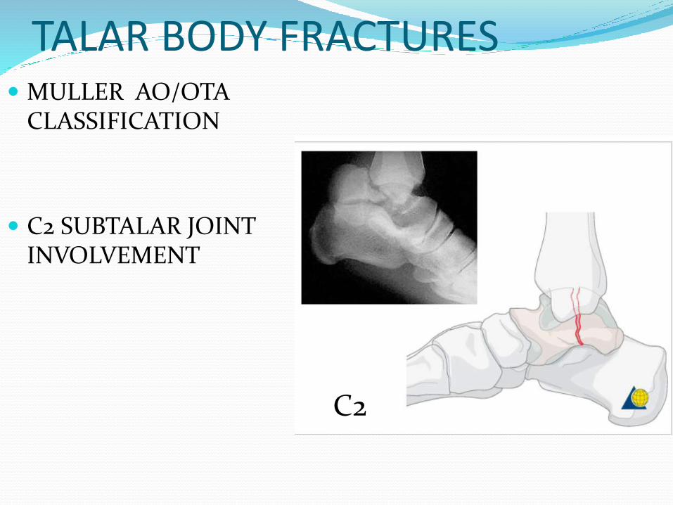
TALAR BODY FRACTURES MULLER AO/OTA
CLASSIFICATION
fracture are grouped according to increasing severity with increasing treatment difficulty and worst prognosis
C1- osteochondralinjuries with ankle joint involvement
C 1
TALAR BODY FRACTURES MULLER AO/OTA
CLASSIFICATION
C2 SUBTALAR JOINT INVOLVEMENT
C2
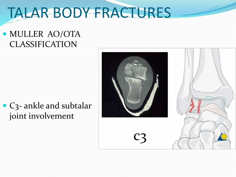
TALAR BODY FRACTURES MULLER AO/OTA
CLASSIFICATION
C3- ankle and subtalar joint involvement
c3

TALAR BODY FRACTURE TREATMENT
OPERATIVE
SURGICAL APPROACH – ANTEROMEDIAL APPROACH WITH MEDIAL MALLEOLUS OSTEOTOMY
.
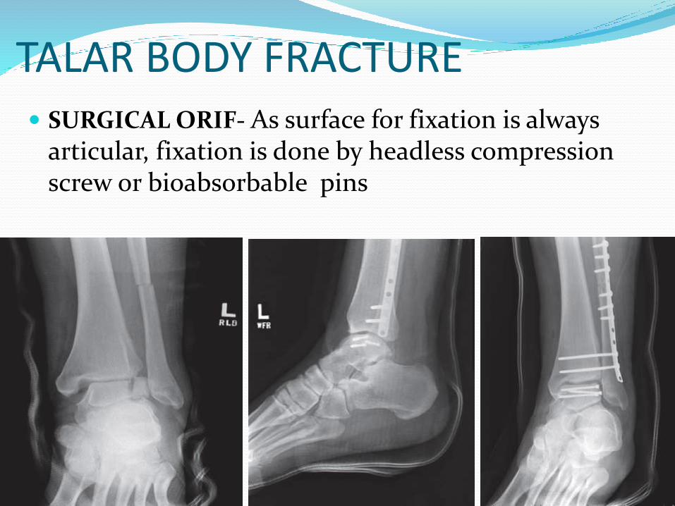
TALAR BODY FRACTURE SURGICAL ORIF- As surface for fixation is always
articular, fixation is done by headless compression screw or bioabsorbable pins
TALAR BODY FRACTURETREATMENT COMMINUTED FRACTURES OF BODY
Difficult to treat
Accurate replacement of fragments is near impossible
Long term results- bad
IN SUCH CASES TALECTOMY ALONG WITH CALCANEOTIBIAL FUSION IS PREFFERRED.
GIVES PATIENT PAINLESS AND STABLE WALKING FOOT
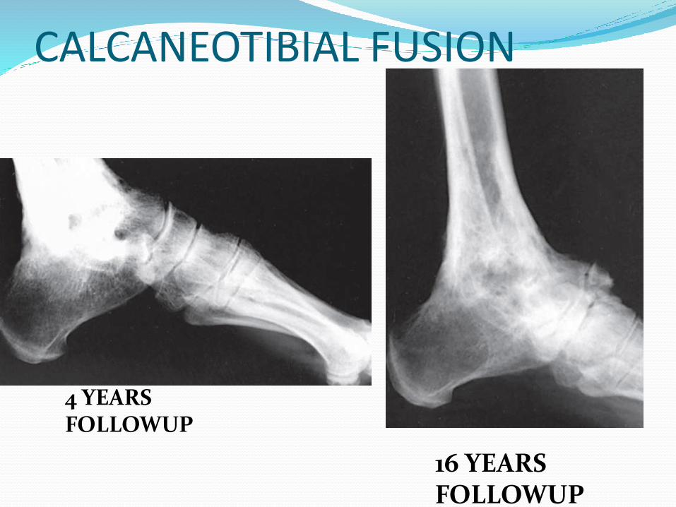
CALCANEOTIBIAL FUSION
4 YEARS FOLLOWUP
16 YEARS FOLLOWUP
TALAR BODY FRACTURE
PROBLEMS FACED WITH TALOCALCANEAL FUSION
DECREASE IN HEIGHT AND THE RIGIDITY OF ANKLE JOINT
BLAIR SUGGESTED ALTERNATIVE PROCEDURE-
TIBIOTALAR ARTHRODESIS
TALAR BODY FRACTURE
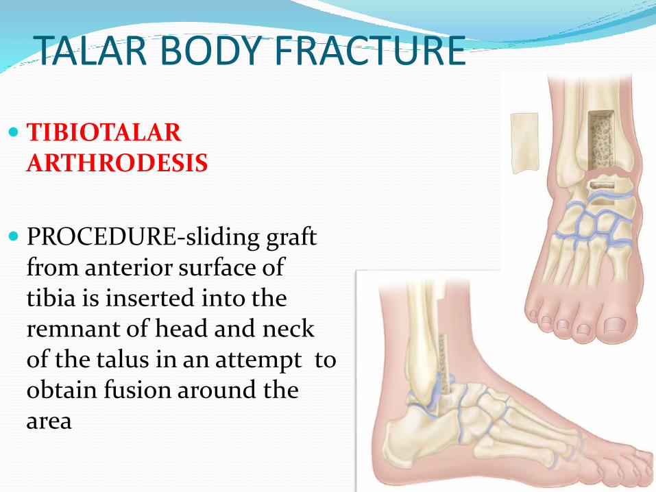
TIBIOTALAR ARTHRODESIS
PROCEDURE-sliding graft from anterior surface of tibia is inserted into the remnant of head and neck of the talus in an attempt to obtain fusion around the area
TALAR BODY FRACTURE ADVANTAGES OF TIBIO TALAR ARTHRODESIS OVER
CALACANEOTIBIAL FUSION
Position of foot is unchanged
Weight bearing thrust is placed on more or less normal undisturbed joint tissue.
No shortening
After surgery- still slight flexion and extention of the foot on leg , the two subtalar facets and talonavicular joint is possible.
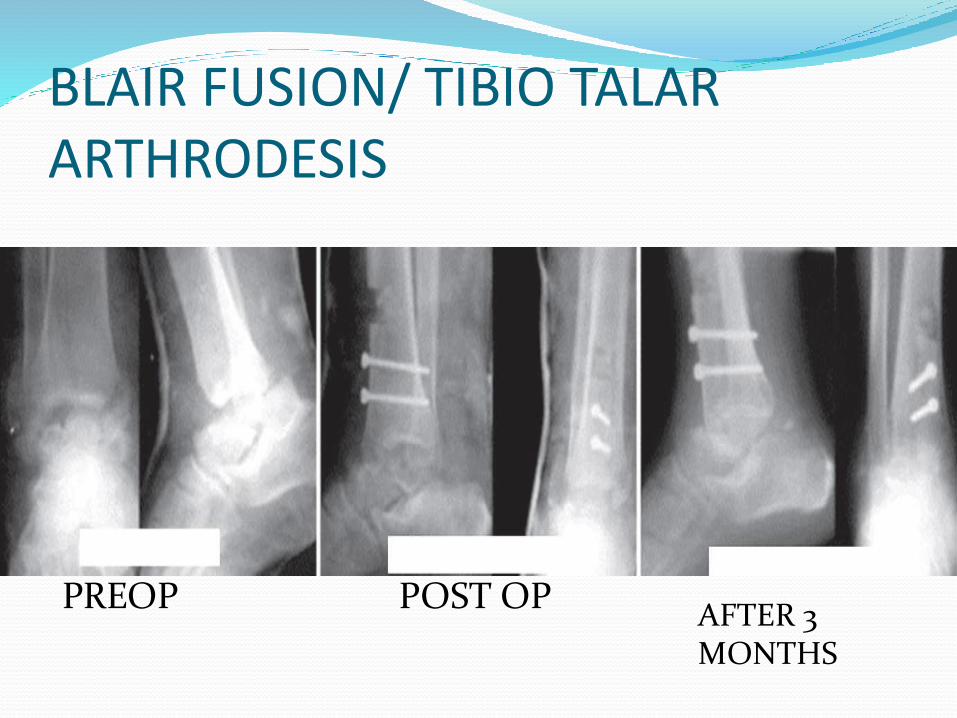
BLAIR FUSION/ TIBIO TALAR ARTHRODESIS
PREOP POST OPAFTER 3 MONTHS

Talus head fracture
Incidence- 5 to 10 % of talar injuries
Mechanism of injury-
axially directed loading and compression of talar head
Dorsal compression fracture of anterior tibialplafond
• injuries to calcaneocuboid and subtalar joint are common with these injuries
TREATMENTTALAR HEAD FRACTURE
PRINCIPLES
Maintainance of alignment of dorsomedial arch of foot.
Prevention of talonavicular joint incongruity and instability
Reduction of displaced talar head fragment
TREATMENTTALAR HEAD FRACTURE Fracture without displacement
Well molded short leg cast for 6 weeks
Weight bearing is started at 6 weeks
TREATMENTTALAR HEAD FRACTURE
Displaced fractures and those associated with joint subluxation or dislocation
ORIF
Small comminuted segments can be excised
Larger fragments are reduced with screws ranging from 2.0 to 3.5 mm
TALAR HEAD FRACTURECOMPLICATIONS AND PROGNOSIS
TALONAVICULAR ARTHRITIS IN DISPLACED FRACTURE
Conservatively managed with longitudianal arch support shoe
If conservative fails then talonavicular arthrodesis releivessymptoms
NONUNION- UNCOMMON
MALUINION - TALONAVICULAR JOINT SUBLUXATION
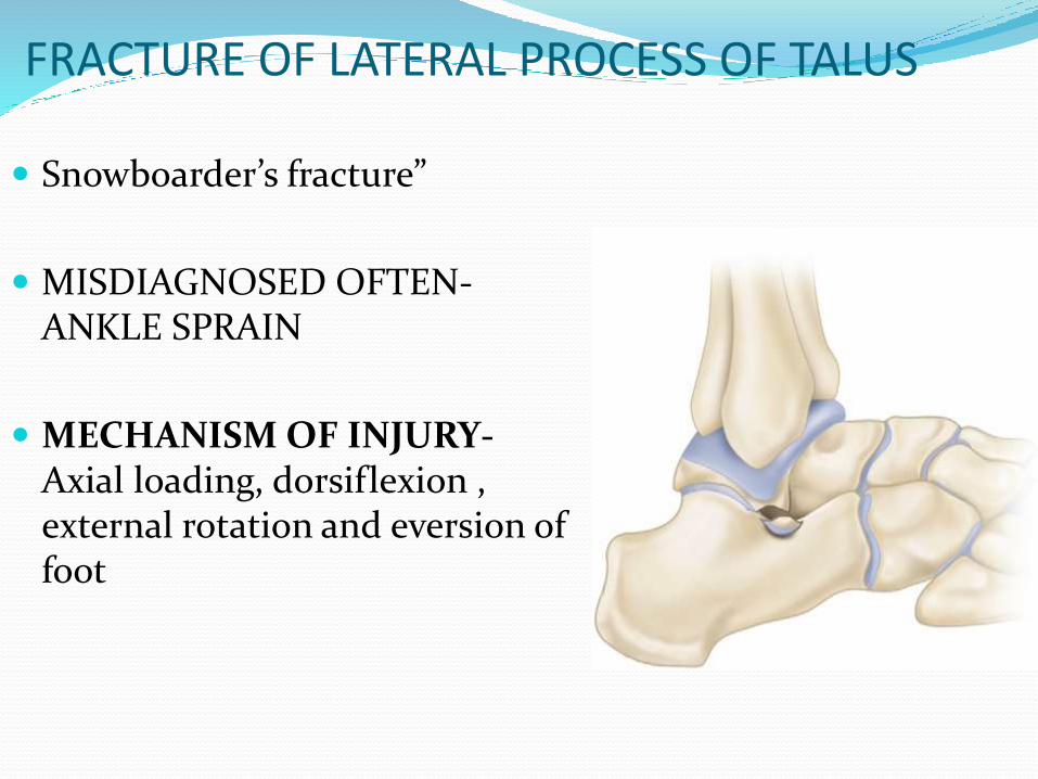
FRACTURE OF LATERAL PROCESS OF TALUS
Snowboarder’s fracture”
MISDIAGNOSED OFTEN-ANKLE SPRAIN
MECHANISM OF INJURY-Axial loading, dorsiflexion , external rotation and eversion of foot
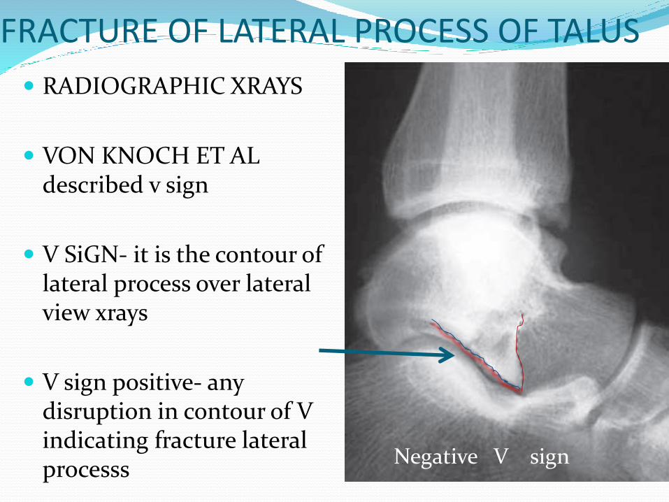
FRACTURE OF LATERAL PROCESS OF TALUS
RADIOGRAPHIC XRAYS
VON KNOCH ET AL described v sign
V SiGN- it is the contour of lateral process over lateral view xrays
V sign positive- any disruption in contour of V indicating fracture lateral processs
Negative V sign
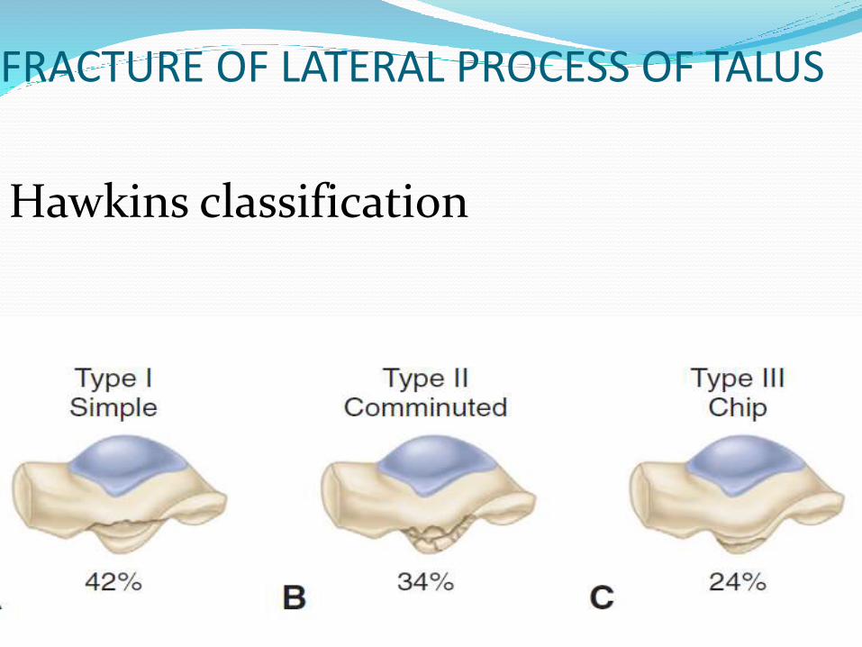
FRACTURE OF LATERAL PROCESS OF TALUS
Hawkins classification
FRACTURE OF LATERAL PROCESS OF TALUS - treatment Type I fractures can be treated in a non weight-bearing
cast for 6 weeks, unless they are displaced or involve a significant portion of the talar side of the posterior facet, in which case they should be treated by ORIF.
Type II fractures benefit from débridement of fraturefragments
Type III fractures- treated conservatively with cast application
If non union occurs the debridement of fragments is advised
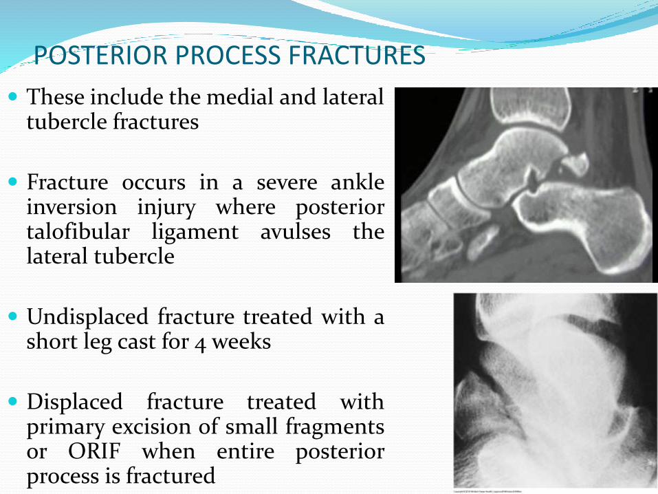
POSTERIOR PROCESS FRACTURES
These include the medial and lateraltubercle fractures
Fracture occurs in a severe ankleinversion injury where posteriortalofibular ligament avulses thelateral tubercle
Undisplaced fracture treated with ashort leg cast for 4 weeks
Displaced fracture treated withprimary excision of small fragmentsor ORIF when entire posteriorprocess is fractured
REFERENCES Rockwood and Green’s Fractures in Adults; 7th
Edition; Volume 2
Watson – Jones fractures and Joint injuries, 7th
Edition
Campbell’s Operative orthopaedics; 11th Edition;Volume 4
Grays Anatomy for students

THANK YOU!!!