case report talar cyst - jscimed central · • talus. case report. talar cyst. adam g. miller*...
TRANSCRIPT
CentralBringing Excellence in Open Access
JSM Foot and Ankle
Cite this article: Miller AG (2016) Talar Cyst. JSM Foot Ankle 1(2): 1009.
*Corresponding authorAdam G. Miller, Beacon Orthopaedics and Sports Medicine, 500 E Business Way, Cincinnati, OH 45241, USA, Tel: 513-354-3700; Fax: 513-354-3705; Email:
Submitted: 14 March 2016
Accepted: 20 August 2016
Published: 10 September 2016
Copyright© 2016 Miller
OPEN ACCESS
Keywords•Talar cyst•Geode•Case report•Bone graft•Aneurysmal bone cyst•Talus
Case Report
Talar CystAdam G. Miller*Beacon Orthopaedics and Sports Medicine, 500 East Business Way, USA
Abstract
Large talar cysts are typically associated with trauma and communicate with an osteochondral lesion. Aneurysmal bone cyst is a rare etiology of a large talar cyst. While there are many graft and surgical options, treatment consists of curettage of the talar lesion and grafting the associated talar cyst. This report presents a case of large talar body cyst without significant disruption of the talar weight bearing surface including: diagnosis, treatment and outcome.
INTRODUCTIONLarge talar cysts typically accompany a large osteochondral
lesion (OCD) resulting in pain and dysfunction. This results from remote trauma to the ankle. OCD can initially be treated with immobilization in CAM walker boot or cast; this has shown efficacy with pain and function improvement 45% of the time [1]. Lesions that are large can be seen on plain radiograph; however, understanding the true architecture and size of the lesion requires evaluation with computerized axial tomography (CT) or magnetic resonance imaging (MRI). Talar lesions that are small and minimally symptomatic have been found to remain relatively stable over time while larger and more painful lesions progress [2]. For symptomatic osteochondral lesions of the talus, surgery often is attempted.
Most literature discusses treatment of the surface lesion and its associated defect in the talar body. While much has been made of osteochondral lesions, less has been written on large subchondral and talar body cysts with relatively intact talar domes. Small defects have been treated with retrograde drilling with positive results [3]. Large lesions may not arise from trauma and necessitate a tumor evaluation. One such benign tumor is an aneurysmal bone cyst (ABC). There are few case reports of ABC in the talus and their treatment [4-6]. This case report describes one patient with a large talar cyst with minimal talar dome involvement.
CASE REPORTThe patient is a 33-year-old 9-weeks pregnant female who
presented with right ankle pain. She could not recall a specific injury. She noticed this initially 6 months ago, and since then she has had vague pain progressive in nature. Over the last few months prior to evaluation the pain became more continuous. This pain came on with axial impact or plantarflexion of the ankle. She had limited her activities. She was initially seen by another physician and a large cystic lesion was immediately noticed in her talus on radiograph. An MRI was ordered and the patient’s care was transferred to my clinic.
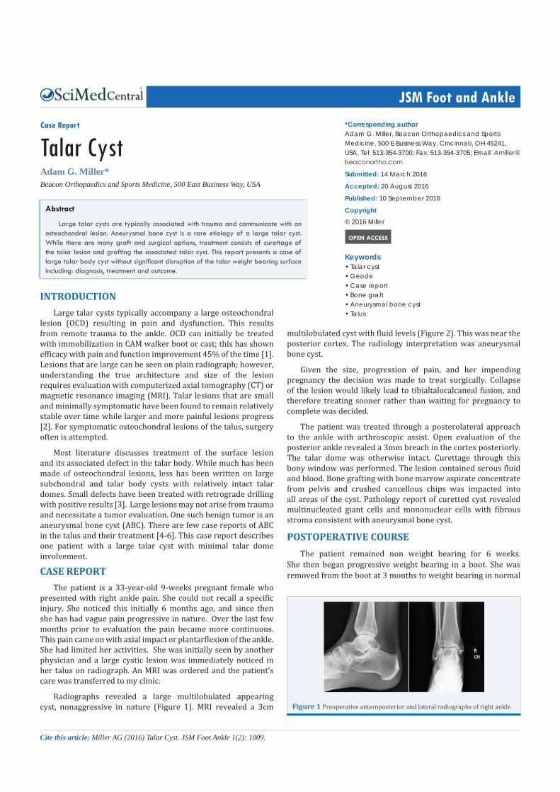
Radiographs revealed a large multilobulated appearing cyst, nonaggressive in nature (Figure 1). MRI revealed a 3cm
multilobulated cyst with fluid levels (Figure 2). This was near the posterior cortex. The radiology interpretation was aneurysmal bone cyst.
Given the size, progression of pain, and her impending pregnancy the decision was made to treat surgically. Collapse of the lesion would likely lead to tibialtalocalcaneal fusion, and therefore treating sooner rather than waiting for pregnancy to complete was decided.
The patient was treated through a posterolateral approach to the ankle with arthroscopic assist. Open evaluation of the posterior ankle revealed a 3mm breach in the cortex posteriorly. The talar dome was otherwise intact. Curettage through this bony window was performed. The lesion contained serous fluid and blood. Bone grafting with bone marrow aspirate concentrate from pelvis and crushed cancellous chips was impacted into all areas of the cyst. Pathology report of curetted cyst revealed multinucleated giant cells and mononuclear cells with fibrous stroma consistent with aneurysmal bone cyst.
POSTOPERATIVE COURSEThe patient remained non weight bearing for 6 weeks.
She then began progressive weight bearing in a boot. She was removed from the boot at 3 months to weight bearing in normal
Figure 1 Preoperative anteroposterior and lateral radiographs of right ankle.
CentralBringing Excellence in Open Access
Miller (2016)Email:
JSM Foot Ankle 1(2): 1009 (2016) 2/2
shoes. Her pregnancy was uncomplicated. At one year she was allowed to commence impact activities and running (Figure 3). She continues to have no pain at this time point.
DISCUSSIONLarge talar cystic lesions pose a significant problem.
Treatment of long-bone ABC has been curettage and bone graft. Large talar lesions propose unique challenges including access to the cyst and potential for collapse. This case presented the best access from the posterior ankle. Upon exploration, a defect already existed in the region of the steida process. In more aggressive giant cell tumors, fusion has been performed with success [7]. A primary fusion would lead to significant morbidity in a young active female. As a primary surgery, an initial attempt at salvage with bone graft is warranted considering the secondary options.
A large ABC of the talus is a rare lesion [8]. This case reports represents one such lesion. The differential for such a lesion consists of a simple bone cyst, osteochondral lesion creating geode in the talus, aneurysmal bone cyst, and giant cell tumor. If there is clinical concern, fine needle aspiration can be used for pathologic diagnosis prior to surgery. The less destructive appearance, fluid
levels on MRI, and pathology with multinucleated giant cells combine to provide the diagnosis. Pathology of an ABC can be similar to a giant cell tumor; however, the lesion tends to be more destructive [9].
The most recognized etiology of an ABC is local vascular disruptions [5]. One previous case report has suggested that this type of lesion may be trauma related [6]. While impossible to confirm, our case may be represent such a lesion. One single sagittal cut of the MRI revealed possible breach of the talar dome cortex near the posterior process. Recognizing this could be an area of OCD from posterior impingement and local trauma with resultant bleeding into the cyst. The true etiology of these lesions is unknown.
With the relative success reported of curettage and bone grafting of these lesions, there has been a wide array of postoperative protocols. Twelve weeks non weight bearing has been recommended [5]. Our case report was able to return to progressive weight bearing in a CAM walker boot at 6 weeks with full weight bearing in a boot at 9 weeks. Patient’s pain and symptoms should guide progression.
CONCLUSIONLarge talar cysts are typically associated with trauma and
communicate with an osteochondral lesion. Aneurysmal bone cyst is a rare etiology of large talar cyst. Surgical treatment should attempt curettage and bone graft with particular attention to limiting trauma and destruction with surgical approach. This has shown success in limited case reports.
REFERENCES1. Tol JL, Struijs PA, Bossuyt PM, Verhagen RA, van Dijk CN. Treatment
strategies in osteochondral defects of the talar dome: a systematic review. Foot Ankle Int. 2000; 21: 119-126.
2. Klammer G, Maquieira GJ, Spahn S, Vigfusson V, Zanetti M, Espinosa N. Natural history of nonoperatively treated osteochondral lesions of the talus. Foot Ankle Int. 2015; 36: 24-31.
3. Zengerink M, Struijs PA, Tol JL, van Dijk CN. Treatment of osteochondral lesions of the talus: a systematic review. Knee Surg Sports Traumatol Arthrosc. 2010; 18: 238-246.
4. El-Moatasem el HM, Abdel-Rahman M, Eid MA. Extended curettage and adjuvant therapy for benign tumors of the talus. Foot (Edinb). 2015; 25: 79-83.
5. Sharma S, Gupta P, Sharma S, Singh M, Singh D. Primary aneurysmal bone cyst of talus. J Res Med Sci. 2012; 17:1192-1194.
6. Soreff J. Aneurysmal bone cyst of the talus. Acta Orthop Scand. 1976; 47: 358-360.
7. Dhillon MS, Singh B, Gill SS, Walker R, Nagi ON. Management of giant cell tumor of the tarsal bones: a report of nine cases and a review of the literature. Foot Ankle. 1993; 14: 265-272.
8. Pollandt K, Werner M, Delling G. Tumors of the footbones- a report from the Hamburg Bone Tumor Registry. Z Orthop Ihre Grenzgeb. 2003; 141: 445-451.
9. Sharma S, Wani IH, Gupta N, Mahajan N, Salaria AQ. Giant cell tumor of talus: a case report. Cases J. 2009; 2: 74.
Miller AG (2016) Talar Cyst. JSM Foot Ankle 1(2): 1009.
Cite this article
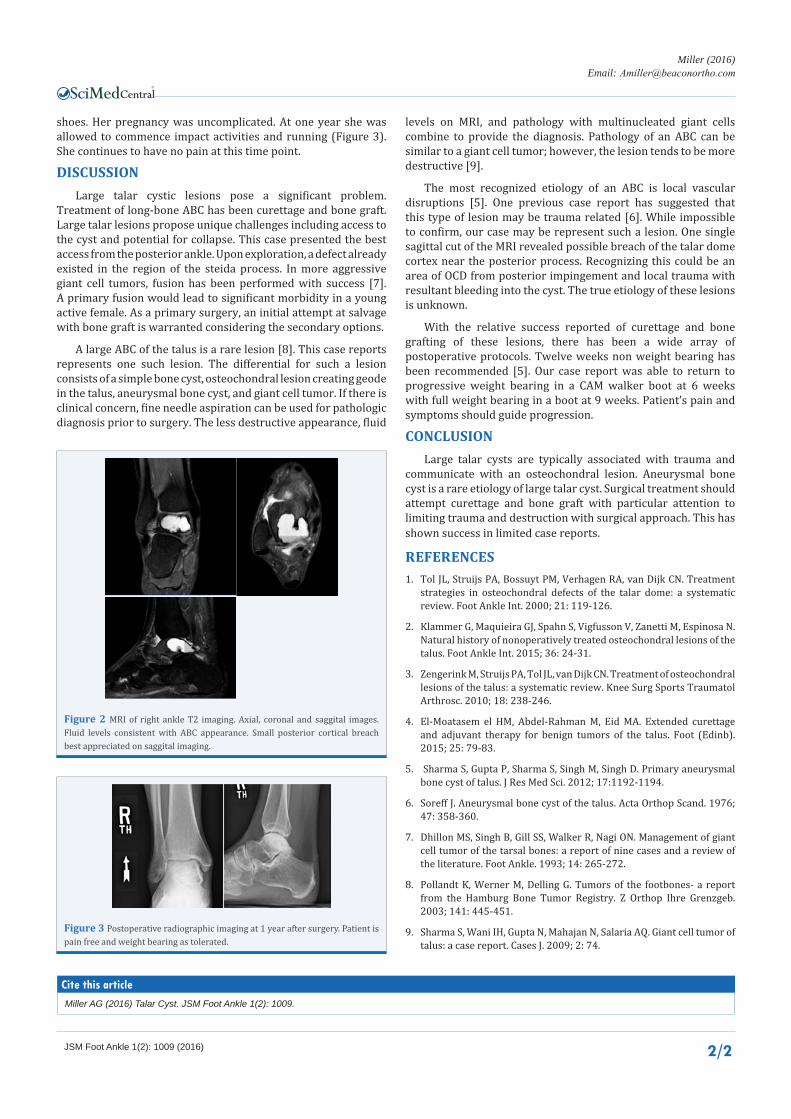
Figure 2 MRI of right ankle T2 imaging. Axial, coronal and saggital images. Fluid levels consistent with ABC appearance. Small posterior cortical breach best appreciated on saggital imaging.
Figure 3 Postoperative radiographic imaging at 1 year after surgery. Patient is pain free and weight bearing as tolerated.