swiss-pdbviewer introduction-wenwen wang
TRANSCRIPT


SPDBV
PDB
Tools
Selection
Labeling
Display
Measuring
Mutation
Torsions
Coloring
Protein & Component
Structural Alignment
Homology Modeling

1. Download and Install
• Downloading the PDB files in your own PC
PDB home page http://www.rcsb.org/pdb/home/home.do
• Downloading Swiss-PdbViewer
http://spdbv.vital-it.ch/download.html

Protein structure database: Protein data bank (PDB)
• The Protein Data Bank (PDB) is a repository for the 3-D structural data of large biological molecules, such as proteins and nucleic acids. The data, typically obtained by X-ray crystallography or NMR spectroscopy
• The PDB is a key resource in areas of structural biology.
http://www.pdb.org/pdb/home/home.do

Get your target protein in PDB
Example : thymidine kinase (TK)
1
2
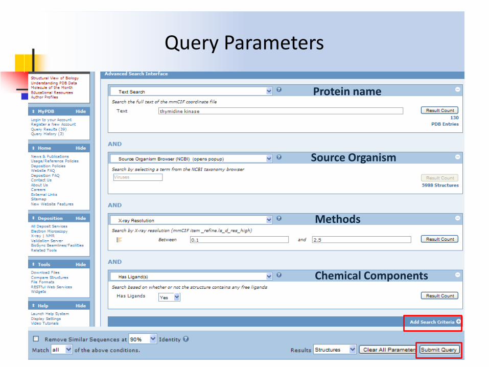
Query Parameters
Protein name
Source Organism
Methods
Chemical Components
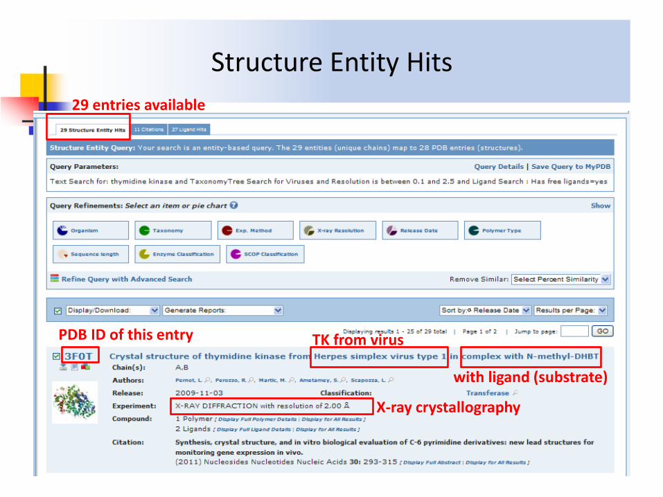
Structure Entity Hits
29 entries available
PDB ID of this entry TK from virus
with ligand (substrate)
X-ray crystallography
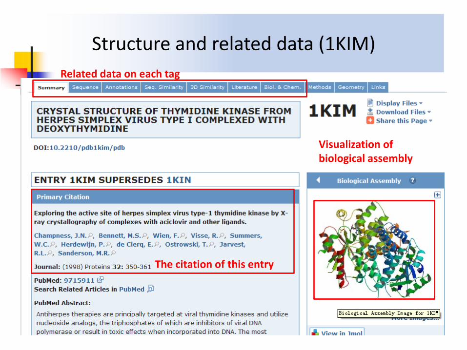
Structure and related data (1KIM)
Related data on each tag
The citation of this entry
Visualization of biological assembly

Ligand component (1KIM)

Sequence data (1KIM)
Sequence ID of 1KIM in UniProtKB
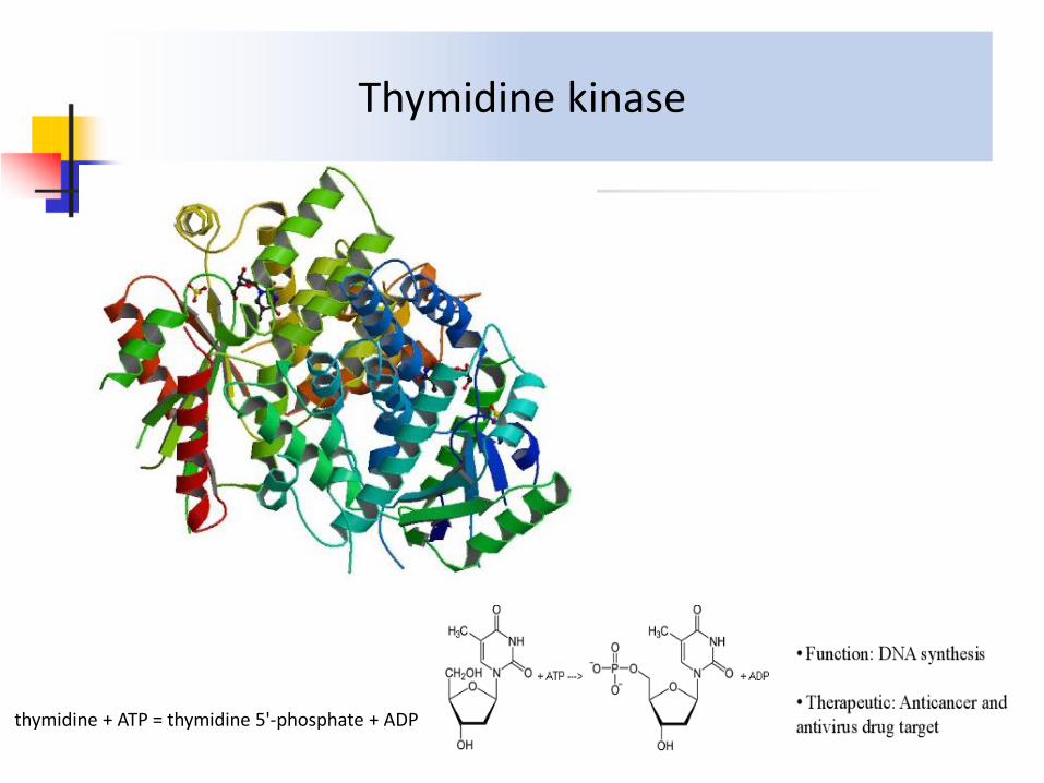
Thymidine kinase
thymidine + ATP = thymidine 5'-phosphate + ADP

Download protein structure file from PDB
1.Save the file in your PC 2.Open the file by PbdViewer

Download and Install PdbViewer
• Download Swiss-PdbViewer
http://spdbv.vital-it.ch/download.html
• Download user guide
http://spdbv.vital-it.ch/Swiss-PdbViewerManualv3.7.pdf
• Tutorial video (English)
http://www.youtube.com/watch?v=nYT5qwtfNew&feature=related
http://www.youtube.com/watch?v=yFE3CAHNkZg

Web page

Install and Run Swiss-PdbViewer

File Operations

2. Workspace
Layer info
Toolbar
Control Panel
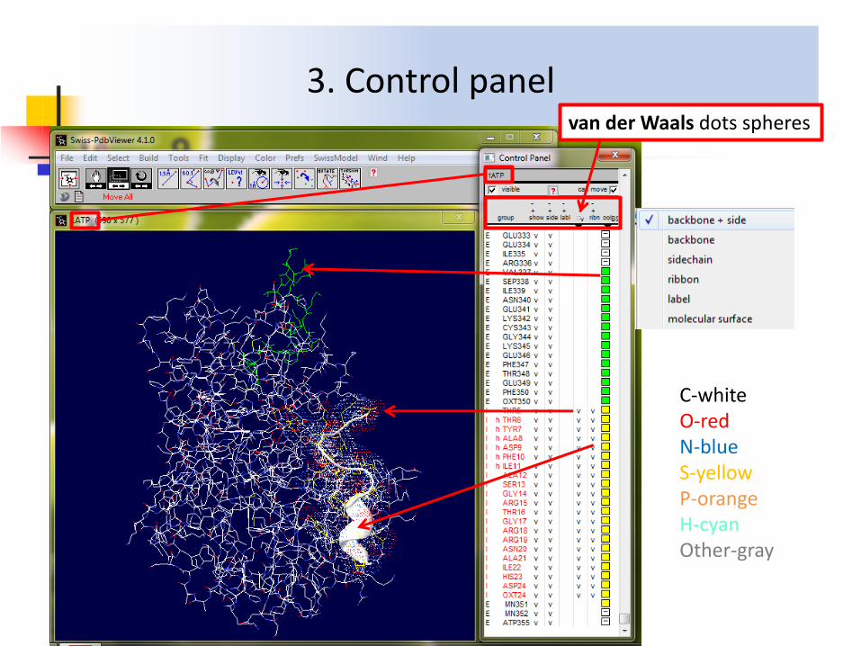
3. Control panel van der Waals dots spheres
C-white O-red N-blue S-yellow P-orange H-cyan Other-gray

3.1 Selection


3.2 Render in Solid 3D
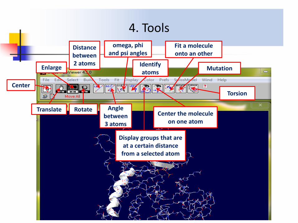
4. Tools
Center
Enlarge
Translate Rotate
Distance between 2 atoms
Angle between 3 atoms
omega, phi and psi angles
Identify atoms
Display groups that are at a certain distance
from a selected atom
Center the molecule on one atom
Fit a molecule onto an other
Mutation
Torsion

4.1 Measure distance between 2 atoms
---Open 1CRN ---Select ---Group Kind ---SS bonds ---Show only ---Side chain ---Label ---Measure the distance between 2 Cα of Cys which have disulphide bonds ---Pick 1st atom ---Pick 2nd atom ---Hit “Esc”

4.2 Measure Angle between 3 atoms
---Press button ---pick center atom ---pick 2nd atom ---pick 3rd atom ---Esc
1
3
2

4.3 Measure of omega, phi and psi angles of the picked amino-acid
---Press “Ctrl” key + button ---pick 4 atoms ---measure the torsion angle of any specific bond ---Esc 1
2
3
4

4.4 Provenance of an atom
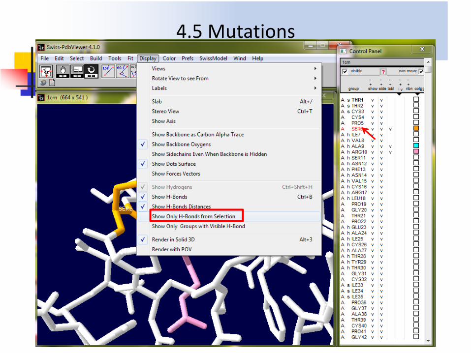
4.5 Mutations

4.5 Mutations

4.5 Mutations
steric hindrances

4.5 Mutations

4.6 Torsions
---Open---1KIM ---select THM1 ---only show THM1 ---label it ---color it orange ---Zoom in & Center ---render in solid 3D ---select one atom and display groups at 6A ---Show & Center ---col---white ---THM1 orange ---Tool ---Compute H-Bonds ---Display ---Show H-Bonds Distance ---only select THM1 ---Show Only H-Bonds from Selection ---Select ---Pick on Screen ---Gln125 & Tyr101 ---color them blue ---Torsion ---pick one atom ---pick 2nd atom

5.1 Coloring Chain
1
2

5.1.1 Coloring Chain acting on Ribbon
1-OR -color -ribbon
1
2

Chain A
Chain B
Chain C
Chain D
5.1.1 Coloring Chain acting on Ribbon

Chain A
Chain B
Chain C
Chain D
5.1.2 Coloring Chain acting on Backbone

OR -color -ribbon
1
2
5.2.1 Color B-factor acting on Ribbon
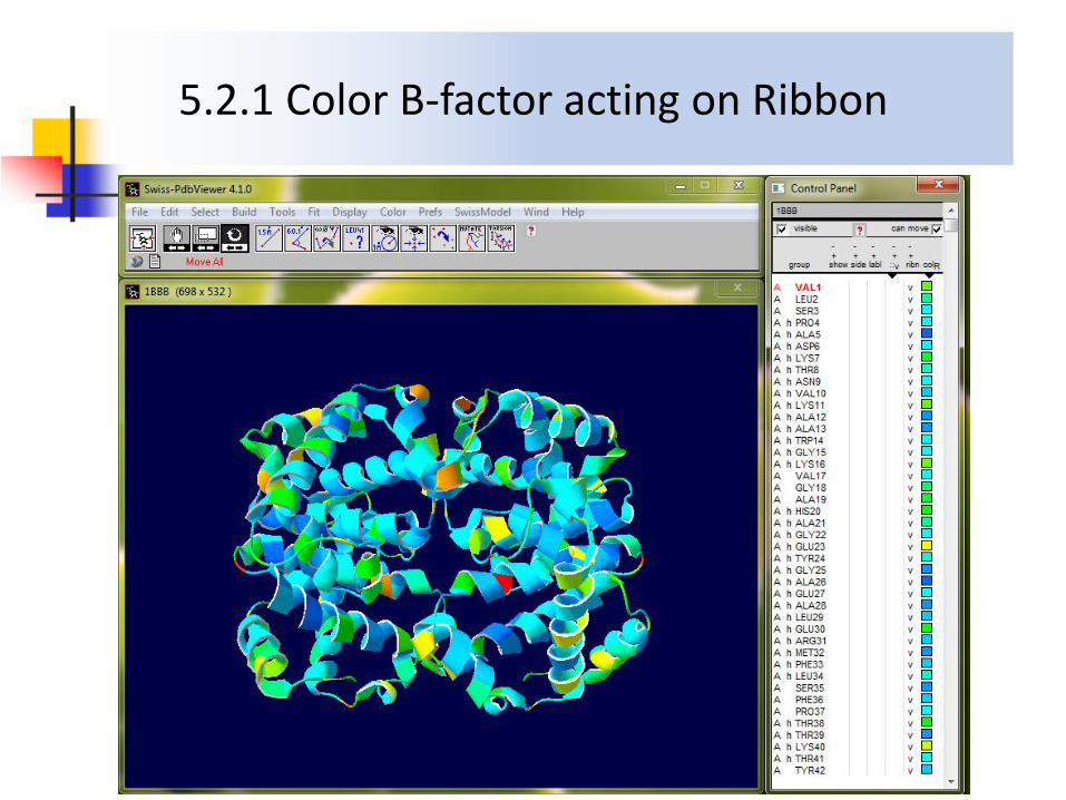
5.2.1 Color B-factor acting on Ribbon

1-OR -color -backbone
1
3 2
5.2.2 Color B-factor acting on Backbone

5.2.2 Color B-factor acting on Backbone

OR -color -ribbon
1
2
5.3 Color in Residue Type acting on Ribbon

5.3 Color in Residue Type acting on Ribbon
Type: Gray-non polar amino acid: Val, Leu, Pro, Ala, Trp, Gly, Met, Phe, Cys, Ile Yellow-polar amino acid: Ser, Thr, Asn, Try, Gln Red-negtive charge (acid amino acid): Asp, Glu Blue-positive charge (basic amino acid): Lys, His, Arg

5.4.1 Color Secondary Structure acting on Ribbon

5.4.1 Color Secondary Structure acting on Ribbon

5.4.2 Color Secondary Structure Succession

5.4.2 Color Secondary Structure Succession

6.1 Enzyme-ligand interaction---Merging
---Loading PDB file of protease ---Color---Chain ---Loading PDB file of inhibitor ---Merging 2 layers ---Alignment window---select protease---Select---All ---select inhibitor ---Edit---Create Merged Layer from Selection (by layer)

6.2 Enzyme-ligand interaction---Active site
---display only groups that are within 10A of the center atom of inhibitor ---Center the molecule ---label them ---Tools ---Compute H-Bonds

6.2 Enzyme-ligand interaction---Active site
---select I121145 in Control Panel ---Display ---Show Only H-Bonds from Selection
1
2

6.2 Enzyme-ligand interaction---Active site
---remove the other labels, only label residues interact with inhibitor, and color them pink ---Display ---Show H-Bonds Distances
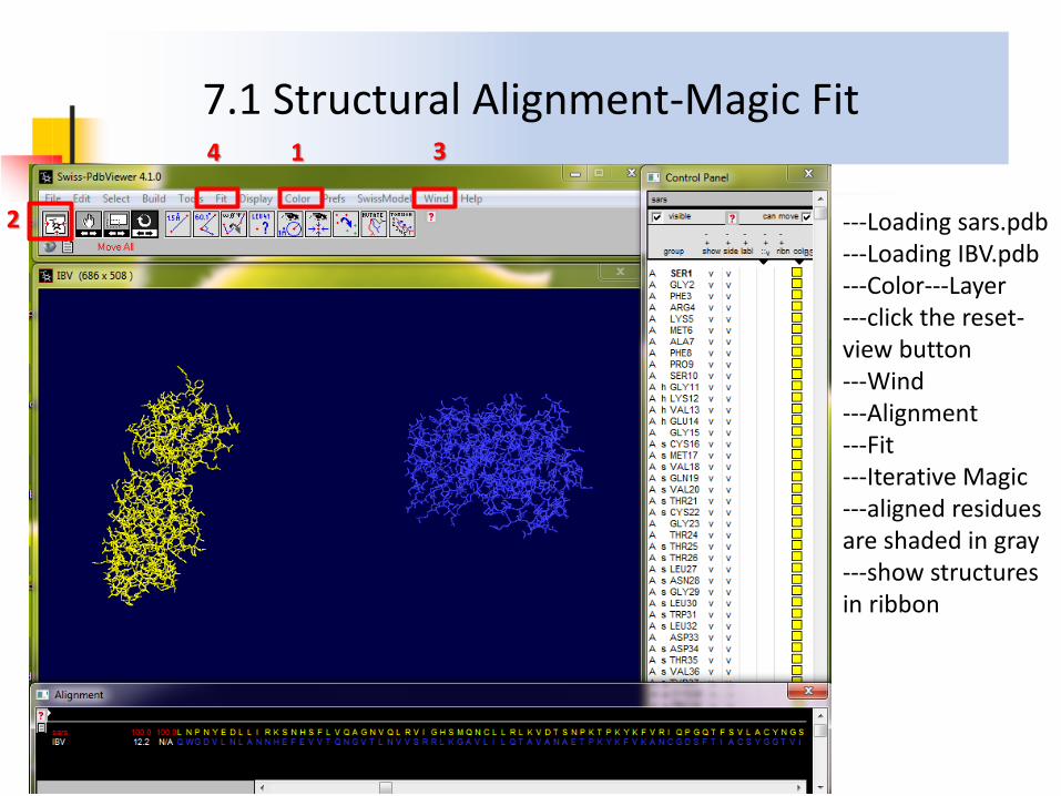
7.1 Structural Alignment-Magic Fit
---Loading sars.pdb ---Loading IBV.pdb ---Color---Layer ---click the reset-view button ---Wind ---Alignment ---Fit ---Iterative Magic ---aligned residues are shaded in gray ---show structures in ribbon
1
2
3 4

7.2 Structural Alignment-show in Ribbon
5

7.3 Structural Alignment vs Sequence Alignment

8.1 Homology Modeling: register with Swiss-Model
http://swissmodel.expasy.org/workspace/index.php?func=account_create1

8.2 Swiss-Model Settings
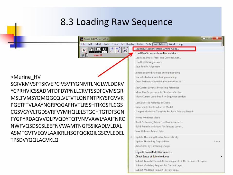
>Murine_HV SGIVKMVSPTSKVEPCIVSVTYGNMTLNGLWLDDKVYCPRHVICSSADMTDPDYPNLLCRVTSSDFCVMSGRMSLTVMSYQMQGCQLVLTVTLQNPNTPKYSFGVVKPGETFTVLAAYNGRPQGAFHVTLRSSHTIKGSFLCGSCGSVGYVLTGDSVRFVYMHQLELSTGCHTGTDFSGNFYGPYRDAQVVQLPVQDYTQTVNVVAWLYAAIFNRCNWFVQSDSCSLEEFNVWAMTNGFSSIKADLVLDALASMTGVTVEQVLAAIKRLHSGFQGKQILGSCVLEDELTPSDVYQQLAGVKLQ
8.3 Loading Raw Sequence

8.4 Load template structure
---loading raw amino acid sequence: Murine_HV.fasta ---loading SARS protease with known 3D structure: 2h2zA.pdb
2h2zA
Murine_HV

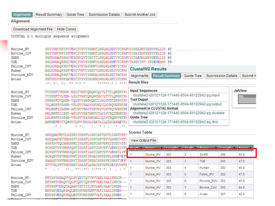
8.5 ClustalW Alignment

8.6 Adjusting manually
3 1
2
4
5
“Ctrl + space bar”

8.7 Model Building


8.8 Template vs Model
2h2zA Murine_HV
