structure protein of - proceedings of the national academy ... · molecular mass and nh2-terminal...
TRANSCRIPT

Proc. Nati. Acad. Sci. USAVol. 81, pp. 5012-5016, August 1984Biochemistry
Structure and molecular arrangement of proteolipid protein ofcentral nervous system myelin
(membrane integration of proteolipid protein/hyposmotic treatment of myelin/trypsinization of myelin membrane/characterization of protein fragments)
WILHELM STOFFEL, HEINZ HILLEN, AND HELMUT GIERSIEFEN
Institut fuer Physiologische Chemie, Universitat zu Koin, Joseph-Stelzmann-Strasse 52, 5000 Koin 41, Federal Republic of Germany
Communicated by Bruce Merrifield, April 18, 1984
ABSTRACT Proteolipid protein (PLP) of central nervoussystem myelin is one of the most hydrophobic integral mem-brane proteins. It consists of a 276-residue-long polypeptidechain with five strongly hydrophobic sequences of 26, 30, 39,12, and 36 residues, respectively, linked by highly charged hy-drophilic sequences. Hyposmotically dissociated bovine myelinmembranes were treated with trypsin. PLP was completelycleaved into smaller fragments, whereas basic myelin proteinremained essentially unaltered. The proteins and tryptic pep-tides of myelin were separated after the removal of the short,water-soluble peptides into three large fragments of 11, 7.3,and 9.0 kDA, respectively. They were characterized by theirmolecular mass and NH2-terminal amino acid sequences,which proved that trypsin cleaved predominantly at Arg-97yielding the 11-kDa fragment from Gly-1 through Arg-97, atArg-126 releasing the 7.3-kDa fragment from Gly-127 throughLys-191, and at Lys-191 releasing the 9-kDa fragment fromThr-192 through Phe-276. We propose that PLP is integratedinto the lipid bilayer of myelin with the NH2 terminus andthree positively charged hydrophilic loops oriented toward theextracytosolic side of the membrane, whereas one stronglynegative hydrophilic loop and the positively charged COOHterminus cover the cytosolic side of the lipid bilayer. Basic my-elin protein remains protected against tryptic cleavage, whichindicates its apposition to the cytosolic side of the membrane.These cleavage sites of trypsin support the suggested orienta-tion of PLP in the myelin membrane and thereby extend ourknowledge about the molecular arrangement of the compo-nents of this membrane. In demyelinating processes mem-brane desintegration could be initiated by proteolysis at theexternal surfaces of proteolipid protein in a similar way as de-scribed here.
Proteolipid protein (PLP), also named lipophilin (1), is themajor structural protein of brain white matter. It accountsfor more than 50% of myelin membrane proteins of the cen-tral nervous system (CNS) and is supposed to maintain themyelin structure and function. PLP has resisted its biochem-ical characterization because of its insolubility and tendencyto aggregate in aqueous solution. These properties explainthe sparse sequence data and slow progress in the structuralelucidation of PLP ever since its discovery (2). Only smallhydrophilic sequences, except a limited hydrophobic se-quence at the COOH-terminal end (3-5), were elaboratedwith the conventional peptide separation techniques.
Recently, new approaches in the separation of hydropho-bic peptides (6-8) enabled us to determine the entire aminoacid sequence of bovine PLP (9). The polypeptide chain con-sists of 276 amino acid residues that are arranged in four longand one short hydrophobic segments, which are linked byhydrophilic highly charged sequences. One fatty acid residue
is attached by an ester bond to a threonine residue within ahydrophilic segment (9). During our structural studies, it alsobecame evident that cysteine residues form disulfide bondsbetween distant hydrophilic sequences. This inevitably leadsto a condensed alignment of these hydrophilic loops at thesurface of the membrane lipid bilayer, associated with a clus-tering of the membrane-spanning and embedded hydropho-bic sequences.The knowledge of the primary structure led to the propos-
al of the alignment in the myelin membrane of CNS withrespect to the main dense line and the intraperiod denseline-the equivalents of the adjacent cytosolic surfaces andthe external surfaces, respectively, in electron microscopy(8, 9). In this communication, experimental evidence in sup-port of this model is given by tryptic cleavage within the hy-drophilic peptide loops oriented toward the external side ofthe myelin membranes. Hyposmotic shock of myelin prepa-ration renders this surface available for enzymatic attack.Three large tryptic polypeptides were formed. Their charac-terization allowed the conclusion that the NH2 terminus andthree positively charged hydrophilic segments are located atthe extracytosolic side of the lipid bilayer, whereas the nega-tively charged loop and the positively charged COOH termi-nus point to the cytosolic side.These results contribute to our understanding of the mo-
lecular architecture of the highly organized myelin mem-brane system and of factors that may affect its stability andfunction in demyelinating processes.
MATERIALS AND METHODSIsolation of Myelin. All procedures were carried out at 4°C.
Myelin was isolated by a modification of the procedure de-scribed by Norton (10). It permits the preparation of myelinon a large scale. The subcortical white matter from a freshbovine brain (300 g) was homogenized in 700 ml of 0.17 Msucrose in a Waring Blendor for 2 min (medium speed). Thesuspension was centrifuged at 5000 x g in a JA-14 Beckmanrotor for 20 min. The supernatant was discarded, and thepellet was resuspended in 600 ml of 0.17 M sucrose andwashed under the same condition twice. The pellet was sus-pended in 80 ml of 0.17 M sucrose and layered on top of a0.67 M sucrose cushion (5 ml) in polyethylene tubes. Myelinbanded in the interphase of the two-step gradient upon cen-trifugation at 40,000 x g (18,500 rpm) in a JA-20 Beckmanrotor. The myelin band was collected, suspended in 200 mlof water, and centrifuged for 30 min at 5000 x g (6000 rpm).The gradient centrifugation and the hyposmotic shock (11)was repeated once. The myelin band was collected.
Trypsin Cleavage. Myelin (100 mg) was suspended in 5 mlof 0.1 M NH4HCO3 buffer (pH 8.4), 0.5 mg of trypsin wasadded, and the suspension was stirred at room temperaturefor 6 hr. The reaction was stopped by adjusting the pH to 4.0
Abbreviations: PLP, myelin proteolipid protein; BMP, basic myelinprotein; CNS, central nervous system.
5012
The publication costs of this article were defrayed in part by page chargepayment. This article must therefore be hereby marked "advertisement"in accordance with 18 U.S.C. §1734 solely to indicate this fact.
Proc. Natl. Acad. Sci. USA 81 (1984) 5013
with acetic acid. The suspension was centrifuged at 5000 x gat 40C, and the pellet was lyophilized. Myelin treated with0.01 M HCl/5% KCl for 16 hr at room temperature also wassubjected to trypsin treatment as described above for hypos-motically shocked myelin.
Isolation of Tryptic Fragments. For delipidation, the lyoph-ilized trypsin-treated myelin was dissolved in 4 ml of acidi-fied chloroform/methanol [2:1 (vol/vol), 1% in aqueousHCl] and dialyzed against 2:1 chloroform/methanol for 4 hrin a Visking dialysis tubing (Serva, Heidelberg). The poly-peptides were precipitated by the addition of 4 ml of petro-leum ether (30-60'C) and centrifuged.
Reductive Carboxymethylation. The pellet was dissolved in5 ml of 0.4 M Tris HCl, pH 8.6/0.08% NaDodSO4, and 0.30mmol (50 mg) of dithiothreitol was added for reduction at40'C for 2 hr. For carboxymethylation, 0.8 mmol of iodoace-tamide was added, and the reaction was continued for 30 minat 40'C. The reaction mixture was dialyzed for 24 hr against0.01 M NH4HCO3 buffer using Spectropor dialysis tubing(exclusion size, 2000 Da) and subsequently was lyophilized.
Separation of Tryptic Fragments. Of the lyophilized pep-tide mixture, 20 mg was extracted twice with 3 ml of 0.01 MHCl at room temperature to remove the water-soluble pep-tides (6-8). The extract contained a minimal amount of pep-tides with molecular masses below 2000 Da. The residue wasdissolved in 500 1.l of concentrated formic acid and was sep-arated by gel permeation high-performance liquid chroma-tography with a mixed-bed column (30 x 2 cm) packed with5-,um-particle-size 1:1:1 Si 100/Si 60/Si 50 (Merck) with 90%formic acid as solvent as described (6-8). The column wascalibrated with the peptides and proteins as indicated in Fig.
W ~ ^ 0
SBpL loft 2
e; F~~~
iCE,~@ o
4. Three fragments generated by trypsin treatment of the my-elin membrane were eluted in distinct peaks at retentiontimes of 9.82 min (15 kDa), 10.32 min (11.5 kDa), and 10.96min (8 kDa). They were further purified by repeated HPLCunder the same conditions for automated Edman degrada-tion (11).Automated Sequence Determination. Edman degradation
(12) was performed in a Beckman Sequenator, model 890 C,using the 0.2 M Quadrol program as described (6-8).NaDodSO4/Polyacrylamide Gel Electrophoresis. Reduced
and carboxymethylated polypeptide and protein samples ofbovine myelin were subjected to NaDodSO4/polyacryl-amide gel electrophoresis with 15%20% acrylamide gels.
Electron Microscopy. Aliquots of untreated myelin .tn(d ofosmotically shocked myelin before and after trypsin treat-ment were fixed in buffered glutardialdehyde and stainedwith buffered 2% osmium tetroxide. A Philips electron mi-croscope (model 300) was used.
RESULTSProtein Structure and Proposed Assembly in the Lipid Bi-
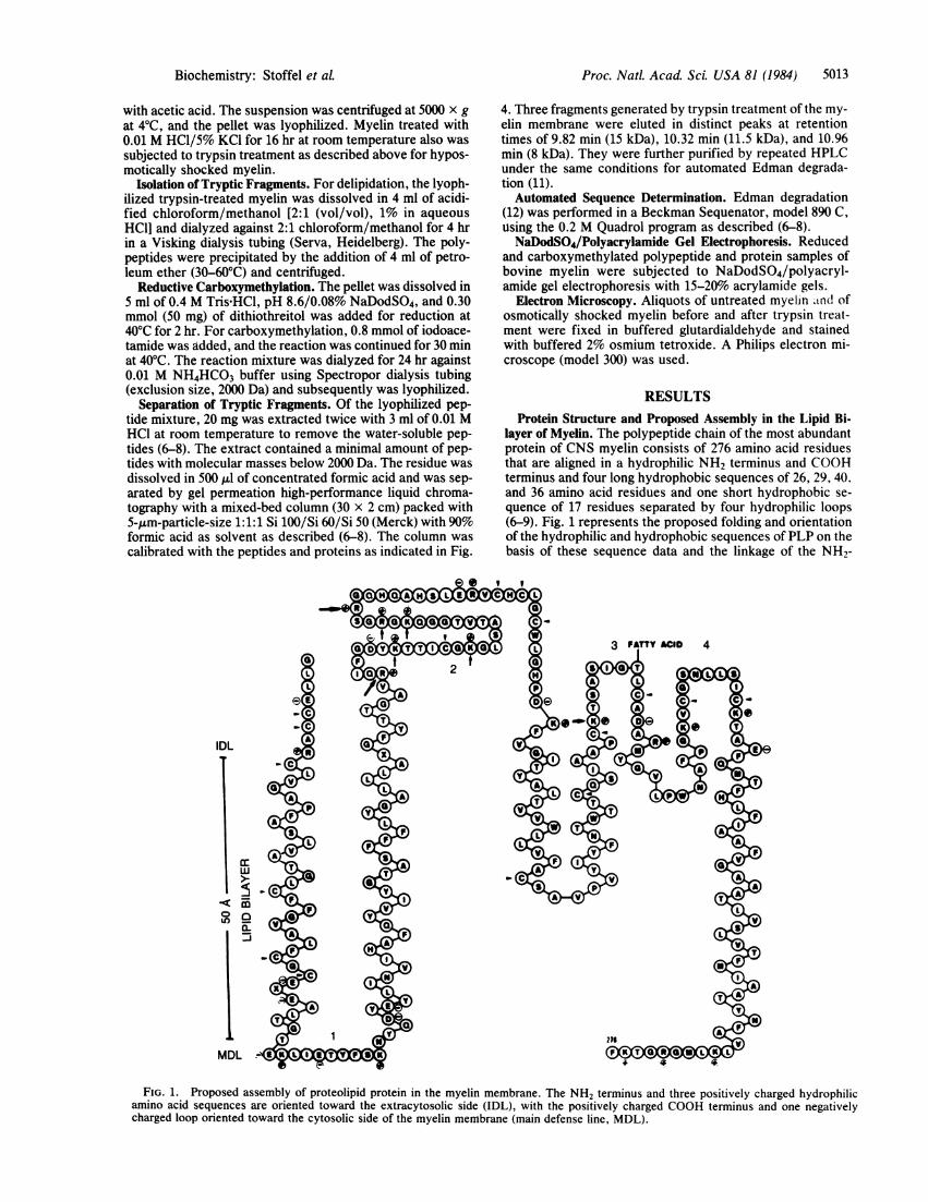
layer of Myelin. The polypeptide chain of the most abundantprotein of CNS myelin consists of 276 amino acid residuesthat are aligned in a hydrophilic NH2 terminus and COORterminus and four long hydrophobic sequences of 26, 29, 40,and 36 amino acid residues and one short hydrophobic se-quence of 17 residues separated by four hydrophilic loops(6-9). Fig. 1 represents the proposed folding and orientationof the hydrophilic and hydrophobic sequences of PLP on thebasis of these sequence data and the linkage of the NH2-
---cm
DaL
MDL
FIG. 1. Proposed assembly of proteolipid protein in the myelin membrane. The NH2 terminus and three positively charged hydrophilicamino acid sequences are oriented toward the extracytosolic side (IDL), with the positively charged COOH terminus and one negativelycharged loop oriented toward the cytosolic side of the myelin membrane (main defense line, MDL).
Biochemistry: Stoffel et aL

Proc. Natl. Acad. Sci. USA 81 (1984)
terminal cysteine-rich sequence to one of the two cysteineresidues (Cys-219 or -227) of the penultimate hydrophilic se-quence and of Cys-219 or -227 to one of the three cysteineresidues in position 138, 140, or 143. Unambiguous evidencefor the orientation of the hydrophilic loop and, therefore,support for the proposed topography should come from theproteolytic attack on the polypeptide chain exposed on theexternal side of the myelin membrane and the identificationof the resulting proteolytic fragments.
Dissociation of Myelin Membrane System by HyposmoticShock and Trypsin Treatment. The tightly spirally wrappedlayers of the myelin membrane were dissociated by hypos-motic shock. This step proved to be essential for an effectiveaccess of trypsin, which was chosen because of the favor-able distribution of lysine and arginine residues in the hydro-philic loops. Fig. 2 shows electron microscopic pictures ofhyposmotically shocked myelin before (Fig. 2 Upper) andafter (Fig. 2 Lower) trypsinization. The dissociation of themyelin layers within the intraperiod dense lines, which cor-responds to the space between adjacent external membranesides, is apparent (arrow in Fig. 2 Upper). The stacked mem-
.4
I
S I
.jfX .':
.11
1,..It.Px
brane appears to be fully detached after trypsin treatment(Fig. 2 Lower).NaDodSO4/Polyacrylamide Gel Electrophoresis. Bovine
myelin exhibited a protein pattern that is similar in manyspecies (Fig. 3, lane 1). The main components are basic my-elin protein (BMP) (18.7 kDa); myelin protein with a molecu-lar mass - 20 kDa, designated DM20 (23 kDa); PLP with anapparent molecular mass of 26 kDa and its dimeric form andaggregates in the high molecular mass range; and the Wolf-gram proteins, designated WP (58-60 kDa). When myelinwas exposed to an acidic medium, PLP aggregated (Fig. 3,lane 2). Trypsin treatment of the hyposmotic myelin prepara-tion (Fig. 3, lane 3) led to the loss of the intensive PLP andDM20 bands but also of thje bands corresponding to aggre-gated PLP. New polypeptide bands, designated A and B inlane 3 of Fig. 3, that were not well resolved appeared in themolecular mass range around and below 10 kDa. The band ofBMP (designated C in lane 3 of Fig. 3) remained unaltered.Trypsin treatment of the acid-exposed myelin membranes,on the other hand, led to the complete degradation of PLP,DM20, and BMP (Fig. 3, lane 4).
Isolation and Characterization of Tryptic Fragments. Afterdelipidation of the trypsin-treated myelin membranes bydialysis against acidic chloroform/methanol, small water-soluble peptides (<2000 Da) were removed by brief extrac-tion with 0.01 M HCL. The residual polypeptide mixture wasseparated by preparative gel permeation HPLC. Fig. 4 a andb shows the resolution of the chromatographic procedurewith marker proteins and peptides: ovalbumin, myoglobin,cytochrome c and its cyanogen bromide (CNBr) fragments,the tripeptide Lys-Tyr-Ser, and tryosine. Three fractionswith retention times of 9.82, 10.32, and 10.96 min, corre-sponding to molecular masses of approximately 17 kDa, 11.0kDa, and 8.5 kDa, respectively, were isolated from trypsin-treated myelin (Fig. 4c). These fractions were collected (be-tween arrows in Fig. 4d) and rechromatographed twice,yielding sufficiently pure fractions for sequence determina-
kDa 1
WP 60-
I.
2 3 4
1P
p
26- _11m)23- "
1 8.7- - C
200nmv .. 4
FIG. 2. Electron microscopy of hyposmotically shocked bovinemyelin before (Upper) and after (Lower) trypsin treatment.
LMW 12
B
A
FIG. 3. NaDodSO4/polyacrylamide gel electrophoresis of pro-
teins of isolated myelin (lane 1), myelin after acid treatment (lane 2),myelin proteins and their fragments after hyposmotic shock andtrypsin treatment (lane 3), and products after trypsinization of acid-treated myelin (lane 4). WP, Wolfgram protein; DM20, myelin pro-
tein of 23 kDa; LMW, low molecular weight proteins of about 12kDa.
PLP
OM 20
BMP
5014 Biochemistry: Stoffel et aL

Proc. NatL. Acad. Sci. USA 81 (1984)
z 0U >-L. n
a t
2 ~n:s y b
K-Y- \
\BrC~~~BrN IIIyt c_r~CN IN+II
ACytc-4NrCN I
c~yt c
t\ogLobiniab.
1600Molecular mass, Da
10.00
a:
E-U)
FIG. 4. Preparative gel permeation HPLC. (a) Elution pattern of calibration peptides and protein from preparative gel permeation HPLCcolumn. (b) Semilogarithmic plot of retention times versus molecular masses of calibration proteins and of myelin polypeptides A, B, and C. (c)HPLC elution pattern of the polypeptide mixture after trypsin treatment. (d) HPLC elution pattern of the purified fractions A, B, and C (20-mgaliquots were separated). The column was 30 x 2 cm with 5-pm 1:1:1 Si 100/Si 60/Si 50; the solvent was 90% formic acid; flow rate was at 4.5ml/min; and UV detection was at 280 nm. Ovalb., ovalbumin; Myogl., myoglobin; Cytc, Cytochrome c; Cytc-BrCN I, II, and HIt, cyanogenbromide fragments I, II, and III of cytochrome c; Y, tyrosine; L-Y, leucine-tyrosine dipeptide; K-Y-S, lysine-tyrosine-serine tripeptide.
tion (Fig. 4d). The band with a retention time of 9.82 mincorresponds to the 17- to 18-kDa protein, identical with basicmyelin protein and traces of the NH2-terminal tryptic frag-ment of proteolipid protein due to incomplete separation.The NH2 terminus of BMP is N-acetylated and, therefore,escapes automatic Edman degradation but was identified by
NaDodSO4/polyacrylamide gel electrophoresis. The band,which was eluted at a retention time of 10.46 min, representsthe 11-kDa fragment. Automated Edman degradation over15 cycles revealed the NH2 terminus to be from Gly-1through Arg-97. Identification of this cleavage site is sup-ported by the molecular mass of the fragment and the amino
15
14r
ai 13*E
0c 12**_pc0)0) 11~
10
9.
us0E-HLn
ACD
c 0 d
Aen
9
c
B
To
E-Uq
P4.l<UusVI0H-
u
5015Biochemistry: Stoffel et al.
f0.-
IHt 11 11 11,11

5016 Biochemistry: Stoffel et al.
acid analysis, which yielded two lysine residues. Two poly-peptides coeluted at retention time of 10.96 min in a 1:1 ratio.They were sequenced in parallel and proved to correspondto the sequence from Gly-127 through Lys-191 (7.3 kDa) andfrom Thr-192 through Phe-276 (9.0 kDa).
DISCUSSIONOur understanding of the molecular structure ofCNS myelinrequires substantial chemical and physical information aboutthe complex lipid and protein components of this membrane.Considerable compositional data have accumulated, whichhave been reviewed (13, 14). The two main proteins BMPand PLP are considered myelin specific (15). They are pres-ent in myelin in approximately equimolar amounts (14, 15).This study describes a topochemical study of the orientationand integration of one of these main protein components ofCNS myelin, PLP, into the lipid bilayer. This approach be-came feasible after we succeeded in determining the com-plete amino acid sequence of this most hydrophobic integralprotein and in having available rapid and efficient proce-dures for the separation and identification of hydrophobicpolypeptides.The model proposed in Fig. 1 concerns the folding of the
five strongly hydrophobic sequences that are bordered bycharged amino acid residues. Three of them span the lipidbilayer and two are harbored in the hydrocarbon layer of themembrane. The hydrophobicity index (16, 17) of the hydro-phobic sequence from Cys-9 to Cys-34 is 2.31, from Tyr-59to Val-96 is 2.72, from Phe-151 to Cys-190 is 2.36, and fromPhe-232 to Leu-267 is 2.35. They are in the range of the hy-drophobicity index of the 23-amino-acid residue sequence ofglycophorin (hydrophobicity index, 2.62) spanning the lipidbilayer (17, 18). Assuming an a-helical conformation of thehydrophobic sequences, about 30 amino acid residues aresufficient to span the 4.6- to 5-nm-thick myelin lipid bilayer(19). The tightly packed membrane layers were dissociatedby hyposmotic shock (11) to make their surfaces accessiblefor the proteolytic attack. Electron microscopy demonstrat-ed that this cleavage occurs in the intraperiod dense line,which corresponds to opposing outer myelin membrane sur-faces. The result of the tryptic cleavage at sites of proteinsegments protruding out of the extracytosolic side of the my-elin membrane are in full agreement with the proposal thatfour of the six hydrophilic sequences, namely, the NH2 ter-minus and loops 2, 3, and 4 (Fig. 1), are oriented toward theextracytosolic side. We were unable to detect fragmentsarising from cleavage at Lys-44 or -48 (residues of hydrophil-ic loop 2); which should be oriented toward the cytosolicside. Our experimental results are incompatible with the in-tegration of PLP in the myelin membrane proposed recentlylargely on our sequence data (20). Further support of thismodel comes from our observation that, in isolated PLP, di-sulfide bonds link the hydrophilic NH2-terminal cysteine-rich sequence to the hydrophilic loops 4 and 2 and also linkloops 2 and 4 (Fig. 1). This is only possible if the correspond-
ing cysteine residues are in appropriate distances at the sameside of the bilayer. In our myelin preparations, BMP wasprotected against the tryptic attack. This gives further evi-dence for the localization of this basic protein in the cyto-plasmic cleft between the myelin bilayers. Our observationis consistent with the inability of nonpermeant chemical la-bels to bind to BMP in intact myelin preparations (21, 22).PLP on the other hand can be 12'I-labeled with lactoperoxi-dase (21). The hydrophilic sequences of PLP on the externalside of the myelin membrane may represent the sites of theproteolytic attack under pathological condition, at which thedesintegration of the membrane starts, which finally leads todemyelination. They also can form the antigenic sites of themyelin membrane.
This work was supported by grants from the Deutsche Fors-chungsgemeinschaft (SFB 74) and the Thyssen Foundation.
1. Boggs, J. M. & Moscarello, M. A. (1978) Biochim. Biophys.Acta 515, 1-21.
2. Folch-Pi, J. & Lees, M. (1951) J. Biol. Chem. 191, 807-817.3. Nussbaum, J. L., Jolles, J. & Jolles, P. (1982) Biochimie 64,
405-410.4. Lees, M. B., Chao, B. H., Laursen, R, A. & L'Italien, J.
(1982) Biochim. Biophys. Acta 702, 1117-124.5. Lees, M. B., Chao, B. H., Liu, L. F. H., Samiullah, M. &
Laursen, R. A. (1983) Arch Biochem. Biophys. 226, 643-656.6. Stoffel, W., Hillen, H., Schroeder, W. & Deutzmanq, R.
(1982) Hoppe-Seyler's Z. Physiol. Chem. 363, 855-864.7. Stoffel, W., Schroeder, W., Hillen, H. & Deutzmann, R.
(1982) Hoppe-Seyler's Z. Physiol. Chem. 363, 1117-1131.8. Stoffel, W., Hillen, H., Schroeder, W. & Deutzmann, R.
(1982) Hoppe-Seyler's Z. Physiol. Chem. 363, 1397-1407.9. Stoffel, W., Hillen, H., Schroeder, W. & Deutzmann, R.
(1983) Hoppe-Seyler's Z. Physiol. Chem. 364, 1455-1466.10. Norton, W. T. (1974) Methods Enzymol. 31, 435 444.11. McIntosh, T. J. & Robertson, J. D. (1976) J. Mol. Biol. 100,
213-217.12. Edman, P. & Begg, G. (1967) Eur. J. Biochem. 1, 80-91.13. Rumsby, M. G. & Crang, A. J. (1977) in The Synthesis Assem-
bly and Turnover of Cell Surface Components, eds. Paste, G.& Nicholson, G. L. (Elsevier/North-Holland, Amsterdam),pp. 247-362.
14. Norton, W. T. (1975) in The Nervous System, ed. Tower,D. B. (Raven, New York), Vol. 1, pp. 467-481.
15. Mehl, E. & Halarris, A. (1970) J. Neurochem. 17, 659-668.16. Tanford, C, (1978) Science 200, 1012-1018.17. Segrest, J. P. & Feldman, R. J. (1974) J. Mol. Biol. 87, 853-
858.18. Tomita, M. & Marchesi, V. T. (1975) Proc. Natl. Acad. Sci.
USA 72, 2964-2968.19. Levine, Y. K. (1975) Prog. Biophys. Mol. Biol. 29, 3-56.20. Laursen, R. A., Samiullah, M. & Lees, M. B. (1983) FEBS
Lett. 161, 71-74.21. Poduslo, J. F. & Braun, P. E. (1975) J. Biol. Chem. 220, 1099-
1105.22. Crang, A. J. & Rumsby, M. G. (1977) Biochem. Soc. Trans. 5,
110-112.
Proc. NatL Acad Sci. USA 81 (1984)