somatomedin ofhuman placenta: solubilization, · pdf fileweights, pls, nh2-terminal amino acid...
TRANSCRIPT

Proc. Natl Acad. Sci. USAVol. 78, No. 7, pp. 4279-4283, July 1981Biochemistry
Somatomedin receptor of human placenta: Solubilization,photolabeling, partial purification, and comparisonwith insulin receptor
(isoreceptors/insulin-like growth factors/growth hormone)
B. BHAUMICK*, R. M. BALA*, AND M. D. HOLLENBERGEndocrine Research Group, Department of Medicine and Department of Pharmacology and Experimental Therapeutics, University of Calgary, Faculty of Medicine,Calgary, Canada T2N 1N4
Communicated by Victor A. McKusick, April 13, 1981
ABSTRACT Using a recently isolated human basic somato-medin (basic SM) similar to insulin-like growth factor I (IGF-I),we studied both the photoaffinity-labeled and unlabeled basic-SMreceptor solubilized from human placental cell membranes. Un-like the result with the insulin receptor, high yields of solublebasic-SM-binding activity are obtained with Triton X-100. The sol-uble basic-SM receptor retains high-affinity (Kd 0.3 nM) pep-tide-specific binding of basic SM, similar to the binding presentin particulate placenta membranes; the receptor exhibits a com-paratively low affinity for insulin(kd 3 FAM). On Sepharose 6B,like the crude soluble insulin receptor, the basic-SM receptormigrates as a species with an apparent Stokes radius of 7.2 nm;unlike the insulin receptor, the basic-SM receptor does not, undersimilar conditions, yield a smaller binding species (apparent Stokesradius 3.8 nm). Upon photoaffinity labeling with "I-labeled basicSM, one principal specifically labeled constituent is detected.Upon gel electrophoresis in the presence of 2-mercaptoethanol,the photolabeled constituent, like the insulin receptor, migratesas a species with an apparent molecular weight of about 140,000;in the absence of reducing agent, a molecular weight greater than240,000 is observed. Lectin-agarose affinity chromatographyyields a 30-fold purification both of the basic-SM-binding activityand the photolabeled constituent. Anti-insulin receptor antibodydoes not appear to precipitate the basic-SM receptor. We concludethat the basic-SM receptor of human placenta is a glycoprotein,remarkably similar to (an isoreceptor) but distinct from the insulinreceptor previously characterized in this tissue.
The somatomedins (SM) are a group of growth hormone-de-pendent, serum-borne, polypeptide growth factors that havein common the ability to stimulate proteoglycan synthesis incartilage and to mimic the actions of insulin in a variety of ex-traskeletal tissues. The somatomedins account for the insulin-like activity in human serum that is not neutralized by anti-in-sulin antibody (so-called nonsuppressible insulin-like activityor NSILA; recently renamed insulin-like growth factors orIGFs). Recent evidence suggests that all of the reported so-matomedins may fall into two main groups based on their iso-electric points. The basic group (pIs greater than 7.4) includesinsulin-like growth factor-I (IGF-I) (1), SM-C (2), and a basicSM that has been purified in our laboratory (3). The acidic-neu-tral group (pI less than 7.4) includes IGF-II (1), SM-A (4), andmultiplication-stimulating activity (5). The basic group of so-matomedins are highly similar in terms of their molecularweights, pls, NH2-terminal amino acid sequences (1-3), andimmunoreactivity (3, 6, 7). IGF-I (similar to our basic SM) andIGF-II have different NH2-terminal amino acid sequences but
share a sequence homology with each other and with proinsulin(8). Thus, there is a complex relationship between insulin, thesomatomedins, and the peptide-specific receptors for each ofthese polypeptides. Although distinct receptors for the soma-tomedins have been detected in fat cells (9), fibroblasts (9, 10),and placenta (11), the. sequence homologies lead to cross-spec-ificity of binding at the receptor sites, so that insulin at highconcentrations (micromolar) can occupy the SM receptors andvice versa.
In view ofthe cross-specificity ofthe ligand recognition prop-erties ofthe receptors for insulin and the SMs, we have becomeinterested in comparing the physicochemical properties of theinsulin receptor with those of the receptors for the SMs. In thepresent work, we report the solubilization, photoaffinity label-ing, characterization, and partial purification of the human pla-centa receptor for human basic SM. Our results indicate a highdegree of physiocochemical similarity between the basic-SMreceptor and the receptor for insulin.
MATERIALS AND METHODSlodination of Basic SM and Insulin. Highly purified basic SM
was isolated as reported (3). This basic SM had a potency of4000units/mg by hypophysectomized rat cartilage bioassay. Insulinwas a gift from Eli Lilly. Peptides were iodinated by a modifi-cation of the chloramine-T method (6) to specific activities of100-150 ,uCi of basic SM and 80-100 pCi of insulin per Ag (1Ci = 3.7 x 10¹⁰ becquerels).
Preparation ofMembranes and Membrane Extract. A crude"microsomal" membrane fraction from term human placentacells was prepared from the placental basal plate by homoge-nization and differential centrifugation, essentially as described(12, 13). Membranes were solubilized (6 mg of membrane pro-tein per ml of detergent solution) for 30 min at 240C with 0.5%Triton X-100 or 0.15-0.5% Ammonyx-LO (Onyx Chemical,Hoboken, NJ) in 50 mM Tris HCl (pH 7.4). The extract wasclarified by centrifugation at 150,000 X g for 70 min at 4°C.Before use, Ammonyx-LO-solubilized membrane preparationswere made 0.5% in Triton X-100 and were dialyzed for 17 hrat 40C against 0.5% Triton X-100 in 50 mM Tris HCl (pH 7.4).
Binding Assay. Solubilized membrane protein (=50 ,ug ofprotein in 0.1 ml) was incubated with radiolabeled basic SM(10,000-100,000 cpm) or insulin ("'100,000 cpm) with or with-out unlabeled basic-SM or insulin (==1 ,ug of unlabeled pep-tide per ml) in Tris HCl buffer (pH 7.4) containing 5 mg of bo-vine serum albumin per ml (final vol, 0.3 ml). Samples in 12x 75 mm polystyrene tubes were equilibrated overnight at 4°C
Abbreviations: IGF, insulin-like growth factor; SM, somatomedin.* Present address: Dept. ofMedicine, University ofSaskatchewan, Sas-katoon, Canada S7N OXO.
The publication costs ofthis article were defrayed in part by page chargepayment. This article must therefore be hereby marked "advertise-ment" in accordance with 18 U. S. C. §1734 solely to indicate this fact.
4279

4280 Biochemistry: Bhaumick et al.
for basic SM and for 60 min at 240C for the measurement of in-sulin binding. Specific ligand binding was measured by thepolyethylene glycol method as described (14).
Photoaffinity Labeling. Basic SM or insulin was first iodi-nated by using a modification of the procedure described bySpivak et al. (15). Glass tubes (12 x 75mm) were freshly coatedwith water-insoluble oxidizing agent 1,3,4,6-tetrachloro-3a,6a-diphenylglycouril (IODO-GEN; Pierce). Basic SM (10 ,g) orinsulin (5 A&g) in 200 ,Al of0.1 M sodium phosphate (pH 7.5) wasadded to the IODO-GEN tube. Carrier-free 125I (1.5-3 mCi;Amersham) was then added (-5 1.l), and the solution was mixedvigorously for 40 sec. The reaction mixture was filtered througha plug of glass wool in a Pasteur pipette and made up to 2 mlwith the sodium. phosphate buffer. A photoaffinity labeling de-rivative was then prepared by the addition of 30 1.l of N-suc-cinimidyl 6-(4'-azido-2'-nitrophenylamino)hexanoate (Pierce;1 mg/ml in dimethyl sulfoxide) as described for epidermalgrowth factor (16) and was used without further purification.The derivative of either basic SM or insulin (200-400 ng/0. 1ml) was incubated with membranes (4mg of protein) in the darkat 40C (total vol, 4 ml) either with or without an excess (=1 Ag/ml) of underivatized peptide. After 45 min, the azide group wasactivated by illumination (two 200-W incandescent bulbs at 10cm) at 40C for 20 min. Photolabeled membrane was collected,washed by centrifugation (48,000 X g for 20 min), and solubi-lized in 0.5-1 ml of buffer containing 1% detergent (either Tri-ton X-100 or Ammonyx-LO). The photolabeled receptor wasthen analyzed by NaDodSOpolyacrylamide gel electropho-resis (7.5% gels; ref. 16) either before or after partial purificationon lectin-agarose columns. Labeled proteins were visualized byautoradiography as described (17).
Immunoprecipitation of Receptor. Rabbit antiserum fromanimals immunized with purified rat liver insulin receptor (LotA410) was provided by S. Jacobs (Burroughs Wellcome, Re-search Triangle Park, NC). Soluble receptor (100 ,ul in deter-gent-containing buffer) was first equilibrated overnight at 4°Cin a total volume of 300 ,ul with radiolabeled ligand (either in-sulin or basic SM) either in the presence or absence ofan excessof the appropriate unlabeled peptide. Anti-receptor antibody(10 ,u1) was then added and the antibody-receptor complex wasallowed to form for 6 hr at 4°C. Second antibody (goat anti-rabbitimmunoglobulin; Calbiochem) was then added (200 ,ul of re-constituted goat antibody; capable of reacting with 8-10 .1d ofrabbit serum), and the immunoprecipitate was allowed to formfor 48-72 hr at 4°C. Control experiments were performed withnonimmune rabbit serum. Radioactivity in the washed immu-noprecipitate was measured by crystal scintillation counting.
RESULTSIn keeping with previous observations (18), extraction of pla-centa membranes with Triton X-100 yielded relatively smallamounts of soluble insulin-binding activity, compared to theinsulin-binding activity present in the membranes prior to solu-bilization; this result suggests a lability of the insulin receptorunder-the conditions of extraction (Table 1). In contrast, morethan 60% ofthe basic-SM-binding activity was recovered in sol-uble form from placenta membranes by using Triton X-100(Table 1); the detergent Ammonyx-LO was equally effective insolubilizing basic-SM-binding activity but, as detailed previ-ously (18), yielded a greater amount of soluble insulin-bindingactivity than did Triton X-100 (data not shown). Thus, thereappears to be a differential solubilization of the receptors forinsulin and basic SM, depending on the detergent used. Moststudies were done with Triton X-100-solubilized basic-SMreceptor.
Table 1. Solubilization of basic-SM- and insulin-bindingactivities by Triton X-100
Basic-SMSample binding, % Insulin binding, %
Supernatant 64 ± 4 9 ± 2Pellet 50 ± 2 47 ± 1Net recovery of binding activity 114 ± 6 56 + 3
Membranes (-6-10 mg) were solubilized for 30 min at 240C in 11ml of 0.5% Triton X-100 in 50 mM Tris-HCl (pH 7.4). The extract wasclarified by centrifugation (150,000 x g for 70 min at 40C), and thepellet was washed twice with detergent-free buffer. The washed pelletwas resuspended in an equal volume of buffer, and aliquots of the sol-uble extract and washed membrane suspension were assayed for ligandbinding; the detergent concentration in the bindingassay was <0.04%.The values represent the average of two independent estimates (± 1/2range) of the percentage of binding present in the supernatant andpellet after detergent extraction, relative to the binding present in theoriginal membrane suspension.
The binding of "LI-labeled basic SM by solubilized receptorparalleled binding by the particulate receptor; binding was sat-urable and of high affinity (Kd 100 pM for the radiolabeledpeptide; Fig. 1A). From the binding-competition curves (Fig.1B), the inhibition constant (14) for the binding of unlabeled
- 0.280
a 0.240
a 0.200
0.160
78 0.120
; 0.080
i0" 0.040
1.0
0.8
0.6
0.4
0.2
I
0 3.0 10 30Basic SM, ng/mlInsulin, pg/ml
100 300
FIG. 1. Binding of 1251-labeled basic SM to receptor from placentamembrane. (A) Binding isotherms were determined for particulatemembrane preparation (a) (100 pg of membrane protein per assaytube) and solubilized membrane preparation (e) (50 ug of protein perassay tube). Separation of bound from free peptide was achieved bycentrifugation (2000 x g for 30 min) for particulate membrane and bythe polyethylene glycol method for solubilized membrane. (B) Bindingcompetition of unlabeled insulin and basic SM for the bindingof 12I-labeled basic SM by solubilized.receptor was measured by the poly-ethylene glycol method. Binding is expressed as the fraction (B/B.) ofradiolabeled basic SM bound in the presence (B) and absence (B.) ofcompeting unlabeledligand. o, SM; a, insulin. Kis = 0.30 nM (SM) and2.80,uM (insulin).
'BI '
Proc. Nad Acad. Sci. USA 78 (1981)

Proc. Natd Acad. Sci. USA 78 (1981) 4281
basic SM to the ligand recognition site was estimated to be about300 pM (19). In contrast, the inhibition constant for unlabeledinsulin in competing for the "2I-labeled basic-SM-binding sitewas about 2.8 ,uM. These values are clearly distinct from thedissociation constants for the binding of insulin (Kd 800 pM)and basic SM (KY versus radiolabeled-insulin binding >2 ,uM;data not shown) to the placenta insulin receptor (18).
Upon chromatography (Sepharose 6B) only one peak ofbasic-SM-binding activity (KAv, 0.32; apparent Stokes radius, 7.2 nm)was detected with either oftwo methods for receptor detection(Fig. 2 A and B). In contrast, in agreement with previous work(18), two peaks of insulin-binding activity with KAVs of0.31 and0.56, corresponding to apparent Stokes radii of about 7.2 and3.8 nm, respectively, were detected in the soluble placentamembrane extracts (data not shown). For both the insulin re-ceptor and the basic-SM receptor, the ligand-binding activityand the photolabeled receptor could be adsorbed to and elutedfrom columns of either concanavalin A-agarose or wheat germagglutinin-agarose; based on the protein contents of the frac-tions eluted from the lectin columns, this procedure resultedin about a 30-fold purification of the basic-SM receptor.
7
x0,
0CY)6
a)_.
&0cl
6
5
4
3
2
1.4
Q 1.2x
o 1.0
^ 0.8-S
i) 0.6-u_)a).0 0.4
N 0.2
By using developed methods (16), it was possible to photo-label both the insulin receptor and the basic-SM receptor inplacenta membranes. Upon electrophoretic analysis and auto-radioraphy, the major soluble constituent photolabeled withthe 1 I-labeled basic-SM photoprobe exhibited a mobility(molecular weight 140,000) very close to that of the photo-labeled insulin receptor (Fig. 3). Photolabeling ofthe basic-SMreceptor was virtually abolished in the presence of an excess ofunlabeled basic SM, but not in the presence ofunlabeled insulin(Fig. 3, channels a-d). In the absence of2-mercaptoethanol, thephotolabeled basic-SM receptor, like the insulin receptor (21,22), migrated upon electrophoresis as a constituent with a mo-lecular weight greater than 240,000 (Fig. 3, channel e). As notedabove, both photolabeled receptors could be adsorbed to andeluted from columns ofconcanavalin A-agarose (Fig. 3, channelsf-i).The results of preliminary studies with anti-insulin receptor
antibody are shown in Table 2. Studies using soluble placentamembrane preparations, containing primarily basic-SM-bind-ing activity and little insulin-binding activity (a Triton X-100extract), revealed that little or no basic-SM-binding activity wasprecipitated with the anti-receptor antibody. In contrast, theanti-receptor antibody was capable of precipitating substantialamounts of insulin receptor either from solubilized rat liver
240
9072
42
0 20 40 60 80 100 120 140 160
Volume, ml
FIG. 2. Chromatography on Sepharose 6B of solubilized SM-bind-ing material. Aliquots (0.2 ml) of the Triton X-100-soluble extract fromplasma membranes (940 ,ug of protein) were subjected to chromatog-raphy on a column (1.5 x 85 cm) of Sepharose 6B equilibrated withphosphate buffer (20) containing 0.1% Triton X-100. (A) Two identicalsamples were analyzed subsequent to equilibration with 12 I-labeledbasic SM (40 fmol per assay tube; 0.1 nM) in the absence (o) or presence(o) of unlabeled basic SM (1 nM). (B) A second aliquot was chroma-tographed first, and the specific binding of 1251-labeled basic SM (15pM) in the effluent fractions was subsequently measured by the poly-ethylene glycol method.
-S
-I_
NU"
a b c d e f g h i
FIG. 3. Electrophoretic analysis and autoradiography of solubleprotein from photoaffinity-labeled placenta membrane. Channels: a-d, analysis of protein that was photolabeled with 125I-labeled basicSMin the absence of an excess unlabeled basic SM (channels a and d), inthe presence of unlabeled basic SM (channel b), and in the presenceof unlabeled insulin (channel c); e, receptor photolabeled with 1251-la-beled basicSM and subjected to electrophoretic analysis in the absenceof 2-mercaptoethanol; f and g, analysis of receptor that was first pho-tolabeled with '25I-labeled basic SM and then solubilized for chro-matographic analysis by using columns of concanavalin A-agarose(300 ul) as described (16), showing the electrophoretic-autoradio-graphic analysis of the unadsorbed fraction (channel f) and the ad-sorbed material that was eluted from the column with 0.2 M a-D-methyl-mannopyranoside (channel g); h and i, analysis of insulin re-ceptor that was first photolabeled with 1251-labeled insulin and thensolubilized for chromatographic analysis with concanavalin A-agarosecolumns (300 ,d), showing the unadsorbed fraction (channel h) and theadsorbed material that was eluted from the column with 0.2 M a-D-methyl-mannopyranoside (channel i). The molecular weight calibra-tion shown x 10-3 applies only to the experiment depicted in channelsa and b; human erythrocyte membrane marker proteins were used forcalibration. Separate electropherograms are depicted by channels aand b, c-e, and f-i. The relative mobility of the photolabeled receptor,determined in separate experiments, did not differ appreciably.
Biochemistry: Bhaumick et al.
4282 Biochemistry: Bhaumick et al.
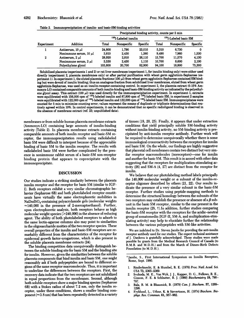
Table 2. Immunoprecipitation of insulin- and basic-SM-binding activitiesPrecipitated binding activity, counts per 5 min
'25I-Labeled insulin '25I-Labeled basic SM
Experiment Addition Total Nonspecific Specific Total Nonspecific Specific
1 Antiserum, 10 /l 24,800 1,790 23,010 5,310 6,730 0Nonimmune serum, 10 1Al 3,810 2,430 1,380 9,490 7,960 1,530
2 Antiserum, 2 Al 28,920 2,810 26,110 15,700 11,570 4,130Nonimmune serum, 2 Al 3,530 2,430 1,110 10,700 8,600 2,100Polyethylene glycol 103,600 20,700 82,900 94,100 18,800 75,300
Solubilized placenta (experiments 1 and 2) or rat liver membranes (experiment 1, for insulin binding only) were either useddirectly (experiment 2, placenta membranes only) or after partial purification with wheat germ agglutinin-Sepharose (ex-periment 1). In experiment 1, the eluted placenta fractions (100 AD) from wheat germ agglutinin-Sepharose contained SM bind-ing but were devoid of insulin binding; thus an analogous fraction from solubilized liver membranes, eluted from wheat germagglutinin-Sepharose, was used as an insulin-receptor-containing control. In experiment 2, the placenta extract (0.15% Am-monyx-LO) contained comparable amounts of both insulin-binding and basic-SM-binding activity as indicatedby the polyethyl-ene glycol assay. This extract (100 Ml) was used directly for the immunoprecipitation experiment. In experiment 1, extractswere equilibrated with 156,000 cpm of '25I-labeled insulin and 87,800 cpm of "I-labeled basic SM; in experiment 2 sampleswere equilibrated with 122,000 cpm of 125I-labeled insulin and 132,000 cpm of 125I-labeled basic SM. Immunoprecipitates werecounted for 5 min to minimize counting error; values represent the means of duplicate or triplicate determinations that rou-tinely agreed within 10%. In control experiments, it can be demonstrated that no specific radioligand binding is observed inthe absence of membrane extract (ref. 23; unpublished data).
membranes or from soluble human placenta membrane extracts(Ammonyx-LO) containing large amounts of insulin-bindingactivity (Table 2). In placenta membrane extracts containingcomparable amounts of both insulin receptor and basic-SM re-ceptor, the immunoprecipitation results using radiolabeledbasic SM were difficult to interpret because of the appreciablebinding of basic SM to the insulin receptor. The results withradiolabeled basic SM were further complicated by the pres-ence in nonimmune rabbit serum of a basic-SM non-receptorbinding protein that appears to coprecipitate with theimmunoprecipitate.
DISCUSSION
Our studies indicate a striking similarity between the placentainsulin receptor and the receptor for basic SM (similar to IGF-I). Both receptors exhibit a very similar chromatographic be-havior (Sepharose 6B) and both photolabeled receptor constit-uents have virtually the same electrophoretic mobilities inNaDodSO4-containing polyacrylamide gels (molecular weights-140,000 in the presence of 2-mercaptoethanol). Further,upon electrophoretic analysis, both receptors behave as highmolecular weight species (>240,000) in the absence ofreducingagent. The ability of both photolabeled receptors to adsorb tothe same lectin-agarose columns also may point to similaritiesin the oligosaccharide moities ofthe two receptor species. Theseoverall properties of the insulin and basic-SM receptors are re-markably different from the characteristics of the receptor forepidermal growth factor-urogastrone, which is also present inthe soluble placenta membrane extracts (24).The binding competition data unequivocally distinguish be-
tween the soluble binding site for basic SM and the binding sitefor insulin. However, given the similarities between the solubleplacenta components that bind insulin and basic SM, one mightreasonably ask if both polypeptides are bound to different re-gions ofthe same receptor macromolecule. Thus, it is importantto underline the differences between the receptors. First, therecovery data indicate that the two receptors are not solubilizedin equal proportions from the membranes. Second, althoughboth soluble receptors show a major binding species (Sepharose6B) with a Stokes radius of about 7.2 nm, only the insulin re-ceptor, under these conditions, shows a smaller binding com-ponent (-'3.8 nm) that has been repeatedly detected in a variety
of tissues (18, 20, 25). Finally, it appears that under extractionconditions that yield principally soluble SM-binding activitywithout insulin-binding activity, no SM-binding activity is pre-cipitated by anti-insulin receptor antibody. Further work willbe required to determine unequivocally whether there is someimmunological crossreactivity between the receptors for insulinand basic SM. On the whole, our findings are highly suggestivethat placental cell membranes contain two distinct but very sim-ilar receptor macromolecules (isoreceptors)-one for insulinand another for basic SM. This result is in accord with other datasuggesting that the receptors for multiplication-stimulating ac-tivity (26) and SM-A (4, 27) are distinct from the receptor forinsulin.We believe that our photolabeling method labels principally
the 140,000 molecular weight or a subunit of the insulin-re-ceptor oligomer described by others (21, 22). Our results in-dicate the presence of a very similar subunit in the basic-SMreceptor. Further studies using peptide-mapping methods todetermine the structural homology between the subunits ofthetwo receptors may establish the presence or absence ofa IB sub-unit in the basic SM receptor, similar to the one present in theinsulin receptor (28, t).In addition, further studies comparingthe basic-SM receptor with the receptors for the acidic-neutralgroup ofsomatomedin (IGF-II, SM-A, and multiplication-stim-ulating activity) may help to elucidate further the relationshipbetween the various polypeptides with SM-like activities.We are indebted to Dr. Steven Jacobs for providing the anti-insulin
receptor antibody used for our studies. The expert technical assistanceof J. Dankova is gratefully acknowledged. These studies were madepossible by grants from the Medical Research Council of Canada (toR.M.B. and M.D.H.) and from the March of Dimes-Birth DefectsFoundation (to M.D.H.).
t Jacobs, S., First International Symposium on Insulin Receptors,Rome, Sept. 1980.
1. Rinderknecht, E. & Humbel, R. E. (1976) Proc. Natd Acad. Sci.USA 73, 2365-2369.
2. Svoboda, M. E., Van Wyk, J. J., Kapper, D. G., Fellows, R. E.,Gissom, F. E. & Schlneter, R. J. (1980) Biochemistry 19, 790-797.
3. Bala, R. M. & Bhaumick, B. (1979) Can. J. Biochem. 57, 1289-1298.
4. Fryklund, L., Uthne, K. & Sievertsson, H. (1974) Biochem. Bio-phys. Res. Commun. 61, 957-962.
Proc. Nad Acad. Sci. USA 78 (1981)
Proc. Nati Acad. Sci. USA 78 (1981) 4283
5. Moses, A. C., Nissley, S. P., Short, P. A., Rechler, M. M. &Podskalny, J. M. (1980) Eur. J. Biochem. 103, 387-400.
6. Bala, R. M. & Bhaumick, B. (1979) J. Clin. EndocrinoL Metab.49, 770-777.
7. Hintz, R. L., Lin, F., Marshall, L. B. & Chang, D. (1980)1. Clin.EndocrinoL Metab. 50, 405-407.
8. Blundell, T. L., Bedarkar, S., Rinderknecht, E. & Humbel, R.E. (1978) Proc. NatL Acad. Sci. USA 75, 180-184.
9. Zapf, J., Schoenle, E. & Froesch, E. R. (1978) Eur. J. Biochem.87, 285-296.
10. Rechler, M. M., Nissley, S. P., Podskalny, M. M., Moses, A. C.& Frykiund, L. (1977) J. Clin. Endocrinot Metab. 44, 820-831.
11. Marshall, R. N., Underwood, L. E., Voina, S. J., Foushee, D.B. & Van Wyk, J. J. (1974) J. Clin. EndocrinoL Metab. 39, 283-292.
12. Cuatrecasas, P. (1972) Proc. NatL Acad. Sci. USA 69, 1277-1281.13. Hock, R. A. & Hollenberg, M. D. (1980) J. BioL Chem. 255,
10731-10736.14. Cuatrecasas, P. & Hollenberg, M. D. (1976) Adv. Protein Chem.
30, 251-451.15. Spivak, J. L., Small, D. & Hollenberg, M. D. (1977) Proc. Nati
Acad. Sci. USA 74, 4633-4635.16. Hock, R. A., Nex0, E. & Hollenberg, M. D. (1979) Nature (Lon-
don) 277, 403-405.
17. Laemmli, U. K. (1978) Nature (London) 272, 370-371.18. Maturo, J. M., III, Shackelford, W. H. & Hollenberg, M. D.
(1978) Life Sci. 23, 2063-2072.19. Cheng, Y.-C. & Prusoff, W. H. (1973) Biochem. Pharmacol 22,
3099-3108.20. Maturo, J. M., III & Hollenberg, M. D. (1978) Proc. NatL Acad.
Sci. USA 75, 3070-3074.21. Jacobs, S., Hazum, E., Shechter, Y. & Cuatrecasas, P. (1979)
Proc. Natt Acad. Sci. USA 76, 4918-4921.22. Pilch, P. F. & Czech, M. P. (1980) J. BioL Chem. 255, 1722-
1731.23. Jacobs, S., Chang, K.-J. & Cuatrecasas, P. (1978) Science 200,
1283-1284.24. Hock, R. A., Nex0, E. & Hollenberg, M. D. (1980)J. BioL Chem.
255, 10737-10743.25. Maturo, J. M., III & Hollenberg, M. D. (1979) Can. J. Biochem.
57, 497-506.26. Hollenberg, M. D. & Fryklund, L. (1977) Life Sci. 21, 943-950.27. King, G. L., Kahn, C. R., Rechler, M. M. & Nissley, S. P. (1980)
J. Clin. Invest. 66, 130-140.28. Czech, M., in Current Views on Insulin Receptors, eds. An-
dreani, D. & DePirro, R. (Academic, New York), in press.
Biochemistry: Bhaumick et al.