structural and mechanistic insights into
TRANSCRIPT

University of Nebraska - LincolnDigitalCommons@University of Nebraska - Lincoln
Biochemistry -- Faculty Publications Biochemistry, Department of
2017
Structural and Mechanistic Insights intoHemoglobincatalyzed Hydrogen Sulfide Oxidationand the Fate of Polysulfide ProductsVictor VitvitskyUniversity of Michigan Medical School
Pramod K. YadavUniversity of Michigan Medical School
Sojin AnUniversity of Michigan Medical School
Javier SeravalliUniversity of Nebraska-Lincoln, [email protected]
Uhn-Soo ChoUniversity of Michigan Medical School
See next page for additional authors
Follow this and additional works at: http://digitalcommons.unl.edu/biochemfacpub
Part of the Biochemistry Commons, Biotechnology Commons, and the Other Biochemistry,Biophysics, and Structural Biology Commons
This Article is brought to you for free and open access by the Biochemistry, Department of at DigitalCommons@University of Nebraska - Lincoln. Ithas been accepted for inclusion in Biochemistry -- Faculty Publications by an authorized administrator of DigitalCommons@University of Nebraska -Lincoln.
Vitvitsky, Victor; Yadav, Pramod K.; An, Sojin; Seravalli, Javier; Cho, Uhn-Soo; and Banerjee, Ruma V., "Structural and MechanisticInsights into Hemoglobincatalyzed Hydrogen Sulfide Oxidation and the Fate of Polysulfide Products" (2017). Biochemistry -- FacultyPublications. 395.http://digitalcommons.unl.edu/biochemfacpub/395

AuthorsVictor Vitvitsky, Pramod K. Yadav, Sojin An, Javier Seravalli, Uhn-Soo Cho, and Ruma V. Banerjee
This article is available at DigitalCommons@University of Nebraska - Lincoln: http://digitalcommons.unl.edu/biochemfacpub/395

Structural and Mechanistic Insights into Hemoglobin-catalyzed Hydrogen Sulfide Oxidation and the Fate ofPolysulfide Products*
Received for publication, January 4, 2017, and in revised form, February 15, 2017 Published, JBC Papers in Press, February 17, 2017, DOI 10.1074/jbc.M117.774943
Victor Vitvitsky‡, Pramod K. Yadav‡, Sojin An‡, Javier Seravalli§, Uhn-Soo Cho‡, and Ruma Banerjee‡1
From the ‡Department of Biological Chemistry, University of Michigan Medical School, Ann Arbor, Michigan 48109 and the§Department of Biochemistry and the Redox Biology Center, University of Nebraska, Lincoln, Nebraska 68588
Edited by F. Peter Guengerich
Hydrogen sulfide is a cardioprotective signaling molecule butis toxic at elevated concentrations. Red blood cells can synthe-size H2S but, lacking organelles, cannot dispose of H2S via themitochondrial sulfide oxidation pathway. We have recentlyshown that at high sulfide concentrations, ferric hemoglobinoxidizes H2S to a mixture of thiosulfate and iron-bound polysul-fides in which the latter species predominates. Here, we reportthe crystal structure of human hemoglobin containing low spinferric sulfide, the first intermediate in heme-catalyzed sulfideoxidation. The structure provides molecular insights into whysulfide is susceptible to oxidation in human hemoglobin but isstabilized against it in HbI, a specialized sulfide-carrying hemo-globin from a mollusk adapted to life in a sulfide-rich environ-ment. We have also captured a second sulfide bound at a postu-lated ligand entry/exit site in the �-subunit of hemoglobin,which, to the best of our knowledge, represents the first directevidence for this site being used to access the heme iron. Hydro-disulfide, a postulated intermediate at the junction betweenthiosulfate and polysulfide formation, coordinates ferric hemo-globin and, in the presence of air, generated thiosulfate. At lowsulfide/heme iron ratios, the product distribution between thio-sulfate and iron-bound polysulfides was approximately equal.The iron-bound polysulfides were unstable at physiological glu-tathione concentrations and were reduced with concomitantformation of glutathione persulfide, glutathione disulfide, andH2S. Hence, although polysulfides are unlikely to be stable in thereducing intracellular milieu, glutathione persulfide could serveas a persulfide donor for protein persulfidation, a posttransla-tional modification by which H2S is postulated to signal.
Hydrogen sulfide (H2S)2-dependent signaling occurs via per-sulfidation (Fig. 1A), a posttranslational modification of protein
cysteine residues that leads to cysteine persulfide (Cys-SSH)formation (1). Persulfidation could occur via the reactionbetween H2S and an oxidized cysteine (e.g. cysteine sulfenicacid) (Fig. 1B). Alternatively, this modification could be cata-lyzed by thiol sulfurtransferases that stabilize Cys-SSH in activesites and transfer the persulfide group to a cysteine thiolate on atarget protein (Fig. 1C) (2). Sulfur sources other than H2S thatcould potentially lead to protein persulfidation include (i) lowmolecular weight persulfides like Cys-SSH or glutathione per-sulfide (GSSH), (ii) sulfurtransferase substrates such as thiosul-fate or mercaptopyruvate, and (iii) hydropolysulfides (hereafterreferred to as polysulfides).
Cystathionine �-synthase and �-cystathionase can synthe-size Cys-SSH (3, 4) in addition to H2S (5, 6). GSSH and thiosul-fate are products of the sulfide oxidation pathway housed in themitochondrion, which converts H2S ultimately to sulfate (7, 8)via reactive sulfur species (2). In bacteria, sulfide quinone oxi-doreductase, the first enzyme in the sulfide oxidation pathway,generates polysulfides, which can serve as a periplasmic sulfurstorage form (9, 10). The product of the mammalian sulfidequinone oxidoreductase is GSSH rather than polysulfides (7, 8).Hence, a biological source of polysulfides in mammals was notknown until recently, when we demonstrated that hemepro-teins such as hemoglobin and myoglobin can support the cata-lytic oxidation of H2S to thiosulfate and polysulfides (Fig. 1D)(11, 12). The ability of human hemoglobin to oxidize sulfidestands in intriguing contrast to other hemoglobins that func-tion as sulfide carriers. Thus, organisms adapted to life in sul-fide-rich environments use specialized hemoglobins (e.g. HbI inLucina pectinata) to bind sulfide and deliver it to endosymbi-onts that utilize sulfide as an energy source (13).
Other systems have been reported to generate polysulfides asside products. For example, in the absence of a sulfur acceptor,mercaptopyruvate sulfur transferase generates polysulfides bycatalyzing repeated sulfur transfers from the substrate, 3-mer-captopyruvate, to an active site cysteine (at pH 9.1). The boundpolysulfide can eventually be released from the enzyme (14).Polysulfides can also be formed in solution (e.g. by the rapidreaction of hypochlorous acid, an oxidant produced by neutro-phils, with sulfide to generate HSCl). The latter can be subse-quently oxidized to a mixture of polysulfides, with HS4S� andHS3S� predicted to be the dominant species at physiologicalpH (15). In principle, polysulfide synthesis could occur via this
* This work was supported in part by National Institutes of Health GrantsGM112455 (to R. B.) and DK111465 (to U.-S. C). The authors declare thatthey have no conflicts of interest with the contents of this article. The con-tent is solely the responsibility of the authors and does not necessarilyrepresent the official views of the National Institutes of Health.
The atomic coordinates and structure factors (code 5UCU) have been depositedin the Protein Data Bank (http://wwpdb.org/).
1 To whom correspondence should be addressed. Tel.: 734-615-5238; E-mail:[email protected].
2 The abbreviations used are: H2S, hydrogen sulfide; Cys-SSH, cysteine persul-fide; CAM, carbamidomethyl; FeIII-Hb, methemoglobin or ferric hemoglo-bin; MSR, methionine synthase reductase; TCEP, tris(2-carboxyethyl)phosphine.
crossmarkTHE JOURNAL OF BIOLOGICAL CHEMISTRY VOL. 292, NO. 13, pp. 5584 –5592, March 31, 2017
© 2017 by The American Society for Biochemistry and Molecular Biology, Inc. Published in the U.S.A.
5584 JOURNAL OF BIOLOGICAL CHEMISTRY VOLUME 292 • NUMBER 13 • MARCH 31, 2017

route if H2S, which is typically present at very low concentra-tions, is transiently increased at sites of inflammation. Finally,H2S can reduce cytochrome c, the electron carrier betweencomplexes III and IV. However, elemental sulfur, the expectedproduct of this reaction, has not been characterized (16).
Polysulfides are considerably more reactive than H2S and, asexpected, elicit cellular effects at lower concentrations. Hence,it is not surprising that polysulfides are increasingly invoked assubstrates for protein persulfidation (17, 18). Depending onwhich sulfur atom in the catenated sulfur chain is attacked bythe protein thiol, a string of posttranslational modificationswould be generated (Fig. 1E). The drawback in using polysul-fides is that nucleophilic attack at the terminal sulfur in a cate-nated sulfur chain must be precisely controlled. Strategies forhow this control is achieved are not known and have not beenaddressed in studies invoking its relevance.
Oxidation of sulfide by ferric myoglobin and hemoglobin togenerate iron-bound polysulfides and thiosulfate representchemically challenging multistep reactions in which the reac-tion intermediates are poorly characterized (11, 12). The intra-cellular milieu is reducing, and it begs the question as towhether the polysulfides formed by hemoglobin evade reactionwith low molecular weight thiols or succumb to reduction.Because red blood cells express mercaptopyruvate sulfurtrans-ferase and synthesize H2S but lack mitochondria, understand-ing the mechanism of sulfide oxidation via the action ofFeIII-Hb assumes even greater importance in this versus inother cell types.
In this study, we have captured the initially formed HS�-FeIII-Hb intermediate by X-ray crystallography in addition to asecond sulfide at the entrance of a postulated ligand accesschannel in the �-subunit of hemoglobin. We have demon-strated that the postulated hydrodisulfide intermediate coordi-nates to ferric heme iron and is a substrate for further oxidation.
We have found that the iron-bound polysulfides are susceptibleto physiologically relevant reductants, revealing that they areunlikely to survive in the reducing conditions found in the cyto-plasm. On the other hand, the predominant persulfide productof the reaction between polysulfides and reductants (i.e. GSSH)might play a role in signaling.
Results
Structure of Human Hemoglobin with Bound Sulfide—Thecrystal structure of human FeIII-Hb incubated with H2S wasdetermined at 1.79 Å resolution (Fig. 2, Table 1). The globinfold structure of the HS�-FeIII-Hb complex is similar to theR-state structure of hemoglobin (19) with C� root mean squaredeviations of �0.2 Å. To verify that the extra density observedat the distal side of the iron is a sulfur atom (Fig. 2A), sulfuranomalous dispersion signals were collected at a 1.77-Å wave-length, and diffraction was recorded to 2.8 Å resolution. Fol-lowing molecular replacement, the sulfur anomalous differencemap was calculated to locate sulfur atoms in hemoglobin. Asshown in Fig. 2B, the sulfur anomalous signal overlaps with theelectron density near the iron atom, confirming the presence ofa sulfur ligand on the distal side of heme.
The distance between the proximal HisF8 (His-87 in the�-subunit and His-92 in the �-subunit) NE2 and the iron atomsis 2.3 Å in both the �- and �-subunits (Fig. 2, C and D). Thebond length between the iron and the sulfur atoms with unre-
FIGURE 1. Schematic showing H2S-derived reactive sulfur species andpersulfidation mechanisms. A, protein persulfidation is a posttranslationalmodification at a cysteine residue. B, persulfidation could occur via the reac-tion of H2S and an oxidized cysteine (e.g. cysteine sulfenic acid). C, oxidationof H2S via the mitochondrial sulfide oxidation pathway generates reactivesulfur species, such as GSSH and thiosulfate (S2O3
2�), which are substrates forthiol sulfurtransferases (TST) that stabilize active site persulfides and cantransfer the outer sulfur to cysteines on target proteins. D, ferric heme-depen-dent oxidation of H2S by hemoglobin (or myoglobin) leads to thiosulfate andiron-bound polysulfide formation. E, the uncatalyzed reaction of cysteine thi-ols on target proteins with polysulfide could lead to the nonspecific transferof one or more sulfur atoms, depending on which sulfur atom in the cate-nated chain is attacked.
FIGURE 2. Crystal structure of the human ferric hemoglobin sulfide com-plex. A, the 2Fo � Fc electron density map (2� contour level) near the hemegroup of the HS�-FeIII-Hb complex in the �-subunit. The density for the�-subunit was similar (not shown). B, the sulfur anomalous difference map(4� contour level) of the HS�-FeIII-Hb complex identified two sulfur atoms atthe distal side of the heme group and at a potential entry/exit point locatednear a path lined by Phe-43 and Phe-46 (PHE path, orange dashed arrow). Thesecond sulfur atom at the surface was only seen in the �-subunit. The CE loopis marked as a red dashed circle. The additional observed electron densitiesbelong to the sulfur atoms of methionine and cysteine residues. C and D,close-up showing the heme group and key residues in the �-subunit (C) andthe �-subunit (D) of FeIII-Hb treated with sulfide. The �- and �-subunits ofhemoglobin are colored dark blue and cyan, respectively. The sulfur ligand isshown as a yellow sphere.
Mechanism of Hemoglobin-catalyzed H2S Oxidation
MARCH 31, 2017 • VOLUME 292 • NUMBER 13 JOURNAL OF BIOLOGICAL CHEMISTRY 5585

strained distance refinement is 2.2 Å in both the �- and �-sub-units. The HS�-FeIII-Hb intermediate forms a hydrogen bondwith the distal HisE7 NE2 (His-58 in the �-subunit and His-63in the �-subunit) with distances of 3.0 and 3.2 Å in the �- and�-subunits, respectively (Fig. 2, C and D).
Inspection of the sulfur anomalous difference map revealedthe presence of another strong anomalous signal, which doesnot belong to the sulfur atoms in cysteine or methionine resi-dues. Located at the surface of the �-subunit near the CE loop,this sulfur atom is involved in hydrogen bonding interactionswith the backbone carbonyl groups of Phe-43, Pro-44, andPhe-46 (Fig. 2B). Interestingly, this sulfur atom is positioned atthe mouth of the PHE path, one of proposed entry/exit sites forthe hemoglobin ligands, CO and O2 (20, 21).
Reactivity of FeIII-Hb with Hydrodisulfide—We have postu-lated the presence of ferrous iron-bound hydrodisulfide (FeII-S-S�) as an intermediate in the globin-catalyzed sulfide oxida-tion reaction coordinate (11, 12). To test the possible formationof this species, we mixed FeIII-Hb with an excess of sodiumhydrodisulfide (Na2S2). A shift in the Soret peak from 405 to421 nm and the appearance of peaks at 543 and 575 nm wereobserved under anaerobic conditions (Fig. 3, A and B). Similarspectral changes were also observed under aerobic conditions(Fig. 3C). This spectrum is similar to that of FeIII-Hb treatedwith sulfide, which shows absorbance maxima at 423, 541, and577 nm (11) and suggests direct coordination of the hydrodis-ulfide to ferric hemoglobin. In the absence of O2, slow reduc-tion to FeII-Hb was observed as evidenced by a shift in the Soretpeak to 429 nm and the appearance of a broad �/� band cen-tered at 554 nm (Fig. 3B). A small increase in absorbance wasalso observed at 617 nm, indicating the formation of a smallproportion of sulfhemoglobin. Aeration of the reaction mixtureled to the formation of the oxy-FeII-Hb, with a Soret peak at 416
nm and �/� bands at 577 and 541 nm (Fig. 3D). Followingexposure to air, thiosulfate formation was observed.
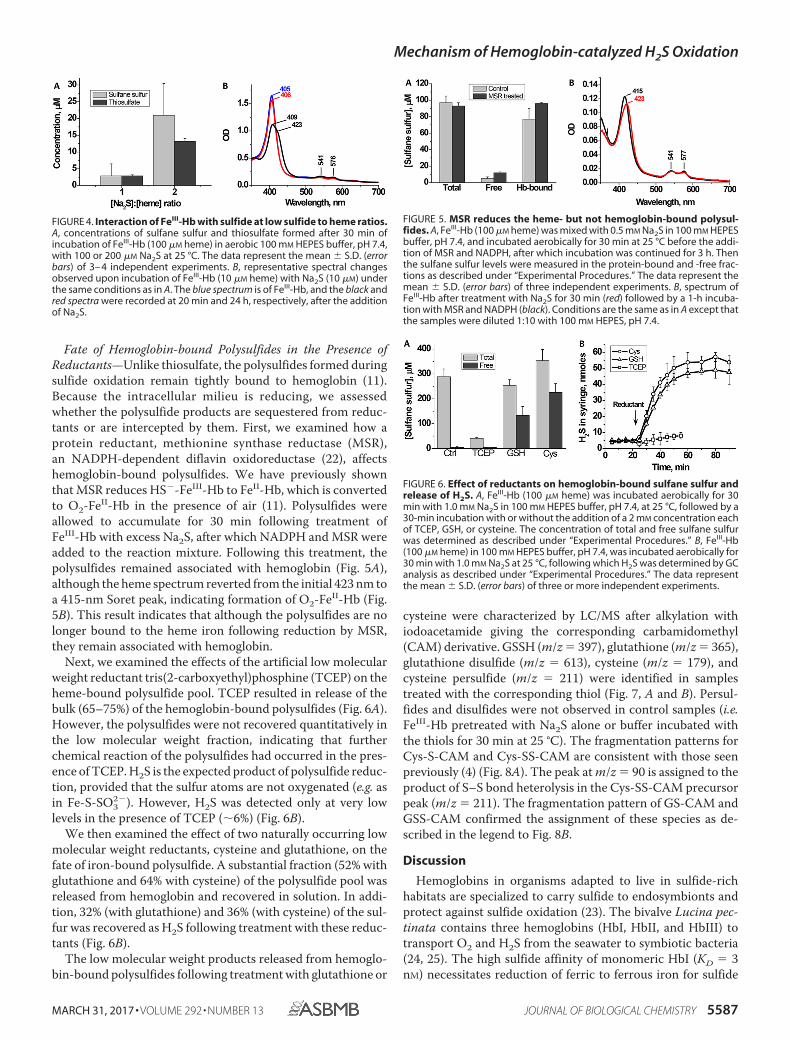
Characterization of Sulfide Oxidation Products at LowH2S/FeIII-Hb—We had previously characterized sulfide oxida-tion products at high H2S/FeIII-Hb ratios (11). However, undercellular conditions, the ratio of H2S to FeIII-Hb is expected to below, and it is not known whether the product distributionbetween thiosulfate and polysulfides would be similar or differ-ent. To address this issue, we assessed the relative concentra-tion of sulfide oxidation products that were formed when H2S/FeIII-Hb ratios were 1:1 or 2:1 (Fig. 4A). Under these conditions,low albeit detectable concentrations of polysulfides (3.0 � 3.6�M (1:1 ratio) and 20.9 � 9.4 �M (2:1 ratio)) and thiosulfate(2.9 � 0.3 �M (1:1 ratio) and 13.2 � 0.8 �M (2:1 ratio)) wereformed (Fig. 4A). These values correspond to the presence of5.8 and 26.4 �M sulfur in the thiosulfate (S2O3
2�) product.The spectrum of FeIII-Hb treated with stoichiometric Na2S is
indicative of the presence of a mixture of species, most likelyFeIII-Hb and HS�-FeIII-Hb (Fig. 4B). The Soret peak diminishesin intensity as it shifts from 405 to 409 nm with a prominentshoulder at 423 nm. In the visible region of the spectrum, thepeaks at 500 and 630 nm associated with FeIII-Hb decrease inintensity upon the addition of Na2S, and �/� bands at 576 and541 nm appear (Fig. 4B, black trace). In contrast, in the presenceof excess Na2S, the Soret peak shifts completely to 423 nm,indicating complete conversion to the HS�-FeIII-Hb species(11). The original FeIII-Hb spectrum was gradually restoredafter prolonged incubation of the sample under aerobic condi-tions (Fig. 4B, red trace).
TABLE 1Crystallographic data collection and refinement statistics
Sulfur-boundhemoglobin
Sulfur-bound hemoglobin(sulfur anomalous)
Data collectionWavelength (Å) 1.1272 1.77Space group P41212 P41212Cell dimensions
a, b, c (Å) 53,77, 53.77,193.254
53.52, 53.52, 191.72
�, �, � (degrees) 90, 90, 90 90, 90, 90Resolution (Å) 50 to 1.8
(1.86 to 1.8)a48.08 to 2.81 (2.82 to 2.81)
Rsym or Rmerge (%) 7.1 (146.3) 6.1 (10.2)Rrim or Rmeas (%) 2.5 (65.4) 6.3 (10.6)I/�I 28.97 (2.80) 34.93 (19.29)Completeness (%) 99.9 (100) 98.9 (93.4)Redundancy 11.9 13.7
RefinementResolution (Å) 37.30 to 1.80No. of reflections 27,405Rwork/Rfree 18.57/22.22No. of atoms
Protein 2458Water 169
B-factorsProtein 41.15Water 46.16
Root mean square deviationsBond lengths (Å) 0.004Bond angles (degrees) 0.688
Protein Data Bank code 5UCUa Values in parentheses are for the highest resolution shell.
FIGURE 3. UV-visible spectral changes in FeIII-Hb by Na2S2. A, FeIII-Hb (10�M heme) in 100 mM HEPES buffer, pH 7.4, was mixed with 100 �M Na2S2 at25 °C under anaerobic conditions. The blue line represents the initial spec-trum of FeIII-Hb, and the red and black spectra were recorded 1 min and 5 h,respectively, after the addition of Na2S2. The spectrum after 5 h is a mixture ofpredominantly FeII-Hb with low levels of sulfhemoglobin (as indicated by the617-nm feature). B, close-up of the visible region of the spectra in A. C, FeIII-Hb(10 �M heme) in 100 mM HEPES buffer, pH 7.4, was mixed with 100 �M Na2S2at 25 °C under aerobic conditions. The blue line represents the initial spectrumof FeIII-Hb, and the red spectrum was recorded 1 min after the addition ofNa2S2. D, spectral shift induced upon exposure of the sample in A to air. Thered spectrum corresponds to the anaerobic sample of FeII-Hb generated in Aafter a 5-h incubation of MetHb with Na2S2, and the black spectrum corre-sponds to O2-FeII-Hb formed upon exposure to air.
Mechanism of Hemoglobin-catalyzed H2S Oxidation
5586 JOURNAL OF BIOLOGICAL CHEMISTRY VOLUME 292 • NUMBER 13 • MARCH 31, 2017
Fate of Hemoglobin-bound Polysulfides in the Presence ofReductants—Unlike thiosulfate, the polysulfides formed duringsulfide oxidation remain tightly bound to hemoglobin (11).Because the intracellular milieu is reducing, we assessedwhether the polysulfide products are sequestered from reduc-tants or are intercepted by them. First, we examined how aprotein reductant, methionine synthase reductase (MSR),an NADPH-dependent diflavin oxidoreductase (22), affectshemoglobin-bound polysulfides. We have previously shownthat MSR reduces HS�-FeIII-Hb to FeII-Hb, which is convertedto O2-FeII-Hb in the presence of air (11). Polysulfides wereallowed to accumulate for 30 min following treatment ofFeIII-Hb with excess Na2S, after which NADPH and MSR wereadded to the reaction mixture. Following this treatment, thepolysulfides remained associated with hemoglobin (Fig. 5A),although the heme spectrum reverted from the initial 423 nm toa 415-nm Soret peak, indicating formation of O2-FeII-Hb (Fig.5B). This result indicates that although the polysulfides are nolonger bound to the heme iron following reduction by MSR,they remain associated with hemoglobin.
Next, we examined the effects of the artificial low molecularweight reductant tris(2-carboxyethyl)phosphine (TCEP) on theheme-bound polysulfide pool. TCEP resulted in release of thebulk (65–75%) of the hemoglobin-bound polysulfides (Fig. 6A).However, the polysulfides were not recovered quantitatively inthe low molecular weight fraction, indicating that furtherchemical reaction of the polysulfides had occurred in the pres-ence of TCEP. H2S is the expected product of polysulfide reduc-tion, provided that the sulfur atoms are not oxygenated (e.g. asin Fe-S-SO3
2�). However, H2S was detected only at very lowlevels in the presence of TCEP (�6%) (Fig. 6B).
We then examined the effect of two naturally occurring lowmolecular weight reductants, cysteine and glutathione, on thefate of iron-bound polysulfide. A substantial fraction (52% withglutathione and 64% with cysteine) of the polysulfide pool wasreleased from hemoglobin and recovered in solution. In addi-tion, 32% (with glutathione) and 36% (with cysteine) of the sul-fur was recovered as H2S following treatment with these reduc-tants (Fig. 6B).
The low molecular weight products released from hemoglo-bin-bound polysulfides following treatment with glutathione or
cysteine were characterized by LC/MS after alkylation withiodoacetamide giving the corresponding carbamidomethyl(CAM) derivative. GSSH (m/z � 397), glutathione (m/z � 365),glutathione disulfide (m/z � 613), cysteine (m/z � 179), andcysteine persulfide (m/z � 211) were identified in samplestreated with the corresponding thiol (Fig. 7, A and B). Persul-fides and disulfides were not observed in control samples (i.e.FeIII-Hb pretreated with Na2S alone or buffer incubated withthe thiols for 30 min at 25 °C). The fragmentation patterns forCys-S-CAM and Cys-SS-CAM are consistent with those seenpreviously (4) (Fig. 8A). The peak at m/z � 90 is assigned to theproduct of S–S bond heterolysis in the Cys-SS-CAM precursorpeak (m/z � 211). The fragmentation pattern of GS-CAM andGSS-CAM confirmed the assignment of these species as de-scribed in the legend to Fig. 8B.
Discussion
Hemoglobins in organisms adapted to live in sulfide-richhabitats are specialized to carry sulfide to endosymbionts andprotect against sulfide oxidation (23). The bivalve Lucina pec-tinata contains three hemoglobins (HbI, HbII, and HbIII) totransport O2 and H2S from the seawater to symbiotic bacteria(24, 25). The high sulfide affinity of monomeric HbI (KD � 3nM) necessitates reduction of ferric to ferrous iron for sulfide
FIGURE 4. Interaction of FeIII-Hb with sulfide at low sulfide to heme ratios.A, concentrations of sulfane sulfur and thiosulfate formed after 30 min ofincubation of FeIII-Hb (100 �M heme) in aerobic 100 mM HEPES buffer, pH 7.4,with 100 or 200 �M Na2S at 25 °C. The data represent the mean � S.D. (errorbars) of 3– 4 independent experiments. B, representative spectral changesobserved upon incubation of FeIII-Hb (10 �M heme) with Na2S (10 �M) underthe same conditions as in A. The blue spectrum is of FeIII-Hb, and the black andred spectra were recorded at 20 min and 24 h, respectively, after the additionof Na2S.
FIGURE 5. MSR reduces the heme- but not hemoglobin-bound polysul-fides. A, FeIII-Hb (100 �M heme) was mixed with 0.5 mM Na2S in 100 mM HEPESbuffer, pH 7.4, and incubated aerobically for 30 min at 25 °C before the addi-tion of MSR and NADPH, after which incubation was continued for 3 h. Thenthe sulfane sulfur levels were measured in the protein-bound and -free frac-tions as described under “Experimental Procedures.” The data represent themean � S.D. (error bars) of three independent experiments. B, spectrum ofFeIII-Hb after treatment with Na2S for 30 min (red) followed by a 1-h incuba-tion with MSR and NADPH (black). Conditions are the same as in A except thatthe samples were diluted 1:10 with 100 mM HEPES, pH 7.4.
FIGURE 6. Effect of reductants on hemoglobin-bound sulfane sulfur andrelease of H2S. A, FeIII-Hb (100 �M heme) was incubated aerobically for 30min with 1.0 mM Na2S in 100 mM HEPES buffer, pH 7.4, at 25 °C, followed by a30-min incubation with or without the addition of a 2 mM concentration eachof TCEP, GSH, or cysteine. The concentration of total and free sulfane sulfurwas determined as described under “Experimental Procedures.” B, FeIII-Hb(100 �M heme) in 100 mM HEPES buffer, pH 7.4, was incubated aerobically for30 min with 1.0 mM Na2S at 25 °C, following which H2S was determined by GCanalysis as described under “Experimental Procedures.” The data representthe mean � S.D. (error bars) of three or more independent experiments.
Mechanism of Hemoglobin-catalyzed H2S Oxidation
MARCH 31, 2017 • VOLUME 292 • NUMBER 13 JOURNAL OF BIOLOGICAL CHEMISTRY 5587

release (24). In contrast, human ferric hemoglobin exhibits arelatively low affinity for sulfide (17 �M) and, in the presence ofO2, catalyzes its conversion to a mixture of thiosulfate and poly-sulfides (11). Sulfide also undergoes spontaneous oxidation atneutral pH, and the product distribution is governed by theratio of sulfide/O2. When the ratio is high, polysulfides pre-dominate, and when it is low, thiosulfate and other oxyanionsare formed (26). Our earlier study on catalytic sulfide oxidationby hemoglobin was performed at high sulfide concentrations,simulating a high sulfide/O2 ratio, and led to formation ofhigher polysulfide than thiosulfate product (11). In this study,we examined the distribution of products at lower sulfide/hemeratios (1:1 and 2:1) and found that polysulfides and thiosulfatewere formed in approximately equal concentration (Fig. 4A).
Following H2S entry, the first step in the hemoglobin-cata-lyzed sulfide oxidation cycle is formation of a ferric sulfideintermediate (HS�-FeIII-Hb) (11). In the crystal structure ofhuman hemoglobin, a sulfide ligand to the heme iron wasobserved in the �- and �-subunits (Fig. 2, C and D). In addition,a second sulfur atom was seen at the surface of the �-subunit ofhemoglobin (Fig. 2B). The extra sulfur atom is located at themouth of the postulated PHE path used by ligands to access theheme iron, which was identified using xenon to fill cavities inthe crystal structure and by atomistic molecular dynamics sim-ulations (20, 21). Simulations revealed that the PHE path is themajor CO escape route from the �-subunit in the R-state ofhemoglobin (20). We conclude that the extra sulfur atom in ourcrystal structure represents the entry/exit point for H2S. To thebest of our knowledge, this is the first direct evidence for the use
of the PHE path by a ligand for accessing the heme. The ligandentry/exit points are predicted to be different in the �- and�-subunits (20, 27, 28), in agreement with our observation thatthe extra sulfur anomalous signal was observed in the �-subunitbut not in the �-subunit.
An Fe–S distance (unrestrained during refinement) of 2.2 Åwas observed in the �- and �-subunits (Figs. 2 (C and D) and9A). By comparison, the crystal structure of ferric sulfide HbIform L. pectinata revealed an Fe–S bond distance of 2.3 Å (Fig.9B). Based on a computational analysis of sulfide-bound to fer-ric myoglobin, different Fe–S bond lengths were predicted,depending on whether the distal ligand is a sulfide anion (2.24Å) or H2S (2.50 Å) (12). Our crystallographic results agree withthe calculated bond lengths for FeIII-HS� (2.24 Å) and HisF8NE2-FeIII (2.12 Å) in a low spin HS�-FeIII complex and indicatethat we have captured the first postulated sulfide oxidationintermediate (11, 12).
In human hemoglobin, the sulfide forms a hydrogen bondwith the His-58 nitrogen (Fig. 9A). In the L. pectinata HbI, thecorresponding interaction involves Gln-64, with a longer andmore flexible side chain (Fig. 9B). Furthermore, in HbI, fourphenylalanine residues form a hydrophobic cage around thesulfide ligand, whereas Gln-64 seals off access to the solvent.The tight aromatic pocket contributes to both the stabilizationof and the high affinity for the sulfide ligand in HbI, propertiessuited for its role as a sulfide carrier. In human hemoglobin,smaller hydrophobic residues (valine and leucine) substitute forthe phenylalanines in HbI (Fig. 9A). These less bulky aminoacids with flexible side chains in the distal heme pocket ofhuman hemoglobin accommodate binding of additional equiv-alents of sulfur and O2 and enable the observed oxidationchemistry. Another structural difference between sulfide-com-plexed HbI and human hemoglobin is in the conformation ofthe CE loop, which we have identified as the entry/exit site forH2S (Fig. 9C). It is not known whether the conformational dif-ference has a bearing on the kinetics of H2S access and/or sul-fide oxidation in human hemoglobin.
Of the two products of hemoglobin-catalyzed sulfide oxida-tion observed in vitro, thiosulfate is released into solution,whereas the polysulfides remain iron-bound. The fact that theintracellular environment is reducing begs the question ofwhether the polysulfides evade reduction by being sequesteredin the distal pocket. We found that MSR, which can lead toheme iron reduction, does not lead to release of polysulfidesinto solution (Fig. 5). The artificial triphosphine reductantTCEP led to the loss of hemoglobin-bound polysulfides, albeitwithout the release of H2S, the expected product of polysulfidereduction. The lack of detectable H2S is explained by the reac-tion of TCEP (Ph3P) with polysulfides to form thiophosphines(Ph3P(S)), described previously (29). In contrast, polysulfideswere released from hemoglobin with formation of H2S in thepresence of the physiologically relevant low molecular weightreductants, cysteine and glutathione (Fig. 6, A and B). The otherproducts of the reaction between glutathione or cysteine andpolysulfides were the corresponding persulfides, GSSH andCys-SSH. These persulfides, in turn, reacted with glutathioneor cysteine, generating H2S and the oxidized products, glutathi-one disulfide and cystine, which were detected by mass spec-
FIGURE 7. Mass spectrometric (LC-MS) analysis of the reaction of cysteineor glutathione with FeIII-Hb treated with Na2S. Hydrophilic interaction liq-uid ion chromatography was used to elute the components, as describedunder “Experimental Procedures.” A, traces are for the 0.5-atomic mass unitwindows for Cys-S-CAM (dashed black line, m/z � 179.25) and Cys-SS-CAM(dotted red line, m/z � 211.25). B, traces are for the 0.5-atomic mass unit win-dows for GS-SG (solid black line, m/z � 613.25), GSH (dashed black line, m/z �308.25), GS-CAM (solid red line, m/z � 365.25), and GSS-CAM (dotted red line,m/z � 397.25). In each panel, the top chromatogram represents samples con-taining reductant only (Cys or GSH), and the bottom one represents samplescontaining FeIII-Hb � Na2S that were treated with reductant. Scans were col-lected as described under “Experimental Procedures,” and masses for theselected ions were extracted using 0.5 Da windows. The peaks labeled withasterisks in A were present in all samples. They are likely to represent Tris(m/z � 122; red) and HEPES (m/z � 241; black), which were present in thesamples.
Mechanism of Hemoglobin-catalyzed H2S Oxidation
5588 JOURNAL OF BIOLOGICAL CHEMISTRY VOLUME 292 • NUMBER 13 • MARCH 31, 2017

trometry (Figs. 7 and 8). In contrast to cysteine (5 �M), theintracellular glutathione concentration in erythrocytes is high(3.2 mM) (30). Our results suggest that polysulfides, if formed inerythrocytes, would react with glutathione, forming GSSH. Thelatter, in turn, could serve as a persulfide donor for proteinpersulfidation, a posttranslational modification used for sulfidesignaling (1). GSSH is a substrate for thiol sulfurtransferases,which could, in turn, catalyze protein persulfidation, as postu-lated (2).
In summary, we provide crystallographic evidence for theroute used by sulfide to access the distal heme site in humanhemoglobin and have captured the first intermediate in thesulfide oxidation reaction (i.e. HS�-FeIII-Hb). We have alsodemonstrated the ability of hydrodisulfide to bind to FeIII-Hband be converted to products, consistent with its proposed rel-evance as an intermediate during sulfide oxidation. The suscep-tibility of iron-bound polysulfides to reduction by glutathionesuggests that GSSH rather than polysulfides might be involvedin sulfide signaling via protein persulfidation.
Experimental Procedures
Materials—Lyophilized human FeIII-Hb, glutathione, cys-teine, NADPH, and sodium sulfide nonahydrate were pur-chased from Sigma; TCEP was from Gold Biotechnology (St.Louis, MO); and sodium disulfide (Na2S2) was from DojindoMolecular Technologies (Rockville, MD). Recombinant humanMSR was prepared as described previously (22).
Measurement of Polysulfides—Sulfane sulfur concentrationwas measured using the cold cyanolysis method as describedpreviously (11). The concentration of sulfane sulfur was esti-mated using a calibration curve prepared using potassium thio-cyanate samples of known concentration.
Treatment of Hemoglobin-Sulfide with MSR—FeIII-Hb (100�M heme concentration in 100 mM HEPES-Na� buffer, pH 7.4)was incubated aerobically for 30 min at 25 °C with Na2S (0.5mM). Then, the reaction mixture was divided equally into twoaliquots. To the first aliquot, MSR and NADPH were added tofinal concentrations of 8 �M and 1 mM, respectively, while thesecond aliquot was left untreated. Both aliquots were incubatedaerobically at 25 °C for 3 h. Formation of O2-FeII-Hb in thereaction mixture was monitored spectrophotometrically. Freeand protein-bound polysulfides were separated using centrifu-gal filters (Amicon Ultracell with a 10 kDa cut-off). Samples(400 – 450 �l) were placed on the filter and centrifuged at10,000 � g and 4 °C for 10 –15 min. The filtrate containing thelow molecular weight sulfane sulfur fraction was collected sep-arately from the protein fraction on the filter, and the concen-tration of sulfane sulfur in each fraction was determined.
Release of H2S from Iron-bound Polysulfides—FeIII-Hb (100�M heme concentration) in 100 mM HEPES-Na� buffer, pH 7.4,was incubated aerobically at 25 °C for 30 min with Na2S (1 mM)to generate iron-bound polysulfides as described (11). Then 0.5ml of the reaction mixture was placed in the barrel of a 20-ml
FIGURE 8. MS/MS spectra of the reactions of cysteine and glutathione with FeIII-Hb treated with sulfide. MS/MS spectra were extracted at 14.3 min forCys-S-CAM (A; m/z � 179), at 14.5 min for GS-CAM (B; m/z � 365, top), at 13.8 min for Cys-SS-CAM (C; m/z � 211) and at 14.3 for GS-S-CAM (D; m/z � 397). Thefragmentation of GS-S-CAM produced peaks corresponding to the loss of glycine (m/z � 322), pyroglutamic acid (m/z � 268), pyroglutamic acid � NH3 (m/z �251), CAM persulfide � CO2 (m/z � 231), pyroglutamic acid � glycyl (m/z � 177), pyroglutamic acid � glycine � CO (m/z � 165), and loss of pyroglutamic acid� glycine � CO � NH3 (m/z � 148). We also observed the glyoxylic acid imine (m/z � 74) and S-S-CAM (m/z � 122) cation peaks. Fragmentation of GS-CAMshows the homologous fragments missing the additional sulfur atom (m/z � 290, 236, 219, 133, and 116). For the fragmentation of the protonated Cys-SS-CAM(m/z � 211), the main products are consistent with glyoxylic acid imine (m/z � 74), protonated 2-thiaglyoxamide (m/z � 90), 3-thia-alanine (m/z � 120),cysteine persulfide imine (m/z � 152), and protonated-3-thio-propenoic acid (m/z � 105) cation. The fragment peak at m/z � 120 is also consistent with theproduct of the loss of formic acid, CO, and NH3 from the persulfide precursor.
Mechanism of Hemoglobin-catalyzed H2S Oxidation
MARCH 31, 2017 • VOLUME 292 • NUMBER 13 JOURNAL OF BIOLOGICAL CHEMISTRY 5589

polypropylene syringe, and the headspace was flushed six timeswith N2 using a three-way stopcock and then filled with N2 to atotal volume (gas plus liquid) of 20 ml. The syringe was kept at25 °C, and 200-�l gas samples were aspirated at the desiredtimes. Within 20 min, the H2S concentration in the gas phasestabilized at the level of �0.25– 0.3 �M. Then the plunger waspushed to remove all but 2 ml of gas from the syringe, and 5 �lof 200 mM reductant (GSH, cysteine, or TCEP) in 100 mM
HEPES-Na� buffer, pH 7.4, was added to the reaction mixture
to obtain a final concentration of 2 mM. The pH of the TCEPsolution was adjusted to 7.0 with saturated potassium carbon-ate. The syringe was filled with N2 to a total volume of 20 ml andincubated at 25 °C. At the desired time intervals, 200-�l ali-quots were removed from the gas phase. The quantity of H2Sin the samples was measured using a gas chromatographequipped with 355 sulfur chemiluminescence detector (GC-SCD) (Agilent) as described (31).
Mass Spectrometric Analysis—FeIII-Hb (100 �M in heme) in100 mM HEPES-Na� buffer, pH 7.4, was incubated with 1 mM
Na2S for 30 min at 25 °C. Then the mixture was divided intothree aliquots. The control sample received no further treat-ment; the second and third aliquots were treated with GSH orcysteine to a final concentration of 2 mM. All three aliquots wereincubated for an additional 30 min at 25 °C. Then the reactionmixtures were filtered using Amicon filters (10 kDa molecularmass cut-off), and the filtrate was incubated with 10 mM iodo-acetamide for 1 h at 25 °C in the dark, frozen, and stored at�80 °C. Control samples containing buffer with 2 mM GSH orcysteine were prepared and treated with iodoacetamide in par-allel. The total and low molecular weight sulfane sulfur concen-tration was measured in all samples before and after incubationwith reductants.
Aliquots (5 �l) of the reaction mixture were injected into a4.6 � 100-mm amide XBridge column (Waters, Milford, MA)and eluted at a flow rate of 0.5 ml/min using Buffers A (20 mM
ammonium acetate, 20 mM ammonium hydroxide, pH 9.0) andB (acetonitrile) and the following steps: isocratic for 2 min with5% A, linear gradient from 5 to 95% A for 15 min, isocratic for 5min with 5% B, and reequilibration for 10 min with 5% A. Theeffluent was coupled to a Sciex 4000 QTrap triple quadrupolemass spectrometer operating in either Q1 scan (MS) or MS2(MS/MS) mode. Other instrument parameters were as follows:curtain gas � 20 liters/min, ion spray voltage � 5500 V, elec-trospray ionization temperature � 650 °C, GS1 � GS2 � 60liters/min, declustering potential � 80 V, entrance potential �10 V, exit potential � 15 V, collisional energy � 30 V (forMS/MS). Scans for MS were from m/z � 5 to 1005 in 1.0 s, andfor MS/MS, they were from m/z � 5 to 650 in 0.25 s.
Reaction of FeIII-Hb with Hydrodisulfide—The experimentswere performed inside an anaerobic chamber (Vacuum Atmo-spheres Co., Hawthorne, CA) with an atmosphere of N2 andcontaining �0.2– 0.5 ppm O2. A stock solution of Na2S2 wasprepared in anaerobic 100 mM Tris-HCl buffer, pH 8.0. Spectrawere monitored following the addition of Na2S2 (100 �M finalconcentration) to FeIII-Hb (10 �M heme) in anaerobic 100 mM
HEPES-Na� buffer, pH 7.4, in a sealed cuvette.X-ray Crystallography of FeIII-Hb in the Presence of H2S—
Crystallization was carried as described previously (19) with thefollowing modifications. Briefly, 50 mg of human FeIII-Hb wasdissolved in 1 ml of 30 mM HEPES-Na�, pH 7.4, and dialyzedovernight against 1 liter of 1.6 M K2HPO4/NaH2PO4 buffer, pH6.7, at 4 °C. The protein was concentrated to 60 mg/ml, and 200�l of the protein solution was mixed with 40 �l of toluene and300 �l of 2.8 M K2HPO4/NaH2PO4 buffer, pH 7.2, from which5-�l drops were placed on coverslips. The latter were invertedon wells containing 2.3 M K2HPO4/NaH2PO4 reservoir buffer,pH 7.2, and sealed. Crystals were obtained at 20 °C in 1 week by
FIGURE 9. Comparison of the crystal structures of sulfide-bound humanhemoglobin and HbI from L. pectinata. The distal heme pocket of the�-subunit human hemoglobin in complex with sulfide (A) and that of L. pec-tinata HbI in complex with sulfide (B). The distal heme site of L. pectinata HbIcontains a “Phe cage” comprising Phe-28 and Phe-68, which are replaced byLeu-29 and Val-62 in human hemoglobin. The interaction between Gln-64 inL. pectinata HbI and sulfide is replaced by His-58 and sulfide in human hemo-globin. C, the conformation of the CE loop (enclosed by the dashed red circle)differs in the structures of HbI and human hemoglobin. The CE loop in humanhemoglobin is postulated to be the entry/exit point for sulfide.
Mechanism of Hemoglobin-catalyzed H2S Oxidation
5590 JOURNAL OF BIOLOGICAL CHEMISTRY VOLUME 292 • NUMBER 13 • MARCH 31, 2017

the vapor diffusion method. The crystals were harvested bycryo-loops and soaked for 1 h at 20 °C in a cryoprotectant solu-tion containing 50 mM Na2S, 360 mM K2HPO4/NaH2PO4buffer, pH 7.2, and 14% (v/v) glycerol. The crystals were flash-frozen in liquid N2 and stored for data collection. The native(1.8 Å resolution) and sulfur anomalous (2.8 Å resolution) datasets of the crystals were collected at the LS-CAT beamline21-ID-D (Advanced Photon Source, Argonne National Labora-tory) at 1.13- and 1.77-Å wavelengths, respectively (Table 1).
Data sets were integrated and scaled using HKL2000. Thespace group and unit cell dimensions of the human HS�-FeIII-Hb complex were P41212 and a � b � 53.77 Å, c � 193.25Å, � � � � � � 90°. The phases of the HS�-FeIII-Hb complexwere obtained by molecular replacement using Phaser (32) withthe known structure of human hemoglobin as a search model(Protein Data Bank code 3D7O) (19). The model was built usingCOOT (33), and refinement calculations were carried out usingthe Phenix.Refine (34). The sulfur anomalous data set collectedat 1.77-Å wavelength (2.8 Å resolution) was processed by XDS(35). After molecular replacement using Phaser (32), the sulfuranomalous difference map of the HS�-FeIII-Hb complex wascalculated using the program phenix.maps (36). All molecularstructure figures were prepared using PyMOL (Schrodinger,LLC, New York).
Author Contributions—V. V. designed and performed the experi-ments. P. K. Y., S. A., and U.-S. C. crystallized hemoglobin with sul-fide and solved its structure. J. S. performed the mass spectrometricanalysis. R. B. helped conceive the experiments, analyzed the data,and co-wrote the manuscript with V. V., U.-S. C., and J. S. Allauthors approved the final version of the manuscript.
References1. Mustafa, A. K., Gadalla, M. M., Sen, N., Kim, S., Mu, W., Gazi, S. K.,
Barrow, R. K., Yang, G., Wang, R., and Snyder, S. H. (2009) H2S signalsthrough protein S-sulfhydration. Sci. Signal. 2, ra72
2. Mishanina, T. V., Libiad, M., and Banerjee, R. (2015) Biogenesis of reactivesulfur species for signaling by hydrogen sulfide oxidation pathways. Nat.Chem. Biol. 11, 457– 464
3. Ida, T., Sawa, T., Ihara, H., Tsuchiya, Y., Watanabe, Y., Kumagai, Y., Sue-matsu, M., Motohashi, H., Fujii, S., Matsunaga, T., Yamamoto, M., Ono,K., Devarie-Baez, N. O., Xian, M., Fukuto, J. M., and Akaike, T. (2014)Reactive cysteine persulfides and S-polythiolation regulate oxidativestress and redox signaling. Proc. Natl. Acad. Sci. U.S.A. 111, 7606 –7611
4. Yadav, P. K., Martinov, M., Vitvitsky, V., Seravalli, J., Wedmann, R., Fili-povic, M. R., and Banerjee, R. (2016) Biosynthesis and reactivity of cysteinepersulfides in signaling. J. Am. Chem. Soc. 138, 289 –299
5. Chiku, T., Padovani, D., Zhu, W., Singh, S., Vitvitsky, V., and Banerjee, R.(2009) H2S biogenesis by cystathionine �-lyase leads to the novel sulfurmetabolites, lanthionine and homolanthionine, and is responsive to thegrade of hyperhomocysteinemia. J. Biol. Chem. 284, 11601–11612
6. Singh, S., Padovani, D., Leslie, R. A., Chiku, T., and Banerjee, R. (2009)Relative contributions of cystathionine �-synthase and �-cystathionase toH2S biogenesis via alternative trans-sulfuration reactions. J. Biol. Chem.284, 22457–22466
7. Hildebrandt, T. M., and Grieshaber, M. K. (2008) Three enzymatic activ-ities catalyze the oxidation of sulfide to thiosulfate in mammalian andinvertebrate mitochondria. FEBS J. 275, 3352–3361
8. Libiad, M., Yadav, P. K., Vitvitsky, V., Martinov, M., and Banerjee, R.(2014) Organization of the human mitochondrial H2S oxidation pathway.J. Biol. Chem. 289, 30901–30910
9. Marcia, M., Ermler, U., Peng, G., and Michel, H. (2009) The structure ofAquifex aeolicus sulfide:quinone oxidoreductase, a basis to understandsulfide detoxification and respiration. Proc. Natl. Acad. Sci. U.S.A. 106,9625–9630
10. Brito, J. A., Sousa, F. L., Stelter, M., Bandeiras, T. M., Vonrhein, C., Teix-eira, M., Pereira, M. M., and Archer, M. (2009) Structural and functionalinsights into sulfide:quinone oxidoreductase. Biochemistry 48, 5613–5622
11. Vitvitsky, V., Yadav, P. K., Kurthen, A., and Banerjee, R. (2015) Sulfideoxidation by a noncanonical pathway in red blood cells generates thiosul-fate and polysulfides. J. Biol. Chem. 290, 8310 – 8320
12. Bostelaar, T., Vitvitsky, V., Kumutima, J., Lewis, B. E., Yadav, P. K., Brunold,T. C., Filipovic, M., Lehnert, N., Stemmler, T. L., and Banerjee, R. (2016)Hydrogen sulfide oxidation by myoglobin. J. Am. Chem. Soc. 138, 8476–8488
13. Pietri, R., Roman-Morales, E., and Lopez-Garriga, J. (2011) Hydrogen sul-fide and hemeproteins: knowledge and mysteries. Antioxid. Redox Signal.15, 393– 404
14. Hylin, J. W., and Wood, J. L. (1959) Enzymatic formation of polysulfidesfrom mercaptopyruvate. J. Biol. Chem. 234, 2141–2144
15. Nagy, P., and Winterbourn, C. C. (2010) Rapid reaction of hydrogen sul-fide with the neutrophil oxidant hypochlorous acid to generate polysul-fides. Chem. Res. Toxicol. 23, 1541–1543
16. Collman, J. P., Ghosh, S., Dey, A., and Decreau, R. A. (2009) Using afunctional enzyme model to understand the chemistry behind hydro-gen sulfide induced hibernation. Proc. Natl. Acad. Sci. U.S.A. 106,22090 –22095
17. Kimura, H. (2015) Signaling of hydrogen sulfide and polysulfides. Anti-oxid. Redox Signal. 22, 347–349
18. Greiner, R., Palinkas, Z., Basell, K., Becher, D., Antelmann, H., Nagy, P.,and Dick, T. P. (2013) Polysulfides link H2S to protein thiol oxidation.Antioxid. Redox Signal. 19, 1749 –1765
19. Yi, J., Safo, M. K., and Richter-Addo, G. B. (2008) The nitrite anion binds tohuman hemoglobin via the uncommon O-nitrito mode. Biochemistry 47,8247– 8249
20. Lucas, M. F., and Guallar, V. (2012) An atomistic view on human hemo-globin carbon monoxide migration processes. Biophys. J. 102, 887– 896
21. Savino, C., Miele, A. E., Draghi, F., Johnson, K. A., Sciara, G., Brunori, M.,and Vallone, B. (2009) Pattern of cavities in globins: the case of humanhemoglobin. Biopolymers 91, 1097–1107
22. Olteanu, H., and Banerjee, R. (2001) Human methionine synthase re-ductase, a soluble P-450 reductase-like dual flavoprotein, is sufficientfor NADPH-dependent methionine synthase activation. J. Biol. Chem.276, 35558 –35563
23. Bagarinao, T., and Vetter, R. D. (1992) Sulfide-hemoglobin interactions inthe sulfide-tolerant salt marsh resident, the California killfish Fundulusparvipinnis. J. Comp. Physiol. B 162, 614 – 624
24. Kraus, D. W., and Wittenberg, J. B. (1990) Hemoglobins of the Lucinapectinata/bacterial symbiosis. I. Molecular properties, kinetics, and equi-libria of reactions with ligands. J. Biol. Chem. 265, 16043–16053
25. Kraus, D. W., Wittenberg, J. B., Lu, J. F., and Peisach, J. (1990) Hemoglo-bins of the Lucina pectinata/bacteria symbiosis. II. An electron paramag-netic resonance and optical spectral study of the ferric proteins. J. Biol.Chem. 265, 16054 –16059
26. Chen, K. Y., and Morris, J. C. (1972) Kinetics of oxidation of aqueoussulfide by O2. Environ. Sci. Technol. 6 529 –537
27. Shadrina, M. S., Peslherbe, G. H., and English, A. M. (2015) Quaternary-linked changes in structure and dynamics that modulate O2 migrationwithin hemoglobin’s gas diffusion tunnels. Biochemistry 54, 5268 –5278
28. Shadrina, M. S., Peslherbe, G. H., and English, A. M. (2015) O2 and watermigration pathways between the solvent and heme pockets of hemoglobinwith open and closed conformations of the distal HisE7. Biochemistry 54,5279 –5289
29. Cumnock, K., Tully, T., Cornell, C., Hutchinson, M., Gorrell, J., Skidmore,K., Chen, Y., and Jacobson, F. (2013) Trisulfide modification impacts thereduction step in antibody-drug conjugation process. Bioconjug. Chem.24, 1154 –1160
30. Raftos, J. E., Whillier, S., and Kuchel, P. W. (2010) Glutathione synthesisand turnover in the human erythrocyte: alignment of a model based on
Mechanism of Hemoglobin-catalyzed H2S Oxidation
MARCH 31, 2017 • VOLUME 292 • NUMBER 13 JOURNAL OF BIOLOGICAL CHEMISTRY 5591
detailed enzyme kinetics with experimental data. J. Biol. Chem. 285,23557–23567
31. Vitvitsky, V., and Banerjee, R. (2015) H2S analysis in biological samplesusing gas chromatography with sulfur chemiluminescence detection.Methods Enzymol. 554, 111–123
32. McCoy, A. J., Grosse-Kunstleve, R. W., Adams, P. D., Winn, M. D., Sto-roni, L. C., and Read, R. J. (2007) Phaser crystallographic software. J. Appl.Crystallogr. 40, 658 – 674
33. Emsley, P., Lohkamp, B., Scott, W. G., and Cowtan, K. (2010) Features anddevelopment of Coot. Acta Crystallogr. D Biol. Crystallogr. 66, 486 –501
34. Adams, P. D., Afonine, P. V., Bunkoczi, G., Chen, V. B., Davis, I. W.,Echols, N., Headd, J. J., Hung, L. W., Kapral, G. J., Grosse-Kunstleve,R. W., McCoy, A. J., Moriarty, N. W., Oeffner, R., Read, R. J., Richard-son, D. C., et al. (2010) PHENIX: a comprehensive Python-based sys-tem for macromolecular structure solution. Acta Crystallogr. D Biol.Crystallogr. 66, 213–221
35. Kabsch, W. (2010) XDS. Acta Crystallogr. D 66, 125–13236. Praznikar, J., Afonine, P. V., Guncar, G., Adams, P. D., and Turk, D. (2009)
Averaged kick maps: less noise, more signal, and probably less bias. ActaCrystallogr. D Biol. Crystallogr. 65, 921–931
Mechanism of Hemoglobin-catalyzed H2S Oxidation
5592 JOURNAL OF BIOLOGICAL CHEMISTRY VOLUME 292 • NUMBER 13 • MARCH 31, 2017