exploration of mechanistic insights of acemetacin in
TRANSCRIPT
Cross Mark
ISSN-1996-918X
Pak. J. Anal. Environ. Chem. Vol. 21, No. 1 (2020) 115 – 124
http://doi.org/10.21743/pjaec/2020.06.14
Exploration of Mechanistic Insights of Acemetacin inMelanogenesis Through Zebrafish Model, Enzyme Kinetics,
Molecular Docking and Simulation Approaches
Hussain Raza*1, Mehar Ali Kazi2, Mubashir Hassan3,Qamar Abbas4 and Sung-Yum Seo1
1College of Natural Sciences, Department of Biological Sciences, Kongju National University, Gongju-si,Chungnam 32588, Republic of Korea.
2Institute of Biochemistry, University of Sindh, Jamshoro 76080, Pakistan.3Institute of Molecular Biology and Biotechnology, The University of Lahore, Lahore 54000 Pakistan.
4Department of Physiology, University of Sindh, Jamshoro 76080, Pakistan.*Coressponding Author Email: [email protected]
Received 09 March 2020, Revised 06 June 2020, Accepted 15 June 2020
--------------------------------------------------------------------------------------------------------------------------------------------Abstract
The present study describes the anti- melanogenesis effect of Acemetacin (ACE). Essentialprotein (melanin) that is vital for the skin for defense from UV rays. In the present research,emerging drug ACE was examined for its melanin inhibition using three different (in vitro, in vivoand computational) methods. ACE showed remarkable potency (IC50 = 0.353 ± 0.003 µM) againsttyrosinase in the comparison of standard, kojic acid (IC50 = 16.841 ± 1.161 µM) and ACEexhibited competitive inhibition. In the in vivo study zebrafish embryos were exposed with 5, 10,15 and 20 µM of ACE and same doses for positive control (Kojic Acid). At 72 h treatment, ACEexpressively (P<0.001) reduced the level of pigmentation (62.89%) at a concentration of 20 µM,relative to that of kojic acid (39.64%). The binding profile of ACE was confirmed by moleculardocking and the stability of the docked complexes was justified by MD simulation. Based on ourresults, it was concluded that ACE possessed good therapeutic potential against melanogenesis bytargeting the tyrosinase.
Keywords: Acemetacin, Tyrosinase inhibition, Melanogenesis, Computational studies, Zebrafish--------------------------------------------------------------------------------------------------------------------------------------------
Introduction
Nowadays, globally, the investment in skinbleaching mediator is approximately increase byup to US$ twenty-three billion by 2020. Asia isobserved to be the biggest market for skinwhitening agents, mainly India, China, Japan andKorea [1]. Human skin, hair and eyes aredependent on the creation of melanin and itsamount, worth and dissemination. Melanin showsan instrumental part in safeguarding the skin incontradiction to dangerous outcomes of UVradiations and oxidative trauma from differentecological contaminants [2]. The natural pigmentmelanin is synthesized from skin layers’ cell
melanocytes via the process of melanogenesis [3,4]. Embryonic neural crests are the stem cells ofmelanoblasts, these unpigmented cells have furtherproceeded towards pigmented melanocytes [5, 6].In the epidermis of the skin, every melanocyte isenclosed by about 36 keratinocytes which are highin the ratio [7].
Melanin plays out an essential part inshielding the human skin however the anomalousunion and high measure of melanin-deliveringcells in various particular organs of the humansand animals lead to many harmful and dangerous
Pak. J. Anal. Environ. Chem. Vol. 21, No. 1 (2020)116
skin diseases, for example, melisma, melanodermaafter inflammation, freckles, and wrinkles.Furthermore, exposure to UV-radiation can causedamage in DNA, mutations in genes, developmentof cancer and destruction of the immune system[8].
Melanosomes, the lysosome likeorganelles, are present in the melanocyte. Theseorganelles produce two types of melanin pigment,one is black-brown or insoluble dark polymerknown as eumelanin present in dim skin and darkhair, and the other is yellow to red and soluble,that is present in freckle skin type and red hair [9].
Melanogenesis is a very specializedprocess and involves multiple sequences ofenzymatic and chemical processes. In thesepathways, three enzymes, namely tyrosinase,dopachrome tautomerase and tyrosinase-relatedprotein-1, are very significant intermediates ofmelanogenesis. In specific, tyrosinase is fullyessential for the synthesis of melanin. Theproduction of melanin started with the conversionof L-tyrosine to L-dihydroxyphenylalanine (L-DOPA) or dopaquinone. L-DOPA acts as thesubstrate in the case of eumelanin andpheomelanin production [10, 11].
The natural decarboxylation ofdopachrome produces 5,6-dihydroxyindole whichquickly oxidizes and jointly produces insolubleblack to brown pigment known as 5,6-dihydrox-yindole-melanin. Conversely, if tyrosinase-relatedprotein 1 is present, dopachrome conversely, in thepresence of tyrosinase-related protein 1,dopachrome will produce dihydroxyindone-2-carboxylic acid (DHICA), which then oxidize andpolymerize to form DHICA-melanin. This type ofpigment is slightly soluble and medium in size andlighter brown in color. In short, the presence ofmelanin activating catalysts and substratesdetermines the types of pigment synthesized.Tyrosinase assumes a significant job in speedcontrolling all the while of oxidation because otherwhole mechanism proceeds with optimum pH[12].
The embryo of zebrafish is a verycommon vertebrate animal model forphysiological and biochemical studies because of
high similarity with mammals, hence the embryoof zebrafish has been developed as a model foranimal experiments [13]. Zebrafish has variousbenefits, such as the production of large amountsof embryos, easy care of embryos, smaller size,and most importantly high efficacy of penetrationof drugs through skin and gills. In the past, severalliterature reports have shown that most of the in-vivo phenotype-based experiments have beenperformed on zebrafish [14-16].
Acemetacin (ACE) is a glycolic acid esterof Indometacin, structurally. ACE is a non-steroidal compound with the potency of treatinginflammation used for the treatment ofosteoporosis, osteopenia, severe gout, toothache,dysmenorrhea, lower back pain and relieving post-operative pain [17, 18].
Although some indole containingcompounds have been reported as tyrosinaseinhibitors [19], yet, as literature search evident thatthere are no significant findings available for ACEas melanogenesis inhibitor. Therefore, consideringthe pharmacological importance of ACE, in thepresent investigation, we have explored itstyrosinase inhibitory potential using threeapproaches; in vitro enzyme inhibition,computational docking and dynamic simulation,and in vivo zebrafish model for determination ofpigment inhibition potency.
Materials and Methods
The analytical grade chemicals andreagents for all experiments were purchased fromSigma-Aldrich Co. Korea.
Mushroom tyrosinase inhibition assay
The potency of ACE against tyrosinasewas investigated by exactly following ourpublished method in various articles [19-21]. Inthe first step, 20 nM of phosphate buffer at pH 6.8was prepared, and 140 µL of the solution wastransferred in each well of the assay plate.Simultaneously, 20 µL of mushroom tyrosinase(target enzyme) was also added from 30 U/mL ofstock solution. In a third step, 20 µL of ACE wastransferred into the assay plate. After the firstincubation of 10 min at 25 ºC, the 20 µL of
Pak. J. Anal. Environ. Chem. Vol. 21, No. 1 (2020) 117
substrate L_DOPA was added from the 0.85 mMstock solution, and the reaction mixture wasincubated for another 20 min at room temperature.Finally, the change in absorbance was recorded atwavelength 475 nm on SpectraMax ABSmicroplate reader. For comparison and assayvalidation, kojic acid was used as a positivecontrol and assay buffer was used as a negativecontrol. For IC50 calculations, all concentrationwas investigated separately and repeated for threetime in order to obtain better accuracy of results.For calculation of IC50 through nonlinearregression GraphPad Prism was used. Byfollowing bellow mentioned equation % inhibitionwas determined.
100Bank
SampleBankinhibition%asesinTyo
Kinetic analysis
The kinetic experiment was carried toreveal the behavior of ACE in tyrosinaseinhibition. Range of doses was applied for theidentification of the pattern of inhibition oftyrosinase by ACE via kinetic evaluation using ourpublished methods [22, 23]. In total, three doses ofACE were investigated, i.e. 0.00, 0.353 and 0.706µM. Various serious of L-DOPA doses were usedfrom 0.0625 to 2 mM for all experiments. The firstincubation and reading period was the same aspresented in the above inhibition method for IC50
calculations. The maximum first velocity wasdetermined using the primary linear phase ofabsorbance for 5 min after mixing the enzymesolution at thirty seconds period. The enzymeblockage pattern was determined using theLineweaver Burk graph of the opposite ofvelocities (1/V) against the opposite of usedsubstrate doses. Another chart was drawn forcalculations of the enzyme inhibition dissociationconstant Ki through 1/V against the ACE doses.
Determination of pigmentation reducing capacityof ACE in embryos of Zebrafish (In Vivo)
The animal experiments were carriedout as accordance with the published methods[21, 24].
Zebrafish farming
The selected animal zebrafishes wereobtained from the local market and maintained inour fish facility laboratory for a period of thirtydays. All conditions were optimized for fishculture as prescribed in literature for zebrafishmaintains. Shrimp larvae were used as the foodand fishes were housed in tanks made up ofthermostatic material. For respiration, growth andbetter health, air and water filtration weremaintained. The fish seed was obtained using thenormal procedure of fish spawning performedusing the light source as a stimulator. All animalexperiments methods were confirmed byDepartmental review Board of Kongju NationalUniversity (IRB NO. 2011-2).
ACE treatment and depigmentation examination
Firstly, the E3 medium was prepared bymixing 5 mM sodium chloride, 0.17 mMpotassium chloride, 0.33 mM calcium chloride and0.33 mM magnesium chloride. Then, the fishembryos were obtained using pipette into an assayplate three to four embryos were transferred ineach well. Subsequently, the ACE solution wasprepared in 0.1% DMSO added into the embryosmedium for nine to seventy-two h post-fertilization. For comparison and assayconfirmation, kojic acid was used as positivecontrol. Then, chorion of embryos was removedand tricaine methanesulfonate MS-222 was usedas anesthesia. Lastly, embryos slide was preparedusing 1% methylcellulose on the whole slide andimages were taken using stereomicroscopepurchased from Nikon, Japan.
Quantification of melanin (In Vivo)
Melanin pigment quantification assay wascarried out by published methods [21,25,26]. Forquantification of melanin and dose selection trailswere performed and a 20 µM dose was chosen foranimal experimentations. The obtained fingerlingfishes were placed in dishes with the help ofdropper, where thirty-three embryos were used inthe plate and dosage with ACE and kojic acid,final volume of each well, adjusted with assay E3medium as 2 mL. After the drug exposure time,anesthetized embryos were cleaned with E3
Pak. J. Anal. Environ. Chem. Vol. 21, No. 1 (2020)118
medium and their eyes were removed from allused embryos and the crude extract was preparedusing centrifugation and homogenization methods.The crude pellet of the extract was mixed in 1 mLof sodium hydroxide and heated at 100 ºC for tenmin. The quantity of melanin was calculated basedon the absorbance at 405 nm and the findings weredetermined with the curve of purchased melaninfrom sigma. Whole experimental methods wererepeated thrice.
Statistical analysis
Values were expressed as the mean ± thestandard error of the mean. The (t-test) wasperformed for the experiments and overall datashowed significance results with the value ofP<0.001.
Computational MethodologyRetrieval of mushroom tyrosinase
For mushroom tyrosinase 3D structure, weused a freely available database of proteinstructures where 2Y9X PDB ID was downloadedas the structure of tyrosinase. The downloadedenzyme was then energy-reduced through theamber force field (gradient algorithm) usingsoftware named UCSF chimera 1.10.1 [27]. Thephysical, chemical and computational application,Ramachandran chart of the enzyme was examinedby the Molprobity database and Ramachandranchart was drawn using Discovery studio 2.1software. The enzyme physical structure andmathematical calculation were determined usingVADAR 1.8 [28].
Grid generation and molecular docking
The molecular docking experiment wasinitiated, a protein preparation wizard was used forthe optimization of the enzyme structure.Mandatory atoms were added into the proteinstructure. The enzyme then reduced to obtain thejoint root mean square deviation (RMSD) of 0.3Aºwith an optimum force field. The publishedliterature was used for the identification of thedynamic region of protein from the crystalstructure of the obtained protein [29,30]. Glidedocking method was used for docking experimentsof ACE with mushroom tyrosinase [23]. ACE wasprojected using the ligprep method in the
Schrödinger Suite. The anticipated bindingdynamisms (docking scores) and conformationallocations of ACE within the energetic area oftyrosinase were also executed using Glide testing.During the docking or simulations, both fractionalplasticity and full plasticity around the energeticlocation deposits are implemented by Glide/SP/XPand induced fit docking (IFD) methods [22].
Molecular dynamics (MD) simulations assay
A Molecular dynamics simulation wasperformed using GROMCS 4.5.4 package forverification of docking clusters [31]. The ACEtopology was investigated using the onlinePRODRG database and enzyme topology wasexamined using GROMOS 53A6 [32].Additionally, the receptor-ligand multiplexes weresolvated and positioned in the cubic packet middlehaving adjusted 9 Å distance. The system chargewas neutralized by adding ions. Energy reduction(nsteps=50,000) was ended by the sharpest lineagetechnique (1000 ps). While energy measurementswere prepared by Particle Mesh Ewald (PME)technique [33], The covalent bond restraints wereconsidered by the linear constraint solver (LINCS)algorithm [34]. Finally, MD arrangement is savedin md.mdp file, period phase for integration wasaccustomed as 0.002 ps, while the final distancefor small range neighbor list (rlist) was attuned at0.8 nm. The MD run was set to 30,000 ps withnsteps 15,000,000 for protein- drug complex andtrajectories files analysis was done by Xmgracetool using diverse instructions such as ‘g_energy’,‘g_rms’, ‘g_rmsf’, ‘g_chi’, ‘g_sas’, and ‘g_gyrate’respectively,(http://plasma-gate.weizmann.ac.il/ Grace/).
Results and DiscussionEnzyme inhibition
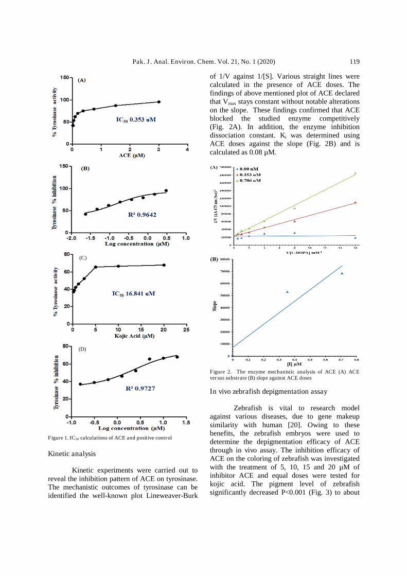
The enzyme inhibition of ACE wasperformed against tyrosinase. To determine theIC50 values of ACE and kojic acid, eightconcentrations were used. The ACE showed a verygood IC50 assessment (0.353±0.003 µM) asequated to that of positive control kojic acid(16.841±1.161). (Fig. 1A-D). Thus, it wasenvisaged that the effect may be due to themultifunctional nature of ACE structure thatmakes this molecule prone to establish somesuperb connections with the energetic side of thetarget protein.
Pak. J. Anal. Environ. Chem. Vol. 21, No. 1 (2020) 119
Figure 1. IC50 calculations of ACE and positive control
Kinetic analysis
Kinetic experiments were carried out toreveal the inhibition pattern of ACE on tyrosinase.The mechanistic outcomes of tyrosinase can beidentified the well-known plot Lineweaver-Burk
of 1/V against 1/[S]. Various straight lines werecalculated in the presence of ACE doses. Thefindings of above mentioned plot of ACE declaredthat Vmax stays constant without notable alterationson the slope. These findings confirmed that ACEblocked the studied enzyme competitively(Fig. 2A). In addition, the enzyme inhibitiondissociation constant. Ki was determined usingACE doses against the slope (Fig. 2B) and iscalculated as 0.08 µM.
Figure 2. The enzyme mechanistic analysis of ACE (A) ACEversus substrate (B) slope against ACE doses
In vivo zebrafish depigmentation assay
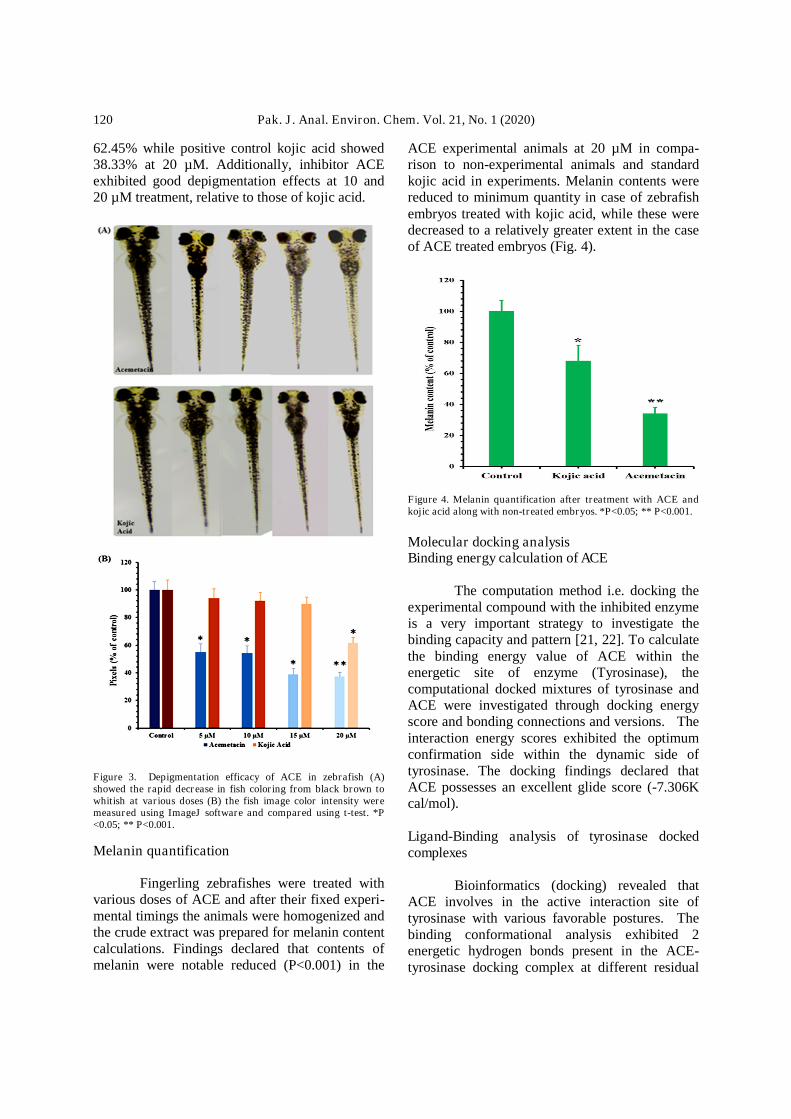
Zebrafish is vital to research modelagainst various diseases, due to gene makeupsimilarity with human [20]. Owing to thesebenefits, the zebrafish embryos were used todetermine the depigmentation efficacy of ACEthrough in vivo assay. The inhibition efficacy ofACE on the coloring of zebrafish was investigatedwith the treatment of 5, 10, 15 and 20 µM ofinhibitor ACE and equal doses were tested forkojic acid. The pigment level of zebrafishsignificantly decreased P<0.001 (Fig. 3) to about
(C)
(D)
Pak. J. Anal. Environ. Chem. Vol. 21, No. 1 (2020)120
62.45% while positive control kojic acid showed38.33% at 20 µM. Additionally, inhibitor ACEexhibited good depigmentation effects at 10 and20 µM treatment, relative to those of kojic acid.
Figure 3. Depigmentation efficacy of ACE in zebrafish (A)showed the rapid decrease in fish coloring from black brown towhitish at various doses (B) the fish image color intensity weremeasured using ImageJ software and compared using t-test. *P<0.05; ** P<0.001.
Melanin quantification
Fingerling zebrafishes were treated withvarious doses of ACE and after their fixed experi-mental timings the animals were homogenized andthe crude extract was prepared for melanin contentcalculations. Findings declared that contents ofmelanin were notable reduced (P<0.001) in the
ACE experimental animals at 20 µM in compa-rison to non-experimental animals and standardkojic acid in experiments. Melanin contents werereduced to minimum quantity in case of zebrafishembryos treated with kojic acid, while these weredecreased to a relatively greater extent in the caseof ACE treated embryos (Fig. 4).
Figure 4. Melanin quantification after treatment with ACE andkojic acid along with non-treated embryos. *P<0.05; ** P<0.001.
Molecular docking analysisBinding energy calculation of ACE
The computation method i.e. docking theexperimental compound with the inhibited enzymeis a very important strategy to investigate thebinding capacity and pattern [21, 22]. To calculatethe binding energy value of ACE within theenergetic site of enzyme (Tyrosinase), thecomputational docked mixtures of tyrosinase andACE were investigated through docking energyscore and bonding connections and versions. Theinteraction energy scores exhibited the optimumconfirmation side within the dynamic side oftyrosinase. The docking findings declared thatACE possesses an excellent glide score (-7.306Kcal/mol).
Ligand-Binding analysis of tyrosinase dockedcomplexes
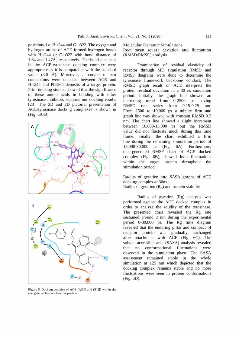
Bioinformatics (docking) revealed thatACE involves in the active interaction site oftyrosinase with various favorable postures. Thebinding conformational analysis exhibited 2energetic hydrogen bonds present in the ACE-tyrosinase docking complex at different residual
Pak. J. Anal. Environ. Chem. Vol. 21, No. 1 (2020) 121
positions, i.e. His244 and Glu322. The oxygen andhydrogen atoms of ACE formed hydrogen bondswith His244 or Glu322 with bond distance of1.64 and 1.47Å, respectively. The bond distancesin the ACE-tyrosinase docking complex wereappropriate as it is comparable with the standardvalue (3.0 Å). Moreover, a couple of π-πconnections were detected between ACE andHis244 and Phe264 deposits of a target protein.Prior docking studies showed that the significanceof these amino acids in bonding with othertyrosinase inhibitors supports our docking results[23]. The 3D and 2D pictorial presentation ofACE-tyrosinase docking complexes is shown in(Fig. 5A-B).
Figure 5. Docking complex of ACE (A)3D and (B)2D within theenergetic section of objective protein
Molecular Dynamic SimulationsRoot mean square deviation and fluctuation(RMSD/RMSF) analysis
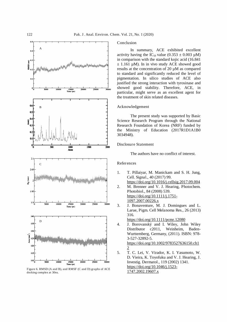
Examination of residual elasticity ofreceptor through MD simulation RMSD andRMSF diagrams were done to determine thetyrosinase framework backbone conduct. TheRMSD graph result of ACE interprets theprotein residual deviation in a 30 ns simulationperiod. Initially, the graph line showed anincreasing trend from 0-2500 ps havingRMSD rate series from 0.15-0.25 nm.From 2500 to 10,000 ps a minute firm andgraph line was showed with constant RMSD 0.2nm. The chart line showed a slight incrementbetween 10,000-15,000 ps but the RMSDvalue did not fluctuate much during this timeframe. Finally, the chart exhibited a firmline during the remaining stimulation period of15,000-30,000 ps (Fig. 6A). Furthermore,the generated RMSF chart of ACE dockedcomplex (Fig. 6B), showed loop fluctuationswithin the target protein throughout thestimulation period.
Radius of gyration and SASA graphs of ACEdocking complex at 30nsRadius of gyration (Rg) and protein stability
Radius of gyration (Rg) analysis wasperformed against the ACE docked complex inorder to analyze the solidity of the tyrosinase.The presented chart revealed the Rg ratesustained around 2 nm during the experimentalperiod 0-30,000 ps. The Rg time diagramrevealed that the enduring pillar and compact ofreceptor protein was gradually unchangedafter attachment with ACE (Fig. 6C). Thesolvent-accessible area (SASA) analysis revealedthat no conformational fluctuations wereobserved in the simulation phase. The SASAassessment remained stable in the wholesimulation at 125 nm which depicted that thedocking complex remains stable and no morefluctuations were seen in protein conformations(Fig. 6D).
B
Pak. J. Anal. Environ. Chem. Vol. 21, No. 1 (2020)122
Figure 6. RMSD (A and B), and RMSF (C and D) graphs of ACEdocking complex at 30ns.
Conclusion
In summary, ACE exhibited excellentactivity having the IC50 value (0.353 ± 0.003 µM)in comparison with the standard kojic acid (16.841± 1.161 µM). In in vivo study ACE showed goodresults at the concentration of 20 µM as comparedto standard and significantly reduced the level ofpigmentation. In silico studies of ACE alsojustified the strong interaction with tyrosinase andshowed good stability. Therefore, ACE, inparticular, might serve as an excellent agent forthe treatment of skin related diseases.
Acknowledgement
The present study was supported by BasicScience Research Program through the NationalResearch Foundation of Korea (NRF) funded bythe Ministry of Education (2017R1D1A1B03034948).
Disclosure Statement
The authors have no conflict of interest.
References
1. T. Pillaiyar, M. Manickam and S. H. Jung,Cell. Signal., 40 (2017) 99.https://doi.org/10.1016/j.cellsig.2017.09.004
2. M. Brenner and V. J. Hearing, Photochem.Photobiol., 84 (2008) 539.https://doi.org/10.1111/j.1751-1097.2007.00226.x
3. J. Bonaventure, M. J. Domingues and L.Larue, Pigm. Cell Melanoma Res., 26 (2013)316.https://doi.org/10.1111/pcmr.12080
4. J. Borovanský and I. Wiley, John WileyDistributor c2011, Weinheim, Baden-Wurttemberg, Germany, (2011). ISBN: 978-3-527-32892-5.https://doi.org/10.1002/9783527636150.ch12
5. T. C. Lei, V. Virador, K. I. Yasumoto, W.D. Vieira, K. Toyofuku and V. J. Hearing, J.Investig. Dermatol., 119 (2002) 1341.https://doi.org/10.1046/j.1523-1747.2002.19607.x
A
B
C
D
Pak. J. Anal. Environ. Chem. Vol. 21, No. 1 (2020) 123
6. E. V. Sviderskaya, S. P. Hill, D.Balachandar, G. S. Barsh and D. C. Bennett,Dev. Dynam., 221 (2001) 373.https://doi.org/10.1002/dvdy.1153
7. G. E. Costin and V. J. Hearing, FASEB J.,21 (2007) 976.https://doi.org/10.1096/fj.06-6649rev
8. J. Y. Yang, J. H. Koo, Y. G. Song, K. B.Kwon, J. H. Lee, H. S. Sohn, B. H. Park, E.C. Jhee, J. W. Park, Acta Pharmacol. Sin.,27 (2006) 1467.https://doi.org/10.1111/j.1745-7254.2006.00435.x
9. S. K. Kim, S. J. Oh, S. Y. Park, W. J. Kim,Y. S. Kim and Y. C. Kim, Pigment. CellMelanoma Res., 31 (2018) 277.https://doi.org/10.1111/pcmr.12658
10. S. M. Hashemi and E. Saeed, Pharm.Biomed. Res., 1 (2015) 1.https://doi.org/10.18869/acadpub.pbr.1.1.1
11. C. Serre, V. Busuttil and J. M. Botto, Int. J.Cosmet. Sci., 40 (2018) 328.https://doi.org/10.1111/ics.12466
12. S. Ito, Pigment Cell Res., 16 (2003) 230.https://doi.org/10.1034/j.1600-0749.2003.00037.x
13. A. Agalou, M. Thrapsianiotis, A. Angelis,A. Papakyriakou, A.L. Skaltsounis, N.Aligiannis and D. Beis, Front. Pharmacol.,9 (2018) 265.https://doi.org/10.3389/fphar.2018.00265
14. J. Rennekamp and R. T. Peterson, Curr.Opin. Chem. Biol., 24 (2015) 58.https://doi.org/10.1016/j.cbpa.2014.10.025
15. N. Tabassum, H. Tai, D. W. Jung and D. R.Williams, Evid. Based. Complement.Alternat. Med., 2015 (2015) 1.https://doi.org/10.1155/2015/287847
16. A. MacRae and R. T. Peterson, Nat. Rev.Drug Discov., 14 (2015) 721.https://doi.org/10.1038/nrd4627
17. R. Leenaraj, D. Manimaran and I. H. Joe, J.Mol. Struct., 1123 (2016) 180.https://doi.org/10.1016/j.molstruc.2016.06.035
18. T. M. Shehata, M. H. Abdallah and M. M.Ibrahim, AAPS. Pharm. Sci. Tech., 16(2015) 375.https://doi.org/10.1208/s12249-014-0233-5
19. R. Qamar, A. Saeed, F.A. Larik, Q. Abbas,M. Hassan, H. Raza and S.Y. Seo, Chem.Biol. Drug Des., 93 (2019) 123.https://doi.org/10.1111/cbdd.13352
20. Q. Abbas, Z. Ashraf, M. Hassan, H.Nadeem, M. Latif, S. Afzal and S.Y. Seo,Drug Des. Devel. Ther., 11 (2017) 2029.https://doi.org/10.2147/dddt.s137550
21. Q. Abbas, H. Raza, M. Hassan, A. R. Phull,S. J. Kim and S. Y. Seo, Chem. Biodiversity,14 (2017) e1700117.https://doi.org/10.1002/cbdv.201700117
22. F. A. Larik, A. Saeed, P. A. Channar, U.Muqadar, Q. Abbas, M. Hassan, S. Y. Seoand M. Bolte, Eur. J. Med. Chem., 141(2017) 273.https://doi.org/10.1016/j.ejmech.2017.09.059
23. A. Saeed, P. A. Mahesar, P. A. Channar, Q.Abbas, F. A. Larik, M. Hassan, H. Raza andS. Y. Seo, Bioorg. Chem., 74 (2017) 187.https://doi.org/10.1016/j.bioorg.2017.08.002
24. T. Y. Choi, J. H. Kim, D. H. Ko, C. H. Kim,J. S. Hwang, S. Ahn, S. Y. Kim, C. D. Kim,J.H. Lee and T. J. Yoon, Pigment Cell Res.,20 (2007) 120.https://doi.org/10.1111/j.1600-0749.2007.00365.x
25. K. D. Hsu, H. J. Chen, C. S. Wang, C. C.Lum, S. P. Wu, S. P. Lin and K. C. Cheng,Sci. Rep., 9 (2016) 32854.https://doi.org/10.1038/srep32854
26. S. H. Baek and S. H. Lee, Exp. Dermatol.,24 (2015) 761.https://doi.org/10.1111/exd.12765
27. F. Pettersen, T. D. Goddard, C. C. Huang,G. S. Couch, D. M. Greenblatt, E. C. Mengand T. E. Ferrin, J. Comput. Chem., 25(2004) 1605.https://doi.org/10.1002/jcc.20084
28. L. Willard, A. Ranjan, H. Zhang, H.Monzavi, R. F. Boyko, B. D. Sykes and D.S. Wishart, Nucleic Acids Res., 31 (2003)3316.https://doi.org/10.1093/nar/gkg565
29. R. A. Friesner, R. B. Murphy, M. P.Repasky, L. L. Frye, J. R. Greenwood, T. A.Halgren, P. C. Sanschagrin and D. T. Mainz,J. Med. Chem., 49 (2006) 6177.https://doi.org/10.1021/jm051256o
Pak. J. Anal. Environ. Chem. Vol. 21, No. 1 (2020)124
30. R. Farid, T. Day, R. A. Friesner and R. A.Pearlstein, Bioorg. Med. Chem., 14 (2006)3160.https://doi.org/10.1016/j.bmc.2005.12.032
31. S. Pronk, S. Páll, R. Schulz, P. Larsson, P.Bjelkmar, R. Apostolov, M. R. Shirts, J. C.Smith, P. M. Kasson, D. Van Der Spoel andB. Hess, Bioinformatics, 29 (2013) 845.https://doi.org/10.1093/bioinformatics/btt055
32. A.W. Schüttelkopf and D. M. F. Van Aalten,Acta Crystallogr. D Biol. Crystallogr., 60(2004) 1355.https://doi.org/10.1107/s0907444904011679
33. H. Wang, F. Dommert and C. Holm, J.Chem. Phys., 133 (2010) 034117.https://doi.org/10.1063/1.3446812
34. S. Amiri, M. S. Sansom and P. C. Biggin,Protein Eng. Des. Sel., 20 (2007) 353.https://doi.org/10.1093/protein/gzm029