stat5 regulation of bcl10 parallels constitutive nfkappab
TRANSCRIPT

Thomas Jefferson University Thomas Jefferson University
Jefferson Digital Commons Jefferson Digital Commons
Department of Cancer Biology Faculty Papers Department of Cancer Biology
1-1-2009
STAT5 regulation of BCL10 parallels constitutive NFkappaB STAT5 regulation of BCL10 parallels constitutive NFkappaB
activation in lymphoid tumor cells. activation in lymphoid tumor cells.
Zsuzsanna S Nagy Department of Biological Sciences, University of Texas at El Paso, El Paso, TX 79968, USA
Matthew J LeBaron Department of Cancer Biology, Thomas Jefferson University, Philadelphia, PA 19107, USA, The Dow Chemical Company, Midland, MI 48674, USA
Jeremy A Ross Department of Biological Sciences, University of Texas at El Paso, El Paso, TX 79968, USA
Abhisek Mitra Department of Biological Sciences, University of Texas at El Paso, El Paso, TX 79968, USA
Hallgeir Rui Department of Cancer Biology, Thomas Jefferson University, Philadelphia, PA 19107, USA
See next page for additional authors
Follow this and additional works at: https://jdc.jefferson.edu/cbfp
Part of the Amino Acids, Peptides, and Proteins Commons, Biological Phenomena, Cell Phenomena,
and Immunity Commons, and the Medical Cell Biology Commons
Let us know how access to this document benefits you
Recommended Citation Recommended Citation
Nagy, Zsuzsanna S; LeBaron, Matthew J; Ross, Jeremy A; Mitra, Abhisek; Rui, Hallgeir; and
Kirken, Robert A, "STAT5 regulation of BCL10 parallels constitutive NFkappaB activation in
lymphoid tumor cells." (2009). Department of Cancer Biology Faculty Papers. Paper 24.
https://jdc.jefferson.edu/cbfp/24
This Article is brought to you for free and open access by the Jefferson Digital Commons. The Jefferson Digital Commons is a service of Thomas Jefferson University's Center for Teaching and Learning (CTL). The Commons is a showcase for Jefferson books and journals, peer-reviewed scholarly publications, unique historical collections from the University archives, and teaching tools. The Jefferson Digital Commons allows researchers and interested readers anywhere in the world to learn about and keep up to date with Jefferson scholarship. This article has been accepted for inclusion in Department of Cancer Biology Faculty Papers by an authorized administrator of the Jefferson Digital Commons. For more information, please contact: [email protected].

Authors Authors Zsuzsanna S Nagy, Matthew J LeBaron, Jeremy A Ross, Abhisek Mitra, Hallgeir Rui, and Robert A Kirken
This article is available at Jefferson Digital Commons: https://jdc.jefferson.edu/cbfp/24
BioMed Central
Page 1 of 16(page number not for citation purposes)
Molecular Cancer
Open AccessResearchSTAT5 regulation of BCL10 parallels constitutive NFκB activation in lymphoid tumor cellsZsuzsanna S Nagy*1, Matthew J LeBaron2,3, Jeremy A Ross1, Abhisek Mitra1, Hallgeir Rui2 and Robert A Kirken1
Address: 1Department of Biological Sciences, University of Texas at El Paso, El Paso, TX 79968, USA, 2Department of Cancer Biology, Thomas Jefferson University, Philadelphia, PA 19107, USA and 3The Dow Chemical Company, Midland, MI 48674, USA
Email: Zsuzsanna S Nagy* - [email protected]; Matthew J LeBaron - [email protected]; Jeremy A Ross - [email protected]; Abhisek Mitra - [email protected]; Hallgeir Rui - [email protected]; Robert A Kirken - [email protected]
* Corresponding author
AbstractBackground: Signal Transducer and Activator of Transcription 5 A and B (STAT5) are key survivalfactors in cells of the lymphoid lineage. Identification of novel, tissue-specific STAT5 regulatedgenes would advance the ability to combat diseases due to aberrant STAT5 signaling. In the presentwork a library of human STAT5 bound genomic elements was created and validated.
Results: Of several STAT5 responsive genomic regulatory elements identified, one was locatedwithin the first intron of the human BCL10 gene. Chromatin immuno-precipitation reactionsconfirmed constitutive in vivo STAT5 binding to this intronic fragment in various human lymphoidtumor cell lines. Interestingly, non-phosphorylated STAT5 was found in the nuclei of Kit225 andYT cells in the absence of cytokine stimulation that paralleled constitutive NFκB activation.Inhibition of the hyperactive JAK3/STAT5 pathway in MT-2 cells via the Mannich-base, NC1153,diminished the constitutive in vivo occupancy of BCL10-SBR by STAT5, reduced NFκB activity andBCL10 protein expression in a dose dependent manner. Moreover, depletion of STAT5 viaselective antisense oligonucleotide treatment similarly resulted in decreased BCL10 mRNA andprotein expression, cellular viability and impaired NFκB activity independent of IL-2.
Conclusion: These results suggest that the NFκB regulator BCL10 is an IL-2-independent STAT5target gene. These findings proffer a model in which un-activated STAT5 can regulate pathwayscritical for lymphoid cell survival and inhibitors that disrupt STAT5 function independent oftyrosine phosphorylation may be therapeutically effective in treating certain leukemias/lymphomas.
BackgroundThe family of mammalian Signal Transducer and Activa-tor of Transcription (STAT) molecules is composed of 7members (STAT1–4, 5A, 5B and 6) which mediates a vari-ety of cellular processes including proliferation, differen-tiation and survival (reviewed in [1]). Current dogma
suggests that STATs are latent factors residing in thecytosol that only become activated following ligand bind-ing to receptors that initially results in the recruitment andactivation of Janus tyrosine kinases (JAKs). JAKs thenphosphorylate tyrosine residues on the receptor that serveas docking sites for SH2 domain-containing STATs and
Published: 26 August 2009
Molecular Cancer 2009, 8:67 doi:10.1186/1476-4598-8-67
Received: 15 July 2009Accepted: 26 August 2009
This article is available from: http://www.molecular-cancer.com/content/8/1/67
© 2009 Nagy et al; licensee BioMed Central Ltd. This is an Open Access article distributed under the terms of the Creative Commons Attribution License (http://creativecommons.org/licenses/by/2.0), which permits unrestricted use, distribution, and reproduction in any medium, provided the original work is properly cited.

Molecular Cancer 2009, 8:67 http://www.molecular-cancer.com/content/8/1/67
Page 2 of 16(page number not for citation purposes)
other signaling molecules. STATs subsequently becometyrosine phosphorylated by JAKs or other tyrosine kinases,disengage from the receptor, form dimers via phosphoty-rosine-SH2 domain interactions, and translocate to thenucleus to initiate gene transcription [2,3].
Mammalian STATs can be classified based in parts ontheir function in promoting various cellular processes. Forexample, STATs 2, 4 and 6 are critical for the immune sys-tem to promote viral defense and Th1 versus Th2 differen-tiation, respectively. Conversely, STATs 1, 3, 5A and 5B aregenerally utilized by cytokines and growth factors thatpromote cellular growth, proliferation or death (reviewedin [1]). The members of this second group are associatedwith cancer formation, including STAT1 [4]. Intriguingly,STAT3 and STAT5 promote cell survival through sharedtarget genes, including Bcl-x and Pim-1 [5-7]. Mice devoidof Stat5a and Stat5b genes have further established theseproteins as important regulators of T-cell function [8,9].Interestingly, IL-2 induced T cell proliferation was mark-edly affected only when both Stat5a and Stat5b genes wereinactivated suggesting that they play redundant roles [9].In addition to lymphocytes, STAT5A and STAT5B act asmajor survival factors for several cell types includingmammary epithelium [10,11] and human prostate can-cers [12]. Cancer cells from certain lymphomas and leuke-mias also display hyper tyrosine-phosphorylated STAT5as a result of chromosomal translocations, deregulatedtyrosine kinases or viral transformation as reviewed in [1].
Chromatin immuno-precipitation has been a widely uti-lized method to study direct transcription factor-DNAinteractions [13] and for identifying transcription factorbinding sites in unknown target genes by cloning cap-tured DNA material [14] generated from a genome-widelibrary that ultimately can be sequenced and located.Alternatively, captured DNA material can be hybridized tomicroarrays representing (i) CpG rich regions of a genomethat are contained in a significant portion of promoter ele-ments [15] or (ii) non-coding regions within whole chro-mosomes [16]. Both of these aforementioned methodshave shed new light onto the biological function, locationand kinetics of transcription factor/DNA binding depend-ent gene expression.
The present study was designed to identify genome-wideimmune specific STAT5 regulated genes. This approachhas shown promise in identifying STAT5 target genes inmouse pro-B cells [17] and human prolactin treated T47-D breast cancer cells [18]. A library of STAT5-boundgenomic fragments was created by cloning and sequenc-ing chromatin immuno-precipitated DNA fragments fromthe human lymphoma cell line, YT. One of thesesequences was identified within an intronic element ofthe BCL10 gene. We showed that STAT5 constitutively
occupied this region in vivo in multiple human lymphoidcell lines. Intriguingly, non-phosphorylated STAT5 waspresent in the nuclei of lymphoid cells that paralleled con-stitutively active NFκB. Disrupting JAK3 activity dimin-ished the in vivo binding of STAT5 to BCL10-SBR in MT-2cells, reduced NFκB activity and BCL10 protein expres-sion. Furthermore, specific STAT5 depletion correlatedwith decreased NFκB DNA-binding, cell viability andBCL10 protein expression in both the presence andabsence of IL-2. Taken together, these findings indicate anovel cross-talk mechanism between the STAT5 and NFκBpathways.
Results and DiscussionGeneration of a library encoding STAT5 Binding RegionsSince STAT5 is critical for maintaining lymphoid cell sur-vival [1], we sought to identify putative target genes thatcould be responsible for this phenotype. In the presentwork a lymphoma-specific library of IL-2-induced STAT5bound genomic elements was generated by cloning chro-matin immuno-precipitated genomic sites directly occu-pied by STAT5 as described in Figure 1A and in theMethods.
Validation of STAT5 chromatin immuno-precipitation in YT cellsIn order to confirm that STAT5 was successfully immuno-precipitated from formaldehyde-treated chromatin, YTcells were stimulated with medium (-) or IL-2 (+) for 30min at 37°C then fixed with formaldehyde. Next, STAT5was immuno-precipitated with antibodies raised againstthe N-terminus (recognizes both STAT5A and STAT5B,lanes c-d) or the C-terminus of either STAT5A (lane e) orSTAT5B (lane f), or normal rabbit serum as control IgG(lane g), separated by SDS-PAGE, and subsequently West-ern blotted with monoclonal anti-STAT5 antibody. Wholecell lysate (1% of IP) was also loaded to demonstrateequal input material for immuno-precipitation (lanes a-b). As shown in Figure 1B, all three antibodies were com-petent to bind STAT5 (indicated by arrow to the right)from fixed cells as compared to the control (lane g). Toconfirm successful capture of genomic elements known tobe occupied by STAT5, qPCR reactions were performedwith primers designed to the region harboring a knownSTAT5 binding site within the human IL2RA enhancer(Positive Regulatory Region PRR III; Figure 1C) [19]. Datapresented in Figure 1C indicated that STAT5 antibody suc-cessfully enriched PRR III as compared to control IgG(nrs).
Next, a library containing STAT5 bound genomic frag-ments was created by amplification and cloning ChIP-edDNA material as described in the Methods. The colonieswere tested for the presence of inserts by direct PCR ampli-fication using vector specific M13 primers (representative

Molecular Cancer 2009, 8:67 http://www.molecular-cancer.com/content/8/1/67
Page 3 of 16(page number not for citation purposes)
(A) Generation of a library encoding STAT5-responsive genomic elements by ChIP-cloningFigure 1(A) Generation of a library encoding STAT5-responsive genomic elements by ChIP-cloning. IL-2 stimulated, for-maldehyde cross-linked YT cells were lysed, sonicated and immuno-precipitated with antibodies to STAT5A or STAT5B. Eluted DNA was ligated to a unidirectional linker (black blocks), amplified and then cloned into pCR II-TOPO vector. Clones containing inserts were identified by sequencing. (B) Successful immuno-precipitation of STAT5 from formaldehyde fixed YT cell lysates. YT cells were stimulated with medium (-) or IL-2 (+) then fixed with formaldehyde. Fixed lysates were immuno-precipitated with antibodies to STAT5 as indicated or normal rabbit serum (IgG CTRL) then Western blotted for STAT5. Molecular weight markers are indicated to the left side of the panel. Input material corresponds to 1% of cell lysate used in the immuno-precipitations. (C) Validation of STAT5 ChIP in YT cells. ChIP assay with C-terminal antibodies to STAT5A and B in combination (αSTAT5 C-term) or IgG control was carried out as described above. The eluted DNA was then used as template in qPCR reactions with primers designed to PRR III. (D) STAT5 bound genomic library captured by ChIP-cloning. Inserts were amplified via PCR using M13 primers prior to sequencing and visualized by agarose gel electro-phoresis (1%). Stars (*) indicate clones without an insert. (E) Nearby gene mapping of the ChIP-clone identified genomic sequences. One hundred and nineteen clones were sequenced, 3 fragments were duplicates and 9 were greater than 300 kb away from any coding region. The remaining sequences that fell within 300 kb from coding regions were analyzed with Cis-Regulatory Element Annotation System (CEAS). The pie chart represents "%" distribution.

Molecular Cancer 2009, 8:67 http://www.molecular-cancer.com/content/8/1/67
Page 4 of 16(page number not for citation purposes)
colonies shown in Figure 1D) prior to sequencing. Onehundred and nineteen clones were sequenced and thegenomic locations analyzed with nearby gene mapping(CEAS) as described in the Methods. Genomic allocationof the clones is depicted in Figure 1E demonstrating themajority of the identified sequences were found inintronic (38%) and enhancer (51%) regions. These dataare in agreement with earlier findings that binding sites oftranscription factors are not restricted to promoterregions, rather, a significant portion of these sites arepresent in introns [16].
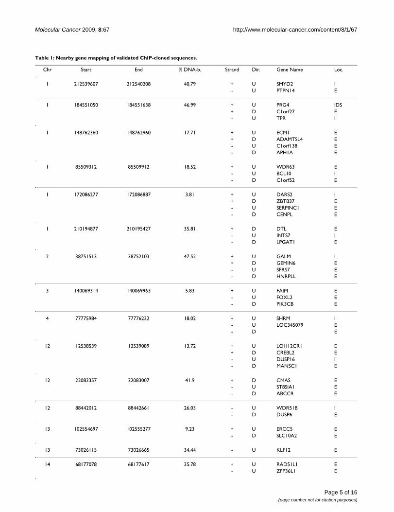
Validation of putative STAT5 binding genomic regions by EMSA-cold competition assaysTo confirm that clones encoding the sequenced genomicelements (Figure 1D) can be bound by STAT5, insertsfrom randomly selected colonies were amplified (from 52clones, 10 representatives shown in Figure 2A, upperpanel) and used in 30–50-fold molar excess as cold com-petitors in EMSA assays employing [32P]-labeled probecorresponding to the STAT5 binding site in the β-caseingene promoter and nuclear extracts from IL-2 stimulatedYT cells (Figure 2B, lower panel) [14]. The results werequantitated by comparing the band intensities of the coldcompetition EMSA reactions to the control reaction (asshown in Figure 2A graph). Of 52 validated clones, 24fragments caused greater than 50% decrease in STAT5DNA-binding intensity to the radioactively labeled probe.Table 1 summarizes the genomic location of the 20 vali-dated clones located within 300 kb of coding sequences asperformed by CEAS (four genomic segments were furtherthan 300 kb from any coding regions).
STAT5 binds an intronic element within the human BCL10 gene in vitroOne putative STAT5 responsive region was identifiedwithin the first intron of the BCL10 gene, a known regula-tor of NFκB activity and an essential positive regulator ofT and B cell development and activation [20]. The BCL10gene is located on chromosome 1 and is composed of fourexons and three introns. The STAT5 binding region wasconfined to the second intron, proximal to the 5' end ofthe third exon which we designated as the BCL10-STAT5Binding Region (BCL10-SBR). To confirm this finding,PCR amplified BCL10-SBR was used as a cold competitorin EMSA assays as described above. Data from two inde-pendent experiments (Figure 2B) showed that BCL10-SBRreduced STAT5 binding to the radioactively labeled probegreater than 80% suggesting that this element was boundby STAT5 in vitro. The genomic region surrounding theSTAT5 binding site in the human CISH promoter was alsoamplified and used as a positive control. BCL10 is anadapter molecule implicated in antigen receptor-medi-ated NFκB signaling by linking to the IκB kinase complex.The relevance of BCL10 mediated NFκB signaling for lym-phoid cells has been described in Bcl10 deficient mice as
T and B cells derived from these animals are nonfunc-tional and exhibit impaired B/T cell receptor signaling, asa consequence of impaired NFκB signaling [20,21]. Theseresults suggest an intriguing cross-talk between the STAT5and NFκB pathways, which are both implicated in malig-nant transformation. [1,22]
STAT5 constitutively occupies BCL10-SBR in vivoCold competition EMSA assays indicated that BCL10-SBRcan bind STAT5 in vitro. Next, we sought to test whetherSTAT5 can also bind this genomic element in vivo. For this
(A) Validation of the STAT5 genomic library by cold compe-tition EMSA analysisFigure 2(A) Validation of the STAT5 genomic library by cold competition EMSA analysis. Ten randomly selected STAT5 bound genomic fragments were amplified with M13 primers (upper panel) and used as cold competitors at 30–50-fold molar excess in EMSA assays using IL-2 stimulated YT nuclear extracts and [32P]-labeled STAT5-probe (lower panel). DNA-binding was expressed as % of control (a reac-tion without cold competitor (-)) as shown in the graph. The PCR amplicon surrounding the STAT5 binding site in the enhancer of the human CISH gene was used as a positive con-trol. (B) Validation of STAT5 binding to BCL10-SBR. PCR amplified BCL10-SBR was used as a cold competitor in EMSA assays employing IL-2 stimulated YT nuclear extracts and [32P]-labeled STAT5-probe (a representative of a BCL10-SBR cold competition EMSA analysis is shown). Band intensi-ties were determined by densitometric analysis. The results presented are an average of two independent experiments for BCL10-SBR and three for CISH.
Molecular Cancer 2009, 8:67 http://www.molecular-cancer.com/content/8/1/67
Page 5 of 16(page number not for citation purposes)
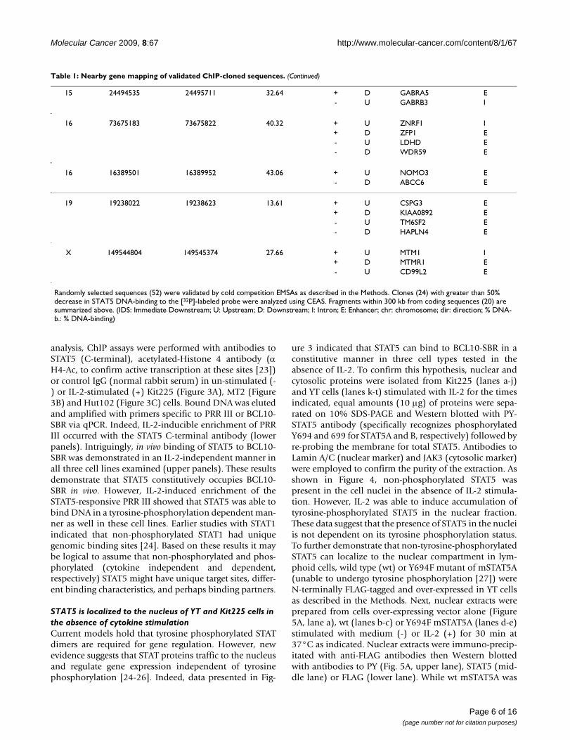
Table 1: Nearby gene mapping of validated ChIP-cloned sequences.
Chr Start End % DNA-b. Strand Dir. Gene Name Loc.
1 212539607 212540208 40.79 + U SMYD2 I- U PTPN14 E
1 184551050 184551638 46.99 + U PRG4 IDS+ D C1orf27 E- U TPR I
1 148762360 148762960 17.71 + U ECM1 E+ D ADAMTSL4 E- U C1orf138 E- D APH1A E
1 85509312 85509912 18.52 + U WDR63 E- U BCL10 I- D C1orf52 E
1 172086277 172086887 3.81 + U DARS2 I+ D ZBTB37 E- U SERPINC1 E- D CENPL E
1 210194877 210195427 35.81 + D DTL E- U INTS7 I- D LPGAT1 E
2 38751513 38752103 47.52 + U GALM I+ D GEMIN6 E- U SFRS7 E- D HNRPLL E
3 140069314 140069963 5.83 + U FAIM E- U FOXL2 E- D PIK3CB E
4 77775984 77776232 18.02 + U SHRM I- U LOC345079 E- D E
12 12538539 12539089 13.72 + U LOH12CR1 E+ D CREBL2 E- U DUSP16 I- D MANSC1 E
12 22082357 22083007 41.9 + D CMAS E- U ST8SIA1 E- D ABCC9 E
12 88442012 88442661 26.03 - U WDR51B I- D DUSP6 E
13 102554697 102555277 9.23 + U ERCC5 E- D SLC10A2 E
13 73026115 73026665 34.44 - U KLF12 E
14 68177078 68177617 35.78 + U RAD51L1 E- U ZFP36L1 E
Molecular Cancer 2009, 8:67 http://www.molecular-cancer.com/content/8/1/67
Page 6 of 16(page number not for citation purposes)
analysis, ChIP assays were performed with antibodies toSTAT5 (C-terminal), acetylated-Histone 4 antibody (αH4-Ac, to confirm active transcription at these sites [23])or control IgG (normal rabbit serum) in un-stimulated (-) or IL-2-stimulated (+) Kit225 (Figure 3A), MT2 (Figure3B) and Hut102 (Figure 3C) cells. Bound DNA was elutedand amplified with primers specific to PRR III or BCL10-SBR via qPCR. Indeed, IL-2-inducible enrichment of PRRIII occurred with the STAT5 C-terminal antibody (lowerpanels). Intriguingly, in vivo binding of STAT5 to BCL10-SBR was demonstrated in an IL-2-independent manner inall three cell lines examined (upper panels). These resultsdemonstrate that STAT5 constitutively occupies BCL10-SBR in vivo. However, IL-2-induced enrichment of theSTAT5-responsive PRR III showed that STAT5 was able tobind DNA in a tyrosine-phosphorylation dependent man-ner as well in these cell lines. Earlier studies with STAT1indicated that non-phosphorylated STAT1 had uniquegenomic binding sites [24]. Based on these results it maybe logical to assume that non-phosphorylated and phos-phorylated (cytokine independent and dependent,respectively) STAT5 might have unique target sites, differ-ent binding characteristics, and perhaps binding partners.
STAT5 is localized to the nucleus of YT and Kit225 cells in the absence of cytokine stimulationCurrent models hold that tyrosine phosphorylated STATdimers are required for gene regulation. However, newevidence suggests that STAT proteins traffic to the nucleusand regulate gene expression independent of tyrosinephosphorylation [24-26]. Indeed, data presented in Fig-
ure 3 indicated that STAT5 can bind to BCL10-SBR in aconstitutive manner in three cell types tested in theabsence of IL-2. To confirm this hypothesis, nuclear andcytosolic proteins were isolated from Kit225 (lanes a-j)and YT cells (lanes k-t) stimulated with IL-2 for the timesindicated, equal amounts (10 μg) of proteins were sepa-rated on 10% SDS-PAGE and Western blotted with PY-STAT5 antibody (specifically recognizes phosphorylatedY694 and 699 for STAT5A and B, respectively) followed byre-probing the membrane for total STAT5. Antibodies toLamin A/C (nuclear marker) and JAK3 (cytosolic marker)were employed to confirm the purity of the extraction. Asshown in Figure 4, non-phosphorylated STAT5 waspresent in the cell nuclei in the absence of IL-2 stimula-tion. However, IL-2 was able to induce accumulation oftyrosine-phosphorylated STAT5 in the nuclear fraction.These data suggest that the presence of STAT5 in the nucleiis not dependent on its tyrosine phosphorylation status.To further demonstrate that non-tyrosine-phosphorylatedSTAT5 can localize to the nuclear compartment in lym-phoid cells, wild type (wt) or Y694F mutant of mSTAT5A(unable to undergo tyrosine phosphorylation [27]) wereN-terminally FLAG-tagged and over-expressed in YT cellsas described in the Methods. Next, nuclear extracts wereprepared from cells over-expressing vector alone (Figure5A, lane a), wt (lanes b-c) or Y694F mSTAT5A (lanes d-e)stimulated with medium (-) or IL-2 (+) for 30 min at37°C as indicated. Nuclear extracts were immuno-precip-itated with anti-FLAG antibodies then Western blottedwith antibodies to PY (Fig. 5A, upper lane), STAT5 (mid-dle lane) or FLAG (lower lane). While wt mSTAT5A was
15 24494535 24495711 32.64 + D GABRA5 E- U GABRB3 I
16 73675183 73675822 40.32 + U ZNRF1 I+ D ZFP1 E- U LDHD E- D WDR59 E
16 16389501 16389952 43.06 + U NOMO3 E- D ABCC6 E
19 19238022 19238623 13.61 + U CSPG3 E+ D KIAA0892 E- U TM6SF2 E- D HAPLN4 E
X 149544804 149545374 27.66 + U MTM1 I+ D MTMR1 E- U CD99L2 E
Randomly selected sequences (52) were validated by cold competition EMSAs as described in the Methods. Clones (24) with greater than 50% decrease in STAT5 DNA-binding to the [32P]-labeled probe were analyzed using CEAS. Fragments within 300 kb from coding sequences (20) are summarized above. (IDS: Immediate Downstream; U: Upstream; D: Downstream; I: Intron; E: Enhancer; chr: chromosome; dir: direction; % DNA-b.: % DNA-binding)
Table 1: Nearby gene mapping of validated ChIP-cloned sequences. (Continued)

Molecular Cancer 2009, 8:67 http://www.molecular-cancer.com/content/8/1/67
Page 7 of 16(page number not for citation purposes)
STAT5 constitutively occupies BCL10-SBR in vivo in Kit225, MT2 and Hut102 cellsFigure 3STAT5 constitutively occupies BCL10-SBR in vivo in Kit225, MT2 and Hut102 cells. Kit225 cells (A) were made quiescent in medium without IL-2 for 24 h, MT2 (B) and Hut102 (C) cells were grown until exhaustion. Cells were then stimu-lated with medium (-) or IL-2 (+) for 30 min at 37°C, then fixed with 1% formaldehyde for 10 min at room temperature and then chromatin immuno-precipitated with antibodies to Acetyl-Histone 4, C-terminal STAT5 or control IgG. The eluted DNA was amplified with primers corresponding to PRR III (black bars) or BCL10-SBR (grey bars). Representative data for MT2, Hut102 (n = 2), and Kit225 cells (n = 3) are shown. Input material represents 5% of immuno-precipitated chromatin.

Molecular Cancer 2009, 8:67 http://www.molecular-cancer.com/content/8/1/67
Page 8 of 16(page number not for citation purposes)
tyrosine-phosphorylated upon IL-2 stimulation, theY694F mutant was not. However, both wt and Y694FmSTAT5A were constitutively present in the cell nucleisuggesting that STAT5 nuclear localization can occur inthe absence of tyrosine phosphorylation. To confirm thatYT cells over-expressing Y694F mSTAT5A retained theability to respond to IL-2, as well as to demonstrate thatSTAT5 nuclear presence was not due to contaminationwith cytosolic proteins, whole nuclear extracts isolatedabove were Western blotted with PY-STAT5 then re-blot-ted with antibodies to STAT5, Lamin A/C (a nuclearmarker) followed by β-actin (cytosolic marker) as shownin Figure 5B. Similar results were obtained with Y699FmSTAT5B (data not shown).
Traditionally, STAT transcription factors were thought toreside in the cytoplasm in the absence of cytokine stimu-lation, and only enter the nucleus to bind DNA and initi-ate gene expression following cytokine engagement[2,28]. However, interesting new evidence suggests thatnuclear-localized non-tyrosine-phosphorylated STATs canregulate gene expression. Indeed, interferon-mediatedgene expression changes in a STAT1-deficient cell linetransfected with a Y699A mutant of STAT1 unable tobecome tyrosine-phosphorylated proved it can initiateconstitutive gene expression [24]. Other recent publica-tions have reported that STAT3 can also induce gene tran-scription in the absence of tyrosine phosphorylation [26].Moreover, non-phosphorylated, nuclear localized STAT6in a non-small cell lung cancer model was shown to drivecyclooxygenase-2 expression independent of its tyrosinephosphorylation status [25]. Our results provide the firstevidence that non-tyrosine-phosphorylated, nuclear-localized STAT5 may also play a similar and critical role ingene regulation in lymphoid cells in the absence of stim-ulation/activation.
NFκB is constitutively active in YT, Kit225 cells and activated human PBMCsSince BCL10 is a positive regulator of NFκB [20], next wesought to test the activation status of NFκB in lymphoidcells. EMSA analysis was performed with either a [32P]-labeled NFκB (lower panel) or STAT5 (upper panel) probeand 5 μg nuclear extracts from YT (Figure 6A, lanes b-d),Kit225 (lanes e-g) cells or naïve (lanes h-j) and activated(lanes k-m) human PBMCs stimulated with medium (-)or IL-2 (+) for 30 min. Figure 6A demonstrated that whileIL-2 was able to induce DNA-binding of STAT5 in YT (lanec), Kit225 (lane f) and PBMCs (lane l), NFκB DNA-bind-ing was constitutive in these cells. Naïve PBMCs, whichdid not respond to IL-2, did not display binding to eitherprobe, thus verifying that constitutive NFκB binding wasnot an artifact resulting from nuclear extraction. To con-firm the specificity of the observed bands, a reaction with-out nuclear extract (Free probe) and cold competitionassays with the corresponding unlabeled probes (c.p.)were also performed (lanes d, g, j and m). To further verifythe specificity of the NFκB bands, antibodies to p50 (Fig-ure 6B, lane c), p65 (lane d) or both (lane e) were used insupershift analyses. Indeed, both p50 and p65 antibodiesresulted in partial supershifts of the NFκB band, whileusing these antibodies in combination resulted in a com-plete supershift. On the contrary, normal goat serum (IgGctrl, lane f) did not result in a supershift of the NFκBbands.
Blockade of the JAK3/STAT5 pathway diminishes in vivo STAT5 binding to BCL10-SBR, impairs NFκB function and reduces BCL10 expressionIn order to confirm that the in vivo binding of STAT5 toBCL10-SBR is responsive to the inhibition of the JAK3/STAT5 pathway, we employed the selective JAK3 inhibitorNC1153 [29]. Although the precise regulation of STAT5
Nuclear localized STAT5 is present in YT and Kit225 cells in the absence of IL-2 stimulationFigure 4Nuclear localized STAT5 is present in YT and Kit225 cells in the absence of IL-2 stimulation. Equal amounts (10 μg) of nuclear and cytosolic proteins (Kit225: lanes a-j, YT: lanes k-t) were resolved on a 10% SDS-PAGE and Western blots performed with PY-STAT5, STAT5, Lamin A/C (nuclear marker) or Jak3 (cytosolic marker) antibodies (indicated to the right). Representative data from three independent experiments are presented.

Molecular Cancer 2009, 8:67 http://www.molecular-cancer.com/content/8/1/67
Page 9 of 16(page number not for citation purposes)
by JAK3 is not yet fully understood, it has been shown thatphosphorylated STAT1 and STAT3 can increase the expres-sion of non-phosphorylated STAT1 and STAT3, respec-tively [30]. Therefore, it was hypothesized that non-phosphorylated STAT5 function could partially beaffected by the inhibition of phosphorylated STAT5. First,the activation status of the JAK3/STAT5 pathway wastested in MT-2 cells treated with ascending amounts ofNC1153 for 24 h as indicated (Figure 7A, lanes c-e) byWestern blotting. Constitutive tyrosine phosphorylationof STAT5 was diminished by NC1153 in a dose dependentmanner as compared to non-treated (NT, lane a) or vehi-cle treated (DMSO, lane b) samples. Equal loading wasconfirmed by re-probing the membrane with antibodies
to STAT5 and GAPDH. Moreover, tyrosine phosphoryla-tion of JAK3 was similarly decreased upon NC1153 treat-ment (data not shown). Next, in vivo binding of STAT5 toPRR III and BCL10-SBR were assessed by ChIP assays andqPCR. As presented in Figure 7B, the occupancy of theseregions by STAT5 was reduced in a dose dependent man-ner upon NC1153 (expressed as fold change of STAT5binding over background (IgG control)). Lastly, the func-tional effect of JAK3 blockade on the expression of BCL10protein and the activation status of NFκB was assessed.Since BCL10 is a known regulator of NFκB signaling inlymphoid cells [20] that is a critical pathway for mediat-ing survival of activated B- and T-cells, it was reasonableto assume that STAT5 depletion mediated decrease ofBCL10 expression might lead to diminished constitutiveNFκB activation. For this assay, MT-2 cells were treatedwith DMSO (Figure 7C, lane a) or ascending concentra-tions of NC1153 for 48 h as indicated (lanes b-d), thenharvested and Western blotted with antibodies to phos-pho-p65/NFκB, p65/NFκB and BCL10. Indeed, data pre-sented in Figure 7C demonstrated that phosphorylationof p65 NFκB on Ser536, an indicator of its enhanced tran-scriptional activity [31], was decreased in parallel toBCL10 protein expression upon NC1153 treatment. Equalloading was confirmed by re-probing the membrane withGAPDH (lower panel). It should be noted that somereduction in the level of total p65 resulted from the treat-ments with higher concentrations of NC1153 that couldbe due to decreased cellular viability at this time point.However, the lowest dose of NC1153 (10 μM) did notaffect total p65 but reduced its Ser536 phosphorylation aswell as BCL10 levels confirming that these reductionswere not due to non-specific treatment effects.
STAT5 depletion reduces BCL10 mRNA and protein expression, decreases the viability of Kit225 leukemia cells and diminishes NFκB DNA binding independently of IL-2 stimulationIn order to test whether STAT5 has a direct role in regulat-ing BCL10 expression and that this effect is independentof cytokines, antisense STAT5 ODN targeting bothSTAT5A and B were utilized. Earlier results demonstratedthat STAT5 is a critical survival factor for activated T-cellsand some lymphoid cell lines. [32] First, Kit225 cells wereleft untreated (Figure 8A and 8B, NT), electroporatedwithout ODN (EP), with 2.5 or 5 μM antisense STAT5ODN (AS STAT5) or 2.5 or 5 μM control ODN (CTRL),cultured in medium without (not shown) or with IL-2 for24 h, and then harvested. Messenger RNA levels of BCL10was measured via qRT PCR with primers specific tohuman BCL10 (Figure 8A) as described in the Methods.STAT5 depleted, but not control treated samples, dis-played reduced BCL10 transcript levels. Next, parallelsamples were lysed, equal amounts of lysates resolved on12% SDS-PAGE and Western blotted with antibodies to
Y694F-mSTAT5A can localize to the nuclei of YT cellsFigure 5Y694F-mSTAT5A can localize to the nuclei of YT cells. (A) YT cells over-expressing vector alone (Vector), wt or Y694F mSTAT5A were stimulated with medium (-) or IL-2 (+) for 30 min at 37°C. Nuclear extracts were prepared and immuno-precipitated with anti-FLAG antibodies, resolved on 7.5% SDS-PAGE then Western blotted with PY antibodies followed by re-blotting with antibodies to STAT5 and FLAG as indicated to the right. (B) Nuclear extracts iso-lated as described above were resolved on a 7.5% SDS-PAGE, Western blotted with PY-STAT5 antibody then re-blotted with antibodies to STAT5, Lamin A/C and β-actin as indicated to the right.

Molecular Cancer 2009, 8:67 http://www.molecular-cancer.com/content/8/1/67
Page 10 of 16(page number not for citation purposes)
BCL10 (Figure 8B). The blot was then re-probed with anti-bodies to STAT5 and GAPDH (as a loading control).Decreased STAT5 expression (Figure 8B, upper panel,lanes c-d) correlated with reduced BCL10 protein levels(middle panel) in a dose dependent manner, whileGAPDH levels were not affected (lower panel). Kit225cells depleted of STAT5 and cultured in the absence of IL-
2 also displayed reduced BCL10 protein levels comparedto controls (data not shown). Taken together, these datafurther support the notion that STAT5 regulates BCL10expression.
Since STAT5 promotes lymphoid cell survival [32,33], cellviability following STAT5 depletion was also assessed by
NFκB is constitutively active in multiple lymphoid cellsFigure 6NFκB is constitutively active in multiple lymphoid cells (A) YT (lanes b-d), quiescent Kit225 (lanes e-g), naïve (lanes h-j) and quiescent activated human PBMCs (lanes k-m) were stimulated with IL-2 for 30 min before nuclear extracts were pre-pared. Equivalent amounts (5 μg) were used in EMSA reactions employing a STAT5 (upper panel) or NFκB radio-labeled probe (lower panel). A reaction without nuclear extract (free probe, lane a) or with unlabeled probes (lanes d, g, j, m) were used to confirm the specificity of the bands. (B) Constitutive NFκB is composed of p50 and p65 subunits in lymphoid cells. Antibodies to p50 (lane c) and p65 (lane d) subunits of NFκB were used either alone or in combination (lane e) for supershift assays in Kit225 nuclear extracts. Control IgG (normal goat serum, lane f) and a reaction without nuclear extract (free probe, lane a) were used to confirm the specificity of the bands. Star (*) indicates a non-specific band.

Molecular Cancer 2009, 8:67 http://www.molecular-cancer.com/content/8/1/67
Page 11 of 16(page number not for citation purposes)
JAK3/STAT5 blockade inhibits constitutive STAT5 tyrosine phosphorylation and BCL10/NFκB activity in MT-2 cellsFigure 7JAK3/STAT5 blockade inhibits constitutive STAT5 tyrosine phosphorylation and BCL10/NFκB activity in MT-2 cells. (A) MT-2 cells were treated with medium, DMSO (0.1%, vehicle control) ascending amounts of NC1153 as indicated for 24 h. Total cell lysates were Western blotted with antibodies as indicated to the right. Molecular weight markers are indicated to the left. (B) JAK3 blockade diminishes BCL10-SBR occupancy by STAT5 in vivo. MT-2 cells were treated with NC1153 as indicated for 10 h. ChIP assays were performed with anti-STAT5 antibody and IgG control from each treatment. STAT5 DNA binding is expressed as fold change over background (IgG control). (C) NC1153 reduces NFκB activity and BCL10 protein expression. MT-2 cells were treated as described above for 48 h. Total cell lysates were Western blotted with antibodies indicated to the right. Molecular weight markers are shown to the left.

Molecular Cancer 2009, 8:67 http://www.molecular-cancer.com/content/8/1/67
Page 12 of 16(page number not for citation purposes)
MTS assays. As shown in Figure 8C, reduced STAT5 andBCL10 expression decreased Kit225 cell viability in a dosedependent manner, regardless of the absence (white bars)or presence (black bars) of IL-2 in the culture medium.These data further suggest that non-cytokine activatedSTAT5 dependent gene regulation may be functionally
important in tumor cell lines such as Kit225. Indeed, IL-2starved Kit225 cells were greater than 90% viable(assessed by trypan blue exclusion) after 72 h, althoughtyrosine phosphorylated STAT5 was abolished within 24h (data not shown). Interestingly, antisense oligonucle-otide depletion of STAT5 resulted in greater than 50%
(A) STAT5 depletion decreases BCL10 mRNA expression in a dose dependent mannerFigure 8(A) STAT5 depletion decreases BCL10 mRNA expression in a dose dependent manner. Kit225 cells were treated with antisense STAT5 (AS STAT5) or control (CTRL) ODN as indicated and cultured for 24 h. Cells were harvested in dupli-cates. One pellet was used for total RNA isolation and cDNA preparation as described in the Methods. QRT-PCR was per-formed with primers specific to human BCL10. (B) STAT5 depletion decreases BCL10 protein expression. Parallel cell pellets were lysed and equal amounts (10 μg) of lysates resolved on 10% SDS-PAGE, then Western blotted with antibodies to BCL10, STAT5, followed by re-probing with antibodies to GAPDH (as a loading control). Representative data from two inde-pendent experiments are shown. (C) STAT5 depletion decreases Kit225 cell viability. Cell viability following electropo-ration (24 h) was assessed using MTS assay as described in the Methods. Cells were cultured either in the absence (white bars) or presence (black bars) of IL-2. Representative data from three independent experiments are shown. (* p < 0.001) (D) Anti-sense STAT5 treatment decreases NFκB DNA binding. Kit225 cells were electroporated with either AS STAT5 (AS STAT5) or control ODN (CTRL) and cultured in medium without (-) or with (+) IL-2 for 24 h. Nuclear proteins were isolated and incubated with [32P]-labeled NFκB probe (indicated to the right). Corresponding un-labeled NFκB probe in 100-fold molar excess (lane e) or free probe (lane f) was used to confirm the binding specificity. % DNA-binding was calculated by normalizing band intensities of STAT5 ODN treated samples to the corresponding CTRL ODN treated samples (100%). Representative data from two independent experiments are shown.

Molecular Cancer 2009, 8:67 http://www.molecular-cancer.com/content/8/1/67
Page 13 of 16(page number not for citation purposes)
reduction in cell viability within 24 h regardless of IL-2(Figure 8C).
These results support the hypothesis that the cell survivalpromoting activities of STAT5 are, at least partially,cytokine independent and targets such as BCL10 may beresponsible for this phenotype. To support this notice, theeffect of STAT5 depletion on NFκB function was assessed.(It should be noted that a lower dose of antisense STAT5ODN (2.5 μM) was employed within the present studiesin order to avoid massive cellular death that followsSTAT5 depletion.) Nuclear proteins were isolated fromSTAT5 antisense or CTRL (2.5 μM) ODN treated Kit225cells at 24 h as described in the Methods and incubatedwith [32P]-labeled NFκB probe. The results presented inFigure 8D showed reduced constitutive DNA binding ofNFκB following STAT5 depletion (lanes a, c) as comparedto control ODN treated samples (lanes b, d). These datasuggest that STAT5 regulates constitutive NFκB signalingin an IL-2-independent manner in Kit225 cells.
In summary, our results demonstrate that STAT5 medi-ated BCL10 expression occurs in the absence or presenceof cytokine stimulation and STAT5 tyrosine phosphoryla-tion. Moreover, these data indicate that STAT5 and NFκBpathways are interconnected and critical for regulatinglymphoid/leukemic cancer cell proliferation/survivalgenes. The functional relevance of these findings is thattherapeutic strategies that seek to disrupt cancer diseaseprogression by blocking STAT tyrosine phosphorylationstatus alone may not prove effective and may be tumor orcell type dependent. Indeed, targeted disruption of tyro-sine and non-tyrosine phosphorylated forms of STAT5may both be required.
MethodsCell culture and treatmentThe human lymphoma cell lines YT [34] and Hut102[35], the human T-cell line MT-2 [36], and leukemia cellline Kit225 [37] were maintained in RPMI-1640 mediumcontaining 10% fetal calf serum, 2 mM L-glutamine andpenicillin-streptomycin (50 IU/ml and 50 μg/ml, respec-tively). Kit225 media was supplemented with 20 U/mlhuman recombinant IL-2 (NCI Preclinical Repository).Prior to IL-2 stimulation, Kit225 cells were made quies-cent for 24 h in their regular medium without IL-2. Cellstimulations were carried out with 10 nM IL-2. Antisenseoligodeoxynucleotides (ODN) were synthesized by ISISPharmaceuticals, Inc. and used as previously described[38].
Chromatin Immuno-precipitationChromatin immuno-precipitation was performed as pre-viously described. [18] Chromatin was immuno-precipi-tated with either anti-STAT5A/B antibody (N-terminal, N-
20, Santa Cruz Biotechnology Inc.); extreme C-terminalSTAT5A and STAT5B mixture [39] or normal rabbit serum(IgG control) (Santa Cruz Biotechnology Inc.,) for 3 h at4°C. DNA was recovered using Qiagen PCR PurificationKit and ultimately eluted with 100 μl 10 mM Tris pH 8.0.To confirm successful chromatin immuno-precipitationin Kit225 cells, PCR amplification of a known STAT5binding element localized 5' to the human IL2RA genewithin the Positive Regulatory Region III [19] (Forward:5'-ACG TCT AGA AAG AAA GTG GTC-3' Reverse: 5'-CTGTCC CTG GAT GAA CCT AGT-3') was performed by quan-titative real time PCR (40 cycles of 30 s at 94°C, 30 s at50°C 30 s at 72°C) and 2× SYBR Green Master Mix fromBioRad on a BioRad iQ5 qPCR machine. BCL10-SBR wasamplified via qPCR with Forward: 5'-CCT GCC ATT ACCTTT GTC ATT AT-3' and Reverse: 5'-GGG AGT GTT CGAAAA ATG-3' primers. Values of transcripts in unknownsamples were obtained by interpolating Ct (PCR cycles tothreshold) values on a standard curve. Standard curveswere prepared from known amounts of purified, PCR-amplified DNA.
Cloning of STAT5 DNA binding regionsThe chromatin immuno-precipitated DNA was bluntended by T4 DNA Polymerase (NEB, according to themanufacturer's recommendations) for 5 min at 37°C andrecovered by purification with Qiagen's PCR PurificationKit. A unidirectional linker was annealed and ligated tothe blunted DNA pool with T4 DNA ligase (Promega) asdescribed earlier [40]. DNA was amplified using the linkeras a primer to generate a sufficient amount to clone intothe pCR II-TOPO vector using TOPO TA Cloning Kit withOne Shot Max Efficiency DH5α-T1 E. coli according to themanufacturer's suggested protocol (Invitrogen). Compe-tent E. coli cells were transformed by heat shock andplated on agarose plates containing ampicillin and S-gal(Sigma). White colonies were checked for DNA inserts byPCR with vector specific M13 primers performed directlyon the colonies according to the manufacturer's protocol(Invitrogen) and visualized on ethidium-bromide stained1% agarose gels. Positive colonies were amplified andplasmids purified with Qiagen's Miniprep Kit. The targetDNA inserts were identified by DNA sequencing usingvector specific M13 primers.
Separation of cytosolic and nuclear proteinsNuclear and cytoplasmic proteins were isolated by a pro-tocol adapted from Panomics, Inc. for their NuclearExtrcation Kit. Nuclear protein concentration was deter-mined by BCA assay (Pierce), aliquoted and either usedimmediately to prepare samples for SDS-PAGE or storedat -70°C. Oligonucleotides corresponding to the β-caseingene promoter for STAT5 (5'-AGA TTT CTA GGA ATT CAATCC-3') and NFκB consensus binding site (5'-AGT TGAGGG GAC TTT CCC AGG C-3') were obtained from Santa

Molecular Cancer 2009, 8:67 http://www.molecular-cancer.com/content/8/1/67
Page 14 of 16(page number not for citation purposes)
Cruz Biotechnology, Inc. and labeled with T4 Polynucle-otide Kinase and [γ-32P]-ATP followed by ethanol precipi-tation. The nuclear extract/DNA binding mixtures wereresolved on 5% native PAGE, dried and exposed to X-rayfilm.
Electromobility Shift Assay and cold competition assayEMSA was performed as described previously [41,42]. Tovalidate the results of ChIP-cloning, randomly selectedclones were amplified by PCR using vector specific M13primers and the products isolated by the Qiagen PCRPurification Kit. DNA integrity was assessed using 1% aga-rose gel. The amplified inserts were used as cold competi-tors at 30–50-fold molar access in EMSA reactions using 5μg IL-2-stimulated YT nuclear extracts and a [32P]-radiola-beled STAT5 DNA binding probe. As a positive control,cold competition was also performed with an amplifiedknown STAT5 binding site located 5' to the human CISHgene (Forward: 5'-CTA TTG GCC CTC CCC GAC CG-3'Reverse: 5'-GGC GAG CTG CTG CCT AAT CC-3') [18] orIL2RA gene (primer sequences indicated above). Theresults were quantitated by Scion Image (Scion Corpora-tion) or Un-Scan-It gel Version 6.1 (Silk Scientific Corpo-ration) densitometry analysis software. Supershiftanalysis was performed with polyclonal anti-p65 andanti-p50 NFκB antibodies from Santa Cruz Biotechnol-ogy, Inc. by incubating the nuclear extract for 1 h at 4°Cprior to the binding reactions.
In silico AnalysesTo determine the localization of the ChIP-cloned frag-ments, plasmids from the positive colonies were isolatedand the inserts sequenced and located within the humangenome by using the UCSC web-tool BLAT at http://genome.ucsc.edu/cgi-bin/hgBlat and Sanger Institute'sEnsemble genome browser at http://www.ensembl.org/index.html. Proximal gene mapping of the genomicsequences up to 300 kb was performed using the Cis-Reg-ulatory Element Annotation System (CEAS) at http://ceas.cbi.pku.edu.cn/index.html.
Viability (MTS) assayCell viability was assessed with MTS reagent (Promega) intriplicates according to the manufacturer's instructions.Three independent experiments were performed. Theerror bars represent the standard deviation.
Cell lysis and Western blottingCell lysis and Western blots with antibodies to JAK3,STAT5A or STAT5B were performed as previouslydescribed [42]. Monoclonal anti-phosphotyrosine STAT5and anti-BCL10 antibodies were obtained from Millipore,monoclonal anti-STAT5 antibody from BD Biosciences,monoclonal anti-GAPDH antibody from RDI, mono-clonal anti-Lamin A/C and polyclonal anti-p65 and anti-p50 NFκB antibodies from Santa Cruz Biotechnology,
Inc., polyclonal anti-Ser536-p65 antibody from Cell Sign-aling, Inc. and all antibodies used at a dilution recom-mended by the manufacturer. Western blots were detectedby enhanced chemiluminescence (ECL). For all samples,total protein was determined by the BCA method (Pierce).
RNA isolation, cDNA synthesis and qRT-PCRTotal RNA was isolated from approximately 4–5 × 10⁶
cells using the RNeasy kit (Qiagen), then DNase treatedand quantitated by measuring OD at 260 nm; cDNA wassynthesized with BioRad's iScript cDNA Synthesis Kit asrecommended by the manufacturer (0.5 μg total RNA/each sample). Quantification based on real-time monitor-ing of amplification was determined using a BioRad iQ5machine and 2× SYBR Green Mastermix (Biorad) withprimers for human BCL10 (NM_003921) as follows: For-ward: 5'-CCCGCTCCGCCTCCTCTCCTT-3', Reverse: 5'-GGCGCTTCTTCCGGGTCCG-3'. Relative numbers ofmRNA molecules were normalized to 18S rRNA to correctfor RNA concentration differences. Samples (cDNA corre-sponding to 5 ng total RNA/well) were run in triplicates in25 μl reaction volumes with one control reaction contain-ing no RT enzyme to test for potential DNA contamina-tion. Values of transcripts in unknown samples wereobtained by interpolating Ct (PCR cycles to threshold)values on a standard curve. Standard curves were preparedfrom serial dilution of non-treated Kit225 cDNA, with 10-fold differences, starting with cDNA corresponding to62.5 ng total RNA/well to 6.25 pg total RNA/well. Toensure that fluorescent signals were specifically generated,a melting curve was obtained as recommended by BioRad.
Plasmids and mutantsExpression plasmids for wild type and Y694F mutantmouse STAT5A were kindly provided by Dr. Hallgeir Ruiand described in [27]. FLAG tagged versions of the cDNAswere subsequently created using pCMV-Tag2B vector(Stratagene), Hind III and Xho I cloning enzymes (NEB),Pfu Ultra High Fidelity DNA Polymerase (Stratagene) andT4 DNA Ligase (NEB). DNA amplification and purifica-tion steps were performed with Qiagen's Plasmid isola-tion and Purification Kits. All steps were carried outaccording to the manufacturers' recommendations. YTcells (2.5 million) were electroporated with an AMAXANucleofector® and Cell Line Nucleofector® Kit T, using 2 μgplasmid (1 μg pCMV-Tag2B and 1 μg pmaxGFP®) and pro-gram O-017, selected with 0.3 mg/ml G418 (Invitrogen)and sorted with a Beckman Coulter Epics Altra Cell Sorter.
Statistical analysesNormalized t-tests were performed using SigmaStat 3.1.
AbbreviationsAS: antisense; BCL10: B-cell leukemia/lymphoma 10;CEAS: Cis-Regulatory Elememnt Annotation System;ChIP: chromatin immuno-precipitation; CISH: cytokine

Molecular Cancer 2009, 8:67 http://www.molecular-cancer.com/content/8/1/67
Page 15 of 16(page number not for citation purposes)
inducible SH2-containing protein [Homo sapiens]; ECL:enhanced chemiluminescence; EMSA: ElectromobilityShift Assay; GAPDH: Glyceraldehyde 3-Phosphate Dehy-drogenase; NFκB: Nuclear Factor kappa B; IL-2R: IL-2Receptor; JAK: Janus Kinase; ODN: Oligodeoxynucle-otide; PRR III: Positive Regulatory Region III; PY: phos-photyrosine; qRT-PCR: quantitative reverse transcriptasepolymerase chain reaction; SIE: sis-inducible element;STAT: Signal Transducer and Activator of Transcription.
Competing interestsThe authors declare that they have no competing interests.
Authors' contributionsZSN designed, carried out experiments, interpreted andanalyzed the results and wrote the manuscript, MJL partic-ipated in the ChIP experiments, JR assisted with the draftof the manuscript, AM generated the Y699F mutant ofmSTAT5B-pCMVTag2B, HR participated in the design ofthe project and RAK designed the project and criticallyrevised the manuscript. All authors read and approved thefinal manuscript.
AcknowledgementsThis work was supported by a grant from the Lizanell and Colbert Coldwell Foundation (to R.A.K.), a pilot project grant to Z.S.N. (NIGMS SCORE pro-gram, grant S06 GM008012-37) and made possible by Grant 5G12RR008124 from the National Center for Research Resources, a com-ponent of the National Institutes of Health. Z.S.N. was supported by the Ken Purvis Training Award.
The Kit225 cells were a kind gift of Dr. James Johnston (Queens University, Belfast, UK). The authors would like to thank Drs. Renato Aguilera and Armando Valera (Border Biomedical Research Center Cell Culture and High Throughput Screening Core Facility, UTEP, El Paso, TX) for their assistance with cell sorting, Drs. James Karras (ISIS Pharmaceuticals, Carlsbad, CA) and Stanislaw Stepkowski (The University of Toledo, Toledo, OH) for providing antisense and control oligonucleotides and Dr. Balint L. Balint (University of Debrecen, Medical and Health Science Center, Debrecen, Hungary) for helpful discussions. NC1153 was generously pro-vided by Dr. Jonathan Dimmock and synthesized by Drs. U. Das and A. Jha (The University of Saskatchewan, Saskatoon, Saskatchewan, Canada).
References1. Nagy ZS, Ross J, Cheng H, Stepkowski SM, Kirken RA: Regulation
of lymphoid cell apoptosis by Jaks and Stats. Crit Rev Immunol2004, 24:87-110.
2. Darnell JE Jr, Kerr IM, Stark GR: Jak-STAT pathways and tran-scriptional activation in response to IFNs and other extracel-lular signaling proteins. Science 1994, 264:1415-1421.
3. Lim CP, Cao X: Structure, function, and regulation of STATproteins. Mol Biosyst 2006, 2:536-550.
4. Kovacic B, Stoiber D, Moriggl R, Weisz E, Ott RG, Kreibich R, LevyDE, Beug H, Freissmuth M, Sexl V: STAT1 acts as a tumor pro-moter for leukemia development. Cancer Cell 2006, 10:77-87.
5. Bromberg JF, Wrzeszczynska MH, Devgan G, Zhao Y, Pestell RG,Albanese C, Darnell JE Jr: Stat3 as an oncogene. Cell 1999,98:295-303.
6. Hoover RR, Gerlach MJ, Koh EY, Daley GQ: Cooperative andredundant effects of STAT5 and Ras signaling in BCR/ABLtransformed hematopoietic cells. Oncogene 2001,20:5826-5835.
7. Horita M, Andreu EJ, Benito A, Arbona C, Sanz C, Benet I, Prosper F,Fernandez-Luna JL: Blockade of the Bcr-Abl kinase activityinduces apoptosis of chronic myelogenous leukemia cells bysuppressing signal transducer and activator of transcription5-dependent expression of Bcl-xL. J Exp Med 2000,191:977-984.
8. Moriggl R, Sexl V, Piekorz R, Topham D, Ihle JN: Stat5 activation isuniquely associated with cytokine signaling in peripheral Tcells. Immunity 1999, 11:225-230.
9. Moriggl R, Topham DJ, Teglund S, Sexl V, McKay C, Wang D,Hoffmeyer A, van Deursen J, Sangster MY, Bunting KD, Grosveld GC,Ihle JN: Stat5 is required for IL-2-induced cell cycle progres-sion of peripheral T cells. Immunity 1999, 10:249-259.
10. Groner B, Hennighausen L: Linear and cooperative signaling:roles for Stat proteins in the regulation of cell survival andapoptosis in the mammary epithelium. Breast Cancer Res 2000,2:149-153.
11. Humphreys RC, Hennighausen L: Signal transducer and activatorof transcription 5a influences mammary epithelial cell sur-vival and tumorigenesis. Cell Growth Differ 1999, 10:685-694.
12. Ahonen TJ, Xie J, LeBaron MJ, Zhu J, Nurmi M, Alanen K, Rui H,Nevalainen MT: Inhibition of transcription factor Stat5 inducescell death of human prostate cancer cells. J Biol Chem 2003,278:27287-27292.
13. Orlando V: Mapping chromosomal proteins in vivo by formal-dehyde-crosslinked-chromatin immunoprecipitation. TrendsBiochem Sci 2000, 25:99-104.
14. Weinmann AS, Bartley SM, Zhang T, Zhang MQ, Farnham PJ: Use ofchromatin immunoprecipitation to clone novel E2F targetpromoters. Mol Cell Biol 2001, 21:6820-6832.
15. Weinmann AS, Yan PS, Oberley MJ, Huang TH, Farnham PJ: Isolatinghuman transcription factor targets by coupling chromatinimmunoprecipitation and CpG island microarray analysis.Genes Dev 2002, 16:235-244.
16. Martone R, Euskirchen G, Bertone P, Hartman S, Royce TE, Lus-combe NM, Rinn JL, Nelson FK, Miller P, Gerstein M, Weissman S,Snyder M: Distribution of NF-kappaB-binding sites acrosshuman chromosome 22. Proc Natl Acad Sci USA 2003,100:12247-12252.
17. Nelson EA, Walker SR, Alvarez JV, Frank DA: Isolation of uniqueSTAT5 targets by chromatin immunoprecipitation-basedgene identification. J Biol Chem 2004, 279:54724-54730.
18. LeBaron MJ, Xie J, Rui H: Evaluation of genome-wide chromatinlibrary of Stat5 binding sites in human breast cancer. Mol Can-cer 2005, 4:6.
19. Kim HP, Kelly J, Leonard WJ: The basis for IL-2-induced IL-2receptor alpha chain gene regulation: importance of twowidely separated IL-2 response elements. Immunity 2001,15:159-172.
20. Ruland J, Duncan GS, Elia A, del Barco Barrantes I, Nguyen L, Plyte S,Millar DG, Bouchard D, Wakeham A, Ohashi PS, Mak TW: Bcl10 isa positive regulator of antigen receptor-induced activationof NF-kappaB and neural tube closure. Cell 2001, 104:33-42.
21. Thome M: CARMA1, BCL-10 and MALT1 in lymphocytedevelopment and activation. Nat Rev Immunol 2004, 4:348-359.
22. Shen HM, Tergaonkar V: NFkappaB signaling in carcinogenesisand as a potential molecular target for cancer therapy. Apop-tosis 2009, 14:348-363.
23. Davie JR, Candido EP: Acetylated histone H4 is preferentiallyassociated with template-active chromatin. Proc Natl Acad SciUSA 1978, 75:3574-3577.
24. Chatterjee-Kishore M, Wright KL, Ting JP, Stark GR: How Stat1mediates constitutive gene expression: a complex of unphos-phorylated Stat1 and IRF1 supports transcription of theLMP2 gene. Embo J 2000, 19:4111-4122.
25. Cui X, Zhang L, Luo J, Rajasekaran A, Hazra S, Cacalano N, DubinettSM: Unphosphorylated STAT6 contributes to constitutivecyclooxygenase-2 expression in human non-small cell lungcancer. Oncogene 2007, 26:4253-4260.
26. Yang J, Chatterjee-Kishore M, Staugaitis SM, Nguyen H, SchlessingerK, Levy DE, Stark GR: Novel roles of unphosphorylated STAT3in oncogenesis and transcriptional regulation. Cancer Res2005, 65:939-947.
27. Yamashita H, Nevalainen MT, Xu J, LeBaron MJ, Wagner KU, ErwinRA, Harmon JM, Hennighausen L, Kirken RA, Rui H: Role of serine

Publish with BioMed Central and every scientist can read your work free of charge
"BioMed Central will be the most significant development for disseminating the results of biomedical research in our lifetime."
Sir Paul Nurse, Cancer Research UK
Your research papers will be:
available free of charge to the entire biomedical community
peer reviewed and published immediately upon acceptance
cited in PubMed and archived on PubMed Central
yours — you keep the copyright
Submit your manuscript here:http://www.biomedcentral.com/info/publishing_adv.asp
BioMedcentral
Molecular Cancer 2009, 8:67 http://www.molecular-cancer.com/content/8/1/67
Page 16 of 16(page number not for citation purposes)
phosphorylation of Stat5a in prolactin-stimulated beta-casein gene expression. Mol Cell Endocrinol 2001, 183:151-163.
28. Ihle JN, Witthuhn BA, Quelle FW, Yamamoto K, Silvennoinen O: Sig-naling through the hematopoietic cytokine receptors. AnnuRev Immunol 1995, 13:369-398.
29. Stepkowski SM, Kao J, Wang ME, Tejpal N, Podder H, Furian L, Dim-mock J, Jha A, Das U, Kahan BD, Kirken RA: The Mannich baseNC1153 promotes long-term allograft survival and sparesthe recipient from multiple toxicities. J Immunol 2005,175:4236-4246.
30. Yang J, Stark GR: Roles of unphosphorylated STATs in signal-ing. Cell Res 2008, 18:443-451.
31. Chen LF, Greene WC: Shaping the nuclear action of NF-kap-paB. Nat Rev Mol Cell Biol 2004, 5:392-401.
32. Behbod F, Nagy ZS, Stepkowski SM, Karras J, Johnson CR, Jarvis WD,Kirken RA: Specific inhibition of Stat5a/b promotes apoptosisof IL-2-responsive primary and tumor-derived lymphoidcells. J Immunol 2003, 171:3919-3927.
33. Nagy ZS, Rui H, Stepkowski SM, Karras J, Kirken RA: A PreferentialRole for STAT5, not Constitutively Active STAT3, in Pro-moting Survival of a Human Lymphoid Tumor. J Immunol2006, 177:5032-5040.
34. Yodoi J, Teshigawara K, Nikaido T, Fukui K, Noma T, Honjo T, Taki-gawa M, Sasaki M, Minato N, Tsudo M, et al.: TCGF (IL 2)-receptorinducing factor(s). I. Regulation of IL 2 receptor on a naturalkiller-like cell line (YT cells). J Immunol 1985, 134:1623-1630.
35. Gazdar AF, Carney DN, Bunn PA, Russell EK, Jaffe ES, Schechter GP,Guccion JG: Mitogen requirements for the in vitro propaga-tion of cutaneous T-cell lymphomas. Blood 1980, 55:409-417.
36. Miyoshi I, Kubonishi I, Yoshimoto S, Shiraishi Y: A T-cell linederived from normal human cord leukocytes by co-culturingwith human leukemic T-cells. Gann 1981, 72:978-981.
37. Hori T, Uchiyama T, Tsudo M, Umadome H, Ohno H, Fukuhara S,Kita K, Uchino H: Establishment of an interleukin 2-dependenthuman T cell line from a patient with T cell chronic lym-phocytic leukemia who is not infected with human T cellleukemia/lymphoma virus. Blood 1987, 70:1069-1072.
38. Nagy ZS, Wang Y, Erwin-Cohen RA, Aradi J, Monia B, Wang LH, Step-kowski SM, Rui H, Kirken RA: Interleukin-2 family cytokinesstimulate phosphorylation of the Pro-Ser-Pro motif of Stat5transcription factors in human T cells: resistance to suppres-sion of multiple serine kinase pathways. J Leukoc Biol 2002,72:819-828.
39. Yamashita H, Xu J, Erwin RA, Farrar WL, Kirken RA, Rui H: Differ-ential control of the phosphorylation state of proline-juxta-posed serine residues Ser725 of Stat5a and Ser730 of Stat5bin prolactin-sensitive cells. J Biol Chem 1998, 273:30218-30224.
40. Oberley MJ, Tsao J, Yau P, Farnham PJ: High-throughput screen-ing of chromatin immunoprecipitates using CpG-islandmicroarrays. Methods Enzymol 2004, 376:315-334.
41. Wilson KC, Finbloom DS: Interferon gamma rapidly induces inhuman monocytes a DNA-binding factor that recognizes thegamma response region within the promoter of the gene forthe high-affinity Fc gamma receptor. Proc Natl Acad Sci USA1992, 89:11964-11968.
42. Kirken RA, Erwin RA, Taub D, Murphy WJ, Behbod F, Wang L, PericleF, Farrar WL: Tyrphostin AG-490 inhibits cytokine-mediatedJAK3/STAT5a/b signal transduction and cellular prolifera-tion of antigen-activated human T cells. J Leukoc Biol 1999,65:891-899.