tyrosine phosphorylation stat5,stat3, januskinases
TRANSCRIPT
Proc. Natl. Acad. Sci. USAVol. 92, pp. 8705-8709, September 1995Immunology
Tyrosine phosphorylation and activation of STAT5, STAT3, andJanus kinases by interleukins 2 and 15JAMES A. JOHNSTON*tt, CHRIS M. BACON*t§, DAVID S. FINBLOOMI, ROBERT C. REES§, DAVID KAPLANII,KYO SHIBUYAII, JOHN R. ORTALDO**, SANJAY GUPTAtt, YI QING CHEN*, JUDY D. GIRItt, AND JOHN J. O'SHEA**Lymphocyte Cell Biology Section, Arthritis and Rheumatism Branch, National Institute of Arthritis and Musculoskeletal and Skin Diseases, National Institutes ofHealth, Bethesda, MD 20892; IDivision of Cytokine Biology, Center for Biologics Evaluation and Research, Food and Drug Administration, Bethesda, MD;**Laboratory of Experimental Immunology, Biological Response Modifiers Program, Division of Cancer Treatment and I1Advanced BioScience Laboratories,National Cancer Institute-Frederick Cancer Research and Development Center, Frederick, MD 21702-1201; ttlmmunex Research and Development Corporation,Seattle, WA 98101; ttDepartment of Medicine, College of Physicians and Surgeons, Columbia University, New York, NY 10032; and §Institute for Cancer Studies,University of Sheffield Medical School, Sheffield, South Yorkshire, S10 2RX, United Kingdom
Communicated by Henry Metzger, National Institutes of Health, Bethesda, MD, June 15, 1995 (received for review March 23, 1995)
ABSTRACT The cytokines interleukin 2 (IL-2) and IL-15have similar biological effects on T cells and bind commonhematopoietin receptor subunits. Pathways that involve Januskinases (JAKs) and signal transducers and activators oftranscription (STATs) have been shown to be important forhematopoietin receptor signaling. In this study we identify theSTAT proteins activated by IL-2 and IL-15 in human T cells.IL-2 and IL-15 rapidly induced the tyrosine phosphorylationofSTAT3 and STAT5, and DNA-binding complexes containingSTAT3 and STAT5 were rapidly activated by these cytokinesin T cells. IL-4 induced tyrosine phosphorylation and activa-tion of STAT3 but not STAT5. JAK1 and JAK3 were tyrosine-phosphorylated in response to IL-2 and IL-S15. Hence, the JAKand STAT molecules that are activated in response to IL-2 andIL-15 are similar but differ from those induced by IL-4. Theseobservations identify the STAT proteins activated by IL-2 andIL-15 and therefore define signaling pathways by which theseT-cell growth factors may regulate gene transcription.
Interleukin 2 (IL-2) is a key growth factor that induces theproliferation and functional differentiation of T lymphocytesand natural killer cells. IL-15 shares many characteristics withIL-2, such as the generation of cytotoxic T cells and lympho-kine-activated killer cells (1-3). Two IL-2 receptor subunits,the , chain (p75) and the common y chain (-yc), are used byIL-15, and Yc is also shared by other cytokine receptors,including the IL-4 receptor (4, 5). Studies also suggest theexistence of an IL-15 receptor subunit distinct from the IL-2receptor subunits (3). The finding that IL-15 and IL-2 sharereceptor subunits and exhibit overlapping biological effectsimplies that they may activate common intracellular substratesbut, as yet, this has not been explored.
Recently, a signal transduction pathway that involves Januskinases (JAKs) and signal transducers and activators of tran-scription (STATs) has been found to be utilized by a numberof growth factors and cytokines that bind members of thehematopoietin receptor family (6, 7). We and others (8-11)have cloned a member of the Janus family (JAK3) and haveshown that JAK3 and JAK1 are functionally coupled to theIL-2 receptor, as well as other receptors that use 'yc (receptorsfor IL-4, -7, and -9). The requisite role of yc in signaling isperhaps best illustrated by the discovery that mutations of thissubunit result in X chromosome-linked severe combined im-munodeficiency (12). Many of these mutations disruptJAK3-,yc interactions, suggesting that this disruption might beimportant in the pathogenesis of this immunodeficiency (13).Given that IL-15 shares this common receptor subunit, it was
important to examine whether IL-15 might also activate JAK3and JAK1.
Studies of the mechanism by which hematopoietin receptorsactivate gene transcription indicate that STAT proteins be-come rapidly tyrosine-phosphorylated in the receptor complexand directly translocate to the nucleus, where they bind DNAand activate transcription (14-17). A family of STAT proteinshas emerged that appears to be involved in cytokine-inducedgene activation. STAT1 and -2 were identified as the initialmembers of this family, six of which have now been cloned.These include STAT3, which is activated by IL-6 (18); STAT5,which is activated by prolactin (19, 20); an IL-4-induced STAT(STAT6) (21); and STAT4, which we have reported to beactivated by IL-12 (22). These findings suggest a model inwhich cytokines, such as IL-15 and IL-2, that bind hemato-poietin receptors will activate JAK/STAT family proteins andhence rapidly induce gene transcription.
In the present study we define the STAT proteins involvedin IL-2 and IL-15 signaling. Treatment of human T cells withIL-15 and IL-2 resulted in the tyrosine phosphorylation ofJAK1 and JAK3. Additionally there was a rapid induction ofDNA-binding complexes that contained STAT3 and STAT5,both of which are tyrosine-phosphorylated. We also deter-mined that IL-4 did not activate STAT5. Therefore, while theseT-cell growth factors (IL-2, -4, and -15) bind a commonreceptor subunit and activate identical JAKs, they activatedistinct STAT proteins.
MATERIALS AND METHODS
Cells and Reagents. Human T lymphocytes were obtainedby Percoll gradient centrifugation from normal donors, fol-lowing informed consent. The purity of the T-cell populationwas 93-96%. Peripheral blood T lymphocytes were activated inRPMI 1640 (Advanced Biotechnologies, Columbia, MD) con-taining gentamicin (100 ,ug/ml), 2 mM L-glutamine (GIBCO),10% heat-inactivated fetal bovine serum (HyClone), andphytohemagglutinin (PHA, 1 ,tg/ml) for 3 days. These cellswere then rested in 0.5% human serum for 4 hr and washed inC02-acidified medium, in order to optimize detection of IL-2and IL-15 signaling. Recombinant human IL-2 was kindlyprovided by Cetus. IL-4, IL-7, and IL-9 were obtained fromPeproTech (Rocky Hill, NJ). IL-15 was obtained from Immu-nex. IL-12 was obtained from Stanley Wolf (Genetics Institute,Cambridge, MA). Interferon a (IFN-a) was obtained fromHoffmann-LaRoche. Rabbit polyclonal anti-JAK1 and mouse
Abbreviations: EMSA, electrophoretic mobility-shift assay; IFN, inter-feron; IL, interleukin; GRR, IFN-,y response element; JAK, Janus kinase;STAT, signal transducer and activator of transcription; PHA, phyto-hemagglutinin.tJ.A.J. and C.M.B. contributed equally to this work.*To whom reprint requests should be addressed.
8705
The publication costs of this article were defrayed in part by page chargepayment. This article must therefore be hereby marked "advertisement" inaccordance with 18 U.S.C. §1734 solely to indicate this fact.
Dow
nloa
ded
by g
uest
on
Nov
embe
r 28
, 202
1
8706 Immunology: Johnston et al.
monoclonal anti-phosphotyrosine 4G10 were purchased fromUpstate Biotechnology (Lake Placid, NY). Rabbit polyclonalanti-JAK3 was made in our laboratory (21). Rabbit polyclonalantisera against STAT1 and STAT2 were kindly provided byChris Schindler (Columbia University, New York). STAT3C-terminal antibody was purchased from Santa Cruz Biotech-nology (Santa Cruz, CA). Rabbit polyclonal anti-STAT5 wasa gift from Andrew Lamer (Food and Drug Administration,Bethesda) and was also obtained from Transduction Labs(Lexington, KY). p94-STAT5 antiserum, which was raisedagainst aa 515-607 of STAT1 and which immunoprecipitatesSTAT5 but not STAT1, was prepared as described (23). As thecommercially available STAT5 antibodies do not immunopre-cipitate STAT5, we have used this antibody for STAT5 im-munoprecipitation and electrophoretic mobility-shift assays(EMSAs).Immunoprecipitation and Immunoblotting. Stimulated T
lymphocytes were washed in ice-cold phosphate-buffered sa-line and lysed in 1% (vol/vol) Triton X-100/300mM NaCl/50mM Tris, pH 7.5, with 2.5 mM p-nitrophenyl guanidinoben-zoate and both leupeptin and aprotinin (Sigma) at 10 ,ug/ml.The lysates were centrifuged at 12,000 x g and the postnuclearsupematants were immunoprecipitated with antisera as statedin the text. The immunoprecipitates were resolved by SDS/PAGE and transferred to Immobilon membrane (Millipore)before immunoblotting with anti-phosphotyrosine. Mem-branes were blocked with 1% (wt/vol) gelatin and sequentiallyincubated with anti-phosphotyrosine (1:2000), biotinylatedgoat anti-mouse IgG (Oncogene Science), and horseradishperoxidase-conjugated streptavidin (Oncogene Science). Sig-nal was detected by enhanced chemiluminescence (ECL; Am-ersham). Membranes that were subsequently reprobed weretreated with 15% H202.EMSA. After treatment with IL-2, IL-4, or IL-15, cell
extracts were prepared as described (24). The EMSA probewas an oligodeoxynucleotide (5'-ACGATGTTTCAAG-GATTTGAGATGTATTTCCCAGAAAAG-3') corre-sponding to the IFN--y response region (GRR) of the Fcyreceptor I gene (25). The extracts were incubated with orwithout antibody for 60 min on ice prior to the addition of32P-labeled probe. After a 10-min incubation with probe, theDNA-binding complexes were resolved by electrophoresis in anondenaturing 6% polyacrylamide gel that was subsequentlydried and exposed to film overnight.
Oligonucleotide Affinity Purification of STATs. Human Tcells that had been activated with PHA for 3 days andstimulated with IL-2, IL-4, or IL-15 for 15 min were lysed in0.1% Triton X-100/10 mM Hepes, pH 7.4/2 mM EDTA/1mM EGTA/400 mM KCl/1 mM dithiothreitol/10% (vol/vol)glycerol/i mM Na3VO4/200 AM phenylmethanesulfonyl flu-oride containing leupeptin and aprotinin at 10 ,ug/ml. Lysateswere incubated with biotinylated GRR oligonucleotide boundto streptavidin-coated agarose for 1 hr. The agarose beadswere then washed, and the eluted protein was resolved bySDS/PAGE and immunoblotted on Immobilon membranewith antibody to STAT3 or STAT5.
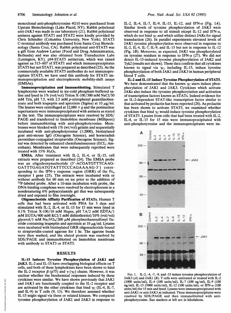
IL-2, IL-4, IL-7, IL-9, IL-15, IL-12, and IFN-a (Fig. 1A).Similar levels of tyrosine phosphorylation of JAK3 wereobserved in response to all stimuli except IL-12 and IFN-a,which do not bind yc and which utilize distinct JAKs for signaltransduction (26). In parallel experiments elevated levels ofJAK1 tyrosine phosphorylation were observed in response toIL-2, IL-4, IL-7, IL-9, and IL-15 but not in response to IL-12(Fig. IB). Moreover, as expected, JAK1 was phosphorylatedon tyrosine residues in response to IFN-a (27). We did notdetect IL-15-induced tyrosine phosphorylation of JAK2 andTyk2 (results not shown). These data confirm that all cytokinesknown to signal via yvc, including IL-15, induce tyrosinephosphorylation of both JAK1 and JAK3 in human peripheralblood T cells.
IL-2 and IL-15 Induce Tyrosine Phosphorylation of STAT5.We have demonstrated that the known -Yc users induce phos-phorylation of JAK1 and JAK3. Cytokines which activateJAKs also induce the tyrosine phosphorylation and activationof transcription factors known as STATs. Indeed evidence foran IL-2-dependent STAT-like transcription factor similar tothat activated by prolactin has been reported (28). As prolactinhas been shown to activate STAT5, we examined whethercytokines that bind lyc would induce tyrosine phosphorylationof STAT5. Lysates from cells that had been treated with IL-2,IL-4, or IL-15 for 15 min were immunoprecipitated withanti-phosphotyrosine and the immunoprecipitates were im-
A °-c0
0
LOlC'JCI q N1-
1J1J
200-
96-_m__ mm ---- JAK3
69-
43-
.~~~~~~~~M
B LOC \l
n * ~J ,J
200-i
. __-iJAK196- : s-, s
RESULTS
IL-15 Induces Tyrosine Phosphorylation of JAK1 andJAK3. IL-2 and IL-15 have overlapping biological effects on Tcells, and both of these lymphokines have been shown to bindthe IL-2 receptor , (p75) and -y (,Yc) chains. However, it wasunclear whether the biochemical responses induced by thesecytokines were similar. We have shown previously that JAK1and JAK3 are functionally coupled to the IL-2 receptor andare activated by the other cytokines that bind -yc (IL-4, IL-7,and IL-9) in T cells (9, 13). We therefore assessed whetherIL-15 might signal via these or related kinases. We comparedtyrosine phosphorylation of JAKi and JAK3 in response to
69 -
FIG. 1. IL-2, -4, -7, -9, and -15 induce tyrosine phosphorylation ofJAK3 (A) and JAKi (B). T cells were untreated or treated with IL-2(1000 units/ml), IL-4 (100 units/ml), IL-7 (100 ng/ml), IL-9 (100ng/ml), IL-15 (5000 units/ml), IL-12 (100 units/ml), or IFN-a (100units/ml) for 15 min and lysed. Lysates were immunoprecipitated withanti-JAK1 or anti-JAK3 as indicated. These immunoprecipitates wereresolved by SDS/PAGE and then immunoblotted with anti-phosphotyrosine. Size markers at left are in kilodaltons.
4.*Mw.ommm4.f
'AMI'lelow
'Modb-.-..odbe-AP.-
Proc. Natl. Acad. Sci. USA 92 (1995)
Dow
nloa
ded
by g
uest
on
Nov
embe
r 28
, 202
1
Proc. Natl. Acad. Sci. USA 92 (1995) 8707
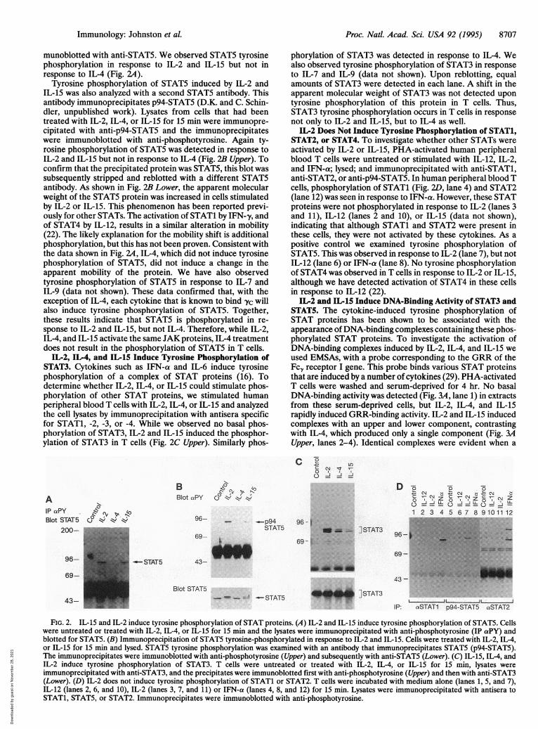
munoblotted with anti-STAT5. We observed STAT5 tyrosinephosphorylation in response to IL-2 and IL-15 but not inresponse to IL-4 (Fig. 2A).
Tyrosine phosphorylation of STAT5 induced by IL-2 andIL-15 was also analyzed with a second STAT5 antibody. Thisantibody immunoprecipitates p94-STAT5 (D.K. and C. Schin-dler, unpublished work). Lysates from cells that had beentreated with IL-2, IL-4, or IL-15 for 15 min were immunopre-cipitated with anti-p94-STAT5 and the immunoprecipitateswere immunoblotted with anti-phosphotyrosine. Again ty-rosine phosphorylation of STAT5 was detected in response toIL-2 and IL-15 but not in response to IL-4 (Fig. 2B Upper). Toconfirm that the precipitated protein was STAT5, this blot wassubsequently stripped and reblotted with a different STAT5antibody. As shown in Fig. 2B Lower, the apparent molecularweight of the STAT5 protein was increased in cells stimulatedby IL-2 or IL-15. This phenomenon has been reported previ-ously for other STATs. The activation of STAT1 by IFN-y, andof STAT4 by IL-12, results in a similar alteration in mobility(22). The likely explanation for the mobility shift is additionalphosphorylation, but this has not been proven. Consistent withthe data shown in Fig. 2A, IL-4, which did not induce tyrosinephosphorylation of STAT5, did not induce a change in theapparent mobility of the protein. We have also observedtyrosine phosphorylation of STAT5 in response to IL-7 andIL-9 (data not shown). These data confirmed that, with theexception of IL-4, each cytokine that is known to bind yc willalso induce tyrosine phosphorylation of STAT5. Together,these results indicate that STAT5 is phosphorylated in re-sponse to IL-2 and IL-15, but not IL-4. Therefore, while IL-2,IL-4, and IL-15 activate the same JAK proteins, IL-4 treatmentdoes not result in the phosphorylation of STAT5 in T cells.
IL-2, IL-4, and IL-15 Induce Tyrosine Phosphorylation ofSTAT3. Cytokines such as IFN-a and IL-6 induce tyrosinephosphorylation of a complex of STAT proteins (16). Todetermine whether IL-2, IL-4, or IL-15 could stimulate phos-phorylation of other STAT proteins, we stimulated humanperipheral blood T cells with IL-2, IL-4, or IL-15 and analyzedthe cell lysates by immunoprecipitation with antisera specificfor STAT1, -2, -3, or -4. While we observed no basal phos-phorylation of STAT3, IL-2 and IL-15 induced the phosphor-ylation of STAT3 in T cells (Fig. 2C Upper). Similarly phos-
phorylation of STAT3 was detected in response to IL-4. Wealso observed tyrosine phosphorylation of STAT3 in responseto IL-7 and IL-9 (data not shown). Upon reblotting, equalamounts of STAT3 were detected in each lane. A shift in theapparent molecular weight of STAT3 was not detected upontyrosine phosphorylation of this protein in T cells. Thus,STAT3 tyrosine phosphorylation occurs in T cells in responsenot only to IL-2 and IL-15, but to IL-4 as well.
IL-2 Does Not Induce Tyrosine Phosphorylation of STAT1,STAT2, or STAT4. To investigate whether other STATs wereactivated by IL-2 or IL-15, PHA-activated human peripheralblood T cells were untreated or stimulated with IL-12, IL-2,and IFN-a; lysed; and immunoprecipitated with anti-STAT1,anti-STAT2, or anti-p94-STAT5. In human peripheral blood Tcells, phosphorylation of STAT1 (Fig. 2D, lane 4) and STAT2(lane 12) was seen in response to IFN-a. However, these STATproteins were not phosphorylated in response to IL-2 (lanes 3and 11), IL-12 (lanes 2 and 10), or IL-15 (data not shown),indicating that although STAT1 and STAT2 were present inthese cells, they were not activated by these cytokines. As apositive control we examined tyrosine phosphorylation ofSTAT5. This was observed in response to IL-2 (lane 7), but notIL-12 (lane 6) or IFN-a (lane 8). No tyrosine phosphorylationof STAT4 was observed in T cells in response to IL-2 or IL-15,although we have detected activation of STAT4 in these cellsin response to IL-12 (22).
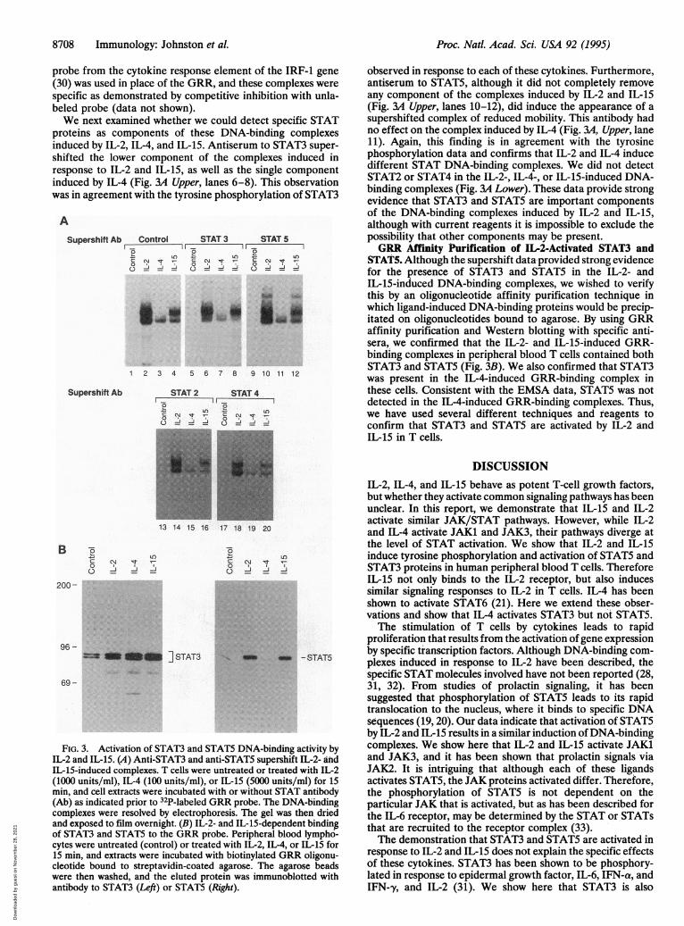
IL-2 and IL-15 Induce DNA-Binding Activity of STAT3 andSTAT5. The cytokine-induced tyrosine phosphorylation ofSTAT proteins has been shown to be associated with theappearance of DNA-binding complexes containing these phos-phorylated STAT proteins. To investigate the activation ofDNA-binding complexes induced by IL-2, IL-4, and IL-15 weused EMSAs, with a probe corresponding to the GRR of theFcz receptor I gene. This probe binds various STAT proteinsthat are induced by a number of cytokines (29). PHA-activatedT cells were washed and serum-deprived for 4 hr. No basalDNA-binding activity was detected (Fig. 3A, lane 1) in extractsfrom these serum-deprived cells, but IL-2, IL-4, and IL-15rapidly induced GRR-binding activity. IL-2 and IL-15 inducedcomplexes with an upper and lower component, contrastingwith IL-4, which produced only a single component (Fig. 3AUpper, lanes 2-4). Identical complexes were evident when a
cC C
Isto
B &Blot aPY C} Iz
96-
69- j
- ..-p94 96 - ISTAT5 u i]_.I STAT3
_~~~~~~~~6 6- ........_ _LNkl
rC N C ~Z C C~JZDo o00 L
1 2 3 4 5 6 7 8 9101112..i .tv,iigii'i . ......
96 -
69 -43-
43 -
Blot STAT5]STAT3
w - = -STAT5L ,L L .. _--
IP: aSTAT1 p94-STAT5 aSTAT2
FIG. 2. IL-15 and IL-2 induce tyrosine phosphorylation of STAT proteins. (A) IL-2 and IL-15 induce tyrosine phosphorylation of STAT5. Cellswere untreated or treated with IL-2, IL-4, or IL-15 for 15 min and the lysates were immunoprecipitated with anti-phosphotyrosine (IP aPY) andblotted for STAT5. (B) Immunoprecipitation of STAT5 tyrosine-phosphorylated in response to IL-2 and IL-15. Cells were treated with IL-2, IL-4,or IL-15 for 15 min and lysed. STAT5 tyrosine phosphorylation was examined with an antibody that immunoprecipitates STAT5 (p94-STAT5).The immunoprecipitates were immunoblotted with anti-phosphotyrosine (Upper) and subsequently with anti-STAT5 (Lower). (C) IL-15, IL-4, andIL-2 induce tyrosine phosphorylation of STAT3. T cells were untreated or treated with IL-2, IL-4, or IL-15 for 15 min, lysates wereimmunoprecipitated with anti-STAT3, and the precipitates were immunoblotted first with anti-phosphotyrosine (Upper) and then with anti-STAT3(Lower). (D) IL-2 does not induce tyrosine phosphorylation of STAT1 or STAT2. T cells were incubated with medium alone (lanes 1, 5, and 7),IL-12 (lanes 2, 6, and 10), IL-2 (lanes 3, 7, and 11) or IFN-a (lanes 4, 8, and 12) for 15 min. Lysates were immunoprecipitated with antisera toSTAT1, STAT5, or STAT2. Immunoprecipitates were immunoblotted with anti-phosphotyrosine.
-N0
6,v ~, 40vl4 *
AlP ctPYBlot STAT5
200-
96-
69-
43-
- STAT5
Immunology: Johnston et al.
":": ......
a 401-todb
Dow
nloa
ded
by g
uest
on
Nov
embe
r 28
, 202
1
8708 Immunology: Johnston et al.
probe from the cytokine response element of the IRF-1 gene(30) was used in place of the GRR, and these complexes werespecific as demonstrated by competitive inhibition with unla-beled probe (data not shown).We next examined whether we could detect specific STAT
proteins as components of these DNA-binding complexesinduced by IL-2, IL-4, and IL-15. Antiserum to STAT3 super-shifted the lower component of the complexes induced inresponse to IL-2 and IL-15, as well as the single componentinduced by IL-4 (Fig. 3A Upper, lanes 6-8). This observationwas in agreement with the tyrosine phosphorylation of STAT3
A
Supershift Ab Control STAT 3 STAT 52 L O LOS C' 'Tcm_N t N0 -t C N t
o 0 0
LuU-f I.i-
1 2 3 4 5 6 7 8 9 10 11 12
Supershift Ab STAT 2 STAT 40
LO
* __44i _ _
B to
0
200
96 -
UL)N llr T
13 14 15 16 17 18 19 20
CM %0
_ _
== ON _ ] STAT3
LO)
-J
u)
*: :.:. : ..:.:.::.:: ::.:. ..
::e:e.... .:.:::.
*:
ov _ _ - STAT5
69-
FIG. 3. Activation of STAT3 and STAT5 DNA-binding activity byIL-2 and IL-15. (A) Anti-STAT3 and anti-STAT5 supershift IL-2- andIL-15-induced complexes. T cells were untreated or treated with IL-2(1000 units/ml), IL-4 (100 units/ml), or IL-15 (5000 units/ml) for 15min, and cell extracts were incubated with or without STAT antibody(Ab) as indicated prior to 32P-labeled GRR probe. The DNA-bindingconmplexes were resolved by electrophoresis. The gel was then driedand exposed to film overnight. (B) IL-2- and IL-15-dependent bindingof STAT3 and STAT5 to the GRR probe. Peripheral blood lympho-cytes were untreated (control) or treated with IL-2, IL-4, or IL-15 for15 min, and extracts were incubated with biotinylated GRR oligonu-cleotide bound to streptavidin-coated agarose. The agarose beadswere then washed, and the eluted protein was immunoblotted withantibody to STAT3 (Left) or STAT5 (Right).
observed in response to each of these cytokines. Furthermore,antiserum to STAT5, although it did not completely removeany component of the complexes induced by IL-2 and IL-15(Fig. 3A Upper, lanes 10-12), did induce the appearance of asupershifted complex of reduced mobility. This antibody hadno effect on the complex induced by IL-4 (Fig. 3A, Upper, lane11). Again, this finding is in agreement with the tyrosinephosphorylation data and confirms that IL-2 and IL-4 inducedifferent STAT DNA-binding complexes. We did not detectSTAT2 or STAT4 in the IL-2-, IL-4-, or IL-15-induced DNA-binding complexes (Fig. 3A Lower). These data provide strongevidence that STAT3 and STAT5 are important componentsof the DNA-binding complexes induced by IL-2 and IL-15,although with current reagents it is impossible to exclude thepossibility that other components may be present.GRR Affinity Purification of IL-2-Activated STAT3 and
STAT5. Although the supershift data provided strong evidencefor the presence of STAT3 and STATS in the IL-2- andIL-15-induced DNA-binding complexes, we wished to verifythis by an oligonucleotide affinity purification technique inwhich ligand-induced DNA-binding proteins would be precip-itated on oligonucleotides bound to agarose. By using GRRaffinity purification and Western blotting with specific anti-sera, we confirmed that the IL-2- and IL-15-induced GRR-binding complexes in peripheral blood T cells contained bothSTAT3 and STAT5 (Fig. 3B). We also confirmed that STAT3was present in the IL-4-induced GRR-binding complex inthese cells. Consistent with the EMSA data, STAT5 was notdetected in the IL-4-induced GRR-binding complexes. Thus,we have used several different techniques and reagents toconfirm that STAT3 and STAT5 are activated by IL-2 andIL-15 in T cells.
DISCUSSIONIL-2, IL-4, and IL-15 behave as potent T-cell growth factors,but whether they activate common signaling pathways has beenunclear. In this report, we demonstrate that IL-15 and IL-2activate similar JAK/STAT pathways. However, while IL-2and IL-4 activate JAKi and JAK3, their pathways diverge atthe level of STAT activation. We show that IL-2 and IL-15induce tyrosine phosphorylation and activation of STAT5 andSTAT3 proteins in human peripheral blood T cells. ThereforeIL-15 not only binds to the IL-2 receptor, but also inducessimilar signaling responses to IL-2 in T cells. IL-4 has beenshown to activate STAT6 (21). Here we extend these obser-vations and show that IL-4 activates STAT3 but not STAT5.The stimulation of T cells by cytokines leads to rapid
proliferation that results from the activation ofgene expressionby specific transcription factors. Although DNA-binding com-plexes induced in response to IL-2 have been described, thespecific STAT molecules involved have not been reported (28,31, 32). From studies of prolactin signaling, it has beensuggested that phosphorylation of STAT5 leads to its rapidtranslocation to the nucleus, where it binds to specific DNAsequences (19,20). Our data indicate that activation of STAT5by IL-2 and IL-15 results in a similar induction ofDNA-bindingcomplexes. We show here that IL-2 and IL-1i activate JAK1and JAK3, and it has been shown that prolactin signals viaJAK2. It is intriguing that although each of these ligandsactivates STAT5, the JAK proteins activated differ. Therefore,the phosphorylation of STAT5 is not dependent on theparticular JAK that is activated, but as has been described forthe IL-6 receptor, may be determined by the STAT or STATsthat are recruited to the receptor complex (33).The demonstration that STAT3 and STAT5 are activated in
response to IL-2 and IL-15 does not explain the specific effectsof these cytokines. STAT3 has been shown to be phosphory-lated in response to epidermal growth factor, IL-6, IFN-a, andIFN-y, and IL-2 (31). We show here that STAT3 is also
Proc. Natl. Acad. Sci. USA 92 (1995)
Dow
nloa
ded
by g
uest
on
Nov
embe
r 28
, 202
1
Proc. Natl. Acad. Sci. USA 92 (1995) 8709
activated by IL-4, IL-7, IL-9, and IL-15. Likewise, we andothers have shown that STAT5 can be activated by a numberof cytokines, including IL-2, IL-7, IL-9, IL-15, thrombopoietin(unpublished observation), and prolactin. However, thesetranscription factors may be of importance in many cell types,perhaps activating genes essential for cell growth and prolif-eration. For example, we find that the megakaryocyte prolif-erative factor thrombopoietin activates STAT3 and STAT5.We show here that the T-cell growth factors IL-2, IL-7, andIL-15 all activate STAT3 and STAT5. Therefore, transcriptioncomplexes that include STAT3 and STAT5 may be of generalimportance to promote cell proliferation in T cells,megakaryocytes, and perhaps other cells. The challenge re-mains to relate the activation of specific STATs to the biolog-ical effects of these cytokines and to identify their roles in geneactivation.Although much of our evidence clearly demonstrates that
the family of cytokines that use Yc activate common signalingmolecules such as JAK1, JAK3, and STAT3, it also suggeststhat distinct signals are activated by IL-4. IL-4 induces thephosphorylation of a number of unique substrates such asIL-4-STAT (21) and the IRS-1-like molecule 4PS (34). Simi-larly, IL-4 and IL-7 fail to activate Shc in T cells (35, 36). Againthe recruitment of different signaling molecules to the recep-tors may contribute to the distinct downstream responses toeach cytokine.The activation of distinct STATs provides a mechanism by
which STATs could contribute to IL-2- or IL-4-specific geneactivation in T cells. STAT3 forms heterodimers with otherSTAT molecules (18), and perhaps the various STAT com-plexes formed are an important mechanism by which signaldiversity is elicited. An important goal will be to understandhow the STATs contribute to gene activation in T cells.
IL-15 binds to subunits of the IL-2 receptor and has bio-logical activity resembling that of IL-2. However, while thesecytokines bind the same 13 and y receptor subunits, IL-15 maybind a distinct receptor chain. Evidence for this comes fromthe IL-3-dependent 32D cell line, which expresses the com-plete IL-2 receptor and proliferates strongly in response toIL-2, but not at all in response to IL-15 (3). Since the precisestructure of the IL-15 receptor is unknown, we cannot becertain that IL-2 and IL-15 signal identically in all respects.However, our data indicate that IL-2 and IL-15, consistentwith their biological effects, have highly similar signalingsubstrates in human peripheral blood T cells.
In summary, our results demonstrate that STAT3 andSTAT5 are rapidly activated in response to IL-2 and IL-15 butthat STAT5 is not activated in response to IL-4. This, coupledwith the identification of the JAKs activated by these cyto-kines, identifies novel pathways by which transcriptional acti-vation may occur. Understanding how IL-2, IL-4, and IL-15regulate gene expression is a critical issue in T-cell activation.The demonstration that these key cytokines activate STAT3and STAT5 will provide important insights into the mecha-nisms of gene regulation by these cytokines.
Note Added in Proof. Our findings agree with those of Lin et al. (37),Hou et al. (38), and Fujii et al. (39) that have been published sincesubmission of this article.
We thank William Bere and Anna Mason for expert technicalassistance. We thank Drs. Dan McVicar and Chris Schindler forreading the manuscript. C.M.B. receives a Ph.D. Research Grant fromthe University of Sheffield and is the recipient of a U.S.-U.K.Education Commission (Fulbright Commission) Cancer Studentship.C.M.B. and R.C.R. are supported by the Yorkshire Cancer ResearchCampaign, U.K.
1. Grabstein, K., Eisenman, J., Shanebeck, K., Rauch, C., Srinivasan, S., Fung,V., Beers, C., Richardson, J., Schoenbom, M. A., Ahdieh, M., Johnson, L.,
Alderson, M. R., Watson, J. D., Anderson, D. M. & Giri, J. G. (1994)Science 264, 965-968.
2. Carson, E. C., Giri, G. G., Lindemann, M. J., Linett, L., Ahdieh, M.,Paxton, R., Anderson, D., Eisemann, J., Grabstein, K. & Caliguiri, M. A.(1994) J. Exp. Med. 180, 1395-1403.
3. Giri, G. G., Ahdieh, M., Eisenman, J., Shanebeck, K., Grabstein, K.,Kumaki, S., Namen, A., Park, L. S., Cosman, D. & Anderson, D. (1994)EMBO J. 13, 2822-2830.
4. Kondo, M., Takeshita, T., Ishii, N., Nakamura, M., Watanabe, S., Arai, K-i.& Sugamura, K. (1993) Science 262, 1874-1877.
5. Russell, S. M., Keegan, A. D., Harada, N., Nakamura, Y., Noguchi, M.,Leland, P., Friedmann, M. C., Miyajima, A., Puri, R. K., Paul, W. E. &Leonard, W. J. (1993) Science 262, 1880-1883.
6. Darnell, J. E., Kerr, I. M. & Stark, G. R. (1994) Science 264, 1415-1421.7. Ihle, J. N. & Kerr, I. M. (1995) Trends Genet. 11, 69-74.8. Kawamura, M., McVicar, D. W., Johnston, J. A., Blake, T. B., Chen, Y.-Q.,
Lal, B. K., Lloyd, A. R., Kelvin, D. J., Staples, J. E., Ortaldo, J. R. &O'Shea, J. J. (1994) Proc. Natl. Acad. Sci. USA 91, 6374-6378.
9. Johnston, J. A., Kawamura, M., Kirken, R. A., Chen, Y. Q., Blake, T. B.,Shibuya, K., Ortaldo, J. R., McVicar, D. W. & O'Shea, J. J. (1994) Nature(London) 370, 151-153.
10. Witthuhn, B. A., Silvennoinen, O., Miura, O., Lai, K. S., Cwik, C., Liu, E. T.& Ihle, J. N. (1994) Nature (London) 370, 153-157.
11. Takahashi, T. & Shirasawa, T. (1994) FEBS Lett. 342, 124-128.12. Noguchi, M., Yi, H., Rosenblatt, H. M., Filipovich, A. H., Adelstein, S.,
Modi, W. S., McBride, 0. W. & Leonard, W. J. (1993) Cell 73, 147-157.13. Russell, S. M., Johnston, J. A., Noguchi, M., Kawamura, M., Bacon, C. M.,
Friedmann, M., Berg, M., Witthuhn, B. A., Goldman, A. S., Schmalsteig,F. C., Ihle, J. N., O'Shea, J. J. & Leonard, W. J. (1994) Science 266,1042-1045.
14. Darnell, J. E., Jr., Kerr, I. M. & Stark, G. R. (1994) Science 264, 1415-1421.15. Improta, T., Schindler, C., Horvath, C. M., Kerr, I. M., Stark, G. R. &
Darnell, J. E., Jr. (1994) Proc. Natl. Acad. Sci. USA 91, 4776-4780.16. Muller, M., Laxton, C., Briscoe, J., Schindler, C., Improta, T., Darnell,
J. E. J., Stark, G. R. & Kerr, I. M. (1993) EMBO J. 12, 4221-4228.17. Schindler, C., Shuai, K., Prezioso, V. R. & Darnell, J. E. J. (1992) Science
257, 809-813.18. Zhong, Z., Wen, Z. & Darnell, J. E., Jr. (1994) Science 264, 95-98.19. Gouilleux, F., Wakao, H., Mundt, M. & Groner, B. (1994) EMBO J. 13,
4361-4369.20. Burdon, T. G., Demmer, J., Clark, A. J. & Watson, C. J. (1994) FEBS Lett.
350, 177-182.21. Hou, J., Schindler, U., Henzel, W. J., Ho, T. C., Brasseur, M. & McKnight,
S. L. (1994) Science 265, 1701-1705.22. Bacon, C. M., Petricoin, E. F., Rees, R. C., Larner, A. C., Johnston, J. A.
& O'Shea, J. J. (1995) Proc. Natl. Acad. Sci. USA 92, 7307-7311.23. Rothman, P., Kreider, B., Azam, M., Levy, D., Wegenka, U., Eilers, A.,
Decker, T., Horn, F., Kashleva, H., Ihle, J. & Schindler, C. (1994) Immunity1, 457-468.
24. Finbloom, D. S., Petricoin, E. F., Hackett, R. H., David, M., Feldman,G. M., Igarashi, K., Fibach, E., Weber, M. J., Thorner, M. O., Silva, C. M.& Lamer, A. C. (1994) Mol. Cell. Biol. 14, 2113-2118.
25. Pearse, R. N., Feinman, R. & Ravetch, J. V. (1991) Proc. Natl. Acad. Sci.USA 88, 11305-11309.
26. Bacon, C. M., McVicar, D. W., Ortaldo, J. R., Rees, R. C., O'Shea, J. J. &Johnston, J. A. (1994) J. Exp. Med. 181, 399-404.
27. Velazquez, L., Fellous, M., Stark, G. R. & Pellegrini, S. (1992) Cell 70,313-322.
28. Gilmour, K. C. & Reich, N. C. (1994) Proc. Natl. Acad. Sci. USA 91,6850-6854.
29. Larner, A. C., David, M., Feldman, G. M., Igarashi, K., Hackett, R. H.,Webb, D. S., Sweitzer, S. M., Petricoin, E. F., III, & Finbloom, D. S. (1993)Science 261, 1730-1733.
30. Pine, R., Canova, A. & Schindler, C. (1994) EMBO J. 13, 158-167.31. Neilsen, M., Svejgaard, A., Skov, S. & Odum, N. (1994) Eur. J. Immunol.
24, 3082-3086.32. Beading, C., Guschin, D., Witthuhn, B. A., Ziemeski, A., Ihle, J. A., Kerr,
I. M. & Cantrell, D. A. (1994) EMBO J. 13, 5605-5615.33. Stahl, N., Farrugella, T. G., Boulton, T. G., Zhong, Z., Darnell, J. E. &
Yancopoulos, G. D. (1995) Science 267, 1349-1353.34. Wang, L. M., Keegan, A. D., Li, W. & Lienhard, G. E. (1993) Proc. Natl.
Acad. Sci. USA 90, 4032-4036.35. Welham, M. J., Duronio, V., Leslie, K. B., Bowtell, D. & Schrader, J. W.
(1994) J. Biol. Chem. 269, 21165-21176.36. Dorsch, M., Hock, H. & Diamantstein, T. (1994) Eur. J. Immunol. 24,
2049-2054.37. Lin, J. X., Migone, T. S., Tsang, M., Friedmann, M., Weatherbee, J. A.,
Zhou, L., Yamauchi, A., Bloom, E. T., John, S. & Leonard, W. J. (1995)Immunity 2, 331-339.
38. Hou, J., Schindler, U., Henzel, W. J., Wong, S. C. & McKnight, S. L. (1995)Immunity 2, 321-329.
39. Fujii, H., Nakagawa, Y., Schindler, U., Kawahara, A., Mori, H., Gouilleux,F., Groner, B., Ihle, J. N., Minami, Y., Miyazaki, T. & Taniguchi, T. (1995)Proc. Natl. Acad. Sci. USA 92, 5482-5486.
Immunology: Johnston et al.
Dow
nloa
ded
by g
uest
on
Nov
embe
r 28
, 202
1