standardized reporting of prostate mri: comparison … · pi-rads sum score was calculatedas the...
TRANSCRIPT
RESEARCH ARTICLE
Standardized Reporting of Prostate MRI:Comparison of the Prostate ImagingReporting and Data System (PI-RADS)Version 1 and Version 2Susanne Tewes1*, Nikolaj Mokov2, Dagmar Hartung1, Volker Schick3, Inga Peters4,
Peter Schedl3, Stefanie Pertschy1, Frank Wacker1, Gotz Voshage2, Katja Hueper1
1 Institute for Diagnostic and Interventional Radiology, Hannover Medical School, Hannover, Germany,
2 Institute for Diagnostic and Interventional Radiology, Klinikum der Region Hannover, Hannover, Gehrden,
Germany, 3 Clinic for Urology, Klinikum der Region Hannover, Hannover, Gehrden, Germany,
4 Department of Urology and Urologic Oncology, Hannover Medical School, Hannover, Germany
Abstract
Introduction
Objective of our study was to determine the agreement between version 1 (v1) and v2 of
the Prostate Imaging Reporting and Data System (PI-RADS) for evaluation of multipara-
metric prostate MRI (mpMRI) and to compare their diagnostic accuracy, their inter-observer
agreement and practicability.
Material and Methods
mpMRI including T2-weighted imaging, diffusion-weighted imaging (DWI) and dynamic
contrast-enhanced imaging (DCE) of 54 consecutive patients, who subsequently under-
went MRI-guided in-bore biopsy were re-analyzed according to PI-RADS v1 and v2 by two
independent readers. Diagnostic accuracy for detection of prostate cancer (PCa) was
assessed using ROC-curve analysis. Agreement between PI-RADS versions and observ-
ers was calculated and the time needed for scoring was determined.
Results
MRI-guided biopsy revealed PCa in 31 patients. Diagnostic accuracy for detection of PCa
was equivalent with both PI-RADS versions for reader 1 with sensitivities and specificities
of 84%/91% (AUC = 0.91 95%CI[0.8–1]) for PI-RADS v1 and 100%/74% (AUC = 0.92 95%
CI[0.8–1]) for PI-RADS v2. Reader 2 achieved similar diagnostic accuracy with sensitivity
and specificity of 74%/91% (AUC = 0.88 95%CI[0.8–1]) for PI-RADS v1 and 81%/91%
(AUC = 0.91 95%CI[0.8–1]) for PI-RADS v2. Agreement between scores determined with
different PI-RADS versions was good (reader 1: κ = 0.62, reader 2: κ = 0.64). Inter-observer
agreement was moderate with PI-RADS v2 (κ = 0.56) and fair with v1 (κ = 0.39). The time
PLOS ONE | DOI:10.1371/journal.pone.0162879 September 22, 2016 1 / 13
a11111
OPENACCESS
Citation: Tewes S, Mokov N, Hartung D, Schick V,
Peters I, Schedl P, et al. (2016) Standardized
Reporting of Prostate MRI: Comparison of the
Prostate Imaging Reporting and Data System (PI-
RADS) Version 1 and Version 2. PLoS ONE 11(9):
e0162879. doi:10.1371/journal.pone.0162879
Editor: Jeroen Hendrikse, Universitair Medisch
Centrum Utrecht, NETHERLANDS
Received: April 29, 2016
Accepted: August 30, 2016
Published: September 22, 2016
Copyright: © 2016 Tewes et al. This is an open
access article distributed under the terms of the
Creative Commons Attribution License, which
permits unrestricted use, distribution, and
reproduction in any medium, provided the original
author and source are credited.
Data Availability Statement: All relevant data are
within the paper and its Supporting Information
files.
Funding: KH and ST received funding from the
Junge Akademie program Hannover Medical
School. KH has a research collaboration with
Siemens Healthcare outside the work submitted.
FW receives institutional grants from Siemens
Healthcare and Promedicus Ltd and Delcath
Systems Inc. outside the work submitted. The
funders had no role in study design, data collection

required for building the PI-RADS score was significantly lower with PI-RADS v2 compared
to v1 (24.7±2.3 s vs. 41.9±2.6 s, p<0.001).
Conclusion
Agreement between PI-RADS versions was high and both versions revealed high diagnos-
tic accuracy for detection of PCa. Due to better inter-observer agreement for malignant
lesions and less time demand, the new PI-RADS version could be more practicable for clini-
cal routine.
Introduction
The use of multiparametric (mp) MRI for the detection and characterization of prostatelesions has evolved over the last 10 years. mpMRI protocols combining information of mor-phology with high spatial resolution (T2-weighted turbo spin echo imaging = T2 TSE), celldensity (diffusionweighted imaging = DWI) and vascularization (dynamic contrast-enhanced imaging = DCE) provide high diagnostic accuracy for the detection of clinicallysignificant prostate cancer (PCa) [1–8]. In addition, MRI is increasingly used for targetedprostate biopsy, which leads to improved detection of significant PCa [9–11].Consensus has been reached that standardization of imaging and reporting of prostate MRI
is important to ensure high diagnostic quality, reproducible MRI results and applicability ofprostate MRI for multicenter studies. In 2012 an expert consensus group of the European Soci-ety of Urogenital Radiology (ESUR) introduced the version 1 (v1) of the Prostate ImagingReporting and Data System (PI-RADS) [12]. Since then, PI-RADS v1 has been clinicallyapplied and evaluated in several clinical studies. mpMRI PI-RADS v1 scores showed high diag-nostic accuracy for the detection of PCa and high inter-observer agreement [13–16]. However,some limitations of PI-RADS v1 have been recognized in the past years. With this first version,it was not clearly defined how exactly scores should be determined and combined and this gaveroom for individual interpretation and therefore to variability in the application of PI-RADSv1 [13]. Additionally, various types of perfusion curves that can occur in the prostate led toconfusion; there is great heterogeneity in enhancement characteristics of prostate cancers andthere is also great heterogeneity in perfusion characteristics of benign prostate lesion [17].Including perfusion scores in the overall PI-RADS led to higher scores in benign lesions.Therefore, the American College of Radiology (ACR), the ESUR, and the AdMeTech Founda-tion established a Steering Committee to further develop, update and simplify PI-RADS underconsideration of ongoing research in an effort to make PI-RADSmore globally acceptable.This resulted in the updated PI-RADS version 2 (v2) [17].The purpose of this study was to determine the agreement between PI-RADS v1 and v2 for
evaluation of multiparametric prostate MRI and to compare their diagnostic accuracy, theirinter-observer agreement and practicability.
Material and Methods
Patients
Data of 69 consecutive patients who underwentmpMRI of the prostate and subsequently anMRI-guided in-bore biopsy betweenDecember 2012 and December 2014 were retrospectivelyanalyzed. 15/69 patients were excluded from the analysis due to an incomplete MRI protocol
Comparison of PI-RADS Version 1 and Version 2
PLOS ONE | DOI:10.1371/journal.pone.0162879 September 22, 2016 2 / 13
and analysis, decision to publish, or preparation of
the manuscript.
Competing Interests: The authors confirm that this
does not alter our adherence to PLOS ONE policies
on sharing data and materials.

or distinct artifacts in one or more MRI sequences. Out of 54 patients, 18 patients had at leastone negative pre-biopsy and 36 had no pre-biopsy. Mean age (±standard deviation) was 69.6±9.6 years, mean PSA was 8.7±4.9 μg/L and mean prostate volume was 52.1±24.7 ml. Thestudy was approved by the ethics committee of the Hannover Medical School.
Multiparametric MRI
Multiparametric MRI was acquired according to ESUR guidelines [1, 12] on a 3 Tesla system(Magnetom Skyra, Siemens Healthcare, Erlangen, Germany) using an 18-channel body coiland a spine coil. In order to reduce bowel movement, all patients received an intravenous injec-tion of 20 mg butylscopolamine (Buscopan 20 mg, Boehringer, Ingelheim, Germany) prior tothe examination. T2 TSE sequences were acquired in transverse, sagittal and coronal orienta-tion with FOV = 220 x 220 mm2, matrix = 300 x 512, TR> 3000 ms, TE> 90 ms. For DWIfour b-values = 0, 200, 400,�800 s/mm2 were used. Other sequence parameters wereFOV = 320 x 320 mm2, matrix = 120 x 160, TR> 4500 ms and TE> 70 ms. For calculation ofADCmaps, a monoexponentialmodel was used and all b-values were included. For DCE avibe sequence was acquired in transverse plane: FOV = 260 x 260 mm2, matrix = 135 x 190,TR = 5 ms, TE = 1.5 ms, temporal resolution = 7.2 s using Gadovist as contrast agent in aweight-adapted standard dose of 0.1 mmol/kg bodyweightwith an injection rate of 2.7 ml/s(Table 1).
MRI-guided in-bore biopsy
The MRI-guided in-bore biopsy was performed on the sameMRI system as diagnostic imaging(Magnetom Skyra, Siemens Healthcare, Erlangen, Germany) using an 18-channel body coiland a spine coil integrated into the scanner. Patients with high clinical probability of PCa and/or equivocal or suspicious MRI findings, as diagnosed by the radiologists who primarilyreported the study, underwent biopsy. 65 prostate lesions in 54 patients were successfully biop-sied. Patients were placed in prone position. A needle guide connected to the arm of a portablebiopsy device (Dyna-TRIMDevice, Invivo International PC Best, Netherlands) was insertedrectally after topic local anesthesia (Lidocaine, Instillagel 40 ml, Farco-Pharma, Köln, Ger-many) was applied. T2-weighted images (HASTE or TSE) were acquired in transverse and sag-ittal planes. Biopsy was planned using the Dyna-CADworkstation and Dyna-CAD software(Invivo International PC Best, Netherlands) and the determined needle position was adjustedmanually. 2 cores were taken per lesion with an MRI-compatible 18-gauge fully automaticbiopsy gun. The needle position inside the lesion was verified using T2 HASTE sequences. Noadditional systematic biopsy was performed.
Table 1. Parameters of mpMRI.
T2 TSE DWI DCE (T1 vibe)
TR (ms) >3000 >4500 5
TE (ms) >90 >70 1.5
FoV (mm2) 220x220 320x320 260x260
matrix 300x512 120x160 135x190
slice thickness (mm) 3mm 4mm 3.6mm
b-values (s/mm2) - 0, 200, 400,�800 -
DCE = dynamic contrast enhancement, DWI = diffusion weighted imaging, TSE = turbo spin echo.
doi:10.1371/journal.pone.0162879.t001
Comparison of PI-RADS Version 1 and Version 2
PLOS ONE | DOI:10.1371/journal.pone.0162879 September 22, 2016 3 / 13
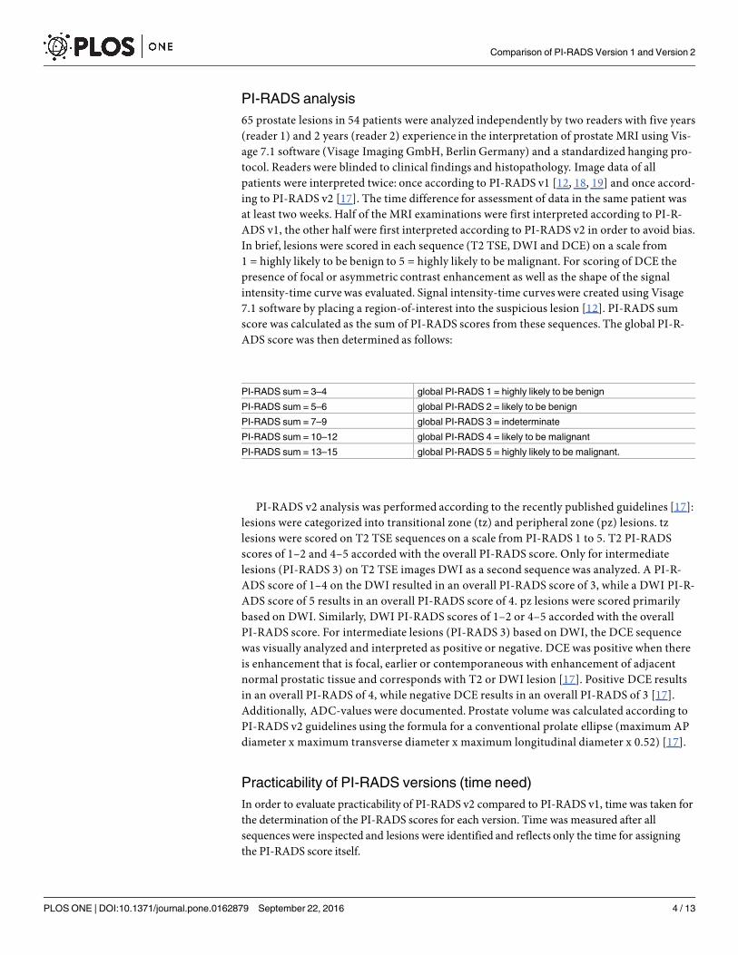
PI-RADS analysis
65 prostate lesions in 54 patients were analyzed independently by two readers with five years(reader 1) and 2 years (reader 2) experience in the interpretation of prostate MRI using Vis-age 7.1 software (Visage Imaging GmbH, Berlin Germany) and a standardized hanging pro-tocol. Readers were blinded to clinical findings and histopathology. Image data of allpatients were interpreted twice: once according to PI-RADS v1 [12, 18, 19] and once accord-ing to PI-RADS v2 [17]. The time difference for assessment of data in the same patient wasat least two weeks. Half of the MRI examinations were first interpreted according to PI-R-ADS v1, the other half were first interpreted according to PI-RADS v2 in order to avoid bias.In brief, lesions were scored in each sequence (T2 TSE, DWI and DCE) on a scale from1 = highly likely to be benign to 5 = highly likely to be malignant. For scoring of DCE thepresence of focal or asymmetric contrast enhancement as well as the shape of the signalintensity-time curve was evaluated. Signal intensity-time curves were created using Visage7.1 software by placing a region-of-interest into the suspicious lesion [12]. PI-RADS sumscore was calculated as the sum of PI-RADS scores from these sequences. The global PI-R-ADS score was then determined as follows:
PI-RADS v2 analysis was performed according to the recently published guidelines [17]:lesions were categorized into transitional zone (tz) and peripheral zone (pz) lesions. tzlesions were scored on T2 TSE sequences on a scale from PI-RADS 1 to 5. T2 PI-RADSscores of 1–2 and 4–5 accorded with the overall PI-RADS score. Only for intermediatelesions (PI-RADS 3) on T2 TSE images DWI as a second sequence was analyzed. A PI-R-ADS score of 1–4 on the DWI resulted in an overall PI-RADS score of 3, while a DWI PI-R-ADS score of 5 results in an overall PI-RADS score of 4. pz lesions were scored primarilybased on DWI. Similarly, DWI PI-RADS scores of 1–2 or 4–5 accorded with the overallPI-RADS score. For intermediate lesions (PI-RADS 3) based on DWI, the DCE sequencewas visually analyzed and interpreted as positive or negative. DCE was positive when thereis enhancement that is focal, earlier or contemporaneous with enhancement of adjacentnormal prostatic tissue and corresponds with T2 or DWI lesion [17]. Positive DCE resultsin an overall PI-RADS of 4, while negative DCE results in an overall PI-RADS of 3 [17].Additionally, ADC-values were documented. Prostate volume was calculated according toPI-RADS v2 guidelines using the formula for a conventional prolate ellipse (maximum APdiameter x maximum transverse diameter x maximum longitudinal diameter x 0.52) [17].
Practicability of PI-RADS versions (time need)
In order to evaluate practicability of PI-RADS v2 compared to PI-RADS v1, time was taken forthe determination of the PI-RADS scores for each version. Time was measured after allsequences were inspected and lesions were identified and reflects only the time for assigningthe PI-RADS score itself.
PI-RADS sum = 3–4 global PI-RADS 1 = highly likely to be benign
PI-RADS sum = 5–6 global PI-RADS 2 = likely to be benign
PI-RADS sum = 7–9 global PI-RADS 3 = indeterminate
PI-RADS sum = 10–12 global PI-RADS 4 = likely to be malignant
PI-RADS sum = 13–15 global PI-RADS 5 = highly likely to be malignant.
Comparison of PI-RADS Version 1 and Version 2
PLOS ONE | DOI:10.1371/journal.pone.0162879 September 22, 2016 4 / 13

Statistical analysis
For statistical analysis, GraphPad Prism software versions 5 (GraphPad Software, Inc., USA) aswell as SPSS Statistics version 21 (SPSS, IBM, Chicago, IL, USA) were used. As clinical dataand PI-RADS scores were not normally distributed as determined by the Kolmogorov-Smirnovtest and PI-RADS scores represent ordinal variables, patients with and without biopsy-provenPCa were compared using the non-parametricMann-Whitney U test. ADC values were nor-mally distributed and were compared with the unpaired t-test between groups with and with-out PCa. The time needed for assigning the PI-RADS was not normally distributed and werecompared with the non-parametricWilcoxon test.Diagnostic performance of mpMRI PI-RADS scores was determined for the dominant
lesion in each patient that was biopsied by MRI-guided in-bore biopsy. Results of MRI-guidedbiopsy were considered as the reference for this study. Receiver operating characteristic (ROC)curve analysis for PI-RADS v1 and v2 was performed separately for each reader as well as forthe tz and pz, using histopathological results of MRI-guided in-bore biopsy as the gold stan-dard. Youden-selected thresholds were determined, and sensitivity and specificity of MRIPI-RADS scores at the threshold were recorded. The agreement between PI-RADS versionsand the inter-observer agreement for each version was determined using Cohen’s kappa statis-tics. The agreement was defined excellent (κ>0.81), good (κ = 0.61–0.80), moderate (κ = 0.41–0.60), fair (κ = 0.21–0.40) and poor (κ�0.20) [20]. Values are given as mean ± standard devia-tion (SD). P-values <0.05 were considered statistically significant.
Results
Comparison of patient data and MRI parameters with and without PCa
MRI-guided in-bore biopsy revealed PCa in 31/54 patients (57%), 3 patients with Gleason 9, 1patient with Gleason 8, 3 patients with Gleason 7b, 4 patients with Gleason 7a, 20 patients withGleason 6. According to D’Amico criteria, 8 out of 20 patients with Gleason 6 had significantprostate cancer. In 40 patients the dominant lesion was located in the pz; 26/40 of these lesionswere positive for PCa. In 14 patients the dominant lesion was located in the tz; 5/14 of these werepositive for PCa. No significant difference in PSA level was observed in patients with and withoutPCa (9.3±5.9 vs. 7.8±3.3 μg/l, p = 0.3). Prostate volume was significantly lower in the group withPCa (46.9±25.8 vs. 59.2±21.6ml, p<0.05). PI-RADS scores were higher in patients with histolog-ically proven PCa with both PI-RADS versions (PI-RADS v1: 4.3±0.8 vs. 2.4±1.0, p<0.001; PI-R-ADS v2: 4.2±0.7 vs. 2.3±1.0, p<0.001). ADC values were significantly lower in tumor-positivelesion (0.8±0.2 vs. 1.2±0.2 10−3 mm2/s, p<0.001, Table 2). An example of PI-RADS scoring in a
Table 2. Comparison of patient data and MRI parameters in patients with and without prostate cancer.
parameter patients with histologically proven PCa
n = 31
patients without histologically proven PCa
n = 23
p-value
patient data age (years) 72.6±8.8 65.4±9.2 <0.01
PSA (μg/L) 9.3±5.9 7.8±3.3 0.3
MRI
parameters
Prostate volume (ml) 46.9±25.8 59.2±21.6 <0.05
PI-RADS v1 4.3±0.8 2.4±1.0 <0.001
PI-RADS v2 4.2±0.7 2.3±1.0 <0.001
ADC value
(10−3 mm2/s)
0.8±0.2 1.2±0.2 <0.001
Values are given as mean ± standard deviation. ADC = apparent diffusion coefficient, DCE = dynamic contrast-enhanced imaging, DWI = diffusion weighted
imaging, MRI = magnetic resonance imaging, PCa = prostate carcinoma, PI-RADS = Prostate Imaging Reporting and Data System, PSA = prostate specific
antigen.
doi:10.1371/journal.pone.0162879.t003
Comparison of PI-RADS Version 1 and Version 2
PLOS ONE | DOI:10.1371/journal.pone.0162879 September 22, 2016 5 / 13

patient with biopsy proven PCa (Gleason 3+4 = 7a) is given in Fig 1. An overviewof PI-RADSscores assigned according to PI-RADS version 1 and version 2 is given in Tables 3 and 4.
Diagnostic accuracy of mpMRI PI-RADS scores version 1 and version 2
Sensitivity and specificity for the detection of PCa were 100% and 74%, respectively, at theYouden-selected cut-off PI-RADS�3 with v2 and 84% and 91%, respectively, at a Youden-selected cut-off PI-RADS�4 with v1 for the experienced reader. Youden-selected thresholdswere similar for the less experienced reader, who achieved nearly equivalent sensitivities andspecificitieswith PI-RADS v2 (Table 5, Fig 2).In addition, diagnostic accuracy for pz and tz lesions was analyzed separately. With PI-R-
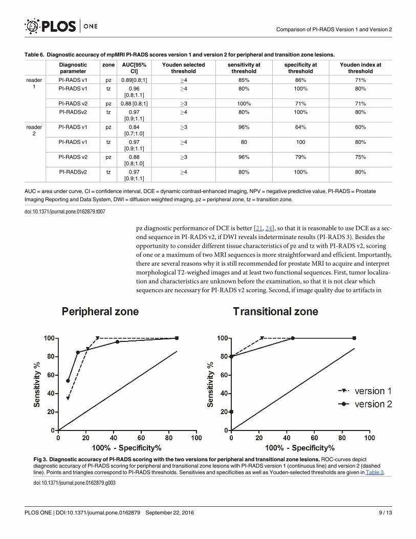
ADS v2 scores best diagnostic accuracy in the pz was reached with a cut-off�3 at a Youden-Index of 71% (sensitivity 100%, specificity 71%), while in the tz a cut-off�4 revealed highestdiagnostic accuracy (sensitivity 80%, specificity 100%, Youden-Index 80%). With PI-RADS v1Youden-selected cut-off was�4 for both pz and tz lesions (Table 6, Fig 3).
Practicability of PI-RADS scoring (time need)
When comparing the time needed to determine the PI-RADS score after having evaluated theentire examination, the experienced reader needed 24.7±2.3 seconds for scoring according toPI-RADS v2 and 41.9±2.6 seconds for PI-RADS v1 (p<0.001). For intermediate lesions (PI-R-ADS 3), which require analysis of two sequences for PI-RADS v2, no significant difference inthe time need was found with 37.1±6.2 seconds for v2 and 48.2±5.1 seconds for v1 (p = 0.2).For PI-RADS 4/5 lesions (19.5±2.1 vs. 38.8±3.1 seconds, p<0.001) as well as for PI-RADS 1/2
Fig 1. Example of PI-RADS scoring with version 1 and 2 in a patient with prostate cancer. mpMRI of an
82-year-old man with elevated PSA of 8.2 μg/l. The dominant lesion is located in the apex of the prostate in the pz
and measures 12 x 18 x 17 mm. According to PI-RADS v2, DWI is the leading sequence. As the lesion has high
signal intensity on b1000 with corresponding strong ADC reduction of 0.5 10−3 mm2/s as well as a diameter >15
mm the lesion was scored PI-RADS 5, highly likely to be malignant. No other sequence is required for scoring
according to PI-RADS v2. With PI-RADS v1 a score for each sequence needs to be determined. For this patient
the following scores were assigned: T2 TSE PI-RADS 5, DWI PI-RADS 5, DCE PI-RADS 5. This results in in a
PI-RADS sum of 15 and a global PI-RADS 5. In this patient the time need for PI-RADS scoring with v2 after
inspection of all images was 9 seconds, while it was 59 seconds with v1. MRI-guided in-bore biopsy revealed
Gleason 3+4 = 7 tumor.
doi:10.1371/journal.pone.0162879.g001
Comparison of PI-RADS Version 1 and Version 2
PLOS ONE | DOI:10.1371/journal.pone.0162879 September 22, 2016 6 / 13

lesions (22.5±3.5 vs. 42.1±6.4 seconds, p<0.05) the time need was significantly shorter forPI-RADS v2 compared to v1.
Agreement between PI-RADS versions and between observers
The agreement between PI-RADS v1 and v2 scores was good for both readers for all lesions(reader 1: κ = 0.62, reader 2: κ = 0.64), moderate when considering only malignant lesion(reader 1: κ = 0.54, reader 2: κ = 0.53) and moderate when for benign lesions (reader 1: κ =0.54, reader 2: κ = 0.60).The agreement between the experienced and less experienced reader (inter-observer agree-
ment) with PI-RADS v1 was fair (κ = 0.39) for all lesions, poor for biopsy proven malignantlesions (κ = 0.14) and moderate for benign lesions (κ = 0.50). With PI-RADS v2 the agreementwas moderate for all lesions (κ = 0.56) and for malignant lesions (κ = 0.56) and fair for benignlesions (κ = 0.26).
Discussion
We showed that the updated PI-RADS v2 for evaluation of mpMRI of the prostate had highdiagnostic accuracy for detection of PCa with equivalent sensitivities and specificities com-pared to PI-RADS v1. The agreement between PI-RADS v1 and v2 scores was good. The inter-observer agreement for malignant lesions was better with PI-RADS v2 than with PI-RADS v1and the time needed for PI-RADS scoring was significantly shorter for PI-RADS v2 indicatinga better practicability.Rising acceptance of mpMRI by urologists depends on high diagnostic accuracy for detec-
tion of significant PCa and reproducible interpretation. Therefore, standardized analysis andreporting of prostate MRI with comprehensible and clearly defined criteria are required. Theinitial version of the PI-RADS scoring (version 1) revealed high diagnostic accuracy in severalstudies [13] and good inter-observer agreement [15]. In the present study, comparing the
Table 3. PI-RADS scores assigned according to v1.
Reader 2 all
PI-RADS 1 PI-RADS 2 PI-RADS 3 PI-RADS 4 PI-RADS 5
Reader 1 PI-RADS 1 2 1 0 0 0 3
PI-RADS 2 3 6 2 0 0 11
PI-RADS 3 1 3 6 2 0 12
PI-RADS 4 0 0 4 7 1 12
PI-RADS 5 0 0 1 8 7 16
all 6 10 13 17 8 54
doi:10.1371/journal.pone.0162879.t004
Table 4. PI-RADS scores assigned according to v2.
Reader 2 all
PI-RADS 1 PI-RADS 2 PI-RADS 3 PI-RADS 4 PI-RADS 5
Reader 1 PI-RADS 1 1 2 0 0 0 3
PI-RADS 2 2 9 3 0 0 14
PI-RADS 3 0 3 3 1 0 7
PI-RADS 4 0 1 3 15 0 19
PI-RADS 5 0 0 0 3 8 11
all 3 15 9 19 8 54
doi:10.1371/journal.pone.0162879.t005
Comparison of PI-RADS Version 1 and Version 2
PLOS ONE | DOI:10.1371/journal.pone.0162879 September 22, 2016 7 / 13

updated PI-RADS scoring (version 2) with the initial version 1, we found equivalent diagnosticaccuracy and a good agreement between the PI-RADS versions. The updated PI-RADS v2 pro-vides a more detailed description on the assessment of prostate MRI with clearly defined crite-ria for PI-RADS scoring and representative images for each PI-RADS score and each sequenceseparately for pz and tz lesions [17]. As it was done with PI-RADS v1, the complete mpMRIexamination (T2, DWI and DCE) has to be acquired and inspected completely. Major renewalsin PI-RADS v2 are that the relevance of MRI-sequences is weighted depending on the localiza-tion of the lesion in the pz or tz and that scoring in one sequence is sufficient for most lesions.Only for indeterminate lesions (PI-RADS 3) scoring in a second sequence is required. In PI-R-ADS v2 the most important sequence for diagnosis of significant PCa is DWI, being the leadingsequence for the pz and the second sequence for the tz. This development is based on previousresearch showing that DWI provides highest accuracy for PCa detection for pz and tz lesions, ifonly one sequence is considered [21–23]. For tz tumors the combination of T2 and DWI, asused in PI-RADS v2, has been reported to have highest diagnostic accuracy [21], while addingDCE could not improve tumor detection in treatment-naïve prostates [24]. In contrast, in the
Table 5. Diagnostic accuracy of mpMRI PI-RADS scores version 1 and version 2.
Diagnostic
parameter
reader AUC[95%
CI]
Youden selected
threshold
sensitivity at
threshold
specificity at
threshold
Youden index at
threshold
PI-RADS v1 1 0.91[0.8;1] �4 84% 91% 75%
PI-RADS v1 2 0.88[0.8;1] �4 74% 91% 65%
PI-RADS v2 1 0.92 [0.8;1] �3 100% 74% 74%
PI-RADSv2 2 0.91 [0.8;1] �3 81% 91% 72%
AUC = area under curve, CI = confidence interval, DCE = dynamic contrast-enhanced imaging, PI-RADS = Prostate Imaging Reporting and Data System,
DWI = diffusion weighted imaging. Reader 1 represents the experienced reader with 5 years experience in the interpretation of prostate MRI and reader 2
represents the less experienced reader with 2 years experience.
doi:10.1371/journal.pone.0162879.t006
Fig 2. Diagnostic accuracy of PI-RADS scoring with the two versions for experienced and unexperienced readers. ROC-curves show diagnostic
accuracy of PI-RADS scoring for an experienced (reader 1) and less experienced reader (reader 2) with PI-RADS version 1 (continuous line) and version 2
(dashed line). Points and triangles correspond to PI-RADS thresholds. Sensitivies and specificities as well as Youden-selected thresholds are given in
Table 2.
doi:10.1371/journal.pone.0162879.g002
Comparison of PI-RADS Version 1 and Version 2
PLOS ONE | DOI:10.1371/journal.pone.0162879 September 22, 2016 8 / 13
pz diagnostic performance of DCE is better [21, 24], so that it is reasonable to use DCE as a sec-ond sequence in PI-RADS v2, if DWI reveals indeterminate results (PI-RADS 3). Besides theopportunity to consider different tissue characteristics of pz and tz with PI-RADS v2, scoringof one or a maximum of twoMRI sequences is more straightforward and efficient. Importantly,there are several reasons why it is still recommended for prostate MRI to acquire and interpretmorphological T2-weighed images and at least two functional sequences. First, tumor localiza-tion and characteristics are unknown before the examination, so that it is not clear whichsequences are necessary for PI-RADS v2 scoring. Second, if image quality due to artifacts in
Table 6. Diagnostic accuracy of mpMRI PI-RADS scores version 1 and version 2 for peripheral and transition zone lesions.
Diagnostic
parameter
zone AUC[95%
CI]
Youden selected
threshold
sensitivity at
threshold
specificity at
threshold
Youden index at
threshold
reader
1
PI-RADS v1 pz 0.89[0.8;1] �4 85% 86% 71%
PI-RADS v1 tz 0.96
[0.8;1.1]
�4 80% 100% 80%
PI-RADS v2 pz 0.88 [0.8;1] �3 100% 71% 71%
PI-RADSv2 tz 0.97
[0.9;1.1]
�4 80% 100% 80%
reader
2
PI-RADS v1 pz 0.84
[0.7;1.0]
�3 96% 64% 60%
PI-RADS v1 tz 0.97
[0.9;1.1]
�4 80 100 80%
PI-RADS v2 pz 0.88
[0.8;1.0]
�3 96% 79% 75%
PI-RADSv2 tz 0.97
[0.9;1.1]
�4 80% 100% 80%
AUC = area under curve, CI = confidence interval, DCE = dynamic contrast-enhanced imaging, NPV = negative predictive value, PI-RADS = Prostate
Imaging Reporting and Data System, DWI = diffusion weighted imaging, pz = peripheral zone, tz = transition zone.
doi:10.1371/journal.pone.0162879.t007
Fig 3. Diagnostic accuracy of PI-RADS scoring with the two versions for peripheral and transitional zone lesions. ROC-curves depict
diagnostic accuracy of PI-RADS scoring for peripheral and transitional zone lesions with PI-RADS version 1 (continuous line) and version 2 (dashed
line). Points and triangles correspond to PI-RADS thresholds. Sensitivies and specificities as well as Youden-selected thresholds are given in Table 3.
doi:10.1371/journal.pone.0162879.g003
Comparison of PI-RADS Version 1 and Version 2
PLOS ONE | DOI:10.1371/journal.pone.0162879 September 22, 2016 9 / 13
one sequence is insufficient (e.g. distortion in DWI often occurs with hip implants), the othertwo sequences can still be used for PI-RADS scoring, as the new version suggests a score if onesequence is missing [17]. Third, in pre-treated prostates following radiation therapy, focal ther-apy or endourethral treatment detection of PCa is often more challenging and treatmentrelated signal changes have to be considered. In this situation other sequences than suggestedin the PI-RADS v2 might be helpful. Therefore, in our study, PI-RADS scores were assignedafter the entire examination was closely evaluated and artifacts were excluded.Sensitivities and specificities of both PI-RADS versions in our study were comparable to
those reported previously for PI-RADS v1 in studies that accurately used PI-RADS scoring andhad pooled sensitivity of 82% (95%CI; 0.7–0.9) and specificity of 82% (95% CI; 0.7–0.9) [13–16, 25–28]. In a recent study, 95% of PCa foci�0.5 ml were correctly identifiedwith PI-R-ADSv2 using whole-mount pathology as reference standard [29]. Youden-selected PI-RADSthresholds in our study varied between PI-RADS�3 and�4 depending on PI-RADS versionand tumor localization. The calculated threshold for best diagnostic accuracy of all lesions withversion 2 was�3, while it was�4 with version 1. However, the number of patients might betoo low to draw any further conclusions from this discrepancy between PI-RADS versions. Inaddition, recommendations for patient management based on mpMRI PI-RADS scores havenot been proposed so far and are explicitly not included in the PI-RADS v2 document [17].Development of reliable recommendations based on PI-RADS scores needs further researchwith large prospectivemulticenter trials and long-term follow-up of patients.The inter-observer agreement between experienced and less experienced readers in our
study was moderate for PI-RADS v2 (κ = 0.56) and fair for PI-RADS v1 (κ = 0.39) when con-sidering all lesions. A recent study, evaluating PI-RADS v2, also found moderate interreaderagreement for five interpreters when considering all lesions [30]. Rosenkrantz et al. reportedthat inter-observer agreement (using PI-RADS v1) between experienced readers was better(concordance correlation coefficient 0.61–0.68) than between experienced and inexperiencedreaders (concordance correlation coefficient 0.34–0.48) for all lesions [31], indicating thatreader agreement is dependent on experiencewith prostate MRI. Schimmoeller et al. showed agood inter-reader agreement for tumor-positive lesions and a moderate to good interreaderagreement for benign lesions with PI-RADS v1 [15]. An explanation for the fact that inter-observer agreement is better for tumor-positive lesions might be that various types of benignchanges of prostate tissue occur, which are often difficult to differentiate from tumor lesions.In our study, when considering only biopsy-proven PCa lesions, inter-observer agreement withPI-RADS v2 (κ = 0.56) was better than with PI-RADS v1 (κ = 0.14). This may be explained bya more detailed and clear definition of PI-RADS scores with v2. For example PI-RADS 4 and 5lesions with v2 are clearly differentiated by the lesion size (diameter�15 mm results in PI-R-ADS 5), the presence of definite extraprostatic extension or invasive behavior.Another important finding of our study is that the time needed for assigning the PI-RADS
score itself was significantly shorter with v2 compared to v1, which is due to the fact that formost prostate lesions (i.e. PI-RADS 1,2, 4 and 5 lesions) scoring of only one sequence for v2instead of three sequences for v1 is necessary. Accordingly, for PI-RADS 3 lesions, requiringtwo sequences for PI-RADS v2 scoring, the time difference was less pronounced and statisticalsignificancewas not reached. Furthermore, producing a signal intensity-time curve is notrequired for DCE analysis with version 2, which saves additional time. Furthermore, the newlyintroduced sectormap of the prostate with 39 regions is more intuitive as the regions arenamed according to their location and are not numbered [17]. Therefore, our study suggeststhat PI-RADS v2 might indeed be more practicable and easier to implement into clinical rou-tine. Note that the time needed to evaluate the entire prostate MRI and to identify the lesionswas not included in the time needed for assigning the PI-RADS score.
Comparison of PI-RADS Version 1 and Version 2
PLOS ONE | DOI:10.1371/journal.pone.0162879 September 22, 2016 10 / 13

Limitations of our study are that MRI-guided in-bore biopsy was used as a reference stan-dard, no additional systematic biopsy was performed and no long-term follow-up was avail-able. Therefore, false negative biopsy results cannot be excluded, which has to be consideredwhen interpreting diagnostic accuracy results. However, the primary objective of our work wasto directly compare the two PI-RADS versions in terms of inter- and intra-observer variability,practicability and their diagnostic performance.In conclusion, standardized evaluation of prostate MRI according to PI-RADS v2 showed
high diagnostic accuracy for the detection of PCa, equivalent to PI-RADS v1. The agreementbetween the two PI-RADS versions was good. Of note, inter-observer agreement for tumor-positive lesions was better with PI-RADS v2 and shorter time was needed for scoring accordingto PI-RADS v2 indicating better practicability for clinical practice.
Supporting Information
S1 Table. Patient details.Pat. no. = patient number, v1 = PI-RADS version 1, v2 = PI-RADSversion 2, r1 = reader 1, r2 = reader 2, pca = prostate carcinoma, PSA = prostate specific anti-gen, pz = peripheral zone, tz = transitional zone, ADC = apparent diffusion coefficient.(DOCX)
Author Contributions
Conceptualization: ST NMDH KH.
Data curation: ST NMKH.
Formal analysis: ST NMKH.
Funding acquisition: ST KH.
Investigation: ST NMVS IP PS FWGV KH.
Methodology:ST NMVS PS FWGV KH.
Project administration: ST KH.
Resources:NMVS PS FWGV KH.
Supervision:DH FWKH.
Validation: DH KH.
Visualization: ST NMKH.
Writing – original draft: ST NMKH.
Writing – review& editing: ST NMDHVS IP PS SP FWGV KH.
References1. Dickinson L, Ahmed HU, Allen C, Barentsz JO, Carey B, Futterer JJ, et al. Magnetic resonance imag-
ing for the detection, localisation, and characterisation of prostate cancer: recommendations from a
European consensus meeting. European urology. 2011; 59(4):477–94. doi: 10.1016/j.eururo.2010.12.
009 PMID: 21195536
2. de Rooij M, Hamoen EH, Futterer JJ, Barentsz JO, Rovers MM. Accuracy of multiparametric MRI for
prostate cancer detection: a meta-analysis. AJR American journal of roentgenology. 2014; 202
(2):343–51. doi: 10.2214/AJR.13.11046 PMID: 24450675
Comparison of PI-RADS Version 1 and Version 2
PLOS ONE | DOI:10.1371/journal.pone.0162879 September 22, 2016 11 / 13

3. Turkbey B, Pinto PA, Mani H, Bernardo M, Pang Y, McKinney YL, et al. Prostate cancer: value of multi-
parametric MR imaging at 3 T for detection—histopathologic correlation. Radiology. 2010; 255(1):89–
99. doi: 10.1148/radiol.09090475 PMID: 20308447
4. Rastinehad AR, Baccala AA Jr., Chung PH, Proano JM, Kruecker J, Xu S, et al. D’Amico risk stratifica-
tion correlates with degree of suspicion of prostate cancer on multiparametric magnetic resonance
imaging. The Journal of urology. 2011; 185(3):815–20. doi: 10.1016/j.juro.2010.10.076 PMID:
21239006
5. Ahmed HU, Kirkham A, Arya M, Illing R, Freeman A, Allen C, et al. Is it time to consider a role for MRI
before prostate biopsy? Nature reviews Clinical oncology. 2009; 6(4):197–206. doi: 10.1038/nrclinonc.
2009.18 PMID: 19333226
6. Heidenreich A, Bastian PJ, Bellmunt J, Bolla M, Joniau S, van der Kwast T, et al. EAU guidelines on
prostate cancer. part 1: screening, diagnosis, and local treatment with curative intent-update 2013.
European urology. 2014; 65(1):124–37. doi: 10.1016/j.eururo.2013.09.046 PMID: 24207135
7. Mazaheri Y, Shukla-Dave A, Muellner A, Hricak H. MR imaging of the prostate in clinical practice.
Magma. 2008; 21(6):379–92. doi: 10.1007/s10334-008-0138-y PMID: 18795354
8. Hoeks CM, Schouten MG, Bomers JG, Hoogendoorn SP, Hulsbergen-van de Kaa CA, Hambrock T,
et al. Three-Tesla magnetic resonance-guided prostate biopsy in men with increased prostate-specific
antigen and repeated, negative, random, systematic, transrectal ultrasound biopsies: detection of clini-
cally significant prostate cancers. European urology. 2012; 62(5):902–9. doi: 10.1016/j.eururo.2012.
01.047 PMID: 22325447
9. Valerio M, Donaldson I, Emberton M, Ehdaie B, Hadaschik BA, Marks LS, et al. Detection of Clinically
Significant Prostate Cancer Using Magnetic Resonance Imaging-Ultrasound Fusion Targeted Biopsy:
A Systematic Review. European urology. 2014.
10. Schoots IG, Roobol MJ, Nieboer D, Bangma CH, Steyerberg EW, Hunink MG. Magnetic Resonance
Imaging-targeted Biopsy May Enhance the Diagnostic Accuracy of Significant Prostate Cancer Detec-
tion Compared to Standard Transrectal Ultrasound-guided Biopsy: A Systematic Review and Meta-
analysis. European urology. 2014.
11. Baco E, Ukimura O, Rud E, Vlatkovic L, Svindland A, Aron M, et al. Magnetic resonance imaging-trans-
ectal ultrasound image-fusion biopsies accurately characterize the index tumor: correlation with step-
sectioned radical prostatectomy specimens in 135 patients. European urology. 2015; 67(4):787–94.
doi: 10.1016/j.eururo.2014.08.077 PMID: 25240973
12. Barentsz JO, Richenberg J, Clements R, Choyke P, Verma S, Villeirs G, et al. ESUR prostate MR
guidelines 2012. European radiology. 2012; 22(4):746–57. doi: 10.1007/s00330-011-2377-y PMID:
22322308
13. Hamoen EH, de Rooij M, Witjes JA, Barentsz JO, Rovers MM. Use of the Prostate Imaging Reporting
and Data System (PI-RADS) for Prostate Cancer Detection with Multiparametric Magnetic Resonance
Imaging: A Diagnostic Meta-analysis. European urology. 2014.
14. Rosenkrantz AB, Kim S, Lim RP, Hindman N, Deng FM, Babb JS, et al. Prostate cancer localization
using multiparametric MR imaging: comparison of Prostate Imaging Reporting and Data System (PI-
RADS) and Likert scales. Radiology. 2013; 269(2):482–92. doi: 10.1148/radiol.13122233 PMID:
23788719
15. Schimmoller L, Quentin M, Arsov C, Lanzman RS, Hiester A, Rabenalt R, et al. Inter-reader agreement
of the ESUR score for prostate MRI using in-bore MRI-guided biopsies as the reference standard.
European radiology. 2013; 23(11):3185–90. doi: 10.1007/s00330-013-2922-y PMID: 23756958
16. Roethke MC, Kuru TH, Schultze S, Tichy D, Kopp-Schneider A, Fenchel M, et al. Evaluation of the
ESUR PI-RADS scoring system for multiparametric MRI of the prostate with targeted MR/TRUS
fusion-guided biopsy at 3.0 Tesla. European radiology. 2014; 24(2):344–52. doi: 10.1007/s00330-013-
3017-5 PMID: 24196383
17. Radiology ACo. MR Prostate Imaging and Reporting and Data System version 2.0 http://www.acr.org/
Qualitiy-Safety/Resources/PIRADS/2015 [cited 2015 February].
18. Rothke M, Blondin D, Schlemmer HP, Franiel T. [PI-RADS classification: structured reporting for MRI
of the prostate]. RoFo: Fortschritte auf dem Gebiete der Rontgenstrahlen und der Nuklearmedizin.
2013; 185(3):253–61. doi: 10.1055/s-0032-1330270 PMID: 23404430
19. Tewes S, Hueper K, Hartung D, Imkamp F, Herrmann TR, Weidemann J, et al. Targeted MRI/TRUS
fusion-guided biopsy in men with previous prostate biopsies using a novel registration software and
multiparametric MRI PI-RADS scores: first results. World journal of urology. 2015.
20. Landis JR, Koch GG. The measurement of observer agreement for categorical data. Biometrics. 1977;
33(1):159–74. PMID: 843571
21. Schimmoller L, Quentin M, Arsov C, Hiester A, Buchbender C, Rabenalt R, et al. MR-sequences for
prostate cancer diagnostics: validation based on the PI-RADS scoring system and targeted MR-guided
Comparison of PI-RADS Version 1 and Version 2
PLOS ONE | DOI:10.1371/journal.pone.0162879 September 22, 2016 12 / 13
in-bore biopsy. European radiology. 2014; 24(10):2582–9. doi: 10.1007/s00330-014-3276-9 PMID:
24972954
22. Nagel KN, Schouten MG, Hambrock T, Litjens GJ, Hoeks CM, ten Haken B, et al. Differentiation of
prostatitis and prostate cancer by using diffusion-weighted MR imaging and MR-guided biopsy at 3 T.
Radiology. 2013; 267(1):164–72. doi: 10.1148/radiol.12111683 PMID: 23329653
23. Osugi K, Tanimoto A, Nakashima J, Shinoda K, Hashiguchi A, Oya M, et al. What is the most effective
tool for detecting prostate cancer using a standard MR scanner? Magnetic resonance in medical sci-
ences: MRMS: an official journal of Japan Society of Magnetic Resonance in Medicine. 2013; 12
(4):271–80.
24. Delongchamps NB, Rouanne M, Flam T, Beuvon F, Liberatore M, Zerbib M, et al. Multiparametric
magnetic resonance imaging for the detection and localization of prostate cancer: combination of T2-
weighted, dynamic contrast-enhanced and diffusion-weighted imaging. BJU international. 2011; 107
(9):1411–8. doi: 10.1111/j.1464-410X.2010.09808.x PMID: 21044250
25. Junker D, Schafer G, Edlinger M, Kremser C, Bektic J, Horninger W, et al. Evaluation of the PI-RADS
scoring system for classifying mpMRI findings in men with suspicion of prostate cancer. BioMed
research international. 2013; 2013:252939. doi: 10.1155/2013/252939 PMID: 24396825
26. Komai Y, Numao N, Yoshida S, Matsuoka Y, Nakanishi Y, Ishii C, et al. High diagnostic ability of multi-
parametric magnetic resonance imaging to detect anterior prostate cancer missed by transrectal 12-
core biopsy. The Journal of urology. 2013; 190(3):867–73. doi: 10.1016/j.juro.2013.03.078 PMID:
23542406
27. Portalez D, Mozer P, Cornud F, Renard-Penna R, Misrai V, Thoulouzan M, et al. Validation of the Euro-
pean Society of Urogenital Radiology scoring system for prostate cancer diagnosis on multiparametric
magnetic resonance imaging in a cohort of repeat biopsy patients. European urology. 2012; 62
(6):986–96. doi: 10.1016/j.eururo.2012.06.044 PMID: 22819387
28. Quentin M, Schimmoller L, Arsov C, Rabenalt R, Antoch G, Albers P, et al. 3-T in-bore MR-guided
prostate biopsy based on a scoring system for target lesions characterization. Acta radiologica. 2013;
54(10):1224–9. doi: 10.1177/0284185113492972 PMID: 23878360
29. Vargas HA, Hotker AM, Goldman DA, Moskowitz CS, Gondo T, Matsumoto K, et al. Updated prostate
imaging reporting and data system (PIRADS v2) recommendations for the detection of clinically signifi-
cant prostate cancer using multiparametric MRI: critical evaluation using whole-mount pathology as
standard of reference. European radiology. 2015.
30. Muller BG, Shih JH, Sankineni S, Marko J, Rais-Bahrami S, George A, et al. Prostate Cancer: Interob-
server Agreement and Accuracy with the Revised Prostate Imaging Reporting and Data System at
Multiparametric MR Imaging. Radiology. 2015:142818.
31. Rosenkrantz AB, Lim RP, Haghighi M, Somberg MB, Babb JS, Taneja SS. Comparison of interreader
reproducibility of the prostate imaging reporting and data system and likert scales for evaluation of mul-
tiparametric prostate MRI. AJR American journal of roentgenology. 2013; 201(4):W612–8. doi: 10.
2214/AJR.12.10173 PMID: 24059400
Comparison of PI-RADS Version 1 and Version 2
PLOS ONE | DOI:10.1371/journal.pone.0162879 September 22, 2016 13 / 13