spinal cord and reflexes
DESCRIPTION
Spinal Cord and Reflexes. Muse: Lecture #9 11/22/10. Spinal cord, nerves and reflexes. Figure 13–1 An Overview of Chapters 13 and 14. Central nervous system (CNS). Peripheral nervous system (PNS). Sensory (afferent) division. Motor (efferent) division. Somatic nervous system. - PowerPoint PPT PresentationTRANSCRIPT
Spinal Cord and Reflexes
Muse: Lecture #9
11/22/10
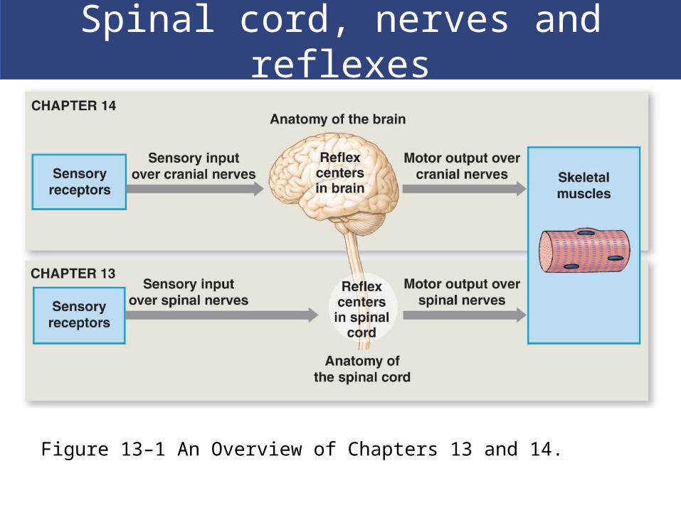
Spinal cord, nerves and reflexes
Figure 13–1 An Overview of Chapters 13 and 14.
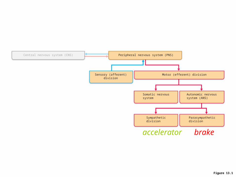
Figure 13.1
Central nervous system (CNS) Peripheral nervous system (PNS)
Motor (efferent) divisionSensory (afferent)division
Somatic nervoussystem
Autonomic nervoussystem (ANS)
Sympatheticdivision
Parasympatheticdivision
accelerator brake
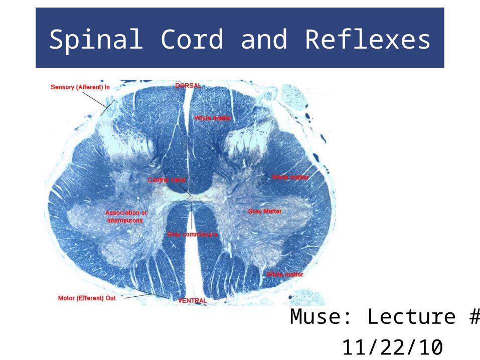
Spinal Cord
Gross Anatomy of the Spinal Cord
About 18 inches (45 cm) long
1/2 inch (14 mm) wide
Ends between vertebrae L1 and L2
Bilateral symmetry
Grooves divide the spinal cord into left and right
Posterior median sulcus: on posterior side
Anterior median fissure: deeper groove on anterior side

Spinal Cord
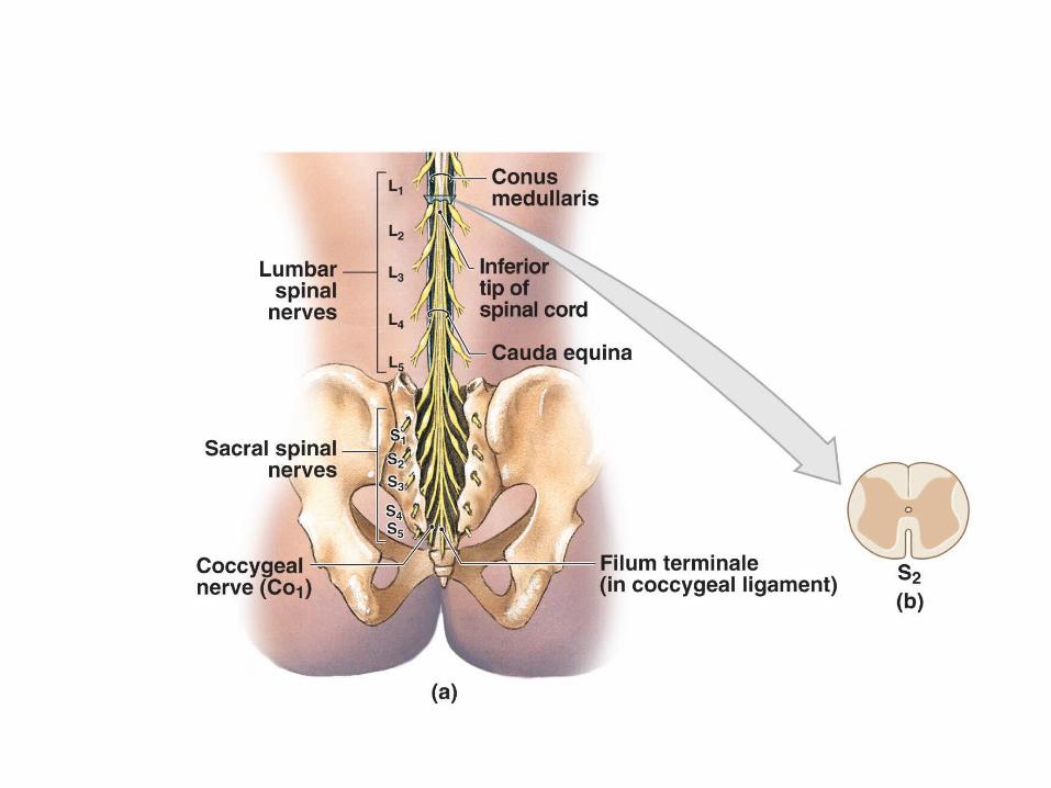
Gross Anatomy of the Spinal Cord The Distal End
Conus medullaris:
– thin, conical spinal cord below lumbar enlargement
Filum terminale:
– thin thread of fibrous tissue at end of conus medullaris
– attaches to coccygeal ligament
Cauda equina:
– nerve roots extending below conus medullaris

Spinal Cord
Figure 13–2 Gross Anatomy of the Adult Spinal Cord.

Spinal CordFigure 13–2 Gross Anatomy of the Adult Spinal Cord.
Made by Honda?

Spinal Cord
31 Spinal Cord Segments Based on vertebrae where spinal nerves
originate
Positions of spinal segment and vertebrae
change with age Cervical nerves:
– are named for inferior vertebra
All other nerves:– are named for superior vertebra

Spinal Cord
Roots Two branches of spinal nerves
Ventral root:
– contains axons of motor neurons
Dorsal root:
– contains axons of sensory neurons
Dorsal root ganglia contain cell bodies of sensory neurons

Spinal Cord
The Spinal Nerve
Each side of spine
Dorsal and ventral roots join
To form a spinal nerve
Mixed Nerves
Carry both afferent (sensory) and efferent (motor)
fibers

Spinal Cord
Figure 13–3 The Spinal Cord and Spinal Meninges

Spinal Cord
Figure 13–3 The Spinal Cord and Spinal Meninges

Spinal Cord
The Spinal Meninges
Specialized membranes isolate spinal cord from
surroundings
Functions of the spinal meninges include
Protect spinal cord
Carry blood supply
Continuous with cranial meninges
Meningitis:
Viral or bacterial infection of meninges

Spinal Cord
The Three Meningeal Layers
Dura mater
Outer layer of spinal cord
Arachnoid mater
Middle meningeal layer
Pia mater
Inner meningeal layer

Spinal Cord
The Dura Mater Tough and fibrous
Cranially Fuses with periosteum of occipital bone
Is continuous with cranial dura mater
Caudally Tapers to dense cord of collagen fibers
Joins filum terminale in coccygeal ligament
The Epidural Space Between spinal dura mater and walls of vertebral canal
Contains loose connective and adipose tissue
Anesthetic injection site

Spinal Cord
The Arachnoid Mater
Middle meningeal layer
Arachnoid membrane
Simple squamous epithelia
Covers arachnoid mater
Spinal Cord
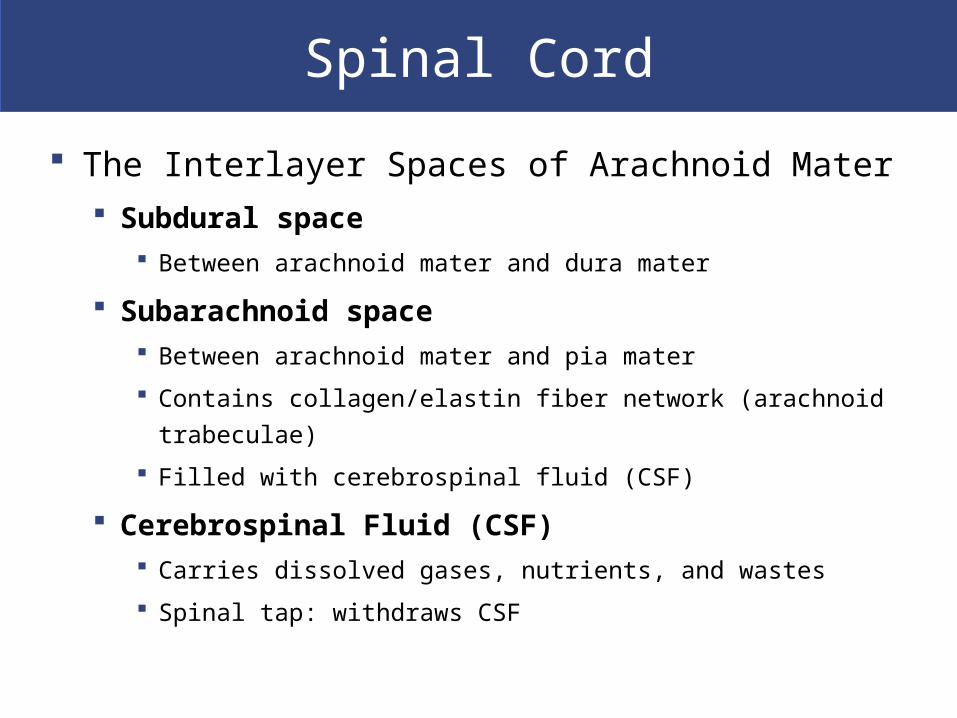
The Interlayer Spaces of Arachnoid Mater Subdural space
Between arachnoid mater and dura mater
Subarachnoid space Between arachnoid mater and pia mater
Contains collagen/elastin fiber network (arachnoid trabeculae)
Filled with cerebrospinal fluid (CSF)
Cerebrospinal Fluid (CSF) Carries dissolved gases, nutrients, and wastes
Spinal tap: withdraws CSF

Spinal Cord
The Pia Mater
Is the innermost meningeal layer
Is a mesh of collagen and elastic fibers
Is bound to underlying neural tissue
Spinal Cord
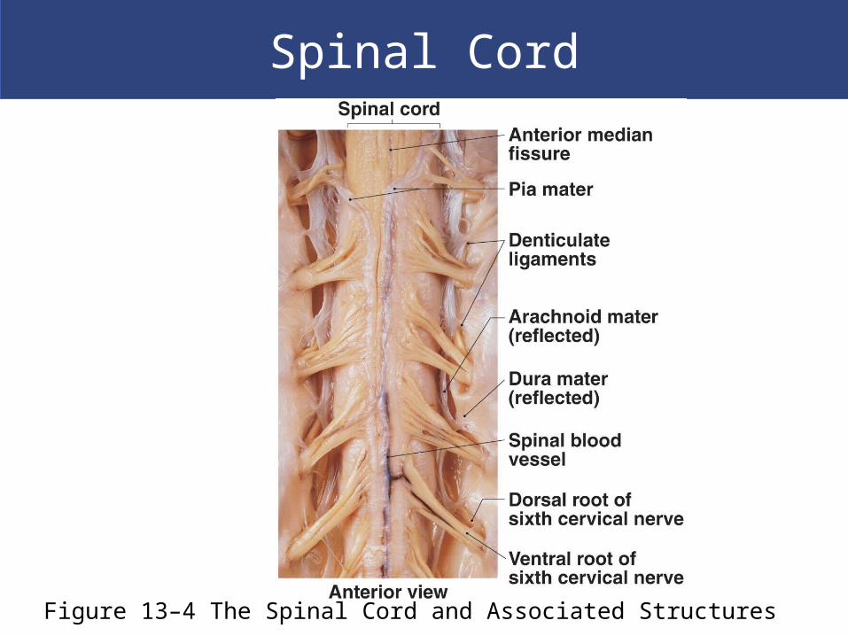
Figure 13–4 The Spinal Cord and Associated Structures

Gray Matter and White Matter
Sectional Anatomy of the Spinal Cord White matter
Is superficial
Contains myelinated and unmyelinated axons
Gray matter Surrounds central canal of spinal cord
Contains neuron cell bodies, neuroglia, unmyelinated axons
Has projections (gray horns)

Gray Matter and White Matter
Organization of Gray Matter The gray horns
Posterior gray horns: contain somatic and visceral sensory nuclei
Anterior gray horns: contain somatic motor nuclei
Lateral gray horns: are in thoracic and lumbar segments; contain visceral motor nuclei
Gray commissures Axons that cross from one side of cord to the other
before reaching gray matter
Gray Matter and White Matter
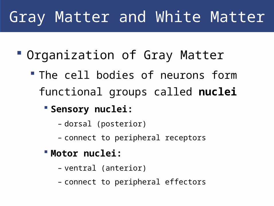
Organization of Gray Matter
The cell bodies of neurons form functional
groups called nuclei
Sensory nuclei:
– dorsal (posterior)
– connect to peripheral receptors
Motor nuclei:
– ventral (anterior)
– connect to peripheral effectors
Gray Matter and White Matter
Control and Location
Sensory or motor nucleus location within the
gray matter determines which body part it
controls

Gray Matter and White Matter
Organization of White Matter
Posterior white columns: lie between posterior gray
horns and posterior median sulcus
Anterior white columns: lie between anterior gray
horns and anterior median fissure
Anterior white commissure: area where axons cross from
one side of spinal cord to the other
Lateral white columns: located on each side of
spinal cord between anterior and posterior columns
Gray Matter and White Matter
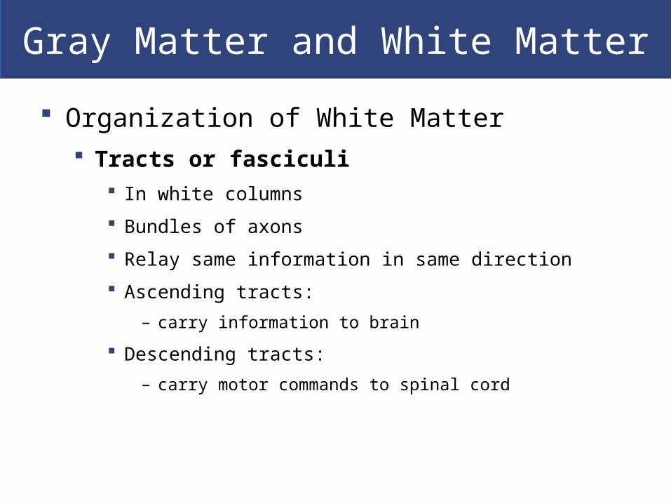
Organization of White Matter Tracts or fasciculi
In white columns
Bundles of axons
Relay same information in same direction
Ascending tracts:
– carry information to brain
Descending tracts:
– carry motor commands to spinal cord

Gray Matter and White Matter
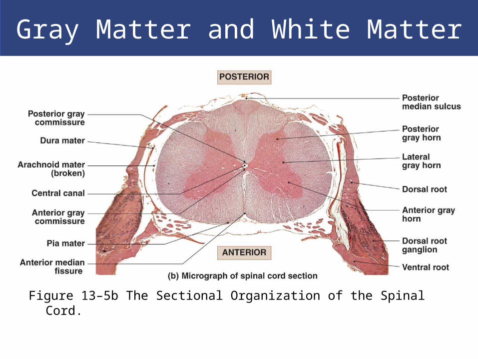
Figure 13–5a The Sectional Organization of the Spinal Cord.
Gray Matter and White Matter
Figure 13–5b The Sectional Organization of the Spinal Cord.

Spinal Cord Summary
Spinal cord has a narrow central canal
Surrounded by gray matter
Containing sensory and motor nuclei
Sensory nuclei are dorsal SounD
Motor nuclei are ventral MoVe

Spinal Cord Summary
Gray matter Is covered by a thick layer of white matter
White matter Consists of ascending and descending axons
Organized in columns
Containing axon bundles with specific functions
Spinal cord is so highly organized It is possible to predict results of injuries to specific
areas
Spinal Nerves and Plexuses
Anatomy of Spinal Nerves
Every spinal cord segment
Is connected to a pair of spinal nerves
Every spinal nerve
Is surrounded by three connective tissue layers
That support structures and contain blood vessels
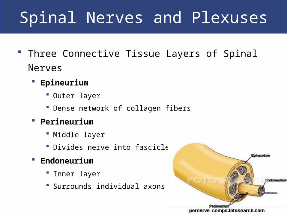
Spinal Nerves and Plexuses
Three Connective Tissue Layers of Spinal Nerves Epineurium
Outer layer
Dense network of collagen fibers
Perineurium
Middle layer
Divides nerve into fascicles (axon bundles)
Endoneurium
Inner layer
Surrounds individual axons

Spinal Nerves and Plexuses
Figure 13–6a A Peripheral Nerve.

Spinal Nerves and Plexuses
Peripheral Nerves
Interconnecting branches of spinal nerves
Surrounded by connective tissue sheaths

Spinal Nerves and Plexuses

Spinal Nerves and Plexuses
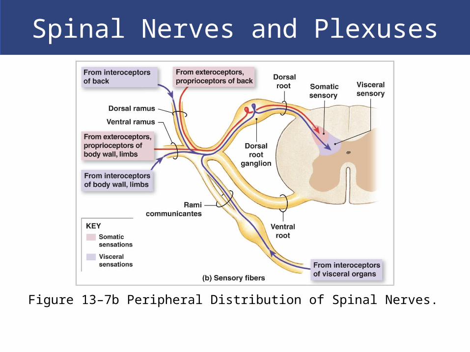
Peripheral Distribution of Spinal Nerves
Sensory nerves
In addition to motor impulses:
– dorsal, ventral, and white rami also carry sensory information
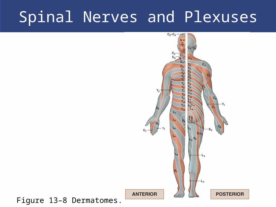
Dermatomes
Bilateral region of skin
Monitored by specific pair of spinal nerves
Spinal Nerves and Plexuses
Figure 13–7b Peripheral Distribution of Spinal Nerves.
Spinal Nerves and Plexuses
Figure 13–8 Dermatomes.

Spinal Nerves and Plexuses
Peripheral Neuropathy
Regional loss of sensory or motor function
Due to trauma or compression
chronic can be due to diabetes
Spinal Nerves and Plexuses
Nerve Plexuses
Complex, interwoven networks of nerve fibers
Formed from blended fibers of ventral rami of
adjacent spinal nerves
Control skeletal muscles of the neck and
limbs

Spinal Nerves and Plexuses
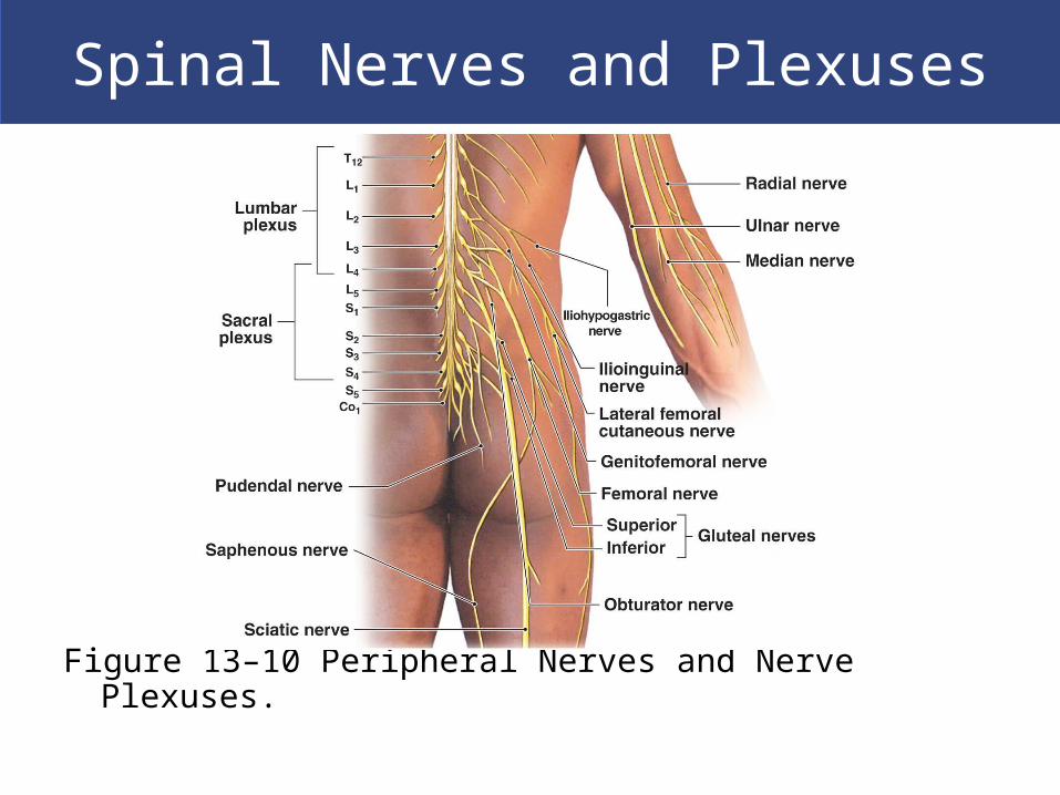
The Four Major Plexuses of Ventral Rami
Cervical plexus
Brachial plexus
Lumbar plexus
Sacral plexus

Spinal Nerves and Plexuses
Figure 13–10 Peripheral Nerves and Nerve Plexuses.
Spinal Nerves and Plexuses
Figure 13–10 Peripheral Nerves and Nerve Plexuses.

Spinal Nerves and Plexuses
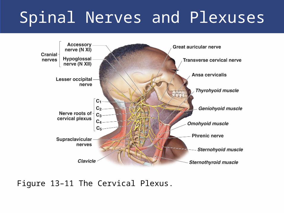
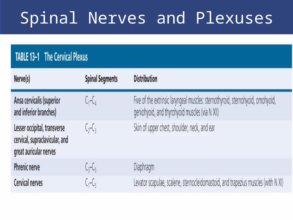
The Cervical Plexus of the Ventral Rami
Includes ventral rami of spinal nerves C1–C5
Innervates neck, thoracic cavity, diaphragmatic
muscles
Major nerve
Phrenic nerve (controls diaphragm)
Spinal Nerves and Plexuses
Figure 13–11 The Cervical Plexus.
Spinal Nerves and Plexuses

Spinal Nerves and Plexuses
The Brachial Plexus of the Ventral Rami
Major nerves of brachial plexus
Musculocutaneous nerve (lateral cord)
Median nerve (lateral and medial cords)
Ulnar nerve (medial cord)
Axillary nerve (posterior cord)
Radial nerve (posterior cord)
Includes ventral rami of spinal nerves C5–T1

Spinal Nerves and Plexuses
Figure 13–12a The Brachial Plexus.

Spinal Nerves and Plexuses
Figure 13–12b The Brachial Plexus.

Spinal Nerves and Plexuses
Figure 13–12c The Brachial Plexus.

Spinal Nerves and Plexuses
Spinal Nerves and Plexuses
The Lumbar Plexus of the Ventral Rami
Includes ventral rami of spinal nerves T12–L4
Major nerves
Genitofemoral nerve
Lateral femoral cutaneous nerve
Femoral nerve

Spinal Nerves and Plexuses
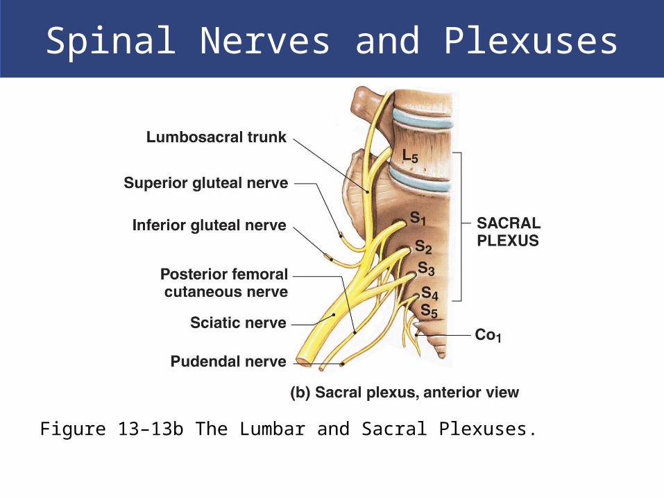
The Sacral Plexus of the Ventral Rami Includes ventral rami of spinal nerves L4–S4
Major nerves Pudendal nerve
Sciatic nerve
Branches of sciatic nerve Fibular nerve
Tibial nerve
3D Rotation of Lumbar and Sacral Plexuses
Spinal Nerves and Plexuses
Figure 13–13b The Lumbar and Sacral Plexuses.

Spinal Nerves and Plexuses
Figure 13–13d The Lumbar and Sacral Plexuses.

Neuronal Pools
Functional Organization of Neurons
Sensory neurons
About 10 million
Deliver information to CNS
Motor neurons
About 1/2 million
Deliver commands to peripheral effectors
Interneurons
About 20 billion
Interpret, plan, and coordinate signals in and out

Neuronal Pools
Neuronal Pools
Functional groups of interconnected neurons
(interneurons)
Each with limited input sources and output
destinations
May stimulate or depress parts of brain or spinal cord
Neuronal Pools
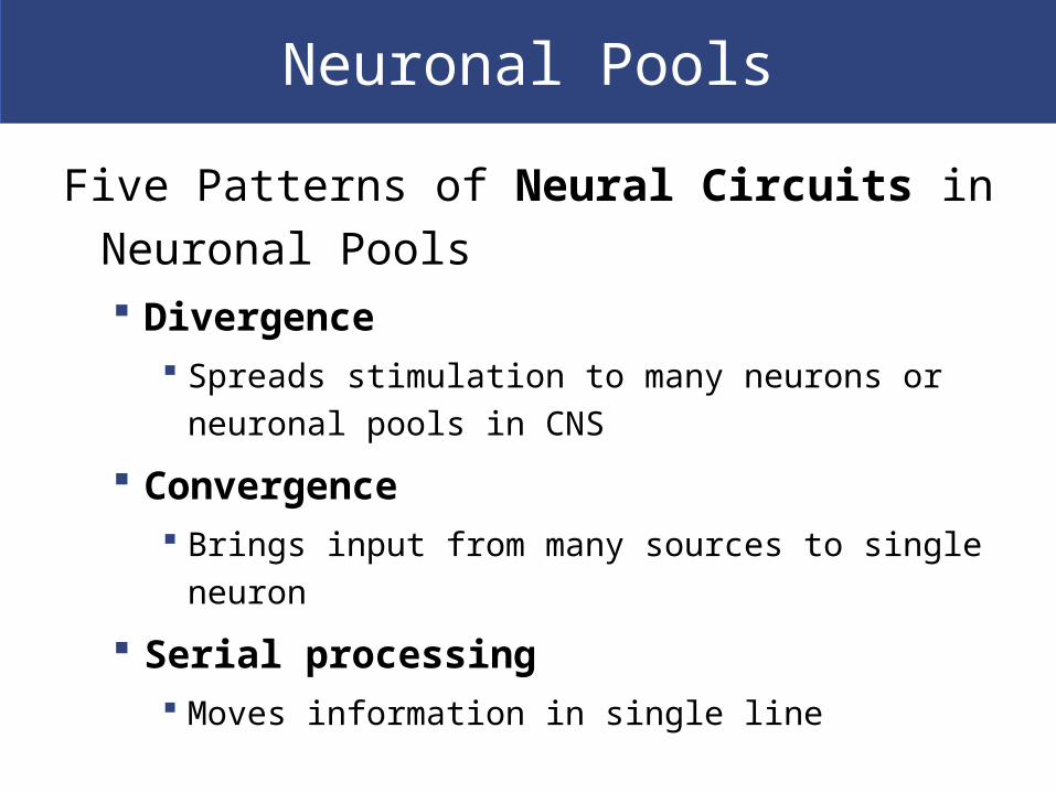
Five Patterns of Neural Circuits in Neuronal
Pools Divergence
Spreads stimulation to many neurons or neuronal
pools in CNS
Convergence Brings input from many sources to single neuron
Serial processing Moves information in single line

Neuronal Pools
Five Patterns of Neural Circuits in Neuronal
Pools Parallel processing
Moves same information along several paths simultaneously
Reverberation Positive feedback mechanism
Functions until inhibited

Neuronal Pools
Figure 13–14 Neural Circuits: The Organization of Neuronal Pools.

Reflexes
Automatic responses coordinated within
spinal cord
Through interconnected sensory neurons,
motor neurons, and interneurons
Produce simple and complex reflexes
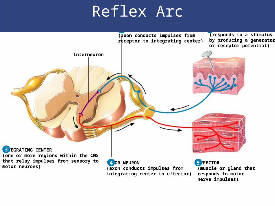
1 SENSORY RECEPTOR(responds to a stimulusby producing a generatoror receptor potential)
1SENSORY NEURON(axon conducts impulses from receptor to integrating center)
SENSORY RECEPTOR(responds to a stimulusby producing a generatoror receptor potential)
2 1SENSORY NEURON(axon conducts impulses from receptor to integrating center)
SENSORY RECEPTOR(responds to a stimulusby producing a generatoror receptor potential)
INTEGRATING CENTER(one or more regions within the CNSthat relay impulses from sensory tomotor neurons)
Interneuron
2
3
1SENSORY NEURON(axon conducts impulses from receptor to integrating center)
SENSORY RECEPTOR(responds to a stimulusby producing a generatoror receptor potential)
INTEGRATING CENTER(one or more regions within the CNSthat relay impulses from sensory tomotor neurons)
MOTOR NEURON(axon conducts impulses fromintegrating center to effector)
Interneuron
2
3
4
1SENSORY NEURON(axon conducts impulses from receptor to integrating center)
SENSORY RECEPTOR(responds to a stimulusby producing a generatoror receptor potential)
INTEGRATING CENTER(one or more regions within the CNSthat relay impulses from sensory tomotor neurons)
MOTOR NEURON(axon conducts impulses fromintegrating center to effector)
EFFECTOR(muscle or gland thatresponds to motornerve impulses)
Interneuron
2
3
4 5
Reflex Arc
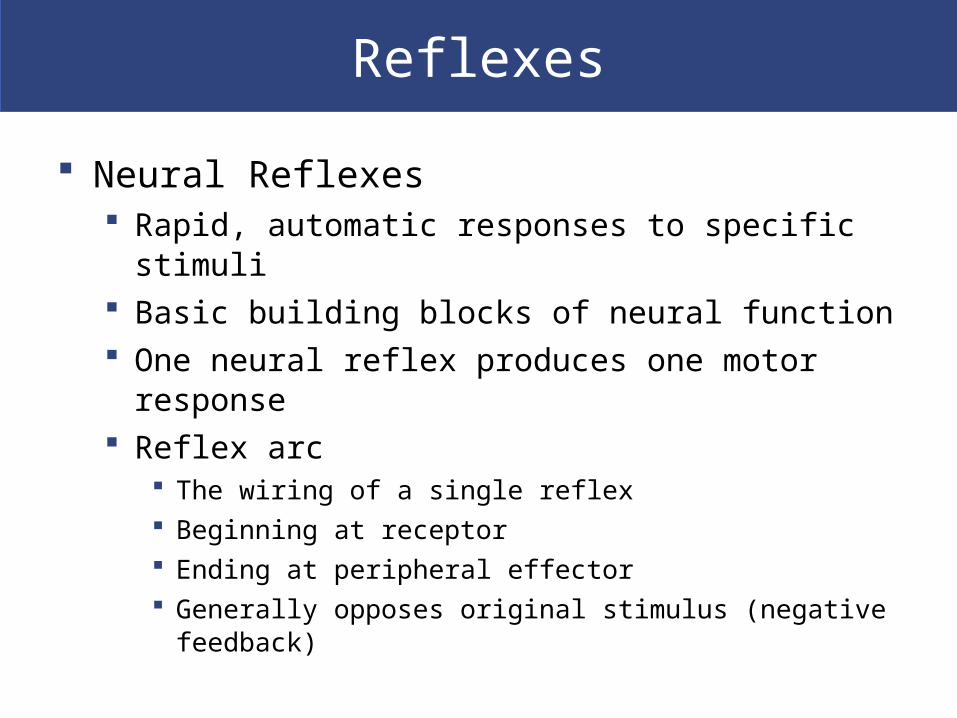
Reflexes
Neural Reflexes Rapid, automatic responses to specific stimuli Basic building blocks of neural function One neural reflex produces one motor response Reflex arc
The wiring of a single reflex Beginning at receptor Ending at peripheral effector Generally opposes original stimulus (negative feedback)

Reflexes
Five Steps in a Neural Reflex Step 1: Arrival of stimulus, activation of receptor
Physical or chemical changes
Step 2: Activation of sensory neuron Graded depolarization
Step 3: Information processing by postsynaptic cell Triggered by neurotransmitters
Step 4: Activation of motor neuron Action potential
Step 5: Response of peripheral effector Triggered by neurotransmitters

Reflexes
Figure 13–15 Events in a Neural Reflex.
Reflexes
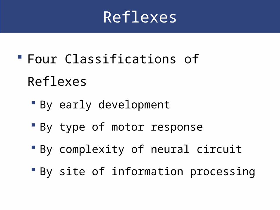
Four Classifications of Reflexes
By early development
By type of motor response
By complexity of neural circuit
By site of information processing
Reflexes
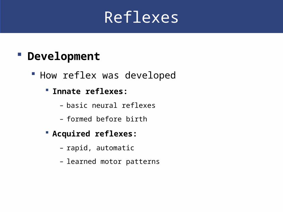
Development
How reflex was developed
Innate reflexes:
– basic neural reflexes
– formed before birth
Acquired reflexes:
– rapid, automatic
– learned motor patterns
Reflexes
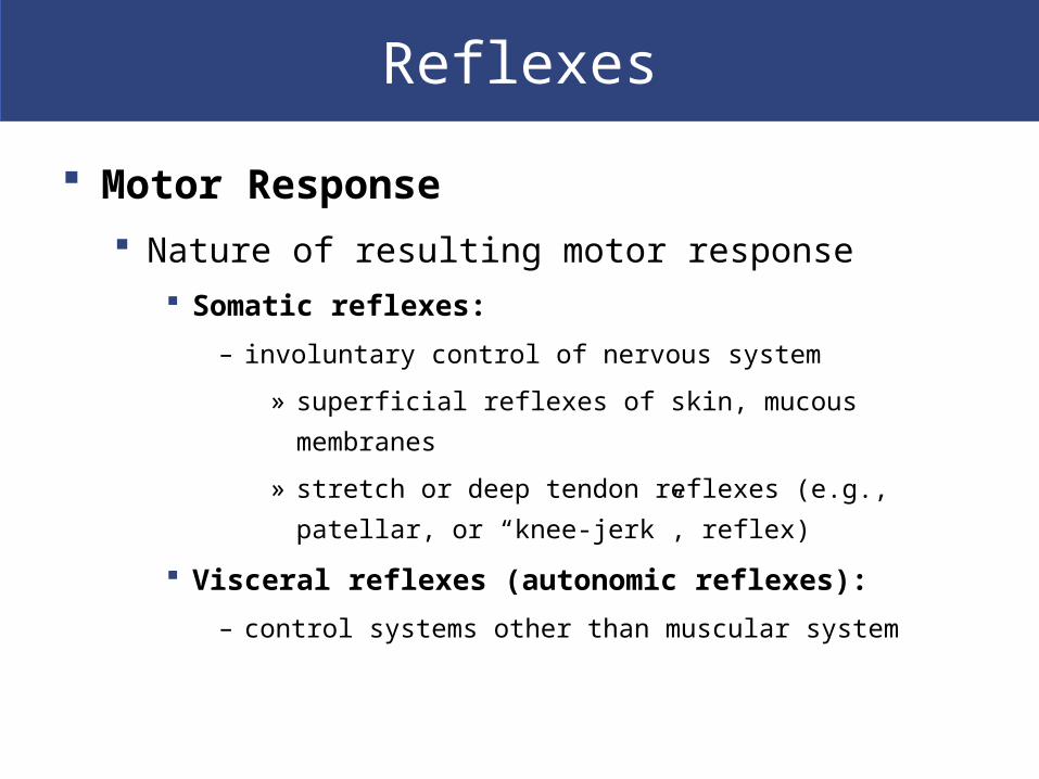
Motor Response
Nature of resulting motor response
Somatic reflexes:
– involuntary control of nervous system
» superficial reflexes of skin, mucous membranes
» stretch or deep tendon reflexes (e.g., patellar, or “knee-
jerk”, reflex)
Visceral reflexes (autonomic reflexes):
– control systems other than muscular system
Reflexes
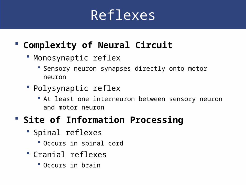
Complexity of Neural Circuit Monosynaptic reflex
Sensory neuron synapses directly onto motor neuron
Polysynaptic reflex At least one interneuron between sensory neuron and motor
neuron
Site of Information Processing Spinal reflexes
Occurs in spinal cord
Cranial reflexes Occurs in brain

Reflexes
Figure 13–16 The Classification of Reflexes.
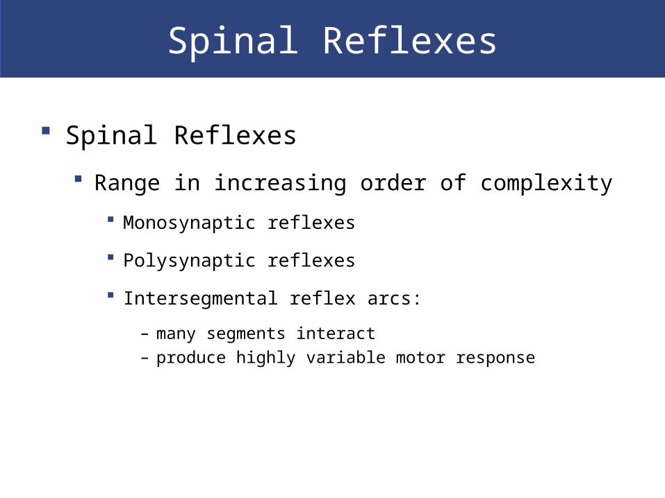
Spinal Reflexes
Spinal Reflexes
Range in increasing order of complexity
Monosynaptic reflexes
Polysynaptic reflexes
Intersegmental reflex arcs:
– many segments interact
– produce highly variable motor response

Spinal Reflexes
Monosynaptic Reflexes
A stretch reflex
Have least delay between sensory input and motor
output:
For example, stretch reflex (such as patellar reflex)
Completed in 20–40 msec
Receptor is muscle spindle

Spinal Reflexes
Figure 13–17 A Stretch Reflex.
Spinal Reflexes
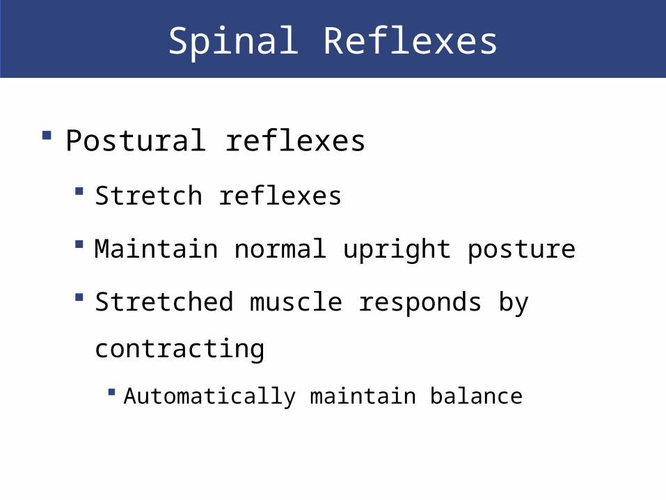
Postural reflexes
Stretch reflexes
Maintain normal upright posture
Stretched muscle responds by contracting
Automatically maintain balance
Spinal Reflexes
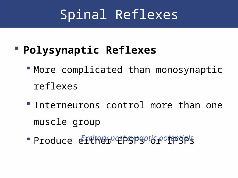
Polysynaptic Reflexes
More complicated than monosynaptic reflexes
Interneurons control more than one muscle
group
Produce either EPSPs or IPSPs
Excitory post synaptic potentials
Spinal Reflexes
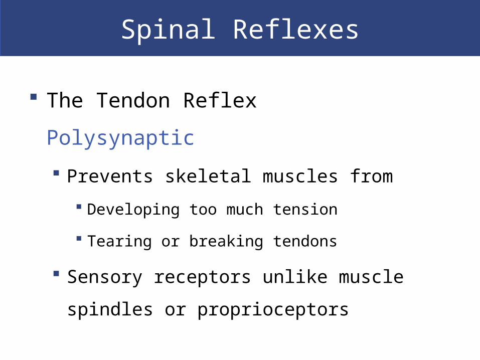
The Tendon Reflex Polysynaptic
Prevents skeletal muscles from
Developing too much tension
Tearing or breaking tendons
Sensory receptors unlike muscle spindles or
proprioceptors

1
Increased tensionstimulatesSENSORYRECEPTOR (tendon)
1
Spinalnerve
SENSORYNEURONexcited
To brain
Increased tensionstimulatesSENSORYRECEPTOR (tendon)
2 ++
1
Within INTEGRATINGCENTER (spinal cord),sensory neuron activatesinhibitory interneuron
Excitatoryinterneuron
Spinalnerve
Inhibitoryinterneuron
SENSORYNEURONexcited
+
To brain
Increased tensionstimulatesSENSORYRECEPTOR (tendon)
++2
3
–
+1
–
Within INTEGRATINGCENTER (spinal cord),sensory neuron activatesinhibitory interneuron
Excitatoryinterneuron
Antagonisticmusclescontract
Spinalnerve
MOTOR NEURONinhibited
Inhibitoryinterneuron
SENSORYNEURONexcited
+
To brain
Increased tensionstimulatesSENSORYRECEPTOR (tendon)
Motor neuron toantagonisticmuscles is excited
+
+
+
+2
3
4
1
–
EFFECTOR(muscle attachedto same tendon)relaxes andrelieves excesstension
Within INTEGRATINGCENTER (spinal cord),sensory neuron activatesinhibitory interneuron
Excitatoryinterneuron
Antagonisticmusclescontract
Spinalnerve
MOTOR NEURONinhibited
Inhibitoryinterneuron
SENSORYNEURONexcited
+
To brain
Increased tensionstimulatesSENSORYRECEPTOR (tendon)
Motor neuron toantagonisticmuscles is excited
+
+
+2
3
45
+

Spinal Reflexes
Withdrawal Reflexes
Move body part away from stimulus (pain or pressure)
For example, flexor reflex:
– pulls hand away from hot stove
Strength and extent of response
Depends on intensity and location of stimulus

Spinal Reflexes
Figure 13–19 A Flexor Reflex.
Spinal Reflexes
Reciprocal Inhibition
For flexor reflex to work
The stretch reflex of antagonistic (extensor)
muscle must be inhibited (reciprocal inhibition) by
interneurons in spinal cord
Spinal Reflexes
Reflex Arcs
Ipsilateral reflex arcs
Occur on same side of body as stimulus
Stretch, tendon, and withdrawal reflexes
Crossed extensor reflexes
Involve a contralateral reflex arc
Occur on side opposite stimulus
Spinal Reflexes
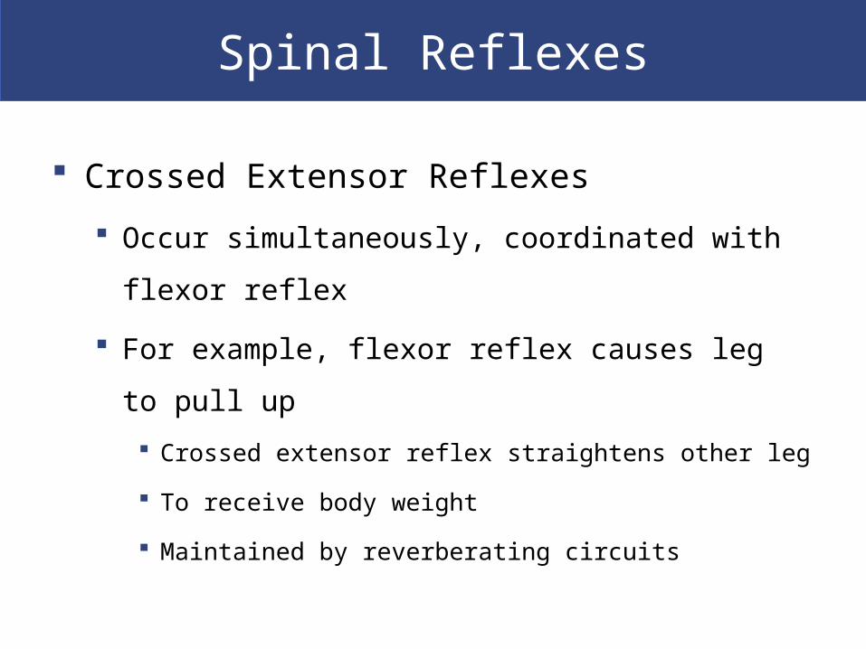
Crossed Extensor Reflexes
Occur simultaneously, coordinated with flexor reflex
For example, flexor reflex causes leg to pull up
Crossed extensor reflex straightens other leg
To receive body weight
Maintained by reverberating circuits
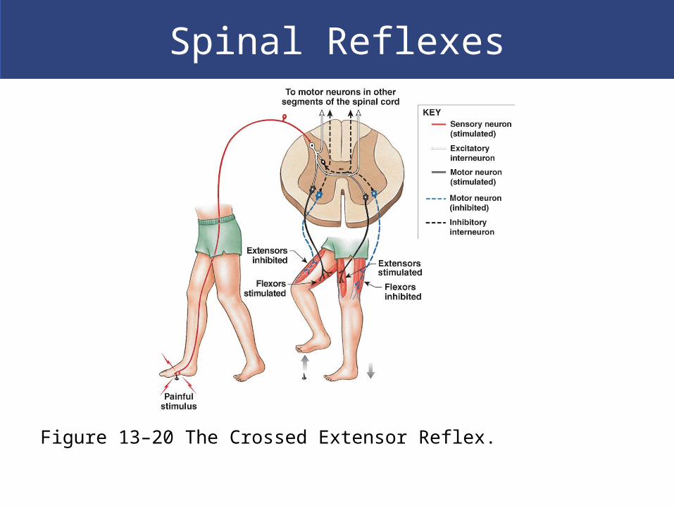
Spinal Reflexes
Figure 13–20 The Crossed Extensor Reflex.
Spinal Reflexes
Five General Characteristics of Polysynaptic Reflexes Involve pools of neurons
Are intersegmental in distribution
Involve reciprocal inhibition
Have reverberating circuits Which prolong reflexive motor response
Several reflexes cooperate To produce coordinated, controlled response
The Brain Can Alter Spinal Reflexes
Integration and Control of Spinal Reflexes
Reflex behaviors are automatic
But processing centers in brain can facilitate or
inhibit reflex motor patterns based in spinal cord

The Brain Can Alter Spinal Reflexes
Voluntary Movements and Reflex Motor
Patterns
Higher centers of brain incorporate lower,
reflexive motor patterns
Automatic reflexes Can be activated by brain as needed
Use few nerve impulses to control complex motor
functions
Walking, running, jumping

The Brain Can Alter Spinal Reflexes
Reinforcement of Spinal Reflexes
Higher centers reinforce spinal reflexes
By stimulating excitatory neurons in brain stem or
spinal cord
Creating EPSPs at reflex motor neurons
Facilitating postsynaptic neurons
The Brain Can Alter Spinal Reflexes
Inhibition of Spinal Reflexes
Higher centers inhibit spinal reflexes by
Stimulating inhibitory neurons
Creating IPSPs at reflex motor neurons
Suppressing postsynaptic neurons

The Brain Can Alter Spinal Reflexes
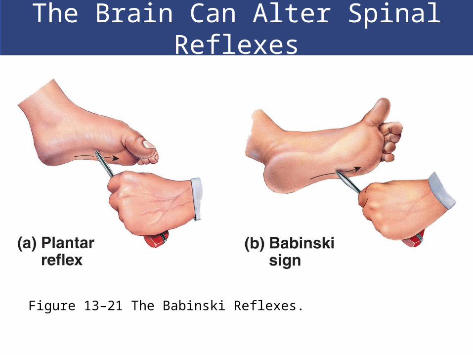
The Babinski Reflexes
Normal in infants
May indicate CNS damage in adults
The Brain Can Alter Spinal Reflexes
Figure 13–21 The Babinski Reflexes.
An Introduction to the Brain and Cranial Nerves
The Adult Human Brain
Ranges from 750 cc to 2100 cc
Contains almost 97% of the body’s neural
tissue
Average weight about 1.4 kg (3 lb)
The Brain
Six Regions of the Brain Cerebrum
Cerebellum
Diencephalon
Mesencephalon
Pons
Medulla oblongata
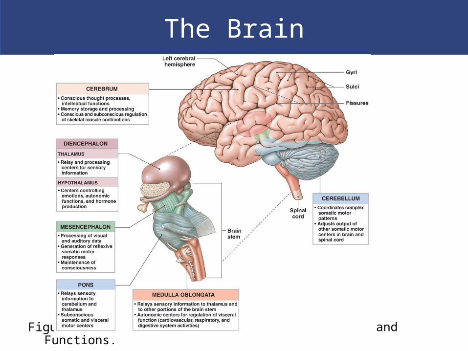
The Brain
Figure 14–1 An Introduction to Brain Structures and Functions.

The Brain
Figure 14–2 Ventricles of the Brain.
Brain Protection and Support
Physical protection
Bones of the cranium
Cranial meninges
Cerebrospinal fluid
Biochemical isolation
Blood–brain barrier

Brain Protection and Support
Cerebrospinal Fluid (CSF)
Surrounds all exposed surfaces of CNS
Interchanges with interstitial fluid of brain
Functions of CSF
Cushions delicate neural structures
Supports brain
Transports nutrients, chemical messengers, and
waste products

Brain Protection and Support
Figure 14–4 The Formation and Circulation of Cerebrospinal Fluid.

Brain Protection and Support
Blood Supply to the Brain
Supplies nutrients and oxygen to brain
Delivered by internal carotid arteries and
vertebral arteries
Removed from dural sinuses by internal
jugular veins

Brain Protection and Support
Figure 21–23 Arteries of the Brain.

Brain Protection and Support
Blood–Brain Barrier
Isolates CNS neural tissue from general circulation
Formed by network of tight junctions
Between endothelial cells of CNS capillaries
Lipid-soluble compounds (O2, CO2), steroids, and
prostaglandins diffuse into interstitial fluid of brain and
spinal cord
Astrocytes control blood–brain barrier by releasing
chemicals that control permeability of endothelium
Brain Protection and Support
Blood–CSF Barrier
Formed by special ependymal cells
Surround capillaries of choroid plexus
Limits movement of compounds transferred
Allows chemical composition of blood and CSF to
differ
Brain Protection and Support
Four Breaks in the BBB Portions of hypothalamus
Secrete hypothalamic hormones
Posterior lobe of pituitary gland Secretes hormones ADH and oxytocin
Pineal glands Pineal secretions
Choroid plexus Where special ependymal cells maintain blood–
CSF barrier
The Medulla Oblongata
The Medulla Oblongata
Allows brain and spinal cord to communicate
Coordinates complex autonomic reflexes
Controls visceral functions
Nuclei in the Medulla
Autonomic nuclei: control visceral activities
Sensory and motor nuclei: of cranial nerves
Relay stations: along sensory and motor pathways
The Medulla Oblongata
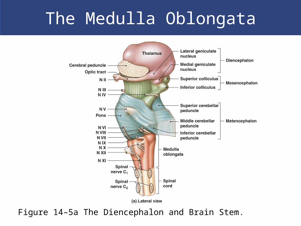
Figure 14–5a The Diencephalon and Brain Stem.
The Medulla Oblongata
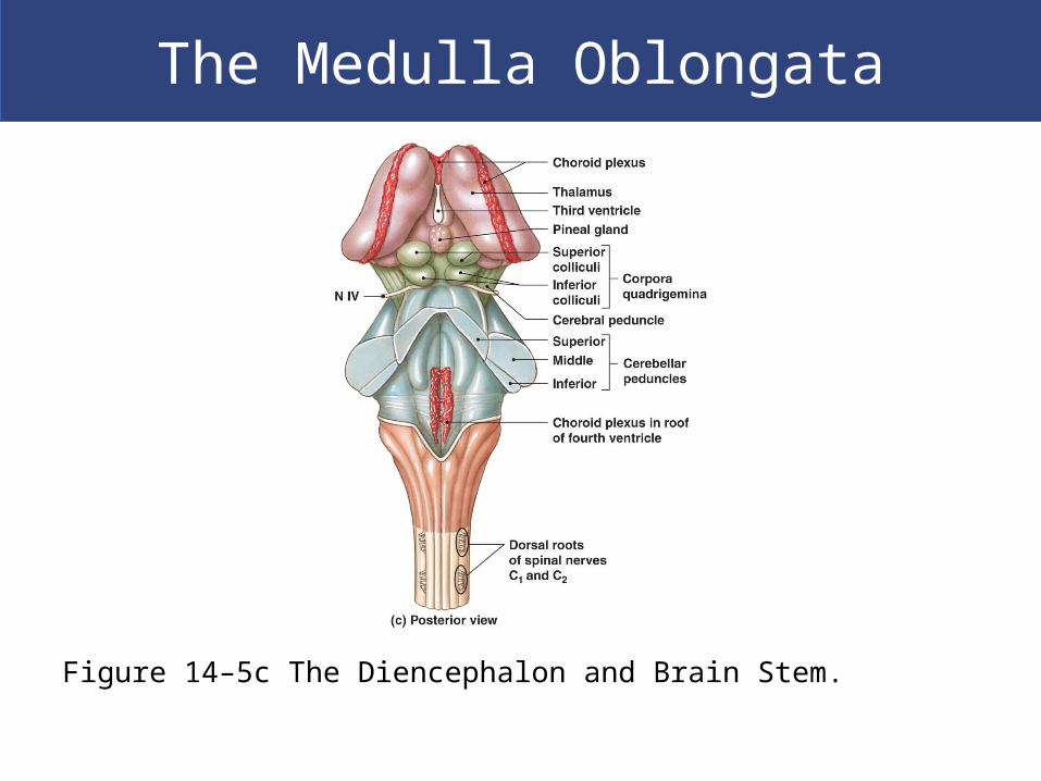
Figure 14–5c The Diencephalon and Brain Stem.
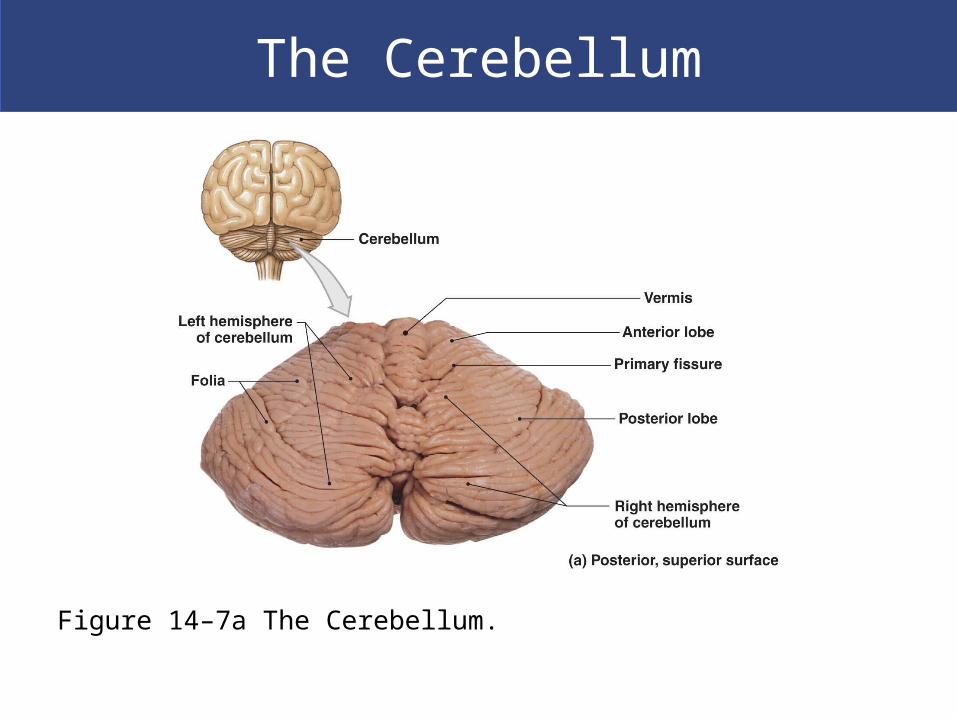
The Cerebellum
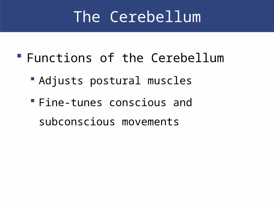
Functions of the Cerebellum
Adjusts postural muscles
Fine-tunes conscious and subconscious
movements
The Cerebellum
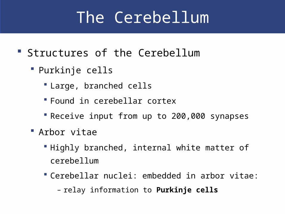
Structures of the Cerebellum
Purkinje cells
Large, branched cells
Found in cerebellar cortex
Receive input from up to 200,000 synapses
Arbor vitae
Highly branched, internal white matter of cerebellum
Cerebellar nuclei: embedded in arbor vitae:
– relay information to Purkinje cells
The Cerebellum
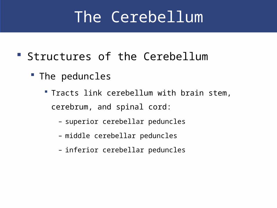
Structures of the Cerebellum
The peduncles
Tracts link cerebellum with brain stem, cerebrum, and spinal
cord:
– superior cerebellar peduncles
– middle cerebellar peduncles
– inferior cerebellar peduncles

The Cerebellum
Disorders of the Cerebellum
Ataxia
Damage from trauma or stroke
Intoxication (temporary impairment)
Disturbs muscle coordination
The Cerebellum
Figure 14–7a The Cerebellum.

The Cerebellum
Figure 14–7b The Cerebellum.

The Diencephalon
Integrates sensory information and motor
commands
Thalamus, epithalamus, and hypothalamus
The pineal gland
Found in posterior epithalamus
Secretes hormone melatonin
The Diencephalon
The Thalamus Filters ascending sensory information for primary
sensory cortex
Relays information between basal nuclei and cerebral
cortex
The third ventricle Separates left thalamus and right thalamus
Interthalamic adhesion (or intermediate mass):
– projection of gray matter
– extends into ventricle from each side
The Diencephalon
The Thalamus
Thalamic nuclei
Are rounded masses that form thalamus
Relay sensory information to basal nuclei and
cerebral cortex
The Diencephalon
The Hypothalamus Mamillary bodies
Process olfactory and other sensory information
Control reflex eating movements
Infundibulum A narrow stalk
Connects hypothalamus to pituitary gland
Tuberal area Located between the infundibulum and mamillary bodies
Helps control pituitary gland function

The Diencephalon
Figure 14–10a The Hypothalamus in Sagittal Section.

The Diencephalon
Eight Functions of the Hypothalamus
Provides subconscious control of skeletal muscle
Controls autonomic function
Coordinates activities of nervous and endocrine
systems
Secretes hormones
Antidiuretic hormone (ADH) by supraoptic nucleus
Oxytocin (OT; OXT) by paraventricular nucleus

The Diencephalon
Eight Functions of the Hypothalamus
Produces emotions and behavioral drives
The feeding center (hunger)
The thirst center (thirst)
Coordinates voluntary and autonomic functions
Regulates body temperature
Preoptic area of hypothalamus
Controls circadian rhythms (day–night cycles)
Suprachiasmatic nucleus

The Limbic System
The Limbic System
Is a functional grouping that
Establishes emotional states
Links conscious functions of cerebral cortex with autonomic
functions of brain stem
Facilitates memory storage and retrieval
The Limbic System
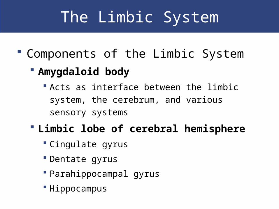
Components of the Limbic System Amygdaloid body
Acts as interface between the limbic system, the
cerebrum, and various sensory systems
Limbic lobe of cerebral hemisphere Cingulate gyrus
Dentate gyrus
Parahippocampal gyrus
Hippocampus
The Limbic System
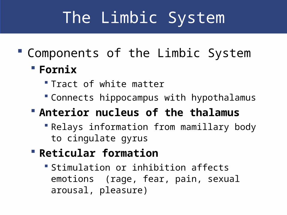
Components of the Limbic System Fornix
Tract of white matter Connects hippocampus with hypothalamus
Anterior nucleus of the thalamus Relays information from mamillary body to
cingulate gyrus
Reticular formation Stimulation or inhibition affects emotions (rage,
fear, pain, sexual arousal, pleasure)

The Limbic System
Figure 14–11a The Limbic System.

The Limbic System
Figure 14–11b The Limbic System.

The Limbic System
The Cerebrum
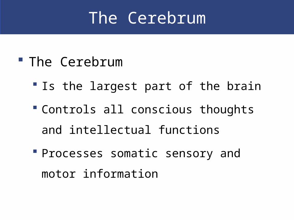
The Cerebrum
Is the largest part of the brain
Controls all conscious thoughts and
intellectual functions
Processes somatic sensory and motor
information

The Cerebrum
Gray matter
In cerebral cortex and basal nuclei
White matter
Deep to basal cortex
Around basal nuclei
The Cerebrum
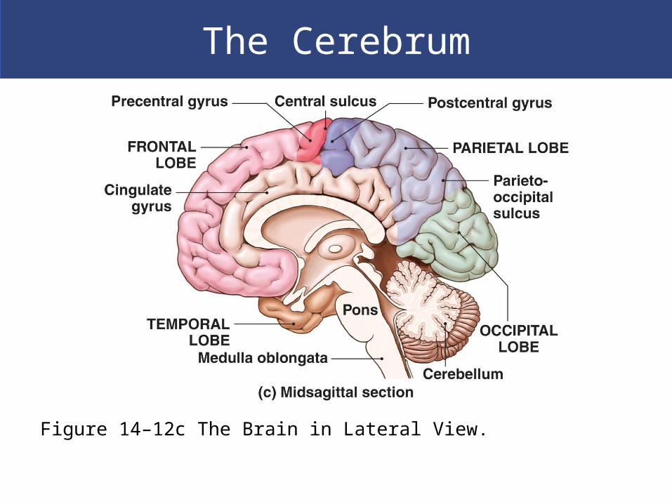
Figure 14–12c The Brain in Lateral View.
The Cerebrum
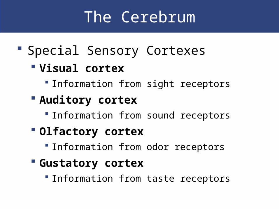
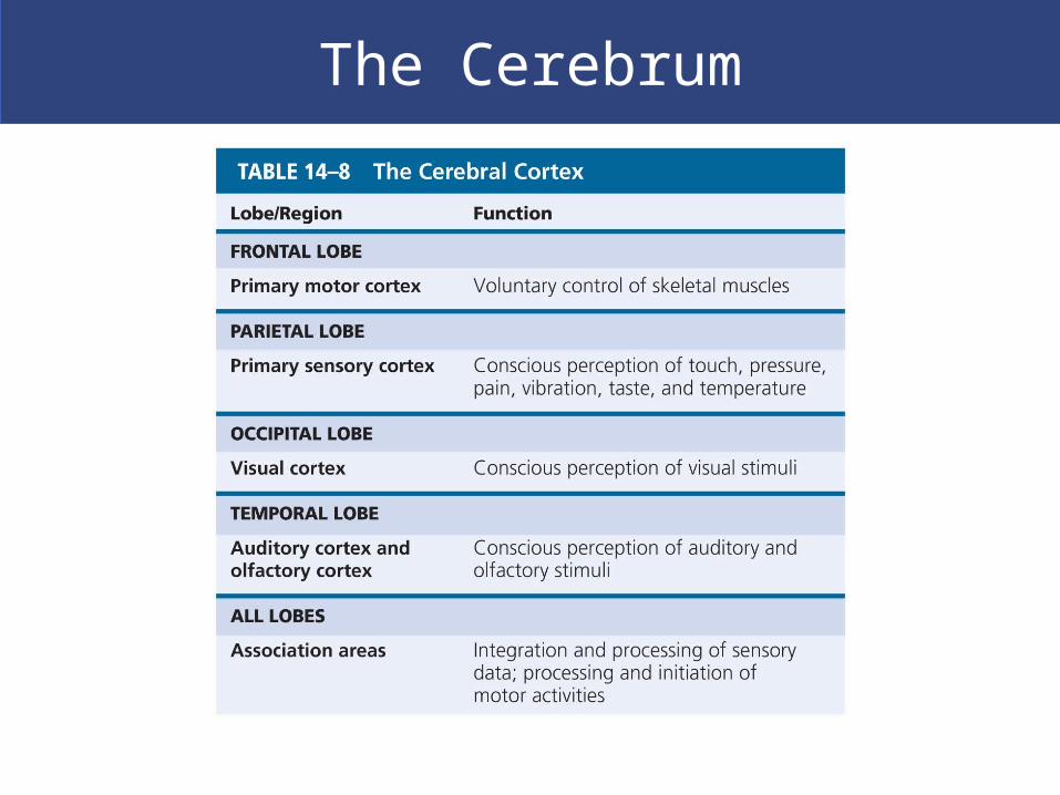
Special Sensory Cortexes Visual cortex
Information from sight receptors
Auditory cortex Information from sound receptors
Olfactory cortex Information from odor receptors
Gustatory cortex Information from taste receptors

The Cerebrum
Figure 14–15a Motor and Sensory Regions of the Cerebral Cortex.
The Cerebrum

The Cerebrum
The Left Hemisphere In most people, left brain (dominant hemisphere)
controls Reading, writing, and math
Decision making
Speech and language
The Right Hemisphere Right cerebral hemisphere relates to
Senses (touch, smell, sight, taste, feel)
Recognition (faces, voice inflections)

The Cerebrum
Figure 14–16 Hemispheric Lateralization.

The Cerebrum
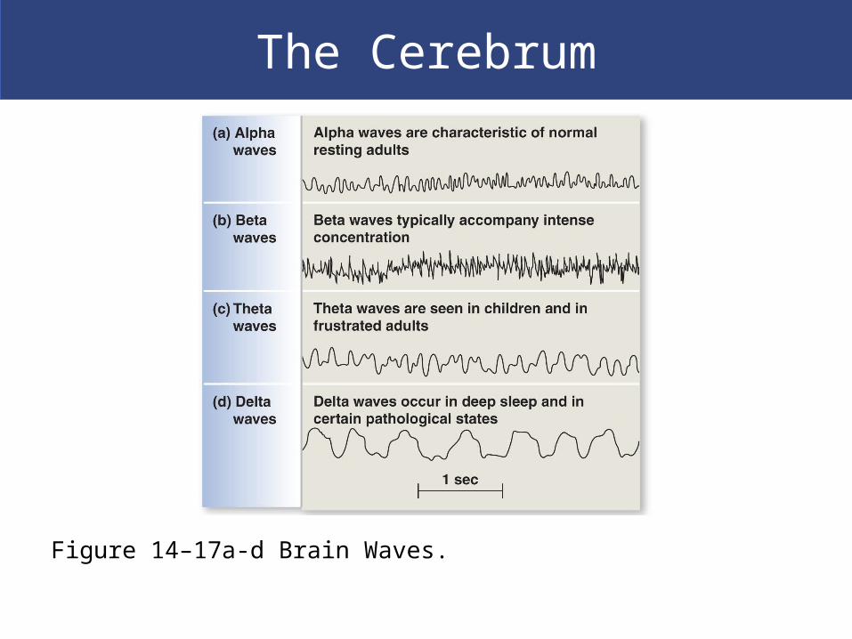
Monitoring Brain Activity
Brain activity is assessed by an
electroencephalogram (EEG)
Electrodes are placed on the skull
Patterns of electrical activity (brain waves) are
printed out
The Cerebrum
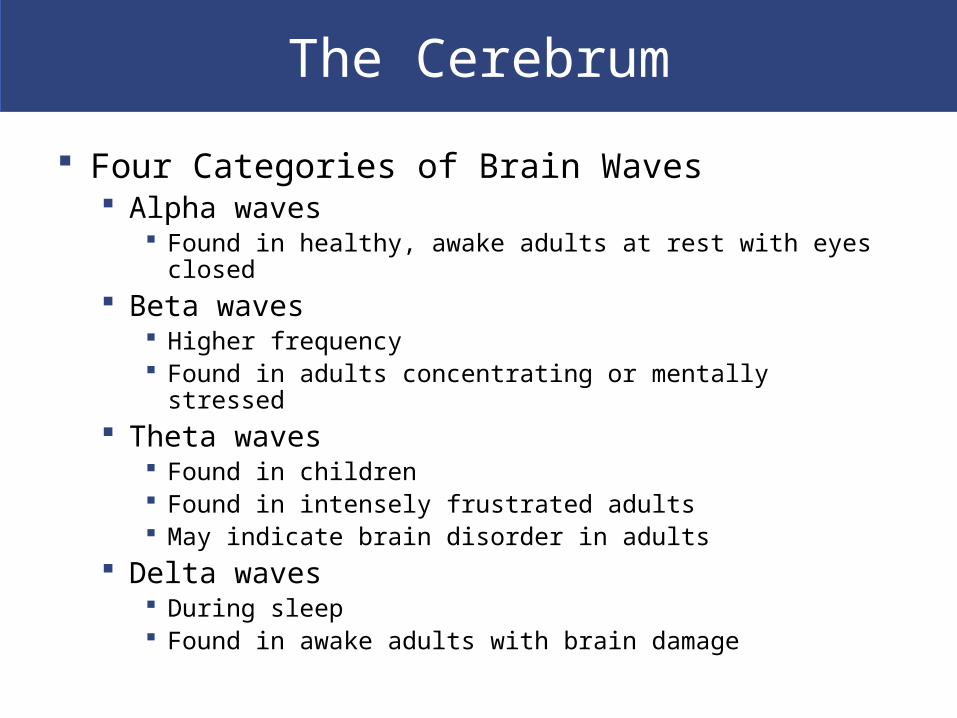
Four Categories of Brain Waves Alpha waves
Found in healthy, awake adults at rest with eyes closed Beta waves
Higher frequency Found in adults concentrating or mentally stressed
Theta waves Found in children Found in intensely frustrated adults May indicate brain disorder in adults
Delta waves During sleep Found in awake adults with brain damage
The Cerebrum
Figure 14–17a-d Brain Waves.
The Cerebrum
Synchronization A pacemaker mechanism
Synchronizes electrical activity between hemispheres
Brain damage can cause desynchronization
Seizure Is a temporary cerebral disorder Changes the electroencephalogram Symptoms depend on regions affected

Cranial Nerves
12 pairs connected to brain
Four Classifications of Cranial Nerves
Sensory nerves: carry somatic sensory information,
including touch, pressure, vibration, temperature, and
pain
Special sensory nerves: carry sensations such as
smell, sight, hearing, balance
Motor nerves: axons of somatic motor neurons
Mixed nerves: mixture of motor and sensory fibers
Cranial Nerves
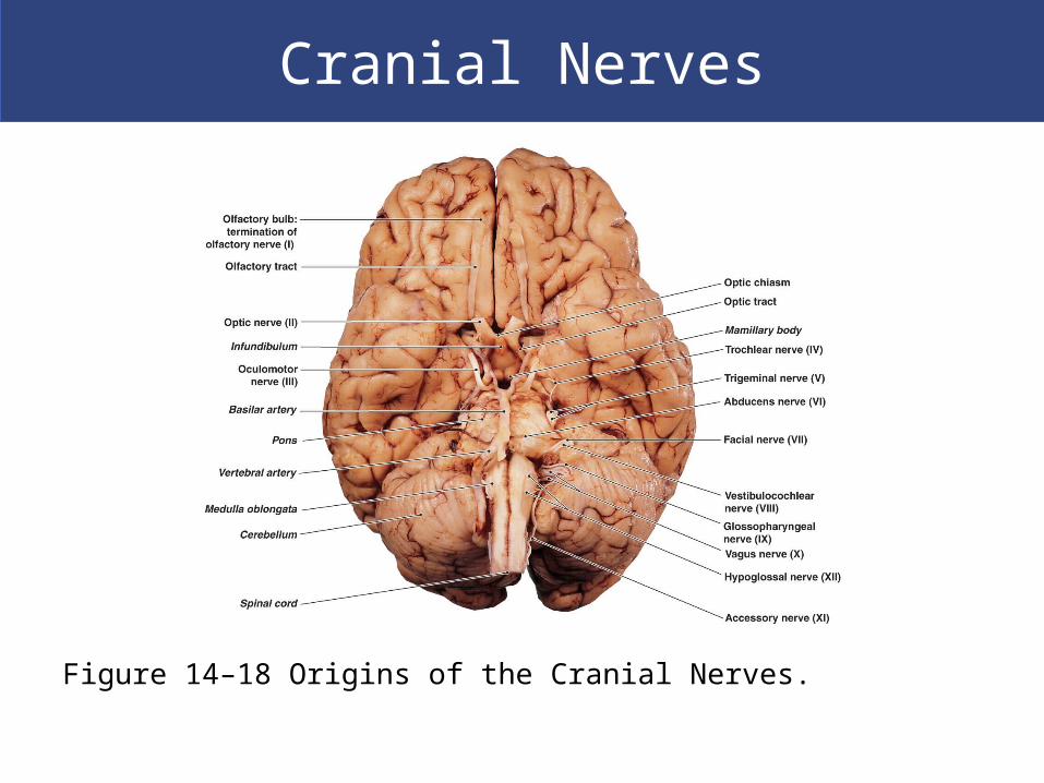
Figure 14–18 Origins of the Cranial Nerves.
Cranial Nerves
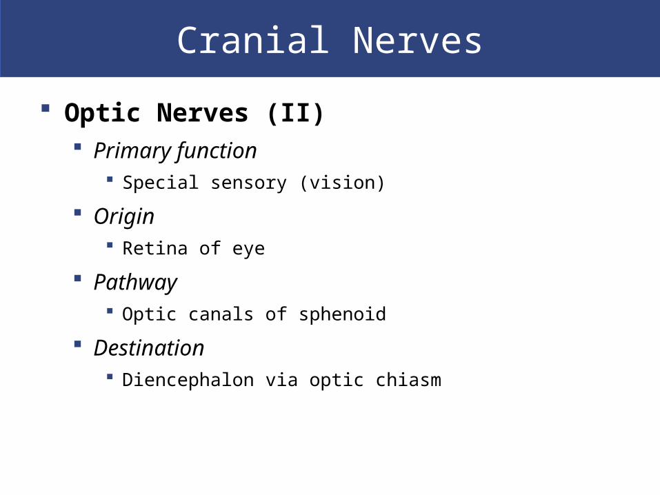
Optic Nerves (II) Primary function
Special sensory (vision)
Origin Retina of eye
Pathway Optic canals of sphenoid
Destination Diencephalon via optic chiasm
Cranial Nerves
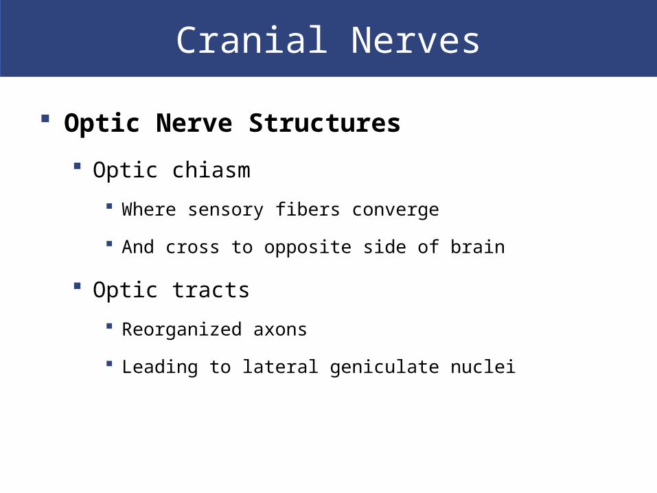
Optic Nerve Structures
Optic chiasm
Where sensory fibers converge
And cross to opposite side of brain
Optic tracts
Reorganized axons
Leading to lateral geniculate nuclei

Cranial Nerves
Figure 14–20 The Optic Nerve.

Cranial Nerves
The Vagus Nerves (X) Primary function
Mixed (sensory and motor) Widely distributed in thorax and abdomen
Origins Sensory:
– part of pharynx– auricle and external acoustic meatus– diaphragm– visceral organs of thoracic and abdominopelvic cavities
Motor: – motor nuclei in medulla oblongata

Cranial Nerves
The Vagus Nerves (X) Pathway
Jugular foramina Between occipital and temporal bones
Destination Sensory:
– sensory nuclei and autonomic centers of medulla oblongata
Visceral motor: – muscles of the palate and pharynx– muscles of the digestive, respiratory, and cardiovascular
systems in thoracic and abdominal cavities

Cranial Nerves
Figure 14–26 The Vagus Nerve.
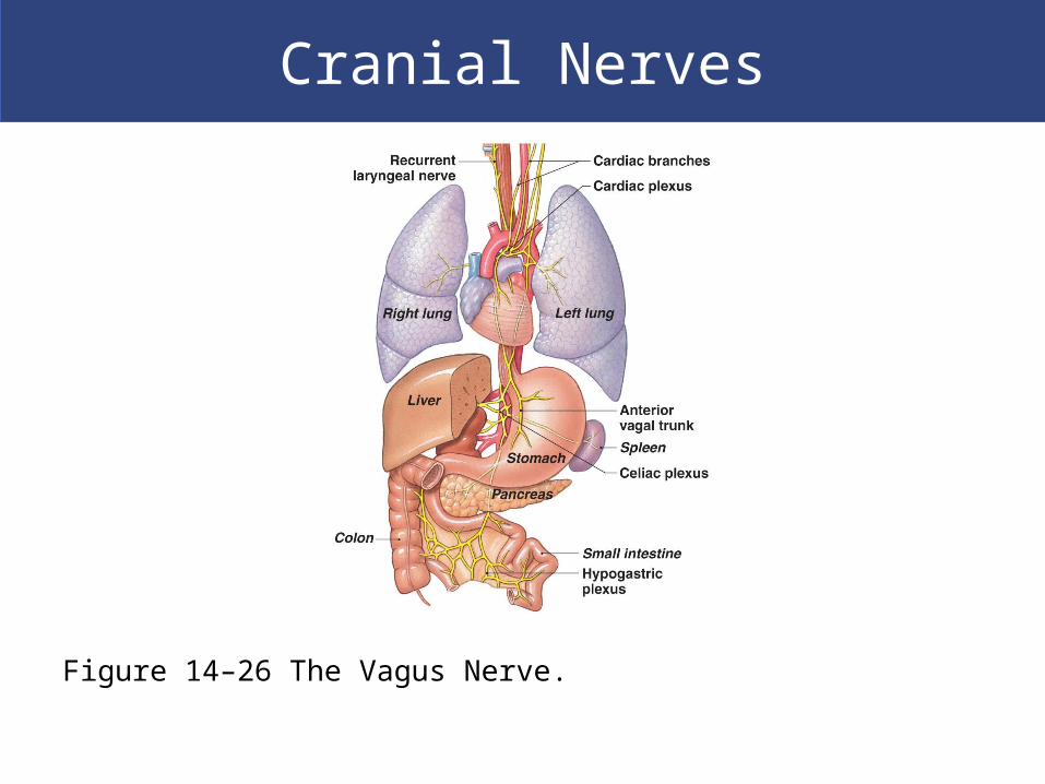
Cranial Nerves
Figure 14–26 The Vagus Nerve.
Cranial Nerves
The Accessory Nerves (XI) Primary function
Motor to muscles of neck and upper back
Origin Motor nuclei of spinal cord and medulla oblongata
Pathway Jugular foramina between occipital and temporal bones
Destination Internal branch:
– voluntary muscles of palate, pharynx, and larynx
External branch: – sternocleidomastoid and trapezius muscles

Cranial Nerves

Cranial Nerves

Cranial Reflexes
Cranial Reflexes
Monosynaptic and polysynaptic reflex arcs
Involve sensory and motor fibers of cranial nerves
Clinically useful to check cranial nerve or brain
damage

Cranial Reflexes