13-1 human anatomy & physiology spinal cord, spinal nerves and somatic reflexes chapter 13 by...
Post on 21-Dec-2015
234 views
TRANSCRIPT
13-1
Human Anatomy & Physiology
Spinal Cord, Spinal Nerves and Somatic Reflexes
Chapter 13
By
Abdul Fellah, Ph.D.
13-2
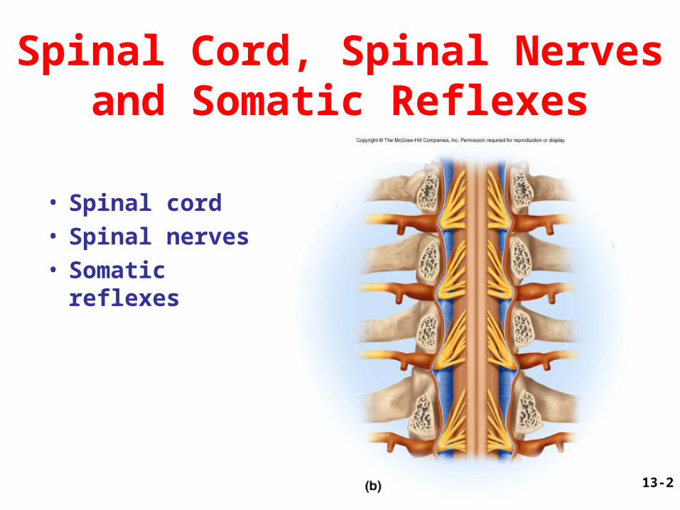
Spinal Cord, Spinal Nerves and Somatic Reflexes
• Spinal cord• Spinal nerves• Somatic reflexes
13-3
Overview of Spinal Cord
• Information highway between brain and body• Extends through vertebral canal from
foramen magnum to L1• Each pair of spinal nerves receives sensory
information and issues motor signals to muscles and glands
• Spinal cord is a component of the Central Nervous System while the spinal nerves are part of the Peripheral Nervous System
13-4
Functions of the Spinal Cord• Conduction– bundles of fibers passing information up and down
spinal cord
• Locomotion– repetitive, coordinated actions of several muscle
groups– central pattern generators are pools of neurons
providing control of flexors and extensors (walking)
• Reflexes– involuntary, stereotyped responses to stimuli
(remove hand from hot stove)– involves brain, spinal cord and peripheral nerves
13-5
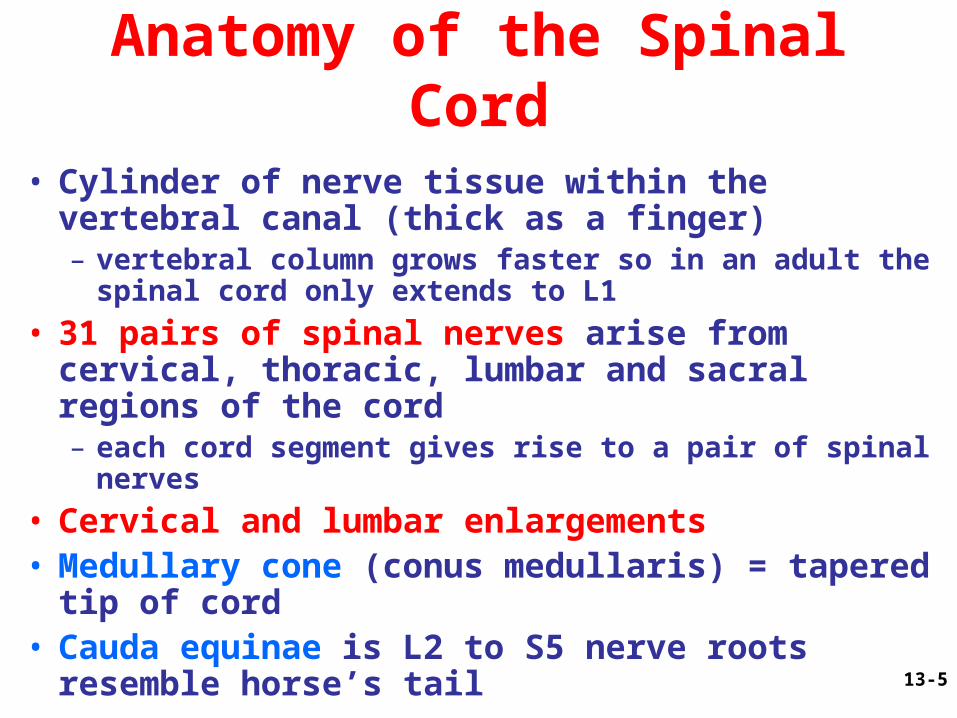
Anatomy of the Spinal Cord
• Cylinder of nerve tissue within the vertebral canal (thick as a finger)– vertebral column grows faster so in an adult the spinal
cord only extends to L1
• 31 pairs of spinal nerves arise from cervical, thoracic, lumbar and sacral regions of the cord– each cord segment gives rise to a pair of spinal
nerves
• Cervical and lumbar enlargements• Medullary cone (conus medullaris) = tapered tip
of cord• Cauda equinae is L2 to S5 nerve roots resemble
horse’s tail
13-6
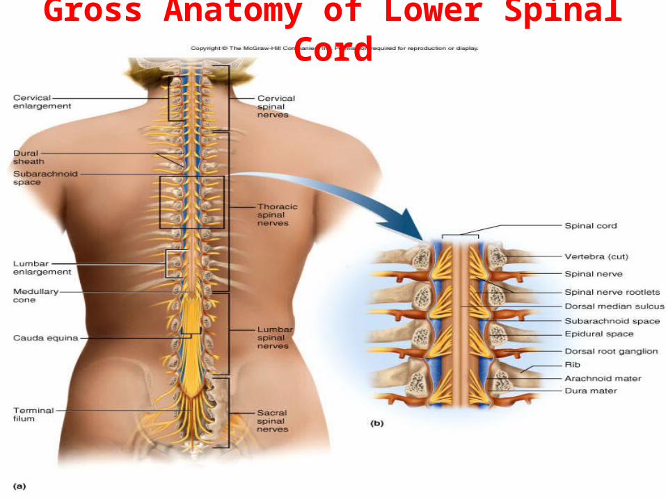
Gross Anatomy of Lower Spinal Cord
13-7
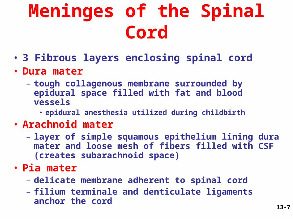
Meninges of the Spinal Cord
• 3 Fibrous layers enclosing spinal cord• Dura mater– tough collagenous membrane surrounded by
epidural space filled with fat and blood vessels• epidural anesthesia utilized during childbirth
• Arachnoid mater– layer of simple squamous epithelium lining dura
mater and loose mesh of fibers filled with CSF(creates subarachnoid space)
• Pia mater– delicate membrane adherent to spinal cord– filium terminale and denticulate ligaments anchor
the cord
13-8
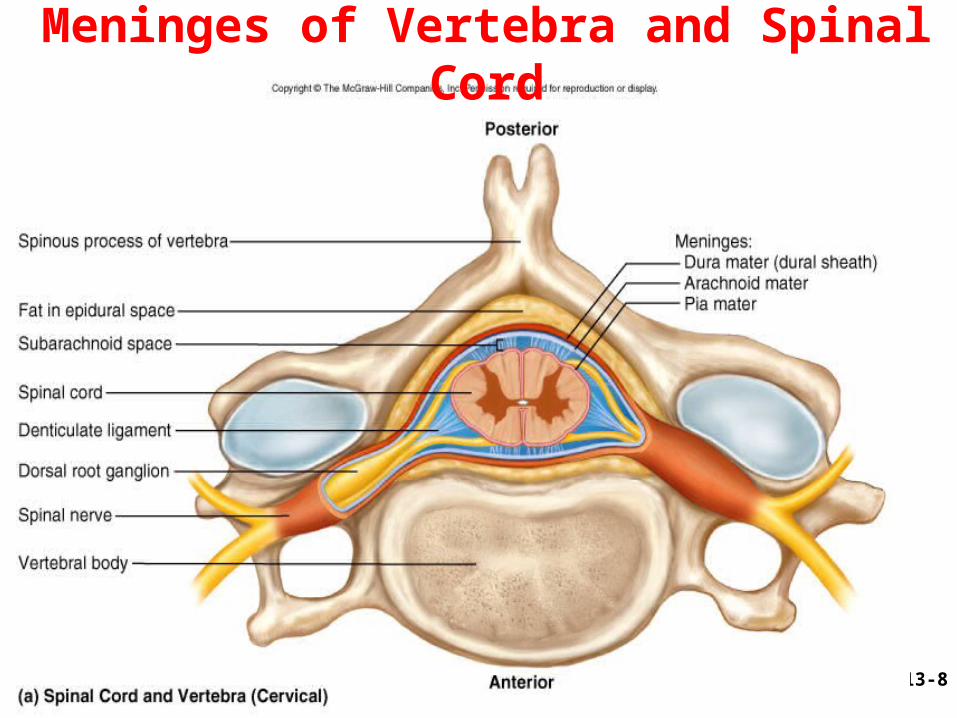
Meninges of Vertebra and Spinal Cord
13-9
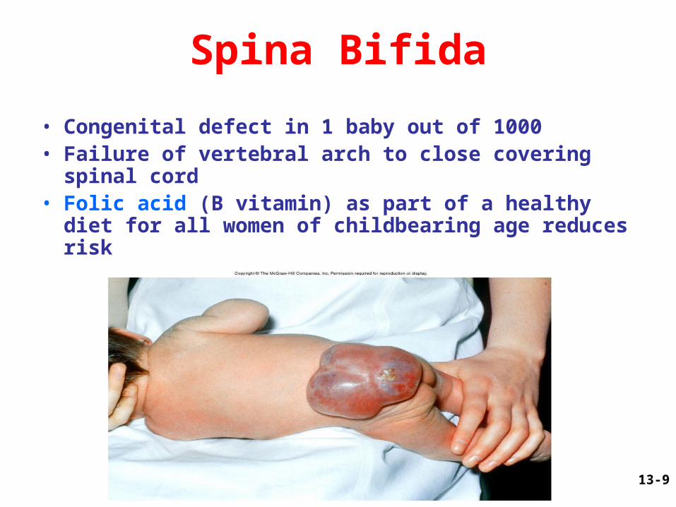
Spina Bifida
• Congenital defect in 1 baby out of 1000• Failure of vertebral arch to close covering spinal
cord• Folic acid (B vitamin) as part of a healthy diet for all
women of childbearing age reduces risk
13-10
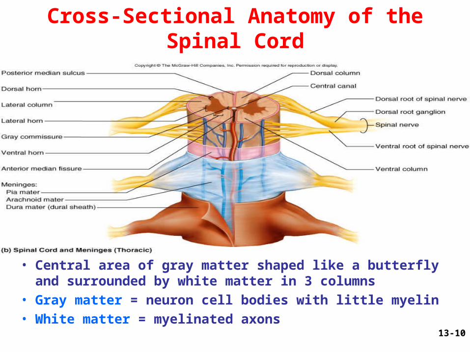
Cross-Sectional Anatomy of the Spinal Cord
• Central area of gray matter shaped like a butterfly and surrounded by white matter in 3 columns
• Gray matter = neuron cell bodies with little myelin• White matter = myelinated axons
13-11
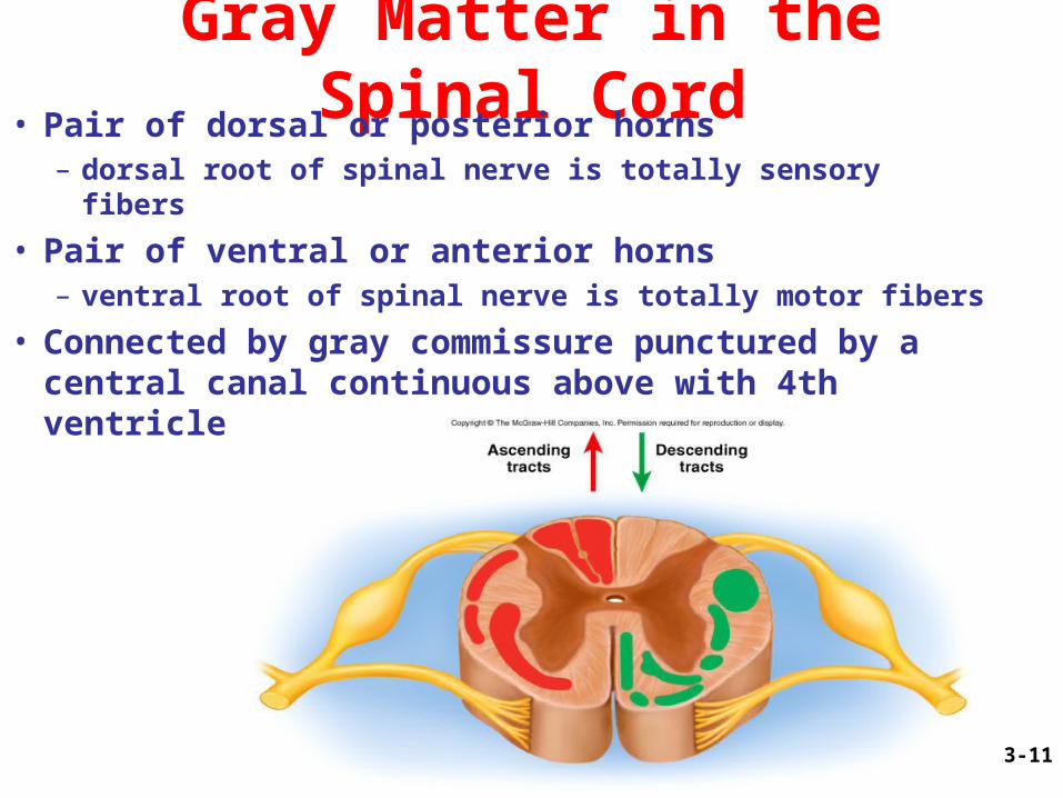
Gray Matter in the Spinal Cord• Pair of dorsal or posterior horns– dorsal root of spinal nerve is totally sensory fibers
• Pair of ventral or anterior horns– ventral root of spinal nerve is totally motor fibers
• Connected by gray commissure punctured by a central canal continuous above with 4th ventricle
13-12
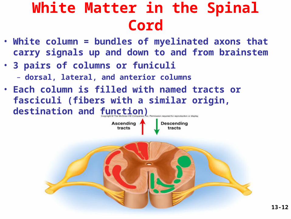
White Matter in the Spinal Cord
• White column = bundles of myelinated axons that carry signals up and down to and from brainstem
• 3 pairs of columns or funiculi– dorsal, lateral, and anterior columns
• Each column is filled with named tracts or fasciculi (fibers with a similar origin, destination and function)
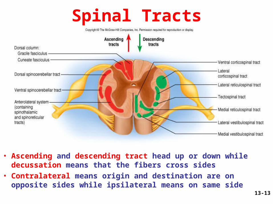
13-13
Spinal Tracts
• Ascending and descending tract head up or down while decussation means that the fibers cross sides
• Contralateral means origin and destination are on opposite sides while ipsilateral means on same side
13-14
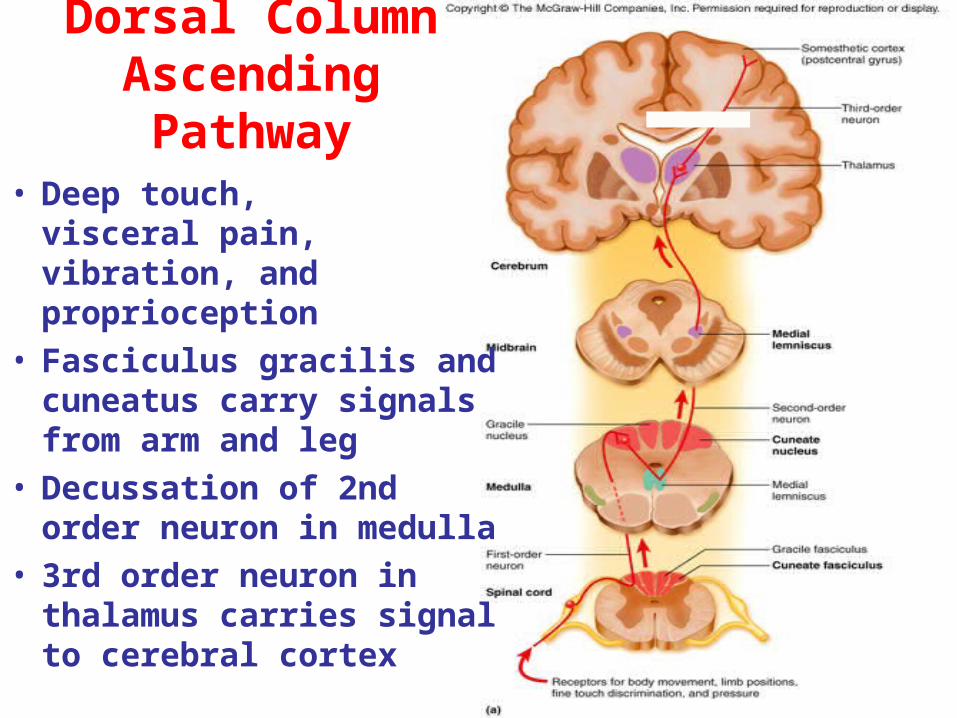
• Deep touch, visceral pain, vibration, and proprioception
• Fasciculus gracilis and cuneatus carry signals from arm and leg
• Decussation of 2nd order neuron in medulla
• 3rd order neuron in thalamus carries signal to cerebral cortex
Dorsal Column Ascending Pathway
13-15
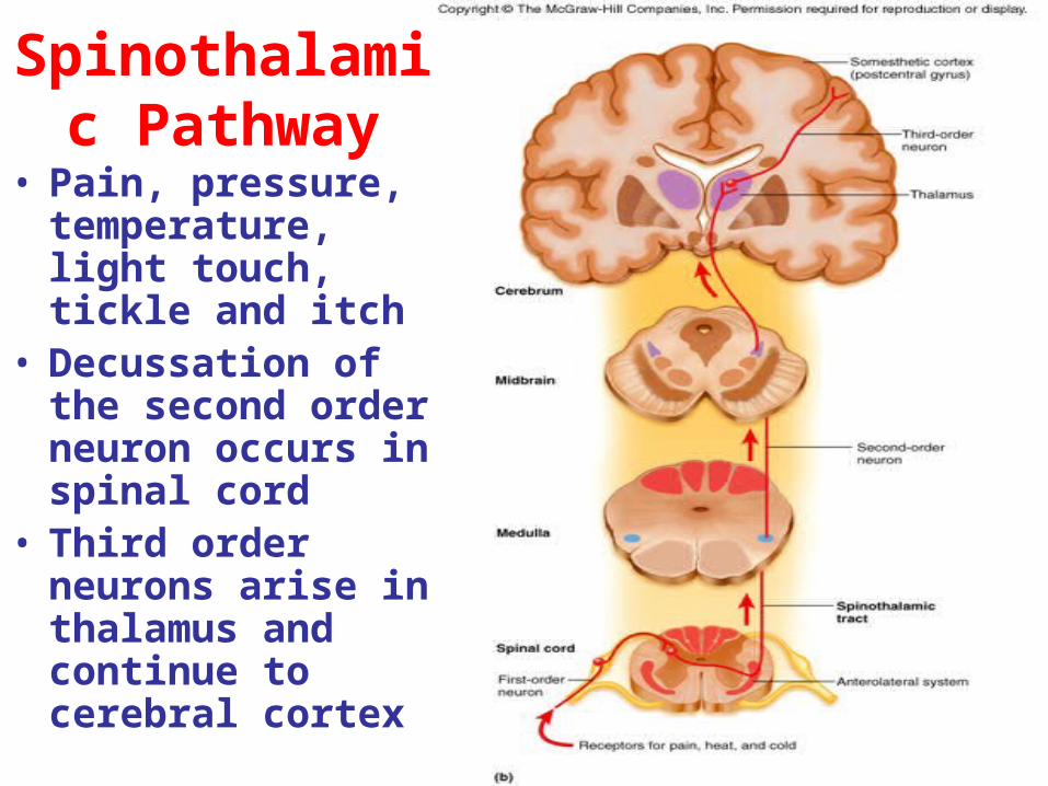
Spinothalamic Pathway
• Pain, pressure, temperature, light touch, tickle and itch
• Decussation of the second order neuron occurs in spinal cord
• Third order neurons arise in thalamus and continue to cerebral cortex
13-16
Spinoreticular Tract
• Pain signals from tissue injury
• Decussate in spinal cord and ascend with spinothalamic fibers
• End in reticular formation (medulla and pons)
• 3rd and 4th order neurons continue to thalamus and cerebral cortex
13-17
Spinocerebellar Pathway
• Proprioceptive signals from limbs and trunk travel up to the cerebellum
• Second order nerves ascend in ipsilateral lateral column
13-18
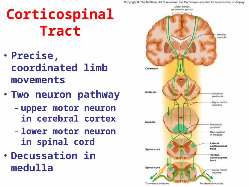
• Precise, coordinated limb movements
• Two neuron pathway– upper motor neuron in
cerebral cortex– lower motor neuron in
spinal cord
• Decussation in medulla
Corticospinal Tract
13-19
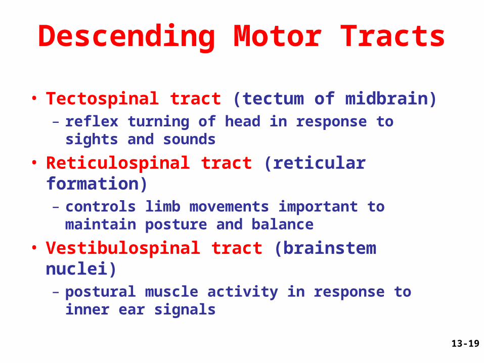
Descending Motor Tracts
• Tectospinal tract (tectum of midbrain)– reflex turning of head in response to sights and
sounds
• Reticulospinal tract (reticular formation)– controls limb movements important to maintain
posture and balance
• Vestibulospinal tract (brainstem nuclei)– postural muscle activity in response to inner ear
signals
13-20
Poliomyelitis and ALS
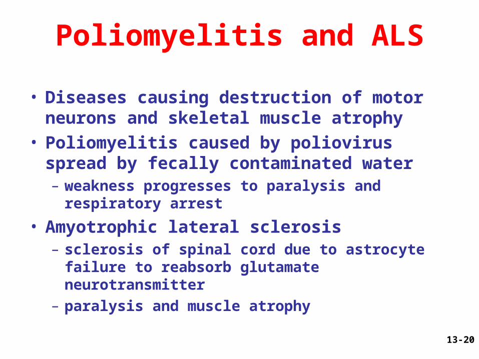
• Diseases causing destruction of motor neurons and skeletal muscle atrophy
• Poliomyelitis caused by poliovirus spread by fecally contaminated water– weakness progresses to paralysis and respiratory
arrest
• Amyotrophic lateral sclerosis– sclerosis of spinal cord due to astrocyte failure to
reabsorb glutamate neurotransmitter– paralysis and muscle atrophy
13-21
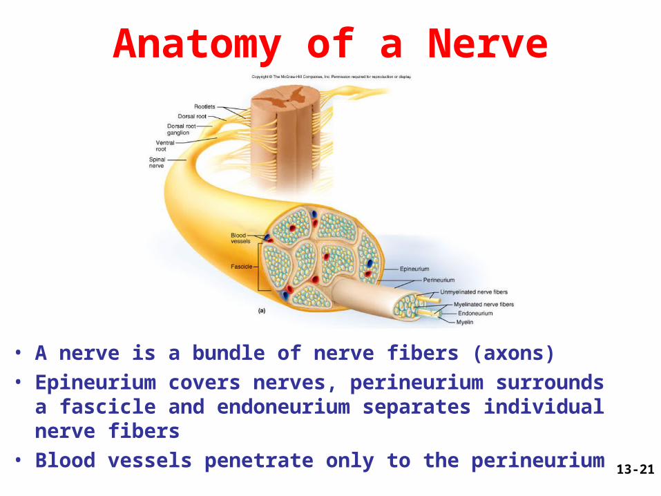
Anatomy of a Nerve
• A nerve is a bundle of nerve fibers (axons)• Epineurium covers nerves, perineurium surrounds a
fascicle and endoneurium separates individual nerve fibers
• Blood vessels penetrate only to the perineurium
13-22
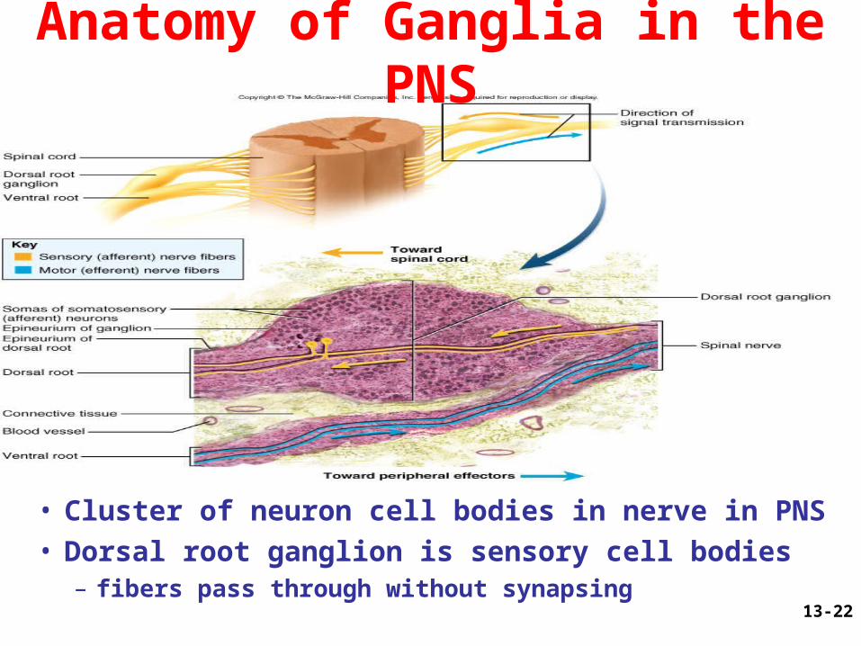
Anatomy of Ganglia in the PNS
• Cluster of neuron cell bodies in nerve in PNS• Dorsal root ganglion is sensory cell bodies – fibers pass through without synapsing
13-23
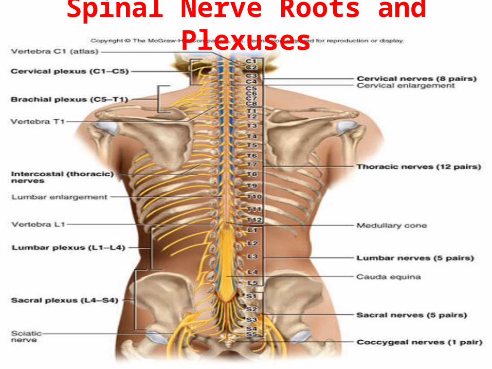
Spinal Nerve Roots and Plexuses
13-24
The Spinal Nerves• 31 pairs of spinal nerves (1st cervical above C1)– mixed nerves exiting at intervertebral foramen
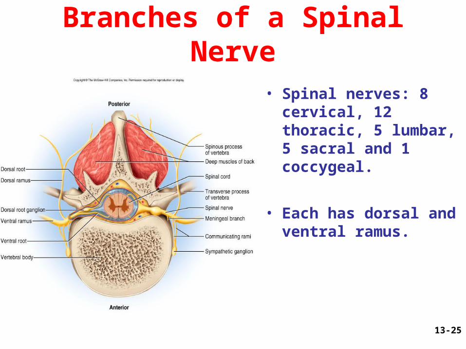
• Proximal branches– dorsal root is sensory input to spinal cord– ventral root is motor output of spinal cord– cauda equina is roots from L2 to C0 of the cord
• Distal branches– dorsal ramus supplies dorsal body muscle and skin– ventral ramus to ventral skin and muscles and limbs– meningeal branch to meninges, vertebrae and
ligaments
13-25
Branches of a Spinal Nerve
• Spinal nerves: 8 cervical, 12 thoracic, 5 lumbar, 5 sacral and 1 coccygeal.
• Each has dorsal and ventral ramus.
13-26
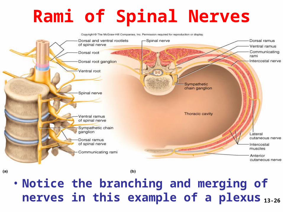
Rami of Spinal Nerves
• Notice the branching and merging of nerves in this example of a plexus
13-27
Shingles
• Skin eruptions along path of nerve
• Varicella-zoster virus (chicken pox) remains for life in dorsal root ganglia
• Occurs after age 50 if immune system is compromised
• No special treatment
13-28
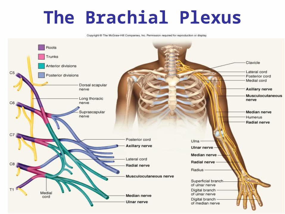
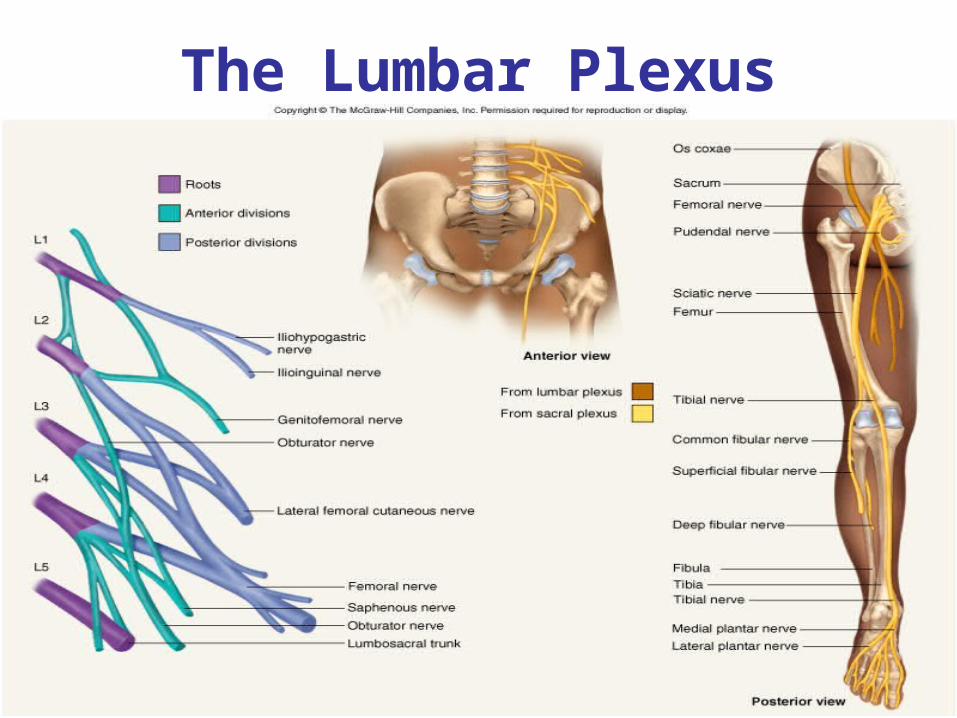
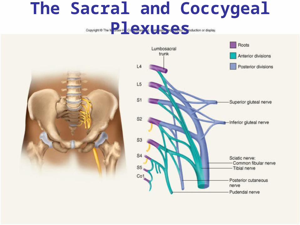
Nerve Plexuses
• Ventral rami branch and anastomose repeatedly to form 5 nerve plexuses– cervical in the neck, C1 to C5
• supplies neck and phrenic nerve to the diaphragm
– brachial in the armpit, C5 to T1• supplies upper limb and some of shoulder and neck
– lumbar in the low back, L1 to L4• supplies abdominal wall, anterior thigh and genitalia
– sacral in the pelvis, L4, L5 and S1 to S4• supplies remainder of lower trunk and lower limb
– coccygeal, S4, S5 and C0
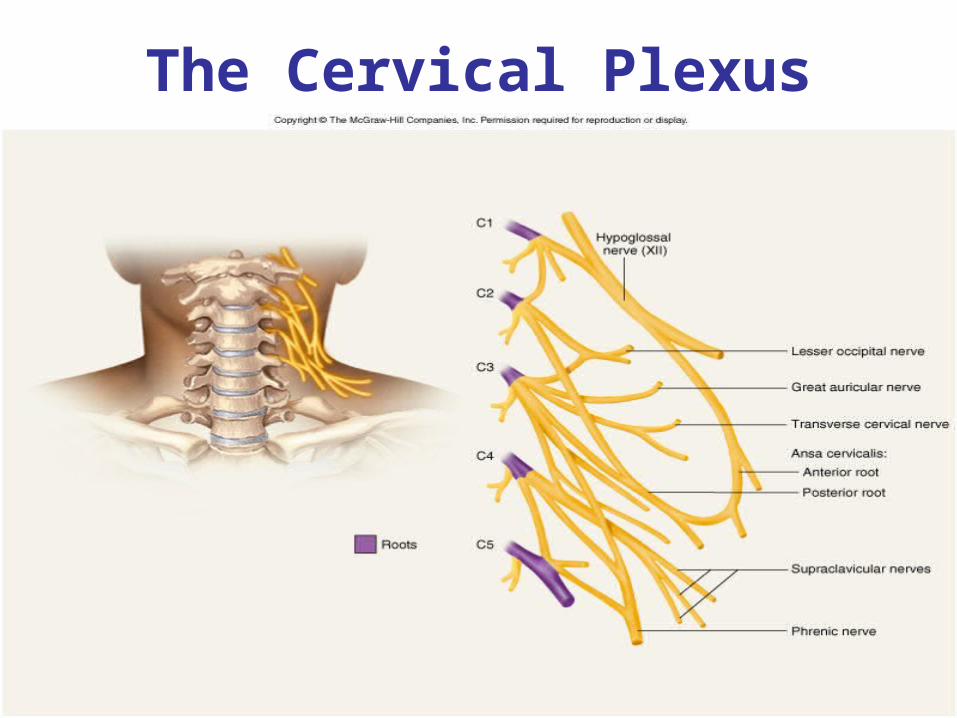
13-29
The Cervical Plexus
13-30
The Brachial Plexus
13-31
The Lumbar Plexus
13-32
The Sacral and Coccygeal Plexuses
13-33
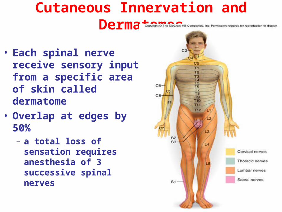
Cutaneous Innervation and Dermatomes
• Each spinal nerve receive sensory input from a specific area of skin called dermatome
• Overlap at edges by 50%– a total loss of sensation
requires anesthesia of 3 successive spinal nerves
13-34
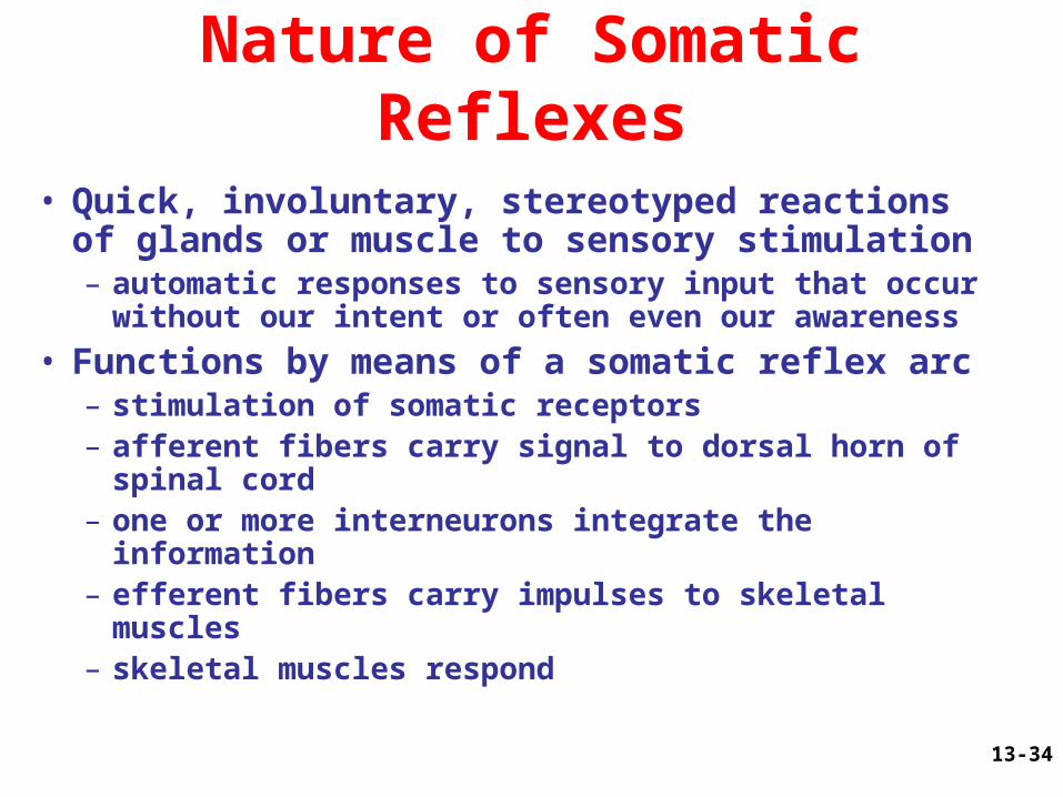
Nature of Somatic Reflexes
• Quick, involuntary, stereotyped reactions of glands or muscle to sensory stimulation – automatic responses to sensory input that occur
without our intent or often even our awareness
• Functions by means of a somatic reflex arc– stimulation of somatic receptors– afferent fibers carry signal to dorsal horn of spinal
cord– one or more interneurons integrate the information– efferent fibers carry impulses to skeletal muscles– skeletal muscles respond
13-35
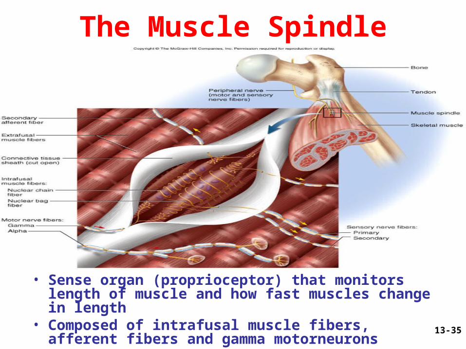
The Muscle Spindle
• Sense organ (proprioceptor) that monitors length of muscle and how fast muscles change in length
• Composed of intrafusal muscle fibers, afferent fibers and gamma motorneurons
13-36
The Stretch (Myotatic) Reflex• When a muscle is stretched, it contracts and
maintains increased tonus (stretch reflex)– helps maintain equilibrium and posture
• head starts to tip forward as you fall asleep• muscles contract to raise the head
– stabilize joints by balancing tension in extensors and flexors smoothing muscle actions
• Very sudden muscle stretch causes tendon reflex– knee-jerk (patellar) reflex is monosynaptic reflex– testing somatic reflexes helps diagnose many
diseases
• Reciprocal inhibition prevents muscles from working against each other
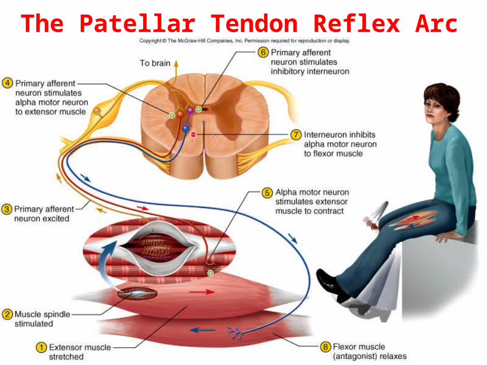
13-37
The Patellar Tendon Reflex Arc
13-38
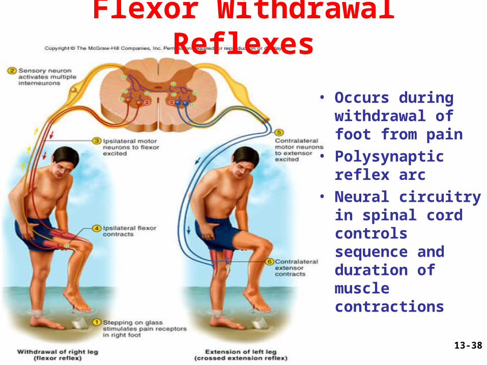
Flexor Withdrawal Reflexes
• Occurs during withdrawal of foot from pain
• Polysynaptic reflex arc
• Neural circuitry in spinal cord controls sequence and duration of muscle contractions
13-39
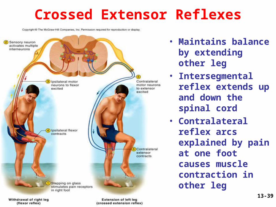
Crossed Extensor Reflexes
• Maintains balance by extending other leg
• Intersegmental reflex extends up and down the spinal cord
• Contralateral reflex arcs explained by pain at one foot causes muscle contraction in other leg

13-40
Golgi Tendon Reflex
• Proprioceptors in a tendon near its junction with a muscle -- 1mm long, encapsulated nerve bundle
• Excessive tension on tendon inhibits motor neuron– muscle contraction
decreased• Also functions when
muscle contracts unevenly
13-41
Spinal Cord Trauma
• 10-12,000 people/ year are paralyzed
• 55% occur in traffic accidents
• This damage poses risk of respiratory failure
• Early symptoms are called spinal shock
• Tissue damage at time of injury is followed by post-traumatic infarction