the spinal cord, spinal nerves and reflexes portions of chapters 13
TRANSCRIPT

The Spinal Cord, spinal nerves and reflexes
Portions of Chapters 13

Reflex arcs, figure 13.3
1. Sensory receptor- distal end of sensory neuron (dendrite) or associated sensory structure
*responds to internal or external stimuli
2. Sensory neuron- nerve impulses propagate from sensory receptor axon of sensory neuron axon terminals located in gray matter of brain or spinal cord

3. Integrating center- one or more regions of CNS gray matter
*single synapse- sensory & motor neuron= monosynaptic. *More often the integration center consists of one or more
interneurons which may relay the impulse to other interneurons or the motor neuron= polysynaptic
Interneuron- axons extend only short distance contact nearby neuron in CNS or ganglia, comprise vast majority of neurons in the body
4. Motor neuron- impulses triggered by integration center propagate out of CNS motor neuron body part that responds:
5. Effector- if skeletal muscle= somatic reflex, if smooth, cardiac muscle or gland=autonomic (visercal) reflex

Reflexes in diagnosis Damage/diseasereflex absent or abnormal Somatic- tapping or stroking:
Patellar- N= extend knee, A= Damage to sensory or motor neuron, s.c. lumbar region
Autonomic- difficult stimulating visceral organs impractical Pupillary light reflex- N= pupils of both eyes diameter if
either exposed to light, A= maybe damage or injury to receptors in brainstem or midbrain
Plantar flexion reflex- gentle stroke of foot sole Babinski sign- toes fan rather than curl (curl= normal sign)
Incomplete myelination in corticospinal tract Damage to descending motor pathway
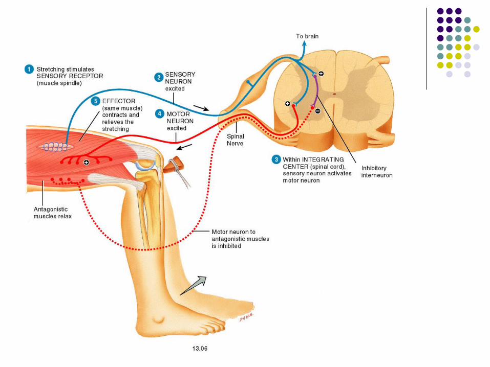
Somatic spinal reflexes
Stretch reflex- fig 13.14, Monosynaptic Contraction of skeletal muscle in response to stretching of
muscle Elicit by tapping tendons: knee, wrist, elbow, ankle Mechanism:
Slight stretch stimulates musle spindles (see figure) Spindles generate one or more AP posterior root Spinal cord integrates & activates neuron in ant gray horn If excitation strong enough, AP sent thru motor neuron axon Ach released, trigger muscle AP, muscle contracts to relieve
the stretch
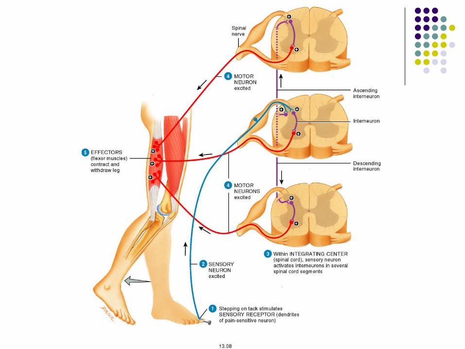
Flexor or withdrawal reflex- fig 13.16, polysynaptic withdrawing from painful stimulus (step on tack) Mechanism:
Stepping on tack stimulates dendrites- pain sensitive neuron
Sensory neuron generates AP spinal cord Spinal cord integrates, activates interneurons that extend to
several spinal cord segments Interneurons activate motor neurons, generate AP Ach released, causes flexor muscles to contract producing
withdrawal

Spinal cord functions
White matter- sensory and motor tracts Highway for conduction of sensory impulse to
brain and motor impulse to effector Gray matter is site of integration Spinal nerves & nerves that branch from
them connect CNS to sensory receptors, muscles, & glands

Cervical enlargement- extends from 4th cervical to 1st thoracic nerves to & from upper limbs arise
Lumbar enlargement- 9th-12th thoracic nerves to & from lower limbs arise inferior to this is a conical portion = conus
medullaris ending at the intervertebral disc between 1st and 2nd lumbar, from this arises: filum terminale- extension of the pia mater
anchoring the spinal cord to coccyx
The roots (pts of attachment of spinal nerve to the spinal cord) are angled downward when arising towards inferior portion of s.c. cauda equina-“horse tail,” roots angle down vertebral
canal from end of the spinal cord, look like hair
Cord appears segmented 31 spinal nerves leave at regular intervals where pairs of spinal nerves arise are spinal segments no segments within cord dividing gray & white matter
Spinal nerves- paths of communication between spinal cord & nerves innervating specific regions
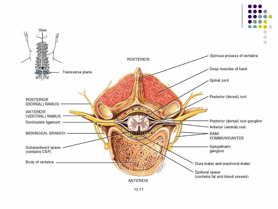
Roots-2 bundles of axons connecting spinal nerves to spinal cord:
A. Posterior or dorsal root- contains only sensory fibers conducting impulses from periphery to CNS-each has swelling = posterior or dorsal root
ganglion-contains cell bodies of sensory neurons
B. Anterior or ventral root- contain axons of motor neurons conducting impulses from CNS to effector organs & cells

Gray matter of spinal cord- cell bodies of neurons, neuroglia, unmyelinated axons, & dendrites of interneurons & motor neurons in CNS- clusters of neuronal cell bodies form
functional groups = nuclei Sensory nuclei Motor nuclei On each side of cord divided into groups- horns
White matter in cord- bundles of myelinated and unmyelinated axons of sensory, inter, and motor neurons Organized into columns…

Gray commissure- center of butterfly connecting 2 lateral masses of gray matter
Central canal- small space in center gray commissure extending entire length of spinal cord Continuous with 4th ventricle
bundles extending long distance up/down= tracts Sensory (ascending) tracts- axons that conduct
nerve impulses toward brain Motor (descending) tracts- axons carry nerve
impulses down cord Both continuous w/sensory & motor tracts in brain

Upper Motor Neurons
Planning, initiating, directing sequences of voluntary movements
Pyramidal (direct) pathway- convey nerve impulses originating in cerebral cortex & are destined to cause precise, voluntary movements of the skeletal muscle
Extrapyramidal (indirect) pathway- convey nerve impulses destined to program automatic movements & help coordinate body movements with visual stimuli, maintain skeletal tone, and posture & equilibrium

Corticospinal tract- white columns in the spinal cord, convey nerve impulses from motor cortex to skeletal muscle
Decussation- cross over to opposite side 90% axons in large motor tracts to opposite side in
medullary pyramids 10% eventually do in the s.c. Then, will synapse with interneuron or LMN
Right cerebral cortex controls left muscles Left cerebral cortex controls right muscles
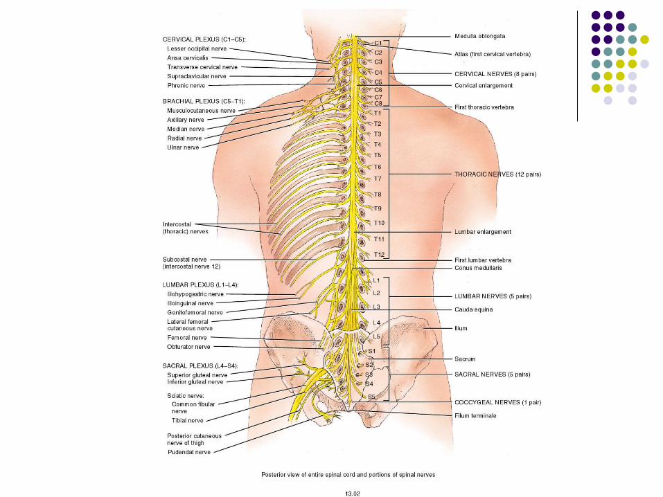
Spinal nerves, figure 13.2
Cord appears segmented: 31 spinal nerves leave at regular intervals areas where pairs of spinal nerves arise are
called spinal segment Spinal nerves- paths of communication
between spinal cord and nerves innervating specific regions of the body.

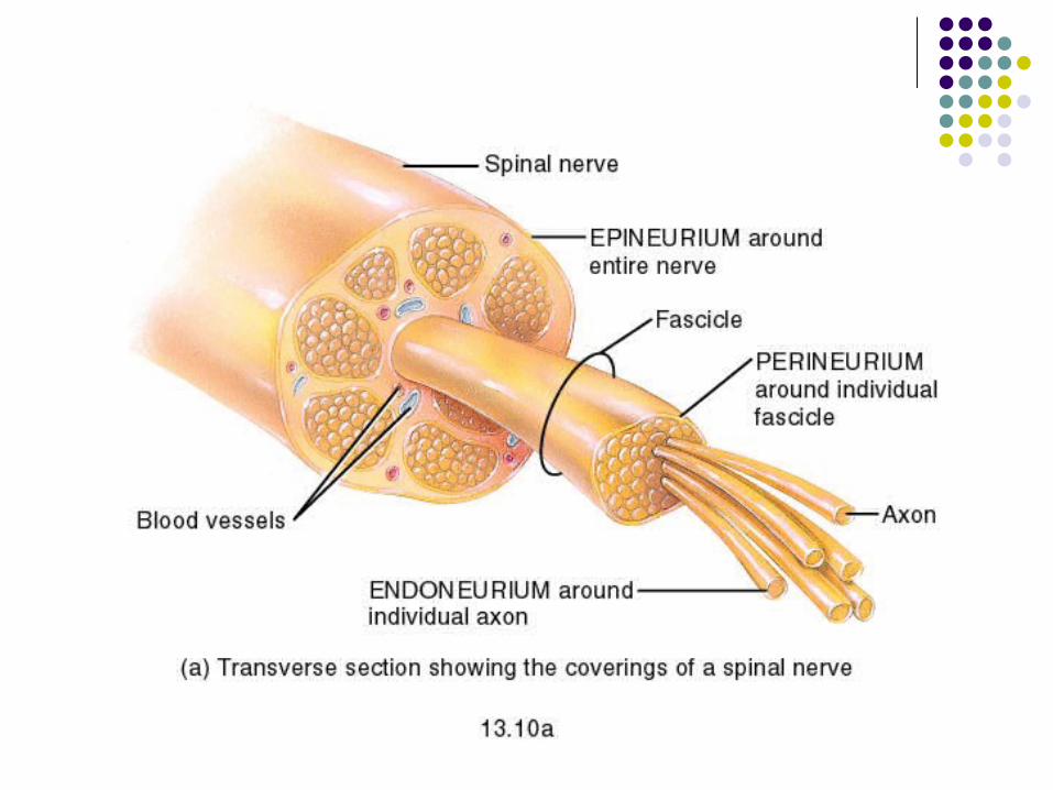
Structure of spinal nerve, 13.4
typical spinal nerve has 2 connections to the cord: posterior root and anterior root
unite at intervertebral foramen to form a spinal nerve Posterior = sensory Anterior = motor mixed nerve
Each spinal and cranial nerve contains layers of protective CT coverings
individual axons (un- & myelinated) are wrapped in endoneurium
fascicles of axons are wrapped in perineurium epineurium covers the entire nerve


5 Spinal nerve plexuses…
Cervical- C1-C4, some C5 Innervates: skin & muscles of head, neck,
shoulder, chest, diaphragm Injuries to phrenic nerve- breathing would stop


Plexus (2)
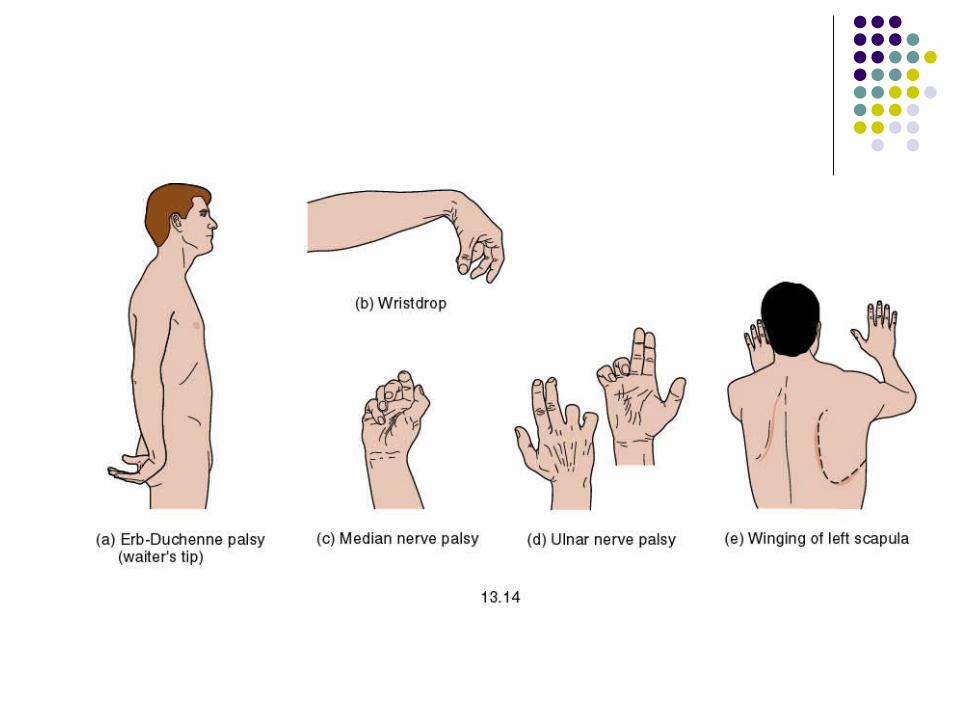
Brachial- C5-C8, T1 Shoulders and upper limbs Injuries to nerves emerging:
Forceful pulling of head from shoulder, loss sensation on lateral arm
Radial nerve- improper deltoid injection. Wrist drop indicates injury (inability to extend wrist & fingers)
Median nerve- pronate forearm, proximal interphalangeal joints (2-3), weakened wrist flexion, adduction & thumb movements
Ulnar nerve- abduct & adduct fingers, clawhand Long thoracic nerve- paralysis of serratus anterior medial
border of scapula protrudes wing like appearance


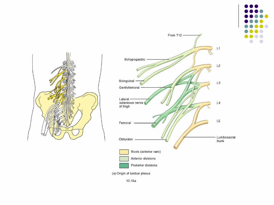
Plexuses (3)
Lumbar- L1-L4 Anterolateral abdominal wall, ext genitals, pt of
lower limbs Injuries:
Femoral nerve- (stab, gunshot wound for ex) inability to extend leg, loss sensation in skin anteromedial aspect of thigh
Obturator nerve- paralysis of adductor muscles of leg, loss of sensation over medial aspect thigh, pressure on by fetus head during pregnancy

Plexus (4)
Sacral- L4-L5, S1-S4 Supplies buttocks, perineum, lower limbs *largest nerve in body (sciatic nerve) arises from Injury- RESULTS in: footdrop- plantar flexed and
inverted Loss of function along anterolateral leg, dorsum of foot
& toes Calcaneovalgus- dorsiflexion and eversion of foot due
to injury to tibial portion of sciatic Loss of sensation to sole
Plexus (5)
Coccygeal- S4-S5, coccygeal nerves Supplies skin in coccygeal region


Disorders: spinal cord Polio- poliomyelitis- cause by virus
Fever; headache; stiff neck, back; deep muscle pain & weak; loss of somatic reflexes
Can cause paralysis by destroying cell body of motor neurons, esp. anterior horn & nuclei of cranial nerves
Can cause death from resp or heart failure Vaccine virtually erradicated polio in US
Meningitis- inflammation of the meninges Can be due to infection by bacteria or virus Bacterial- more serious, treated w/antibiotics (vaccine for)
May be fatal if not treated promptly Viral- no specific treatment, usually resolved in 1-2 weeks

Disorders: spinal cord (2)
Sciatica- injury to sciatic nerve pain- buttocks, posterior, lateral aspect of leg Due to slipped disc, dislocated hip, osteoarthritis
of lumbosacral spine, pressure from uterus (preg), or improper gluteal intramuscular injection
Shingles- acute infection of PNS after chicken pox- virus retreats to posterior root
ganglion If reactivated can send down sensory neurons to
skin