spermatogenesis in vitro
TRANSCRIPT
SPERMATOGENESIS IN VITRO
INDUCTION OF PROLIFERATION, MEIOSIS AND DIFFERENTIATION
Mário Sousa
Lab Cell BiologyInstitute of Biomedical Sciences (ICBAS)
University of [email protected]
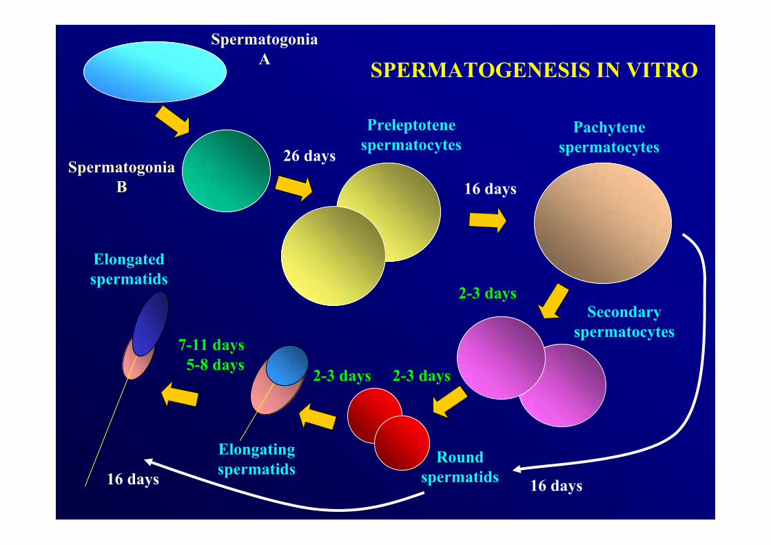
26 days
16 days
16 days16 days
2-3 days
2-3 days
7-11 days5-8 days
SpermatogoniaA
Elongatingspermatids
Elongatedspermatids
SpermatogoniaB
Preleptotenespermatocytes
Pachytenespermatocytes
Secondaryspermatocytes
Roundspermatids
2-3 days
SPERMATOGENESIS IN VITRO
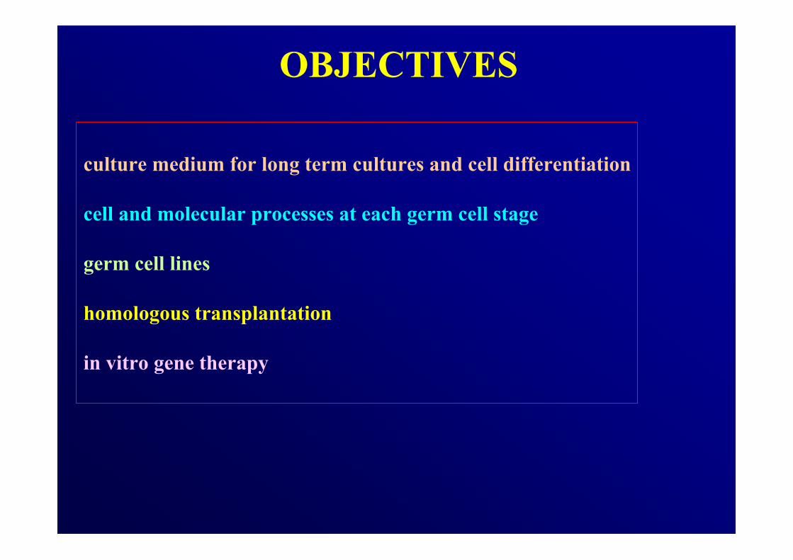
OBJECTIVES
culture medium for long term cultures and cell differentiation
cell and molecular processes at each germ cell stage
germ cell lines
homologous transplantation
in vitro gene therapy

15 anejaculation casesNormal karyotypesAbsence of Y microdeletionsConserved spermatogenesis
Mechanical dissociationErythrocyte lysisEnzymatic digestionCell isolation by micromanipulation
Cell culture:5 CM5 CM + rFSH (25 U/L) 5 rFSH + T (2 µmol/L)
Plated cells:250 S + 100 SGA + 1000 ST1 + 100 ST2
Multiplex-PCRAZF a,b,c
Yq11.2
SY157 (c)
SY254 (c)SY134 (b)
SY142 (b)SY152 (c)
SY14 (SRY) - YpSY84 (a)
SY142 (b)
A B C
600bp
EDM1
M2
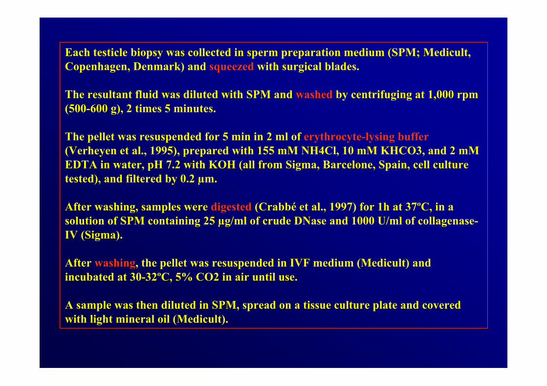
Each testicle biopsy was collected in sperm preparation medium (SPM; Medicult, Copenhagen, Denmark) and squeezed with surgical blades.
The resultant fluid was diluted with SPM and washed by centrifuging at 1,000 rpm (500-600 g), 2 times 5 minutes.
The pellet was resuspended for 5 min in 2 ml of erythrocyte-lysing buffer(Verheyen et al., 1995), prepared with 155 mM NH4Cl, 10 mM KHCO3, and 2 mM EDTA in water, pH 7.2 with KOH (all from Sigma, Barcelone, Spain, cell culture tested), and filtered by 0.2 µm.
After washing, samples were digested (Crabbé et al., 1997) for 1h at 37ºC, in a solution of SPM containing 25 µg/ml of crude DNase and 1000 U/ml of collagenase-IV (Sigma).
After washing, the pellet was resuspended in IVF medium (Medicult) andincubated at 30-32ºC, 5% CO2 in air until use.
A sample was then diluted in SPM, spread on a tissue culture plate and coveredwith light mineral oil (Medicult).
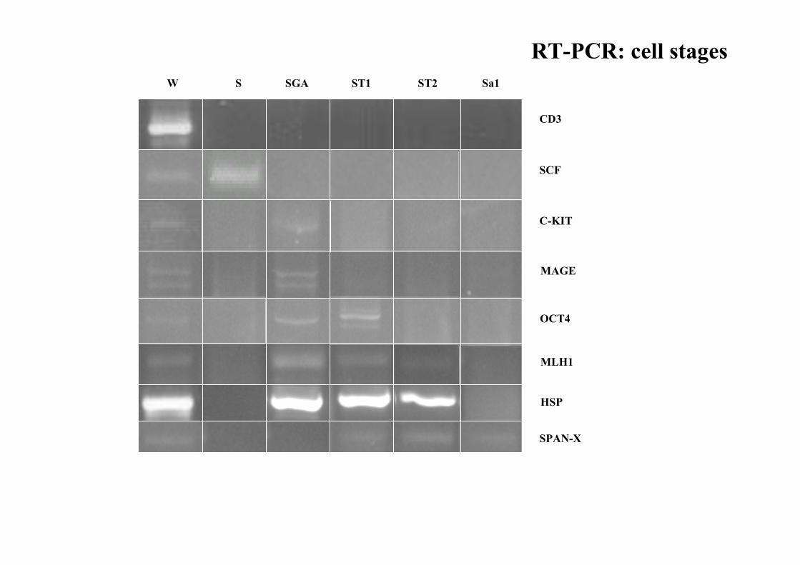
W S SGA ST1 ST2 Sa1
CD3
SCF
C-KIT
MAGE
OCT4
HSP
MLH1
SPAN-X
RT-PCR: cell stages
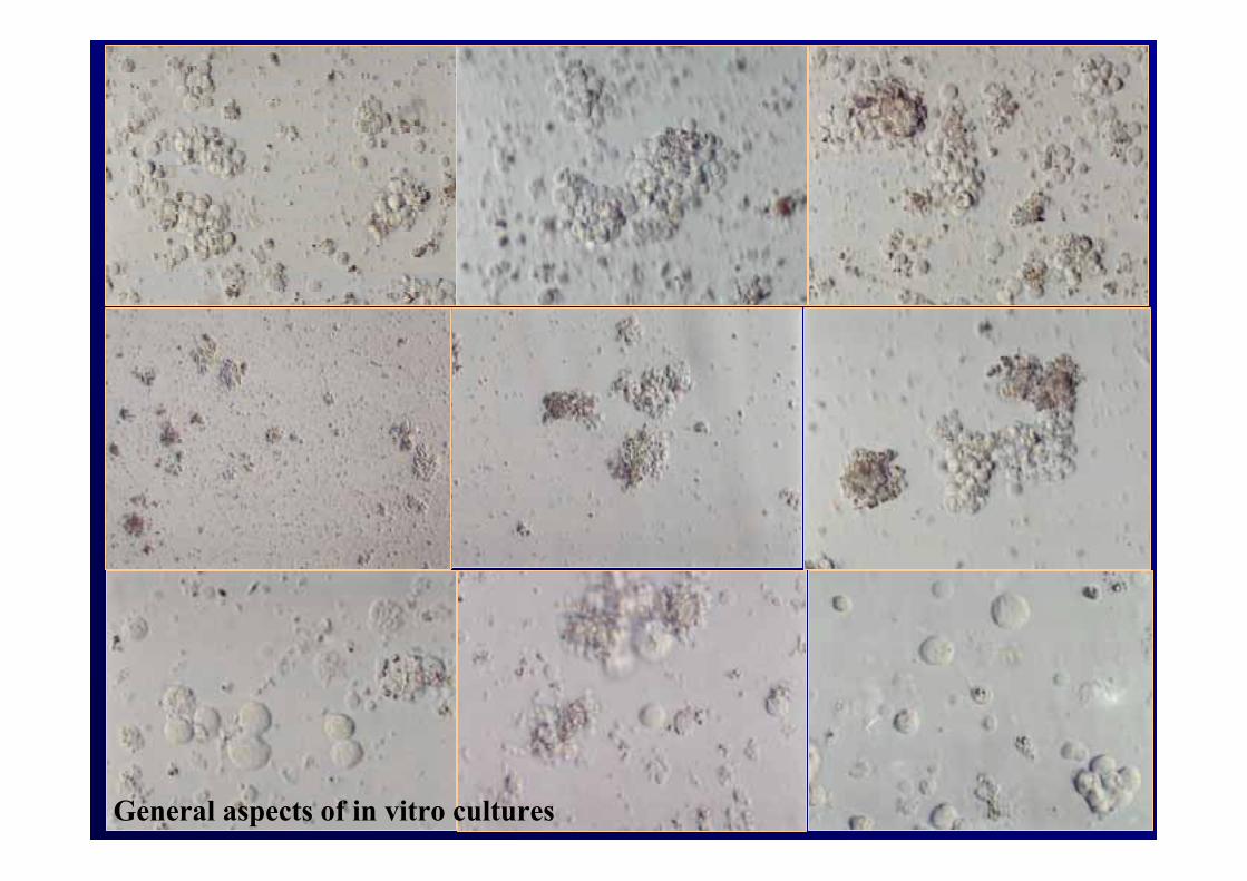
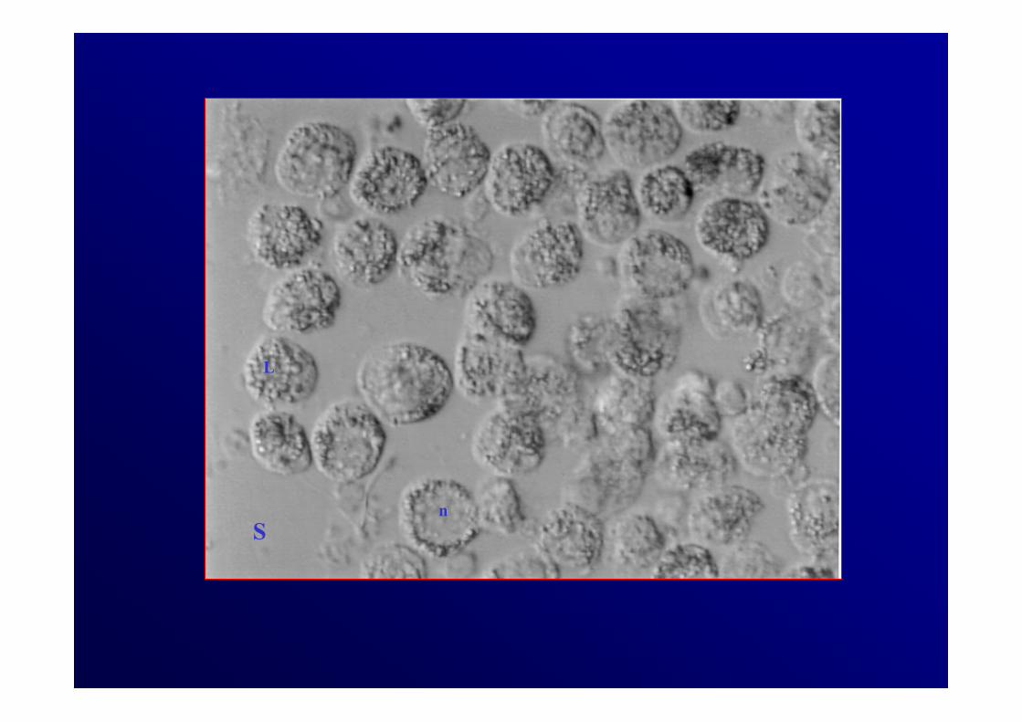
General aspects of in vitro cultures
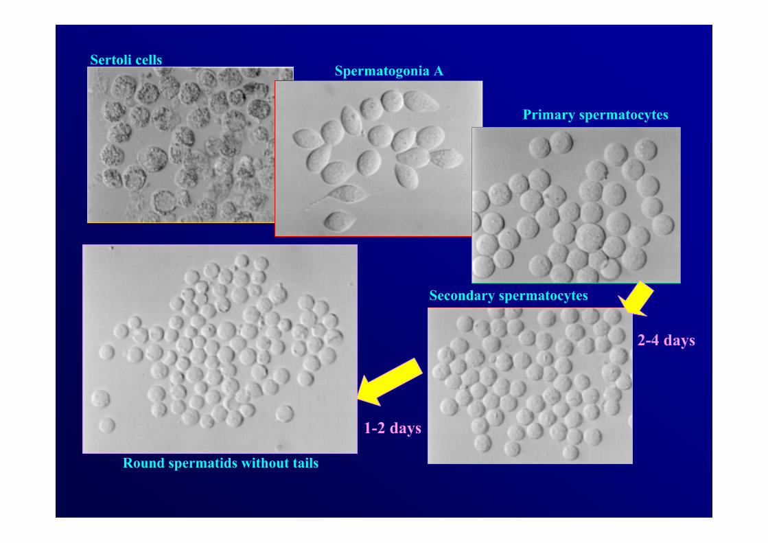
Sertoli cellsSpermatogonia A
Primary spermatocytes
Secondary spermatocytes
Round spermatids without tails
2-4 days
1-2 days
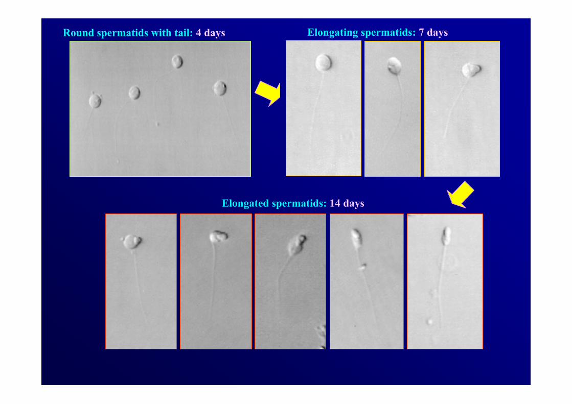
Round spermatids with tail: 4 days Elongating spermatids: 7 days
Elongated spermatids: 14 days
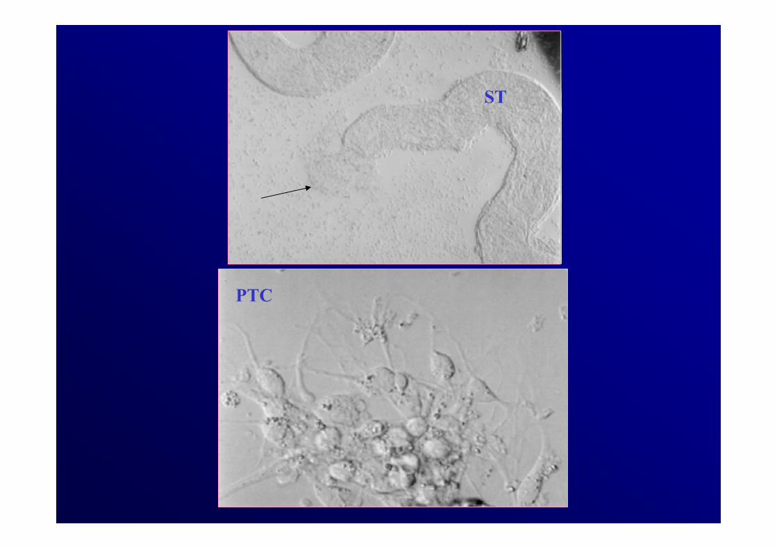
PTC
ST

ST1
SG
S
n
L
Sn
L
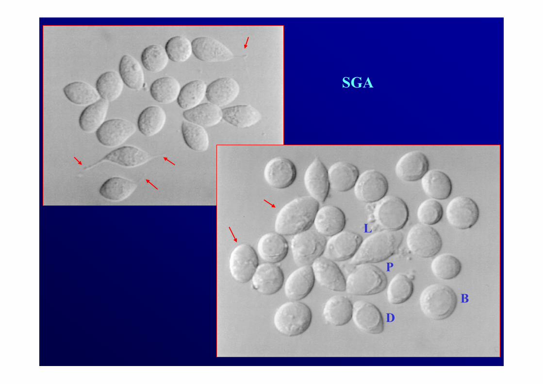
SGA
L
P
DB
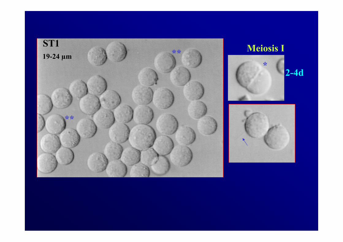
ST1**
**
*19-24 µm
2-4d
Meiosis I
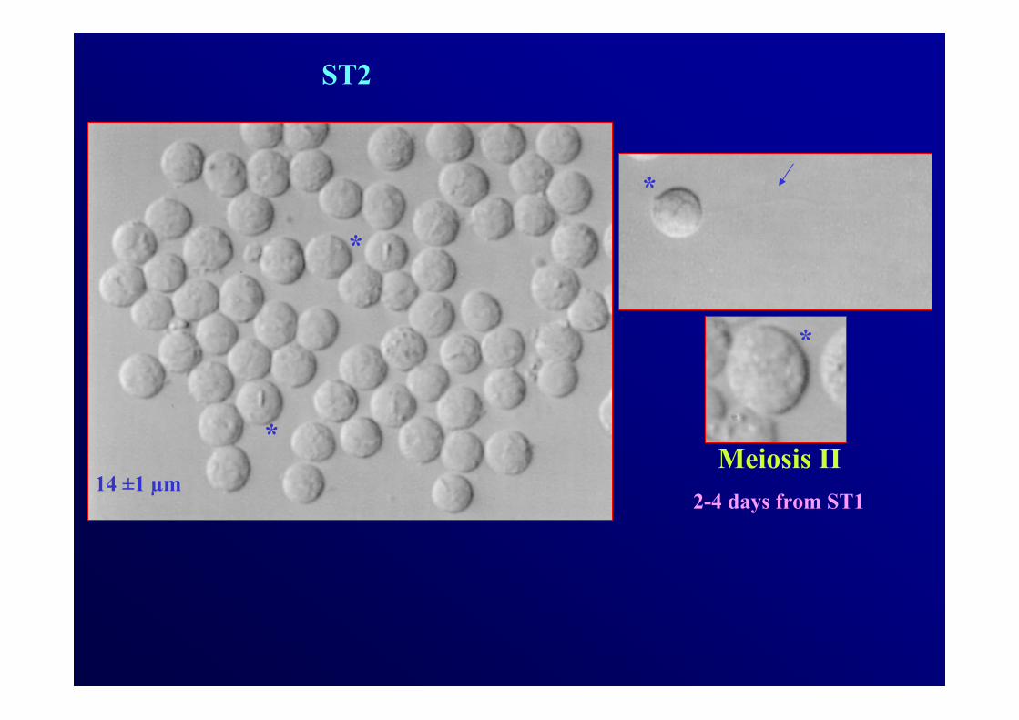
ST2
*
*
*
*
14 ±1 µmMeiosis II
2-4 days from ST1
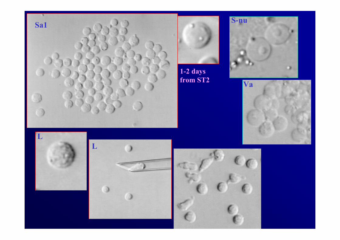
Sa1
L
S-nu
Va
L
1-2 days from ST2
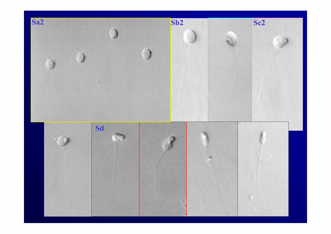
Sa2 Sb2
Sd
Sc2
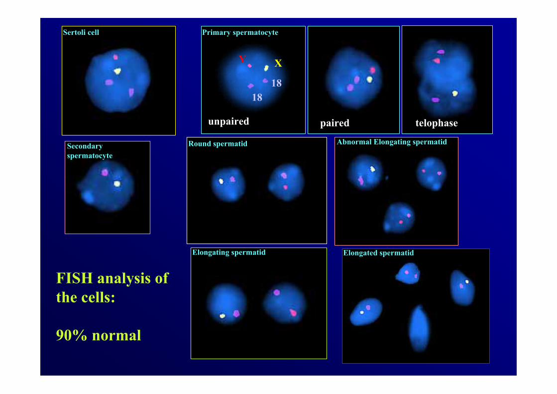
Sertoli cell
Round spermatidSecondaryspermatocyte
Primary spermatocyte
Abnormal Elongating spermatid
unpaired paired telophase
Y X
1818
Elongating spermatid Elongated spermatid
FISH analysis of the cells:
90% normal

HGC: haploid germ cells (Sa1) added to cultures.
T able I . In vitro differentiation of sperm atids.
C ases new Sa1
evolution of Sa1
evolution of Sa2
evolution of
Sb
M edia D G C H G C arrested Sa2 arrested Sb arrested Sd
C M 1000 93 0 81 12 2 10 7 3
C M +F SH 1000 120 30 116 34 7 27 21 6
C M +F SH +T 1000 65 67 61 71 7 64 42 22

For each column:(A) P<0.01 to CM; (B) P<0.05 to CM; (C) P<0.01 to CM and CM+FSH.
Table II. Rates of spermatid in vitro differentiation.
Media Total Sa1
Meiotic index
Spermatid evolution
Sa2 Sb Sd
HGC+
new Sa1 new Sa1/
DGC Sa2/
total Sa1 Sb/
total Sa1 Sb/Sa2 Sd/
total Sa1 Sd/Sb
CM 93 0/1000
(0) 12/93 (12.9)
10/93 (10.8)
10/12 (83.3)
3/93 (3.2)
3/10 (30)
CM+FSH 120 30/1000 (3)A
34/150 (22.7)B
27/150 (18)
27/34 (79.4)
6/150 (4)
6/27 (22.2)
CM+FSH+T 65 67/1000 (6.7)C
71/132 (53.8)C
64/132 (48.5)C
64/71 (90.1)
22/132 (16.7)C
22/64 (34.4)

*
***
****
0
50
100
150
pSa1 new Sa1 Sa2 Sb Sd
Cel
l num
ber
days of culture
2-41-2 4-7 5-160
*
***
****
0
50
100
150
pSa1 new Sa1 Sa2 Sb Sd
Cel
l num
ber
days of culture
2-41-2 4-7 5-160
rFSH+T rFSH CM
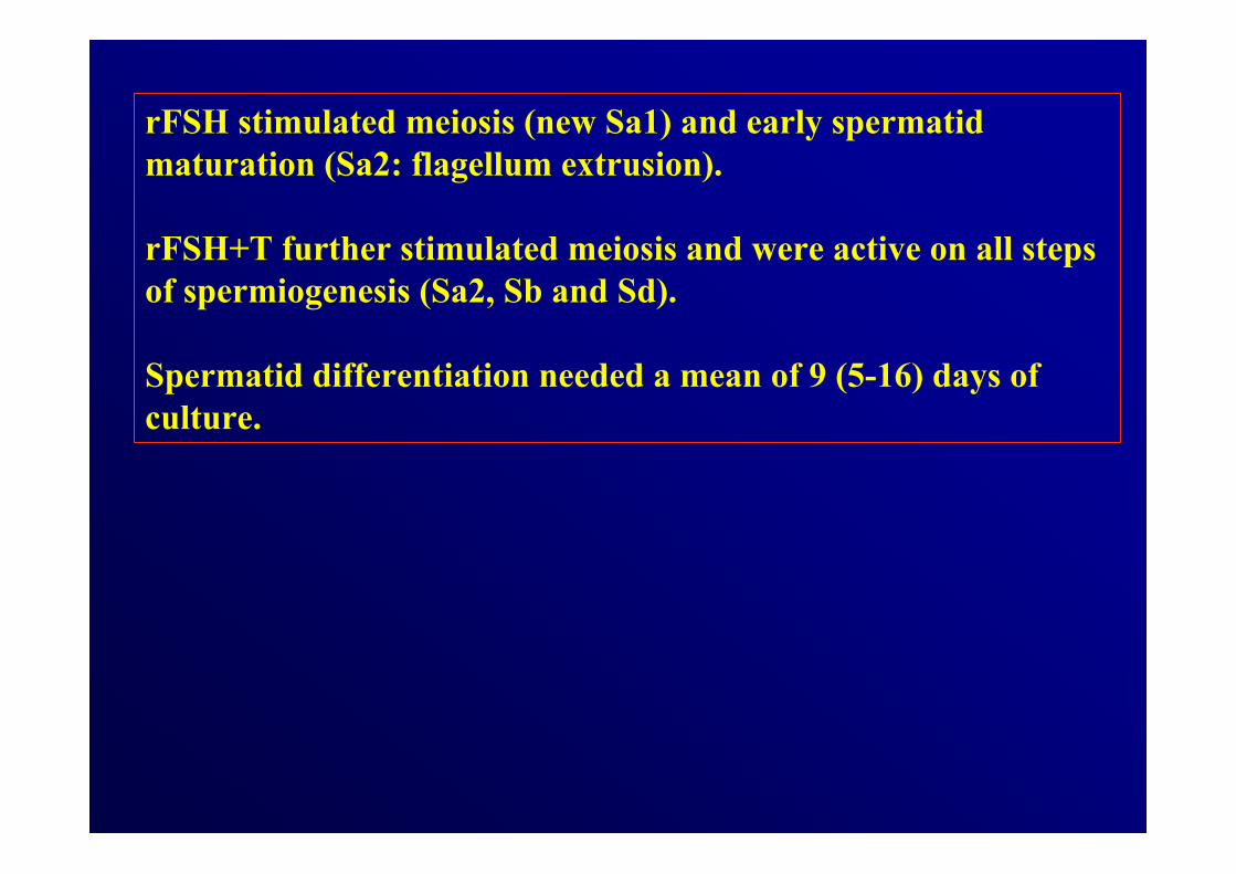
rFSH stimulated meiosis (new Sa1) and early spermatidmaturation (Sa2: flagellum extrusion).
rFSH+T further stimulated meiosis and were active on all steps of spermiogenesis (Sa2, Sb and Sd).
Spermatid differentiation needed a mean of 9 (5-16) days of culture.
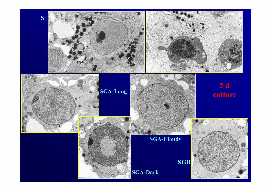
5 dculture
S
SGA-Long
SGA-Dark
SGA-Cloudy
SGB
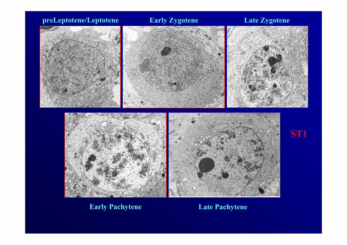
ST1
preLeptotene/Leptotene Early Zygotene Late Zygotene
Early Pachytene Late Pachytene
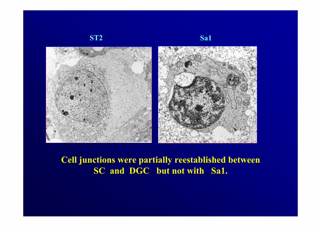
ST2 Sa1
Cell junctions were partially reestablished between SC and DGC but not with Sa1.
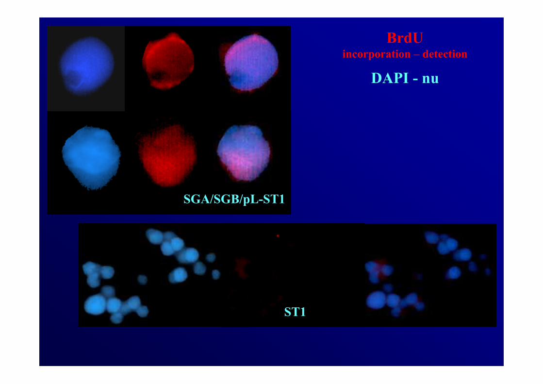
BrdUincorporation – detection
DAPI - nu
ST1
SGA/SGB/pL-ST1

days of culture
Prol
ifera
ti on
DG
C (%
)
*
*
**
0
2
4
6
8
2 6 12
days of culture
Prol
ifera
ti on
DG
C (%
)
*
*
**
0
2
4
6
8
2 6 12
rFSH+T rFSH CM
D2: P<0.01 to CM
Germ cell proliferation appeared stimulated by both hormones during the first
2 days (4%), kept only under rFSH+T by day 6 (1%), and then stopped.
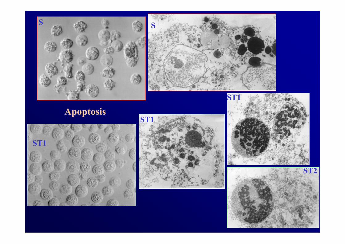
Apoptosis
S
ST1
ST2
S
ST1
ST1
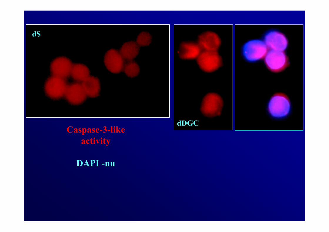
Caspase-3-likeactivity
DAPI -nu
dS
dDGC
*
*
*
**
**
**
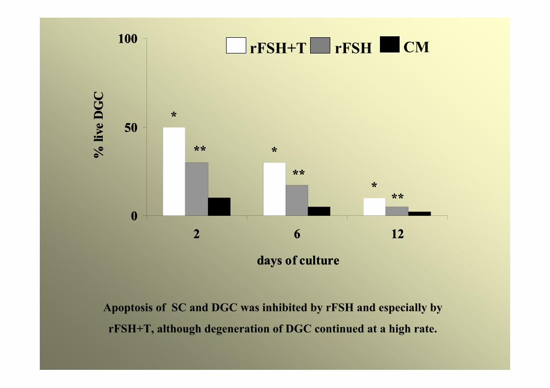
0
50
100
2 6 12
days of culture
% li
veD
GC
*
*
*
**
**
**
0
50
100
2 6 12
days of culture
% li
veD
GC
rFSH+T rFSH CM
Apoptosis of SC and DGC was inhibited by rFSH and especially by
rFSH+T, although degeneration of DGC continued at a high rate.
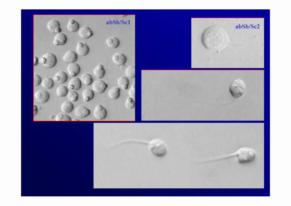
abSb/Sc1abSb/Sc2
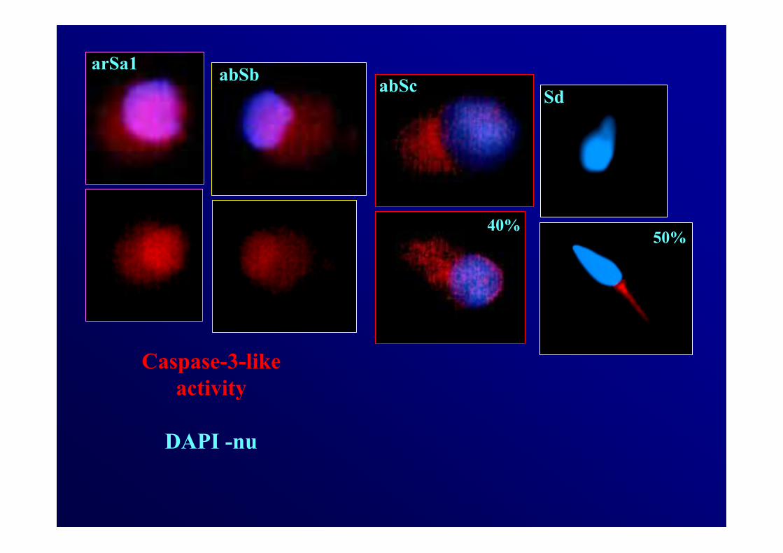
Caspase-3-likeactivity
DAPI -nu
arSa1abSc Sd
50%40%
abSb
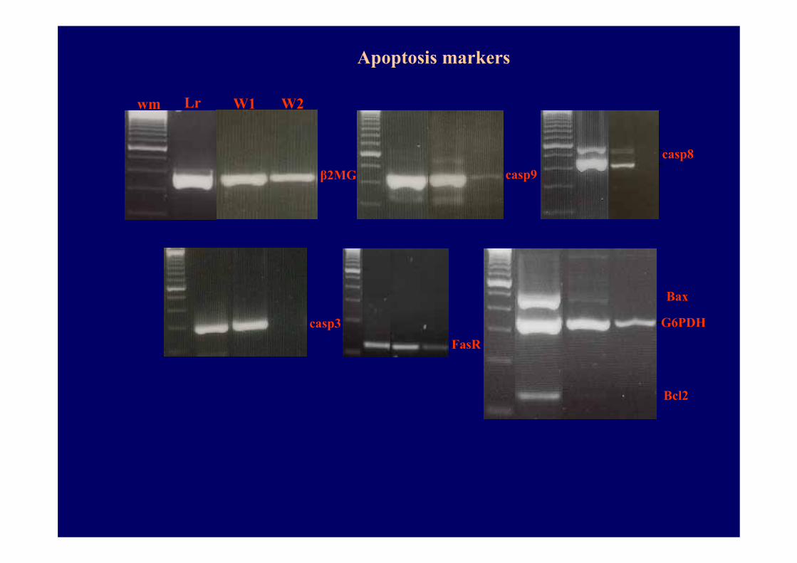
Bax
G6PDH
Bcl2
FasR
casp9casp8
wm Lr W1 W2
β2MG
casp3
Apoptosis markers
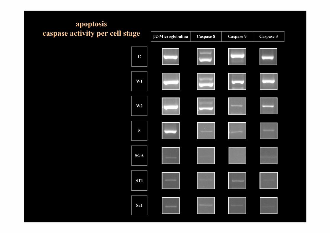
β2-Microglobulina Caspase 8 Caspase 9 Caspase 3
C
W1
W2
S
SGA
ST1
Sa1
apoptosiscaspase activity per cell stage
FINAL CONCLUSIONS
The present data suggest that long term in vitro cocultures of the normal human seminiferous epithelium sustain meiosis (7%) and full germ cell differentiation (17%), at a physiological pace (2-3 weeks).
However, more complex media should be evaluated as the rates of cell proliferation (4%) and meiosis completion (7%) were limited by high levels of DGC apoptosis (70% in the 1st week) and detachment of spermatids from SC intercellular connections.