on spermatogenesis in the bat. -...
TRANSCRIPT

On Spermatogenesis in the Bat.
By
Herlert B. Brown,"University College,. London.
With Plates XXIX and XXIII.
T H E following paper Is the result of an investigation whlciI have made during the past year, in the Physiological Labo-ratory at University College, upon the spermatogenesis of theRat, which, owing to the large size of its spermatozoa, andespecially of the " middle piece," presents peculiar advantagesfor the purpose. Although minor points of difference exist(in the relative length of the " middle piece" of the sper-matozoon and in the shape of the " head") the process Isapparently in all essential points identical with that whichoccurs in other mammals, and hence may be taken as a typeof " mammalian spermatogenesis/'
M e t h o d s of Resea rch .—I have studied the process ofspermatogenesis by means of sections, and by teased prepara-tions.
(1) Sections prepared by the paraffin-shellac method fromthe testis gradually hardened in alcohol, 1. e. by being placedIn alcoholic solution of gradually increasing strength. Thesections were prepared from small pieces of the organ, stainedin bulk, by immersion in Kleinenberg's alcoholic hesmatoxylinsolution for about three hours, the excess of stain being removedby acidulated alcohol, or by prolonged immersion (for a week)in a very dilute watery solution. From sections stained with
VOL. XXV. NEW SKE. Z

344 HEEBEET H. BROWS.
hsematoxylin the history of the process and the structure ofthe nuclei can be made out.
(2) Sections stained with gold. Small pieces of the freshtestis were soaked in chloride of gold solution, I per cent., forone hour, after previous immersion in lemon-juice or diluteformic acid, and exposed to the direct sunlight of midsummerin distilled water acidulated with acetic acid, until the reduc-tion of the gold was complete. Then the pieces of tissue werecarefully and gradually dehydrated with alcohol, and sectionsprepared by the paraffin-shellac method. These preparationsshow in particular the protoplasmic structures and the outlinesof the cells, the nuclei being entirely unstained and appearinglike vacuoles; thus they present a marked contrast to thehsematoxylin-stained sections.
(3) Sections stained with osmic acid, prepared by soakingsmall pieces of the fresh testis in osmic acid solution, 1 percent., for two days, washing with water, and dehydrating carefullywith alcohol. Then the sections were prepared by the paraffin-shellac method, and mounted in balsam.
(4) Teased osmic acid preparations. Small pieces of freshtestis were soaked in osmic acid solution 1 per cent, for twodays, and small pieces of tubules were broken up with needlesin glycerine and water.
Prom these teased preparations the development of thespermatozoa themselves can be best studied.
Nomenclature.1—I shall first describe the different cell-elements contained in a tubule, and give them the nameswhich I propose to adopt in this account, and then trace outwhat I consider to be the history of the development of thespermatozoa.
1 The question of nomenclature presents some difficulty, since so many differ-ent names have been given to the same elements by different observers, whichleads to confusion in the mind of the reader; consequently I have, at thesuggestion of Professor Lankester, avoided the use of such general terms as" spermatoblast," " spermatocyte," &c, and have substituted simple descrip-tive expressions, descriptive of the appearance or function of the elements ofthe tubule. I may here mention that I am much indebted to Professors Schaferand Lankester for their kindness in offering suggestions and advice.

OK SPERHATOGENESIS IN THE EAT. 345
In a section stained with fasematoxylin tubules are seen, cutmore or less transversely, and presenting different appearancesaccording to the stage of development of their contents.Since the process of. development of spermatozoa is a con-tinuous one, it would be possible to take any one stage as a.starting-point for description; but it is, on the whole, moreconvenient to start with the stage represented in fig. 1, inwhich a crop of spermatozoa is fully formed and ready to leavethe tubule.
A tubule in this stage (fig. 1) consists of four layers ofseminal elements, with a basement membrane formed offlattened cells, and these four layers correspond to four genera-tions.
The most external layer immediately within the basementmembrane consists of cells, the nuclei of which are all in theresting condition.
Of these nuclei there are three kinds represented in fig. 1corresponding to three classes of cells.
(1) There are large pale nuclei, the diameter of which isabout 18 ft, each containing a distinct round nucleolus, andbeing bounded by a definite nuclear membrane (fig. 1, e). Inthis stage some of these nuclei are seen resting upon the base-ment membrane, while others are seen extending between thecells of the second layer, and here and there one is foundamong the cells of the third layer. These nuclei belong tosuppor t ing cells, each of which serves for the support of agroup of spermatozoa during their development. The protoplasm of these cells forms a sort of network upon the base-ment membrane, in the meshes of which the other cells of theouter layer are contained, while from the inner extremity ofthe nucleus a protoplasmic process extends towards the sper-matozoa in the lumen of the tube. These cells appear to beconstant among Vertebrata; they have received various namesfrom different investigators, and have had very different func-tions assigned to them.
(2) Besides these supporting ceils there are a considerablenumber of small cells containing oval nuclei, which stain
346 HERBERT H. BROWW.
darkly with hsematoxylin and present the ordinary appearanceof resting nuclei; they contain an irregular coarse chromaticnetwork embedded in a stroma which stains less darkly, andare bounded by a nuclear membrane (fig. 1, b). These I shallcall growing cells.
(3) The other cells in the outer layer, which might easilybe overlooked, are much fewer in number than those last-mentioned, and their nuclei are larger, somewhat paler, andmore homogeneous in appearance, the chromatic substancebeing diffused in a very fine network throughout the nucleus(fig. 1, a). These appear to me to be the cells from which allthe other elements of the tubule, with the exception of thesupporting cells, are derived, and I shall therefore call themspore-cells. Probably they are the direct descendants of theprimitive male ova.
The cells of the second layer are large, and contain largespherical nuclei, which are all in the kinetic condition. Atthis stage the nuclei form for the most part a single row, buthere and there a cell is seen containing two nuclei, one beinginternal to the other. These correspond to the growing cellsof the outer layer of the preceding generation, which havenow attained a considerable size. They are destined to divideby karyokinesis into groups of cells which ultimately becomespermatozoa. They are in fact growing cells which have nearlyfinished growing.
The third layer (fig. 1, c) is composed of smaller cells, threeor four deep, with spherical nuclei which are pale, containingonly a small amount of chromatin. These cells have beencalled spermatoblasts, spermatocytes, &c, but since each cellis destined to develop into a spermatazoon, I shall call themsimply young spermatozoa, or young spermatozoawith spherical nuclei.
The fourth layer is composed of spermatozoa which are justready to leave the tubule. Between the heads of the sperma-tozoa and the cells of the third layer, there are numerousirregular darkly-stained granules1 (fig. 1, x), each granule
1 These bodies are the "seminal granules" which have been so often
OX SPERMA.TOGE5TES1S 1ST THE EAT. 347
being surrounded by a small amount of protoplasmic material.The dark granules are not derived from the destruction ofnuclei, but make their appearance in the protoplasm of thedeveloping spermatozoa at a certain stage of their develop-ment, and are cast off, together with a small amount of unalteredprotoplasm, when the development is nearly completed. Theyappear to consist of a mixture of albuminous and fattymaterial, since in osmic acid preparations their place is takenby a cluster of minute black granules (fig. 15).
A very different appearance is presented by sections preparedby the chloride of gold method (figs. 12, 13, 14). A stage ofdevelopment corresponding to fig. 1 is represented by fig. 12;in this preparation the nuclei are entirely unstained, andresemble clear vacuoles, but the protoplasmic structures andcell-outlines are rendered very conspicuous. The large growingcells of the second layer are seen to contain large granules—the "accessory corpuscles" of Reason—which are darkly-stained by the reagent (fig. 12, b"), and in the small cells ofthe outer layer similar but smaller bodies are seen. Theyoung spermatozoa which form the third layer, also each.contain an accessory corpuscle, which at this stage is embeddedin the protoplasm at the inner part of the cell (fig. 12, c).The fully formed spermatozoa (fig. 12, d) show an obviousdivision into three parts, head, body or middle piece, and tail.The tail is entirely unstained, but the middle piece contains aspiral filament which is darkly stained, and consequently veryconspicuous. It winds in a close spiral round a slightly tintedcore, which is continuous with the tail of the spermatozoon.
noticed; they are produced, not by the destruction of nuclei, but from theprotoplasm of the developing spermatozoa, from which they separate off at alate stage of their development. They are of great interest, since theyappear to represent the polar globules of the ovum, and their separationpossibly means the separation of the female element from thespermatozoa. They are, I believe, constant in mammalia; although owingto the shortness of the middle piece of the spermatozoon in most animalse.g. the dog, rabbit, man, their separation cannot be made out with suchdistinctness as in the case of the rat.

348 HERBERT H. BEOWN.
Dr. Heneage Gibbes1 has observed a spiral filament in thespermatozoa of several animals, e. g. horse, guineapig, andbull, which presents a somewhat similar appearance to thatwhich I have just described; he considered this to correspondto the so-called spiral filament of the newt's spermatozoon,which is an undulating filament attached to the spermatozoonby a fine membrane, which acts as a mesentery; but it isdifficult to see much resemblance between the spiral filamentwhich is stained by the gold method, and is confined to themiddle piece of the spermatozoon, and the long undulatingfilament of the spermatozoon of the newt. I have not yetlooked for this spiral filament in the middle piece of thespermatozoa of other animals, but will endeavour to do soduring the present summer.
His tory of the Process of Spermatogenesis.—Hav-ing thus briefly described the various elements of the seminaltubule, I shall proceed to trace out continuously the historyof the origin and development of the spermatozoa.
The process is a continuous one, i. e. a new crop of cellsmakes its appearance near the basement membrane as eachsuccessive crop of ripe spermatozoa leaves the tubule; and ina section of a single tubule four or five generations of cells atdifferent stages of their development are contained, so thatthe entire process of spermatogenesis occupies a correspondingnumber of cycles. An entire cycle is represented by figs. 1 to10. During this period a crop of spermatozoa leaves thetubule, their place is taken by a similar crop into which theyoung spermatozoa with spherical nuclei of the third layer havedeveloped, the growing cells of the second layer produce bytheir division another generation of young spermatozoa, thesmall growing cells of the outer layer increase in size andbecome the large cells of the second layer, and a new crop ofyoung growing cells makes its appearance in the outer layer.Thus, during a single cycle each of the layers described hasmoved forwards one stage, and the entire process of develop-
1 ' Quart. Journ. JVlic. Soi.'

ON SPEBMATOGENESIS IN THE EAT. 349
meat from the spore-cell to the fully-formed spermatozoawould occupy four cycles, during which time four fresh gene-rations of cells would have been produced; and so the processgoes ou.
The first part of this process, i. e. the origin of the new cropof growing cells, which makes its appearance in the outer layer,is the most difficult to make out. These cells are first seen atthe stage of fig. 9, and are produced by the karyokinetic divisionof larger cells, which are also found in the outer layer (thisdivision by karyokinesis is represented in fig. 8, a"). Theparent cells appear to be derived from the spore-cells by aprocess of division by budding, and not karyokinesis, but it isdifficult to feel certain about this. The spore-cells apparentlyincrease in size, and divide by a process of budding, for thisseems to be the explanation of an appearance such as is repre-sented in fig. 7, a', as if a small outgrowth made its appearancefrom one part of the nucleus, increased until the two partswere about equal in size, and then separated off. Of the twocells which result from this division one divides by karyokinesisand produces growing cells, but the other remains in theresting condition as a spore-cell, destined to repeat the processin the following cycle, and thus perpetuate the production ofspermatozoa in the tubule.
During the time in which a new crop of small-growing cellshas been produced, the growing cells of the previous generationhave been increasing in size, and now, since the new cells haveappeared between them and the wall of the tubule, come toform the second layer. The manner in which this growth takesplace will be understood if they are followed through the seriesrepresented by PI. XXII,1 figs. 1—10, b, b', and fig. 1, b". Infig. 1 these cells are all in the resting condition, and their nucleipresent the ordinary appearance of restiug nuclei, but verysoon they begin to pass into the kinetic condition, the nuclearmembrane disappearing and the chromatin becoming converted
1 Pigs. 3, 8, 9, and 10 are drawn on a somewhat smaller scale than theothers.

350 HEEBERT H. BROWN.
into an Irregular skein of filaments (fig. 2, V). In this con-dition they increase in size without dividing, and graduallyleave the wall of the tubule, until by the division of the spore-cells and formation of a new crop of cells between them andthe basement membrane, they come to form the second row(fig. 8, b'). By the time the stage of fig. 1 is reached the nucleiof these cells have attained their full size, but the protoplasmcontinues to increase in amount. The nuclei are spherical andlarge (diam. 10 n), and present the appearance represented inthe figure (fig. 1, b"). A large granule—the accessory cor-puscle—which stains darkly with chloride of gold, is embeddedin the protoplasm near the nucleus (fig 12, b"). Even at thestage of fig. I a cell is occasionally seen containing two nuclei,one being placed internal to the other; apparently all the cellswhen their growth is completed divide into two, though thenuclei do not as yet show any further karyokinetie changes, sothat at a later stage the cells are arranged in a double row.
Sometimes a nucleus may be seen apparently In the act ofdividing, and cells containing two nuclei become more frequent(fig. 5, b") ; and now the growing cells having reached their fulldevelopment, divide by karyokinesis, the phenomena of whichmay be very well observed, and give to a tubule in this stagea very characteristic appearance (PI. XXII, fig. 6, b'"). Theastral and diastral forms can be readily recognised, and, viewedin profile, the achromatic filaments may also be seen. (Fig- 6represents the appearances presented by these cells under amagnifying power of about 750 diameters.) In chloride ofgold preparations the accessory corpuscle appears to becomebroken up during karyokinesis; perhaps it forms the accessorycorpuscles of the young spermatozoa, and some small granuleswhich stain slightly with hsematoxylin and appear to be pro-duced during karyokinesis may represent this process.
It appears that the cells which result from the karyokinetiedivision of a growing cell do not separate from one anotherentirely, but remain at first united in groups by a very smallamount of the protoplasm of the mother cell. The outlinesof the cells of which these groups are composed cannot be
GIJ SPEBHATOGEXESIS IS THE EAT. 351
made out from sections stained with hfematoxylia, but inchloride of gold preparations the cell outlines are rendered verydistinct (PI. XXIII, fig. 14, c). The nuclei are spherical, of adiameter of 5 p., and are faintly stained by haematoxylin; theypossess a nuclear membrane, and a scanty network of eiiromatin.Between the nucleus and protoplasm of the cell a clear spacemakes its appearance, fitting like a cap over part of the nucleus,and in the neighbourhood of the clear cap a small chromaticgranule is usually to be seen (PL XXII, figs. 8, 9, c).
Stained with chloride of gold, the young spermatozoa presentsan appearance such as is represented in fig. 14. There is asmall accessory corpuscle attached to the nucleus, whichsubsequently becomes separated from it by a small vesiclewhich seems to be derived from, the accessory corpuscle (fig.14, c).
A few minute fatty granules are seen dotted about in theprotoplasm of these cells, in osmic acid preparations, and theaccessory corpuscles are also to be seen (fig. 15). It isdifficult to make out clearly what is the origin of the clearspace which is seen in hagmatoxylin preparations betweenpart of the nucleus and the protoplasm of the cell; apparentlyit corresponds to the small vesicle seen in chloride of gold pre-parations, attaching the accessory corpuscle to the nucleus,which may be increased ia size by the action of alcohol on thefresh cell.
The groups of cells are separated from one another for ashort time by bundles of spermatozoa, which extend betweenthem (fig. 9), but when the spermatozoa have left the sup-porting cells, it becomes difficult to make out a separation intogroups, and there appears to be a layer of cells, three or fourdeep, between the growing cells and the spermatozoa, for nowonly the thin protoplasmic strands of the supporting cellsextend inwards between the groups, to the layer of granules inwhich the heads of the spermatozoa are now embedded (PI.XXII, fig. 10). And now the stage of fig. 1 is again reached. Inthis stage the young spermatozoa present a different appearance;they are apparently free in the tubule, having been set at

352 HEBBEBT H.
liberty by the disintegration of the original groups. Thenucleus now occupies the outer part of each cell, i. e. thatdirected towards the wall of the tubule, and the outer part ofeach nucleus is covered only by the clear cap. At the sametime the chromatin leaves the substance of the nucleus andaccumulates at the nuclear membrane, and chiefly at its outerpart which becomes thickened, while the nuclear membrane inother parts seems to disappear, so that the nuclei appear to bebreaking up. It is apparently such an appearance as this,represented by fig. I, c, which has led V. Ebner and otherobservers who have investigated the spermatogenesis of theRat, to the belief that these cells undergo liquefaction, andproduce the liquid portion of the semen, or serve for thenutrition of the spermatozoa (vide the account of sperma-togenesis given by Professor Schafer in Quain's (Anatomy/vol. ii, ninth edition), and the granules of chromatic materialwhich are found between these cells and the spermatozoa, havebeen attributed to the disintegration of nuclei. The accessorycorpuscle leaves the nucleus at this time, and becomes em-bedded in the protoplasm at the inner part of the cell (fig. 12,c), where it remains inactive during the development of thespermatozoon, and is finally cast off with a residual portion ofthe protoplasm, when the process of development is nearlycompleted.
The young spermatozoa are free in the tubule for a veryshort time only, and they now begin to form groups in con-nection with the supporting cells. The remainder of theirdevelopment takes place in these groups and occupies thefourth and last cycle.
The manner in which the connection between the youngspermatozoa and the supporting cells is brought about is notvery clear; apparently the young spermatozoa congregateround the processes of the supporting cells which extendtowards the lumen of the tubule, and become embedded in theprotoplasm, without of course any fusion between the sub-stance of the young spermatozoa and of the supporting celltaking place; but the appearances presented by the supporting
OK SBEEMATGG-BNESIS IN THE BAT. 853
cells and young spermatozoa at this stage (fig. 1) are difficultto explain.
At this stage some supporting cells are seen which containa more or less conical nucleus, situated In the outer layerupon the wall of the tubule, from which a protoplasmic strandextends Inwards through the third layer of cells as far as thegranules among which the heads of the spermatozoa areembedded; but the nuclei of other supporting cells appear tobe pushing their way towards the lumen of the tubule; theyare elongated in a radial direction, and may be seen betweenthe second and third layers, and even in the middle of the thirdlayer of cells, but farther Inwards than this I have never seenthem. In many cases the nuclei are seen to be connected tothe outer layer by protoplasm, so that probably this Is amigration of the nucleus and not of the entire cell (figs. 1 and1, A). At the same time some of the young spermatozoaappear to move in the opposite direction towards the wall ofthe tubule, so as to occupy the position In the outer layervacated by the supporting nucleus (fig. 1, A) . These appear-ances at first appeared to me to justify the supposition thatthe supporting cells which have finished their work, havingserved for the support of the crop of spermatozoa which hasjust been discharged, are now in their turn being cast off intothe lumen of the tubule, there to undergo disintegration, andthat the cells which are passing outwards towards the wall ofthe tubule are destined to become the new supporting cells,retaining their connection with their brother cells, whichdevelop into spermatozoa; so that according to this view thesupporting cell and the group of spermatozoa which It sup-ports would result from the division of a single cell.
But there are numerous objections to such a view asthis:
1. The young spermatozoa at this stage are apparently free,and not connected together in groups.
2. It is very difficult to believe that, of a group of cells whichare all exactly alike, and are produced by the karyokinetlcdivision of a single cell, that which happens to be most ex-

354 HERBERT H. BEOWX.
ternal should become a supporting cell, while tlie others alldevelop into spermatozoa.
3. This improbability is rendered still more glaring by acomparison with the corresponding elements in the testes of thehedgehog and other animals, for in the hedgehog the nnelei ofthe supporting cells and of the young spermatozoa are remark-ably dissimilar.
4. Nuclei of supporting cells cannot be seen in the lumen ofthe tubule, or in the semen from the vas deferens or epidy-dymis, and the nuclei which have migrated inwards show nosigns of disintegration.
5. In the stage of development immediately succeeding this(vide fig. 3) nuclei of supporting cells are seen, which arequite as large as those in the present stage.
On all these grounds, then, it is impossible to adopt this viewof the origin of the connection between the supporting cellsand the spermatozoa. Probably the migration of the nucleiinto the midst of the young spermatozoa has something to dowith bringing about this connection, and having accomplishedthis the nuclei return to the outer layer.
The grouping of the young spermatozoa becomes more evi-dent as soon as the preceding generation of spermatozoa withthe seminal granules has left the tubule (this stage is repre-sented by fig. 2). Each group contains about ten or twelvespermatozoa. At this time a curious appearance is presentedby the supporting cells themselves; large globules, which stainblack with osmic acid, are seen in the protoplasm near thenuclei; these globules are not simply fat-globules, since theystain very darkly with gentian violet, and in chloride of goldpreparations become quite black from the great affinity whichthey have for the metal (vide fig. 13). In sections stainedwith hasmatoxylin vacuoles are seen in the protoplasm of thesupporting cells, each of which contains a spherical body, whichis slightly stained by the reagent ,• so that they would appearto consist of a mixture of fatty and albuminoid material. Inmany cases these bodies are found indenting the nucleus of thesupporting cell, and sometimes look as though they were
OS SPKBMAIGGE5TESIS I S THE EAT. 355
protruded from the nucleus (vide PL XXII, fig. 2, e andfig. 11).
I was at first inclined to attribute the appearance of thesebodies to a process of disintegration of the nuclei and proto-plasm of the supporting cells taking place at this stage. Ifthis were the case it would be very difficult to understand howthese cells could be so quickly reproduced, for in the next stageof development (fig. 3) the nuclei of the supporting cells arefully developed, and present HO signs either of growth or ofdegeneration, although a few fatty globules may still be seenin their protoplasm. Prof. Schafer, however, pointed out tome that the appearance of these globules is probably due to anincreased nutrition, of the supporting cells taking place at thistime, when they are about to enter upon a new phase ofactivity, and to serve for the support and probably also for thenutrition of a fresh crop of spermatozoa; and this appearsto me to be the most probable explanation. Apparently thesame supporting cell serves for the support of several suc-cessive crops of spermatozoa. I have not, however, been ableto make out in what manner this reproduction takes place, andcan say little about their life-history.
It is much easier to make out the history of the supportingcells in the Elasmobranch testis, which contains in a single trans-verse section every stage of development, from the embryoniccondition to the fully-formed spermatozoa. The result of myown investigations on the testis of the dogfish is in agreementwith the opinion of 1Swaeu and Masquelin, that while thespermatozoa are derived from p r imi t i ve male ova,the support ing cells are descended from foll icularcells, corresponding to the cells of t he Graafianfollicle in t he ovary. It is probable that the same is thecase in the mammal, but in order to make out the origin ofthe supporting cells in the mammal it would be necessary totrace them back to the embryonic condition.
The function of the supporting cells appears to be in great1 " JJtude snr la spermatogenese," par A. Swaea and H. Masqnelln, 'Archives
de Biologie,' torn, iv, fasc. 3,1883.
356 HERBERT H. BROWN.
measure mechanical; they serve the purpose of supporting andkeeping in order the complex testicular epithelium, forming asort of sustentacular framework like that of the Miillerianfibres of the retina. They serve for the support of, and alsoprobably convey nutritive material to the young spermatozoaduring their development, and when this is completed theyexpel them into the lumen of the tubule.
I must now return to the history of the development of thespermatozoa, at the point at which I left off to describe thesupporting cells. "We have at present reached the stage offig. 2 in the fourth cycle.
A young spermatozoon at this stage is somewhat conical inshape, the rounded apex of the cell, which is directed outwards,being occupied by the nucleus.
The nucleus has become oval and projects from the cellprotoplasm so that its outer hemisphere is covered only by theclear cap. As the development progresses the nucleus lengthensout, and projects more and more from the cell, until finallyonly its inner extremity remains embedded in the protoplasm.The projecting part of the nucleus is covered by the clear cap•which increases with it, but when the hooklike form of thenucleus is established (fig. 6, &) the clear cap is no longer tobe seen.
As the nucleus increases in length it diminishes in thickness,and becomes more and more intensely stained by hsematoxylin ;there is no chromatic network, but the chromatin appears tobe uniformly diffused throughout its substance. Before longthe nucleus begins to curve (fig. 4) and the hooklike shape ofthe head of the spermatozoon is established by the time thestage of fig. 6 is reached.
The remainder of the process is occupied chiefly by thedevelopment of the body and tail of the spermatozoon, andcan be studied much more satisfactorily from teased osmic acidpreparations, from which the series of drawings (figs. 16—24)is taken. Growing spermatozoa at an early stage correspondingto fig. 14 are represented by fig. 22, a. They are small cells,elongated in a radial direction and contain a spherical nucleus
OS SPEBMATOGENESIS IN THK EAT. 357
situated, at about the centre of the cell; the accessorycorpuscle is to be seen in these cells attached to the nucleusby a minute vesicle, and another small refracting granule isattached to the nuclear membrane, at the point from whichthe development of the body of the spermatozoon is about tobegin. There are also a few minute fatty granules dottedabout in the protoplasm (fig. 15, c). The manner in.which these cells _develop into spermatozoa is represented byfig. 22.
The nuclear membrane over the outer hemisphere of thenucleus becomes slightly thickened, and at the opposite poleof the nucleus, where the small granule is attached, a finefilament makes its appearance In the protoplasm, and extendsfrom the nucleus to the surface of the cell, where it is pro-longed by a delicate protoplasmic filament, the rudiment ofthe tail of the spermatozoon. At this time the1 accessorycorpuscle breaks away from the nucleus and becomes embeddedin the protoplasm at the Inner part of the cell, where it remainsinactive during the remainder of the process, to be finally castoff with the protoplasmic residue when the development isnearly completed. The protoplasm of the cell now becomescollected entirely at the inner part of the nucleus, leaving theouter hemisphere, upon which is the thickened membrane,covered only by the clear cap (22, d).
This corresponds to the stage of development represented byfigs. 1 and 12, at which the young spermatozoa appear to befree in the tubule. The nuclei of the cells are now commencingto elongate in the radial direction and to take on the oval form,and a small prominence becomes visible at the outer pole ofthe nucleus at the centre of the thickened membrane, the" bouton terminal" of Benson (fig. 22, e). It is at this stagethat the grouping in connection with the supporting cells is firstseen. Fig. 16 represents a group of young spermatozoa em-bedded in the protoplasm of a supporting cell, but the nucleusof the body cannot be seen, and the protoplasm appears to be
1 I have not been able to make oat what the origin of the accessorycorpuscle is. Perhaps it is derived from the nucleus of the growing cell.

358 HEBBEET H. BROWN.
broken off short. This, however, is readily explained, when Itis considered that the tubules were broken up with needles, bywhich process the inner part of the supporting cell contain-ing the young spermatozoa becomes broken off from the basewhich contains the nucleus, and is found to remain adherentto the basement membrane.
The manner in which the oval nucleus becomes transformedinto the head of the spermatozoon will be understood from fig.22. The nucleus increases in length and projects more andmore from the cell, and the thickened part of the nuclearmembrane progresses so as to cover the whole of the pro-jecting portion. Soon the nucleus begins to curve, the curva-ture first appearing near the apes, presumably owing to anincreased growth of one side of the thickened membrane. Asdevelopment goes on the curvature increases, and the denserportion involves more and more of the substance of thenucleus.
The thickening of the nuclear membrane is apparentlydue to a condensation of the nuclear substance and its trans-formation into that of the head of the spermatozoon, which,beginning at the surface and at the outer pole of the nucleus,progresses until the whole nucleus is converted into thespermatozoon head.
During this time the cilium which springs from the innerextremity of the cell has reached a considerable length, butvery little progress has been made with the development ofthe middle piece of the spermatozoon, although the cellprotoplasm has elongated to some extent.. The young spermatozoon is now (fig. 22, /) pyriform inshape, the base of the hooklike nucleus being inserted into thenarrow end of the cell, a long cilium springs from the broadend, and connecting the nucleus to the cilium is a delicatefilament which can only be seen with some difficulty (vide figs.17 and 6, c'). The remainder of the process is occupied chieflyby the development of the middle piece of the spermatozoon(figs. 18—20). The cell protoplasm rapidly elongates andassumes a club-shaped form, since the upper extremity which

ON SPEEMATOGENE8IS IN THE EAT. 859
contains the accessory corpuscle and some fatty granulesremains bulged (figs. 18 and 23). The filament which joinsthe nucleus to the eilium is more plainly seen in the narrowpart of the cell. This lengthening of the spermatozoon isaccompanied by a corresponding movement of the headsdownwards along the protoplasm of the supporting cell, untilwhen the spermatozoa have attained their full length theirheads reach the outer layer, in the neighbourhood of thenucleus, where they remain, until the spermatozoa are castoff into the lumen. A group at this stage is representedby figure 19. The middle piece of the spermatozoon is nowclearly visible passing through the protoplasm, which is col-lected chiefly at its upper end near the junction with the tail.The middle piece is formed out of the protoplasm of the cell,but not, as might be supposed, from the whole of the protoplasm,for a residual portion separates off from the spermatozoon,when its development is nearly completed. This residual partof the protoplasm, which contains the accessory corpuscle andone or two clusters of small fatty granules, gradually accumu-lates in the form of a globule, which separates from the bodyof the spermatozoon. At first the globule remains attached tothe upper part of the body by a short pedicle (a group ofspermatozoa at this stage is represented by fig. 2G; and columnsof spermatozoa in si tu with the globules attached in fig. 15),but before long it breaks away entirely from the spermatozoon.
In sections stained with haematoxylin small chromatic gra-nules make their appearance in the columns of spermatozoa(fig. 9, %), and when the spermatozoa have passed into thelumen these granules, each of which is contained in a smallamount of protoplasmic material, are found detached from thecolumns, and occupying the interval between the heads of thespermatozoa and the cells of the third layer. These bodieshave been previously described as the seminal granules; theyare, in fact, the globules which, as we have just seen, separatefrom the spermatozoa at a late stage of their development, andthe chromatic granules seem to be formed in part by the clustersof fatty granules seen in osmic acid preparations (figs. 10 and
VOL. XXV. NEW SEE. A A

860 HERBERT H. BEOWN.
1, x). This separation of globules is of considerable interestfrom a biological standpoint, for it apparently corresponds tothe separation of polar globules from the ovum, and may re-represent the elimination of the female element from thespermatozoa. Apparently the ova and spermatozoa are derivedfrom similar embryonic cells—the primitive ova, which arehermaphrodite. In a cell which is destined to develop intospermatozoa the male element predominates, and increasesuntil, by the separation of the globules, the spermatozoabecome wholly male, while in the cell which is going to becomean ovum the female element predominates, and by the separa-tion of the polar globules the cell becomes unisexed and readyto be fertilised by the addition of a new male element.
In invertebrate animals the separation of the female elementwould seem to take place at an earlier period during the pro-duction of the spermatozoa, and not, as in the present in-stance, during the development; and this is probably themeaning of the blastophoral body, as described by Blomfieldin the case of the earthworm.1 In this animal the youngspermatozoa undergo their development in groups—" the spermpolyblasts." The " sperm polyblast" is a mulberry-like mass,which results from the repeated division in geometrical pro-gression of a single cell—" the spermatospore/' or male ovum;during each division a certain amount of the protoplasm of themother cell remains behind, connecting together the daughtercells; this residual protoplasm accumulates at the centre of thegroup of cells, so that when the process is completed the spermpolyblast is composed of a central protoplasmic body—the" sperm blastophore," which is covered all over by " sperma-toblasts," or young spermatozoa, which remain planted on theblastophore until their development is completed.
This interpretation of the separation of the globules, andthe comparison with the blastophore of the earthworm, wassuggested to me by Professor Lankester, The blastophore ofthe earthworm, though it has much the same function as the
1 " The Development of the Spermatozoa," part i, " Lumbricus," by J.E. Blomfield, B.A., 'Quart. Journ. Micr. Sei.,'Jan., 1880.
OH SPEBMATOGEXESIS IN THE EAT. 381
vertebrate supporting cell, has a different morphological sig-nificance, the supporting cells being probably derived fromfollicular cells which appear not to be represented in the in-vertebrate testis.
As soon as the separation of the globules has taken place,or even before this, the spermatozoa begin to travel bodilytowards the lumen of the tube. This movement is apparentlyproduced by the supporting cells, which convey the sperma-tozoa inwards to the lumen of the tube, where they finallybecome detached. A supporting cell which is thus casting offits group of spermatozoa, is represented by fig. 21. For ashort time the head of the spermatozoon remains embedded ina protoplasmic envelope, perhaps derived from the supportingcell, and a small granule of protoplasmic material, darklystained in gold preparations, remains for some time at thejunction of the head with the middle piece (fig. 24, b, c), buteventually disappears.
A mature spermatozoon, examined fresh, or mounted in gly-cerine after osmic acid, shows no trace of a division into bodyand tail, appearing to be composed of two parts only, the headand the long tapering body; but by treatment with chlorideof gold, as before described, the division into middle piece andtail is rendered very conspicuous.
Fig. 25 represents a spermatozoon from the epidymis, whichis mounted in glycerine after having been treated with chlo-ride of gold. The middle piece is somewhat swollen andstained by the reduction of the gold, the staining being chieflyconcentrated in a fine spiral fibre, which winds closely roundthis portion. It has a length of about "07 mm., and presents astriking contrast to the tail, which is absolutely unstained;the length of the tail is about -08 mm. The spiral filament,as seen in sections mounted in balsam, has been already de-scribed (fig. 12, d). I have not been able to make out themanner of its development. It is first seen when the sperma-tozoa have reached their full length, before they have begun totravel to the lumen of the tubule (fig. 14).
362 HERBERT H. BROWN.
REVIEW OF THE LITERATURE OF MAMMALIAN SPEKMA-
TOGENESIS.
I will now give a brief account of some of the differentviews upon mammalian spermatogenesis which have been putforward during the last fifteen years, describing chiefly thosewhich are of most interest for the purpose of comparison.
For this I am to a considerable extent indebted to thedigest of the literature upon the subject which is given byEenson in the 'Archives de Biologie3 for 1882, in his paperupon " Mammalian Spermatogenesis."
Von Ebner, in 1871, gave an account of spermatogenesis,taken from a study of the testis of the Bat by means of sec-tions, which has received a good deal of support.
The supporting cells are described under the name of sperma-toblasts, and are considered to be the parent cells of thespermatozoa, which are formed endogenously from the proto-plasm of the spermatoblasts. Von Ebner describes an externallayer, resting upon the wall of the tubule, composed of twokinds of cells, which differ from one another in the appearanceof their nuclei, some of them having large nuclei which con-tain a spherical nucleolus, and others containing small granularnuclei. The cells with the large nuclei are the " spermato-blasts." The protoplasm of each spermatoblast joins that ofits neighbour on each side, to form a sort of protoplasmicnetwork upon the wall of the tubule, in the interstices of whichare contained the small cells with granular nuclei. On theinner side the spermatoblast gives off a protoplasmic process,which extends radially towards the lumen of the tubule. Theinner extremity of this process enlarges and splits up intodigitations, and at the base of each digitation a nucleus de-velops, being formed out of the protoplasm of the mother cell;then the nucleus elongates and becomes the head of a sperma-tozoon, a filament grows out from the extremity of each digi-tation and forms the tail, while the protoplasmic digitationitself is convefted into the middle piece. During their de-

ON SPBBMAIGGBSBSIS IS THE EAT. 368
velopment the heads of the spermatozoa travel downwardstowards the base of the cell, so that they reach the outer layer;on the completion of the process they travel back again, andfinally are thrown off into the lumen. The "round cells,"which occupy the spaces between the columns of spermatozoa,according to von Ebner, serve only for the production of theliquid portion of the semen. ^
It is obvious that this account is due to an erroneous inter-pretation of the appearance of columns of spermatozoa, whichis so conspicuous a feature in the testis of the rat ; therecannot be the least doubt that it is from the " round cells "that the spermatozoa are derived, and that they are notproduced endogenously in the protoplasm of the supportingcells.
Merkel, in 1871, gives a very different account of theprocess to that of von Ebner. He considers that the sperma-tozoa are derived from the small round cells which, accordingto von Ebnor, undergo liquefaction; these become embeddedin cavities which are hollowed out in the protoplasm of thesupporting cells, thus producing the spermatoblasts of vonEbner, and in these supporting ceils undergo development intospermatozoa.
Sertoli, in 1874, gives a much fuller account of the process.He describes fixed, or supporting, and mobile cells, which hedivides into three classes. 1. " Germinative" cells, whichare small, and situated in the outer layer between the bases ofthe supporting cells. 2. " Seminiferous" cells, which arelarger, and form the second layer, and correspond to thegerminative cells of the preceding generation, which haveincreased in size, and become pushed inwards by the formationof a new layer of germinative cells, between them and the wallof the tubule. 3. " Nematoblasts," which are small cellsproduced by the division of the preceding generation ofseminiferous cells, and destined to develop into spermatozoa.Sertoli gives no account of the mode of production of the ger-minative cells.
Lavalette St. George, who has published 'numerous papers

364 HERBERT H. BROWN.
upon spermatogenesis, takes a different view. He considersthat the connection between the spermatozoa and the support-ing cells is primary, both being derived from the division of asingle cell. These cells are situated in the outer layer betweenthe germinative cells of Sertoli, which, according to this ob-server, are follicular, and take no share in the production ofspermatozoa. They divide in a radial direction into two cells,which do not entirely separate from one another. The externalof the two remains in the resting condition attached to the wallof the tubule, and is called the spermatogonium, while theother cell increases in size and its nucleus repeatedly divides,so that a multinucleated mass is produced—the " spermato-gemme •" this becomes segmented, and each segment developsinto a spermatozoon. The cell at the base, or spermatogonium,retains its connection with the spermatozoa until their develop-ment is completed.
Somewhat similar accounts have been given in 1880 byMeyer, in the 'Memoirs of the St. Petersburgh Academie/and by Brissand in the ' Archives de Physiologie.'
Helmann in 1879, and W. Krause in 1881, agree withLavalette St. George in considering the supporting cell andthe spermatozoa to be derived from the same parent cell, butagree with Sertoli that the germinative cells of the outer layerare the progenitors of the spermatozoa. They consider thatone of the nuclei of the spermatogemme migrates towards thewall of the tubule, passing between the cells of the secondlayer to become embedded in the outer layer upon the basementmembrane, and that this nucleus, retaining its connection withthe spermatogemme and increasing in size, becomes the sup-porting nucleus, while the spermatogemme develops into agroup of spermatozoa; so that, according to this view, thereare no follicular cells in the tubules. I myself held for sometime such an opinion as this upon the relation between thespermatozoa and the supporting cell, and have already ex-plained at some length why I felt obliged to give it up.
Klein, in the 'Atlas of Histology/ in 1881, gives an accountof the development of spermatozoa in the dog and some other
ON SPEBMATOGENESIS IN THE EAT. 365
mammals. He describes the development of spermatozoafrom small cells resulting from the division of the inner seminalcells; these daughter-cells are at first free in the tubule, butgradually form fan-shaped groups, which sink between theinner seminal cell towards the wail of the tubule.
Klein has not observed the existence of supporting cells, towhich the groups of spermatozoa are attached, and offers noexplanation, of this grouping.
Sch'afer, in the ninth edition of Quain's ' Anatomy/ in 1882,gives a short account of the testis of the Rat; he attributesto the small cells of the third layer a nutritive function, andconsiders that some of the proliferating cells (the large ceils ofthe second layer) give rise by their division to groups of sper-matozoa, while others form the small cells, which ultimatelybreak down and liquefy.
RensoB, in the ' Archives de Biologie/ 1882, gives a descrip-tion of mammalian spermatogenesis, taken chiefly from a studyof the process in the Eat, which agrees very closely in mostpoints with the result of my own investigations.
Benson traces the origin of the spermatozoa to the smallround cells of the outer layer, which he calls after Sertoli," germinative" cells, and which make their appearance sud-denly in the outer layer upon the basement membrane, buthe has not been able to discover in what manner this aewlayer of cells is produced, and the production of spermatozoaperpetuated.
The germinative cells increase in size, and become in thenest cycle the " seminiferous" cells which form the secondlayer. The seminiferous ceils divide by karyokynesis intogroups of daughter cells, which he calls " cysts." The a cysts"disintegrate, and their component tells, the " nematoblasts," areset free. Soon they contract a connection with the supportingcells, in which they become embedded in groups. Finally,when this development is completed, they are thrown off bythe supporting cells into the interior of the tubule. Rensonalso describes the appearances presented by the nematoblastsduring their development, as studied by teased preparations
866 HERBERT H. BROWN.
and his account of the process agrees very closely with thatwhich I have given, but he does not describe the separation ofthe globules of residual protoplasmic material which takesplace when the development is nearly completed. He describesthe accessory corpuscles, both in the seminiferous cells and inthe nematoblasts, and suggests that they may represent thepolar globules of the ovum.
Swaen and Masquelin, in the ' Archives de Biologie' for1883, give the results of their investigations upon spermato-genesis in the Selachians, the Salamander, and the Mammal.Their account of the process in the mammal was taken from astudy of the testis of the Bull, and agrees in many particularswith that of Reason. These observers give an acccount ofthe manner in which the continual production of succeedinggenerations of spermatozoa is kept up in the tubule, whichpresents some resemblance in principle to the view of theprocess which I have taken, though differing from it in detail.They call the small cells of the outer layer—the germinativecells of Sertoli and Eenson—while they are in the restingcondition '' inactive male ovules." These cells passing into thekinetic condition become the active male ovules, and graduallyleave the wall of the tubule. Before long each cell dividesinto two by karyokinesis in a radial direction; the external ofthe two cells becomes embedded in the outer layer, and passinginto the resting condition, becomes an inactive male ovule,which repeats the process in the succeeding cycle. The othercell, which is internal, increases in size, and finally divides bykaryokinesis into a group of cells, the " spermatogemme."
The cells of which the spermatogemme is composed arecalled spermatocytes, and afterwards, when they have obvi-ously begun the process of development into spermatozoa,receive the name of nematoblasts. The nematoblasts becomeattached to the supporting cell without being first free in thetubule, for the inner extremity of a supporting cell which hasdischarged its spermatozoa fuses with the intercellular materialof the spermatogemme.
The account which I have given of the origin of the growing

ON SPEEMATOGBNESIS IN THE EAT. 367
cells of the outer layer from spore cells, which divide in thefirst instance by a process of budding, and the subsequentdivision of one of the resulting cells by karyokinesis, has notbeen confirmed by any previous investigations. It is possiblethat I may have been misled by the appearance of division bybudding -which is occasionally to be seen in these cells in theouter layer^ and is represented in fig. 7, d, consequently I havebeen much interested in finding a similar method of division bybudding of the nucleus described by Arthur Bolles Lee, in arecentx paper on spermatogenesis in the Appendicularia. Thisobserver suggests a theory to explain the occurrence of the twomethods of cell division, which is an ingenious one, and cer-tainly fits in very well with the account of the process which Ihave given above.
He suggests that the complex method of division by karyo-kinesis is intended to serve for the accurate division of theconstituents of the nucleus between the resulting cells, so thatthe daughter cells, possessing in an equal degree the propertiesof the parent nucleus, exactly resemble one another. On theother hand the method of division by budding consists of anelimination of one part of the nucleus from the remainder, sothat the resulting cells will not exactly resemble one another.
I have described above the spore cell as dividing by budding,and of one of the resulting cells remaining as a spore cell•while the other divides by karyokinesis, so that the result ofthe division by budding is to produce two dissimilar cells. Onthe other hand,, the cell which divides by karyokinesis pro-duces the growing cells which are all precisely alike, and theselater on dividing again by karyokinesis give origin to theyoung spermatozoa which again are ail alike.2
1 " Recherches stir l'ovogenese et spermatogenese chez ies Appendicularia,"par Arthur Bolles Lee, ' Rectiei! Zoologique Snisse,' vol. i, No. 4.
2 It. might be supposed that the mode of separation of the polar globulefrom the nucleus of the ovum goes against this theory; but it appears that,according to van Beneden, this is not a true process of cell division by karyo-kinesis (vide a paper on " E. van Beneden's Researches on the Maturationand Fecundation of the Ovum," by J. T. Cunningham, ' Quart. Joum. Micr.

368 HERBERT H. BROWN.
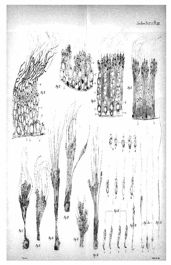
EXPLANATION OF PLATES XXII & XXIII,
Illustrating Mr. Herbert H. Brown's Paper " On Spermato-genesis in the Rat."
PLATE XXII
Represents sections of tubules from the testis of the Rat, stained withhesmatoxylin.
Most of the figures in both Plates are drawn under a magnifying power of750 diameters, but Figs. 3, 8, 9,10,14, and 15 on a slightly smaller scale.
FIGS. 1—10 show consecutive stages in the production of spermatozoa.a. Spore-cell, a'. Ditto, dividing by fission, a". Cells dividing by karyoki-nesis to produce the young growing cells, b. Young growing cell in restingcondition. &'. Ditto, in kinetic condition, b''. Growing cell at a later stage(in the second row), c. Young spermatozoa with spherical nuclei, d. Young-spermatozoa undergoing development, d. Adult spermatozoa, e. Supportingcells, x. Seminal granules.
FIG. 11.—Nuclei of supporting cells, showing the large fatty-albuminoidglobules (stage corresponding to Fig. 2).
PLATE XXIII.
FIGS. 12 —14. — Sections prepared with chloride of gold (lettered asFigs. 1—10).
Fig. 12. Corresponds to Fig. 1.Fig. 13. Ditto to Fig. 5.Fig. 14. Ditto to Fig. 9.
FIG. 15.—Section stained with osmie acid. Stage corresponding to Figs.14 and 9.
FIGS. 16—20.—Groups of developing spermatozoa from osmic acid prepara-tions, mounted in glycerine.
Fig. 16. A group of young spermatozoa with oval nuclei.Fig. 17. Group of young spermatozoa at stage of Fig. 6.Fig. 18. Young spermatozoa slightly more developed, showing elongation
of the protoplasm.
Sci.,' January, 1885, p. 107). In this paper the theory as to the separation ofthe female element from the spermatozoon and the male element from theovum is also brought forward.
ON SPERMA.TOGENESIS IS THE BAT. 369
Fig. 19. Group of young spermatozoa, stage of Fig. 8. (The middlepiece of the spermatozoa has BOW 'reached its full length.)
Fig. 20. A group of spermatozoa, showing the separatioE of the seminalgrannies.
FIG. 21.—A supporting cell casting off its spermatozoa.FIGS. 22—24.—Osmic acid preparation. Separate spermatozoa.
Fig. 22 (a—I). A series showing the development of young spermatozoa,detached from the groups. The gradual transformation of the auciensinto the spermatozoon head is especially shown.
Jig. 23. A spermatozoon at the stage of fig. 18.Fig. 24. Spermatozoa almost fully developed, a. The residual globule is
still attached, h. TMs is thrown off, but the head of the spermatozoonis not yet free. c. There is only a small granule remaining at thejunction of the head and middle piece.
FIG. 25.—A spermatozoon from the epidydymis of the Eat, stained withgold, showing the spiral filament.

Fur 7
,KS.&£r. IW.
mm •
H.H.Es-.-ra Ml.