sonographic demonstration of intracranial hemorrhage in a...
TRANSCRIPT
Case ReportSonographic Demonstration of Intracranial Hemorrhage in aFetus with Hydrops Fetalis due to Rh Alloimmunization afterIntrauterine Intravascular Transfusion: A Case Report andReview of the Literature
Rauf Melekoglu ,1 Ebru Celik,2 and Hasim Kural1
1Department of Obstetrics and Gynecology, Faculty of Medicine, The University of Inonu, 44280 Malatya, Turkey2Department of Obstetrics and Gynecology, Faculty of Medicine, The University of Koc, 34010 Istanbul, Turkey
Correspondence should be addressed to Rauf Melekoglu; [email protected]
Received 11 April 2017; Accepted 21 February 2018; Published 26 March 2018
Academic Editor: Maria Grazia Porpora
Copyright © 2018 RaufMelekoglu et al.This is an open access article distributed under the Creative Commons Attribution License,which permits unrestricted use, distribution, and reproduction in any medium, provided the original work is properly cited.
Intrauterine transfusion is the most common and successful intrauterine procedure for the treatment of fetal anemia due to redcell alloimmunization. Fetal intracranial hemorrhage is a very rare complication of intrauterine transfusion in patients with Rh(D)alloimmunization and it has been demonstrated only in a few case reports in the literature. Herein, we described a case of grade IVintraventricular hemorrhage that was diagnosed following the first intrauterine transfusion and reviewed the literature about thefetal intracranial hemorrhage that occurred after intrauterine intravascular transfusion procedure.
1. Introduction
Intrauterine transfusion (IUT) is the most common andsuccessful intrauterine procedure for the treatment of fetalanemia due to red cell alloimmunization [1]. The beneficialeffect of in utero therapy on perinatal survival has beendemonstrated clearly in several observational studies [2, 3].Despite the dramatic decrease in the IUT requirement due tothe widespread use of prophylactic Rh(D) immune globulin,the procedure continues to be a gold standard for treatmentof severe fetal anemia [4]. While intrauterine intravascu-lar transfusion has remarkable effect on the treatment offetal red blood cell alloimmunization, the total procedure-related complication rate has been reported approximately3.1 percent and commonly indicated as fetal death, neonataldeath, emergency cesarean delivery, infection, and prematurerupture of membranes [5]. Fetal brain injury is a very rarecomplication of IUT that has been demonstrated only ina few case reports in the literature [6]. To the best of ourknowledge, the literature does not include any cases of a gradeIV intraventricular hemorrhage due to IUT.
Herein, we described a case of grade IV intraventricularhemorrhage that was diagnosed following the first IUT and
reviewed the literature about the fetal intracranial hemor-rhage that occurred after intrauterine intravascular trans-fusion procedure in patients with Rh(D) alloimmunization.We searched PubMed, Scopus, Embase, and Google Scholardatabases using the keywords Rh isoimmunization “OR”intrauterine transfusion “AND” intracranial hemorrhage“OR” brain injury “OR” brain damage. We found only twopapers that define three cases of intracranial hemorrhageassociated with intrauterine transfusion due to Rh alloim-munization. The initial platelet value was not noted in thethird case. Author, case number, patient’s age, gestationalage, pretransfusion hemoglobin value, pretransfusion plateletvalue, neurosonogram after the first intrauterine transfusion,and the outcome were summarized in Table 1. In this paper,we have compared our case with the other three cases we havefound in the literature.
2. Case Presentation
A 34-year-old woman, gravida 3, para 2, with a history of anintrauterine death at 32 weeks of gestation due to hydropsfetalis as a result of Rh alloimmunization in the previous
HindawiCase Reports in Obstetrics and GynecologyVolume 2018, Article ID 8412139, 5 pageshttps://doi.org/10.1155/2018/8412139

2 Case Reports in Obstetrics and Gynecology
Table1:Summaryof
ther
eportedfetalintracranialh
emorrhagec
ases
related
tointrauterin
etransfusio
ndu
etoRh
alloim
mun
ization.
Authors
Case
number
Maternalage
Gestatio
nal
age
Pretransfusio
nhemoglobin
value(g/dl)
Pretransfusio
nplatele
tvalue
(/𝜇l)
Neurosono
gram
after
thefi
rst
intrauterin
etransfusio
nOutcome
Ghi
etal.200
4Ca
se1
3020
1.2168000
Intraventricular
andcerebellar
hemorrhage
Term
inationof
pregnancy.Pathologicalconfi
rmationof
cerebellarh
emorrhage
Case
225
231.6
177000
Cerebellarh
emorrhage
Progressiveh
ypop
lasia
ofon
ecerebellarh
emisp
here.
Delivery
at34
weeks
after
sixIU
Ts.N
ormalneurological
developm
entat2
years
Simon
azzietal.
2016
Case
332
224
-Suspicious
cerebellarinfarction
Hem
osiderin
stainingin
thec
erebellum
bilaterally,
reflecting
priorh
emorrhageinpo
stnatalbrainMRI.N
ormal
neurologicaldevelopm
entat14mon
ths
Currentstudy
Case
434
282.9
1540
00
Echo
genicc
ollectionin
ther
ight
lateralventric
leandextend
ingto
thes
urroun
ding
cerebral
parenchymac
ompatib
lewith
gradeIVintraventricular
hemorrhage
Diffusee
chogenicity
extend
ingfro
mtheinferiorleft
caud
ate
nucle
usto
theleft
ventric
lethatleadsleft
ventric
ular
dilatation(in
traventricular
gradeIVhemorrhage).N
ormal
neurologicaldevelopm
entat6
mon
ths

Case Reports in Obstetrics and Gynecology 3
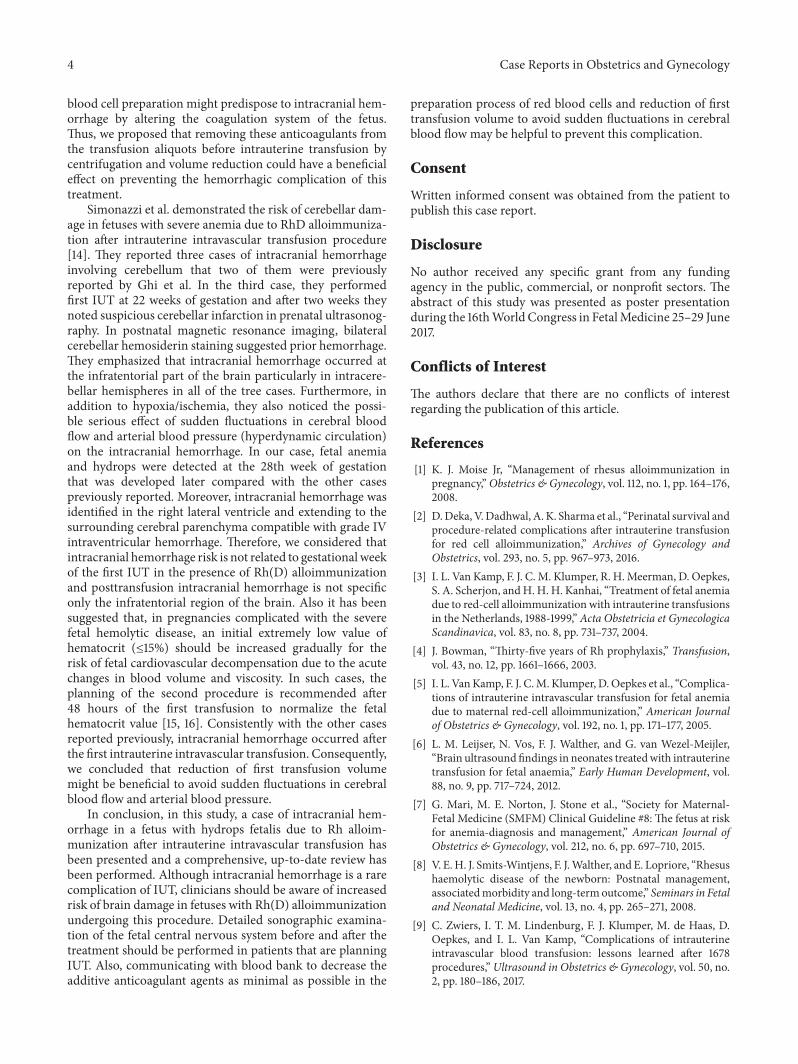
Figure 1: Axial view of the fetal head after intrauterine transfusionprocedure. A large echogenic collection involving right lateral ven-tricle and extending to the surrounding parenchyma that suggestsgrade IV intraventricular hemorrhage.
pregnancywas referred to our center at 13 weeks of gestations.There were no pathologic findings in her physical exami-nation, laboratory findings, and obstetric ultrasonography.Gray-scale and color Doppler ultrasonography evaluationwas performed to detect the findings of anemia and hydropswith a two-week interval starting from the 18th gestationalweek in the Prenatal Diagnosis and Treatment Unit by aprotocol defined by Society for Maternal-Fetal Medicine forthe pregnant women complicated with Rh alloimmunization[7]. Her obstetric follow-up was unremarkable until 29weeks of gestation. At 29 weeks of gestation, mid-cerebralartery peak systolic velocity (MCA-PSV) was detected as80.6 cm/sn [>1.5 multiple of the median (MoM)] in herobstetric Doppler ultrasonography that suggests fetal ane-mia with ascites, cardiomegaly, and pericardial effusion. Anintrauterine intravascular transfusion was performed, andthe hemoglobin concentration before the procedure wasdetected as 2.9 g/dL. 40ml O Rh(D)-negative red blood cellthat was freshly prepared and underwent irradiation andleukodepletion was transfused to the fetus via the umbilicalvein in the portion of the umbilical cord near its insertioninto the placenta by a 22-gauge needle with no complication.Posttransfusion hemoglobin was detected as 8.1 g/dl. The ini-tial and posttransfusion platelet countwas detected in normalrange (154000/𝜇l and 163000/𝜇l, resp.). A fewdays later, ultra-sound examination revealed the presence of an echogeniccollection involving right lateral ventricle and extending tothe surrounding cerebral parenchyma compatible with gradeIV intraventricular hemorrhage (Figure 1). The couple wascounseled, and they opted for the continuation of in uterotherapy. The second IUT was scheduled after ten days. At 30+ 4 weeks of pregnancy, the second IUT was performed. Theinitial hemoglobin value was detected as 4.9 g/dl. Persistentfetal bradycardia was noted during the procedure, and anemergency cesarean section was performed. APGAR score4/7, 1315 g, 43 cm male infant was delivered by cesarean sec-tion. Neonate was transferred to the neonatal intensive careunit. Exchange transfusion, phototherapy, and intravenousimmunoglobulin treatment were applied. Postnatal cranialultrasonography showed diffuse echogenicity extending fromthe inferior left caudate nucleus to the left ventricle that
leads to left ventricular dilatation compatible with intraven-tricular grade IV hemorrhage. The intracranial hemorrhagewas gradually regressed in the subsequent ultrasonographicexaminations and completely disappeared at the end ofthe first month, and the neonate was discharged from thehospital after healing two months after birth. At the time ofwriting this paper, the babywas showing normal neurologicaldevelopment at 6 months.
3. Discussion
Intrauterine transfusion has been reported as the mostsuccessful fetal therapy procedurewith 95%perinatal survivalrate [8].The overall survival rate after this antenatal treatmentprocedure varies with experience of center, development offetal anemia before 20 weeks of gestation, and occurrence offetal hydrops [9]. The presence of fetal hydrops during thefirst IUT reduces the success of the treatment. Lindenburget al. reported the perinatal outcome of 491 fetuses whounderwent 1422 intrauterine intravascular transfusion pro-cedure during the antenatal period. They demonstrated thatperinatal survival rate was 83% and 95% in hydropic and non-hydropic fetuses, respectively [10]. Although IUT contributesto the reduction of perinatal mortality, concerns about theneurological morbidities associated with this procedure havebeen considered only in a few studies [11]. Fetal intracranialhemorrhage as a short-term neurological morbidity wasreported by Ghi et al. for the first time in 2003 [12]. Theydescribed four cases with intracranial hemorrhage related tothe fetal anemia (two immune hydrops due to Rh D alloim-munization, two monochorionic twins complicated with thedeath of the cotwin). Consistently with the case currentlyreported, each of the cases related to Rh alloimmunizationhad very low initial hemoglobin values in the first IUT(1.2 g/dl and 1.6 g/dl, resp.). They suggested that disruptionof intracranial vessels may be responsible in the pathophys-iology of brain injury in severe anemic fetuses and noticedthe importance of fetal neurosonography in pregnancies withsevere anemia due to Rh alloimmunization undergoing IUT.In 2004, the same group reportedmultiplanar neurosonogra-phy results of seven consecutive hydropic fetuses undergoingintrauterine transfusion procedure due to Rh alloimmu-nization [13]. In addition to the previously reported twocases, they described a case of periventricular leukomalaciaand a case of unilateral ventriculomegaly that was noticedafter the first IUT. They speculated that hypoxia/ischemiaand the hyperdynamic circulation in fetal anemia cause thebrain vessel disruption that leads to intracranial hemorrhage.They also considered that altered coagulation due to IUTmight be responsible for intracranial hemorrhage. In ourcase, the initial and posttransfusion platelet values weredetected in the normal range (154000/𝜇l and 163000/𝜇l,resp.), and there was no sign of increasing bleeding timesuch as excessive bleeding from the umbilical cord afterwithdrawal of the needle. Furthermore, we hypothesized thatpreservative-anticoagulant system such as additive solution-1 (AS-1), AS-3, AS-5, citrate-phosphate-dextrose-adenine-1(CPDA-1), citrate-phosphate-dextrose (CPD), and citrate-phosphate-dextrose-dextrose (CP2D) that were used in red
4 Case Reports in Obstetrics and Gynecology
blood cell preparation might predispose to intracranial hem-orrhage by altering the coagulation system of the fetus.Thus, we proposed that removing these anticoagulants fromthe transfusion aliquots before intrauterine transfusion bycentrifugation and volume reduction could have a beneficialeffect on preventing the hemorrhagic complication of thistreatment.
Simonazzi et al. demonstrated the risk of cerebellar dam-age in fetuses with severe anemia due to RhD alloimmuniza-tion after intrauterine intravascular transfusion procedure[14]. They reported three cases of intracranial hemorrhageinvolving cerebellum that two of them were previouslyreported by Ghi et al. In the third case, they performedfirst IUT at 22 weeks of gestation and after two weeks theynoted suspicious cerebellar infarction in prenatal ultrasonog-raphy. In postnatal magnetic resonance imaging, bilateralcerebellar hemosiderin staining suggested prior hemorrhage.They emphasized that intracranial hemorrhage occurred atthe infratentorial part of the brain particularly in intracere-bellar hemispheres in all of the tree cases. Furthermore, inaddition to hypoxia/ischemia, they also noticed the possi-ble serious effect of sudden fluctuations in cerebral bloodflow and arterial blood pressure (hyperdynamic circulation)on the intracranial hemorrhage. In our case, fetal anemiaand hydrops were detected at the 28th week of gestationthat was developed later compared with the other casespreviously reported. Moreover, intracranial hemorrhage wasidentified in the right lateral ventricle and extending to thesurrounding cerebral parenchyma compatible with grade IVintraventricular hemorrhage. Therefore, we considered thatintracranial hemorrhage risk is not related to gestational weekof the first IUT in the presence of Rh(D) alloimmunizationand posttransfusion intracranial hemorrhage is not specificonly the infratentorial region of the brain. Also it has beensuggested that, in pregnancies complicated with the severefetal hemolytic disease, an initial extremely low value ofhematocrit (≤15%) should be increased gradually for therisk of fetal cardiovascular decompensation due to the acutechanges in blood volume and viscosity. In such cases, theplanning of the second procedure is recommended after48 hours of the first transfusion to normalize the fetalhematocrit value [15, 16]. Consistently with the other casesreported previously, intracranial hemorrhage occurred afterthe first intrauterine intravascular transfusion. Consequently,we concluded that reduction of first transfusion volumemight be beneficial to avoid sudden fluctuations in cerebralblood flow and arterial blood pressure.
In conclusion, in this study, a case of intracranial hem-orrhage in a fetus with hydrops fetalis due to Rh alloim-munization after intrauterine intravascular transfusion hasbeen presented and a comprehensive, up-to-date review hasbeen performed. Although intracranial hemorrhage is a rarecomplication of IUT, clinicians should be aware of increasedrisk of brain damage in fetuses with Rh(D) alloimmunizationundergoing this procedure. Detailed sonographic examina-tion of the fetal central nervous system before and after thetreatment should be performed in patients that are planningIUT. Also, communicating with blood bank to decrease theadditive anticoagulant agents as minimal as possible in the
preparation process of red blood cells and reduction of firsttransfusion volume to avoid sudden fluctuations in cerebralblood flow may be helpful to prevent this complication.
Consent
Written informed consent was obtained from the patient topublish this case report.
Disclosure
No author received any specific grant from any fundingagency in the public, commercial, or nonprofit sectors. Theabstract of this study was presented as poster presentationduring the 16thWorld Congress in FetalMedicine 25–29 June2017.
Conflicts of Interest
The authors declare that there are no conflicts of interestregarding the publication of this article.
References
[1] K. J. Moise Jr, “Management of rhesus alloimmunization inpregnancy,”Obstetrics & Gynecology, vol. 112, no. 1, pp. 164–176,2008.
[2] D.Deka, V.Dadhwal, A. K. Sharma et al., “Perinatal survival andprocedure-related complications after intrauterine transfusionfor red cell alloimmunization,” Archives of Gynecology andObstetrics, vol. 293, no. 5, pp. 967–973, 2016.
[3] I. L. Van Kamp, F. J. C. M. Klumper, R. H.Meerman, D. Oepkes,S. A. Scherjon, andH. H. H. Kanhai, “Treatment of fetal anemiadue to red-cell alloimmunization with intrauterine transfusionsin the Netherlands, 1988-1999,” Acta Obstetricia et GynecologicaScandinavica, vol. 83, no. 8, pp. 731–737, 2004.
[4] J. Bowman, “Thirty-five years of Rh prophylaxis,” Transfusion,vol. 43, no. 12, pp. 1661–1666, 2003.
[5] I. L. VanKamp, F. J. C.M. Klumper, D.Oepkes et al., “Complica-tions of intrauterine intravascular transfusion for fetal anemiadue to maternal red-cell alloimmunization,” American Journalof Obstetrics & Gynecology, vol. 192, no. 1, pp. 171–177, 2005.
[6] L. M. Leijser, N. Vos, F. J. Walther, and G. van Wezel-Meijler,“Brain ultrasound findings in neonates treatedwith intrauterinetransfusion for fetal anaemia,” Early Human Development, vol.88, no. 9, pp. 717–724, 2012.
[7] G. Mari, M. E. Norton, J. Stone et al., “Society for Maternal-Fetal Medicine (SMFM) Clinical Guideline #8: The fetus at riskfor anemia-diagnosis and management,” American Journal ofObstetrics & Gynecology, vol. 212, no. 6, pp. 697–710, 2015.
[8] V. E.H. J. Smits-Wintjens, F. J.Walther, and E. Lopriore, “Rhesushaemolytic disease of the newborn: Postnatal management,associatedmorbidity and long-termoutcome,” Seminars in Fetaland Neonatal Medicine, vol. 13, no. 4, pp. 265–271, 2008.
[9] C. Zwiers, I. T. M. Lindenburg, F. J. Klumper, M. de Haas, D.Oepkes, and I. L. Van Kamp, “Complications of intrauterineintravascular blood transfusion: lessons learned after 1678procedures,”Ultrasound in Obstetrics & Gynecology, vol. 50, no.2, pp. 180–186, 2017.

Case Reports in Obstetrics and Gynecology 5
[10] I. T. M. Lindenburg, I. L. Van Kamp, and D. Oepkes, “Intrauter-ine blood transfusion: Current indications and associated risks,”Fetal Diagnosis and Therapy, vol. 36, no. 4, pp. 263–271, 2014.
[11] I. T. Lindenburg, V. E. Smits-Wintjens, J. M. Van Klink etal., “Long-termneurodevelopmental outcome after intrauterinetransfusion for hemolytic disease of the fetus/newborn: TheLOTUS study,” American Journal of Obstetrics & Gynecology,vol. 206, no. 2, pp. 141–e8, 2012.
[12] T. Ghi, G. Simonazzi, A. Perolo et al., “Outcome of antenatallydiagnosed intracranial hemorrhage: case series and review ofthe literature,” Ultrasound in Obstetrics & Gynecology, vol. 22,no. 2, pp. 121–130, 2003.
[13] T. Ghi, L. Brondelli, G. Simonazzi et al., “Sonographic demon-stration of brain injury in fetuses with severe red bloodcell alloimmunization undergoing intrauterine transfusions,”Ultrasound in Obstetrics & Gynecology, vol. 23, no. 5, pp. 428–431, 2004.
[14] G. Simonazzi, D. Bernabini, A. Curti et al., “Fetal cerebellardamage in fetuses with severe anemia undergoing intrauter-ine transfusions,” The Journal of Maternal-Fetal and NeonatalMedicine, vol. 29, no. 3, pp. 389–392, 2016.
[15] N. Papantoniou, S. Sifakis, and A. Antsaklis, “Therapeuticmanagement of fetal anemia: Review of standard practice andalternative treatment options,” Journal of Perinatal Medicine,vol. 41, no. 1, pp. 71–82, 2013.
[16] G. Mari, K. J. Moise, R. L. Deter, and R. J. Carpenter, “Flowvelocity waveforms of the umbilical and cerebral arteries beforeand after intravascular transfusion,” Obstetrics & Gynecology,vol. 75, no. 4, pp. 584–589, 1990.

Stem Cells International
Hindawiwww.hindawi.com Volume 2018
Hindawiwww.hindawi.com Volume 2018
MEDIATORSINFLAMMATION
of
EndocrinologyInternational Journal of
Hindawiwww.hindawi.com Volume 2018
Hindawiwww.hindawi.com Volume 2018
Disease Markers
Hindawiwww.hindawi.com Volume 2018
BioMed Research International
OncologyJournal of
Hindawiwww.hindawi.com Volume 2013
Hindawiwww.hindawi.com Volume 2018
Oxidative Medicine and Cellular Longevity
Hindawiwww.hindawi.com Volume 2018
PPAR Research
Hindawi Publishing Corporation http://www.hindawi.com Volume 2013Hindawiwww.hindawi.com
The Scientific World Journal
Volume 2018
Immunology ResearchHindawiwww.hindawi.com Volume 2018
Journal of
ObesityJournal of
Hindawiwww.hindawi.com Volume 2018
Hindawiwww.hindawi.com Volume 2018
Computational and Mathematical Methods in Medicine
Hindawiwww.hindawi.com Volume 2018
Behavioural Neurology
OphthalmologyJournal of
Hindawiwww.hindawi.com Volume 2018
Diabetes ResearchJournal of
Hindawiwww.hindawi.com Volume 2018
Hindawiwww.hindawi.com Volume 2018
Research and TreatmentAIDS
Hindawiwww.hindawi.com Volume 2018
Gastroenterology Research and Practice
Hindawiwww.hindawi.com Volume 2018
Parkinson’s Disease
Evidence-Based Complementary andAlternative Medicine
Volume 2018Hindawiwww.hindawi.com
Submit your manuscripts atwww.hindawi.com