snip and clip - c.ymcdn.com · to be able to complete procedures for comfort or removal of skin...
TRANSCRIPT
10/3/2014
1
Office skin procedures
Snip and Clip
Mary Barton APN CNS CNP
Disclosures I do not have any relationships that would alter
the content I am sharing with you, including those with any pharmaceutical companies.
Objectives To be able to identify appropriate rashes/lesions/masses to address
in a primary care office.
To be able to competently manage the aberrant skin condition from evaluation to decision making about the results.
To be able to perform the following techniques for skin biopsy: punch, shave or, excisional biopsy.
To be able to complete procedures for skin lesion removal. Specifically: cryo system, excision, and shaving.
To be able to complete procedures for comfort or removal of skin tags, carbuncles/furuncles, aberrant lesions, and sebaceous cysts
10/3/2014
2
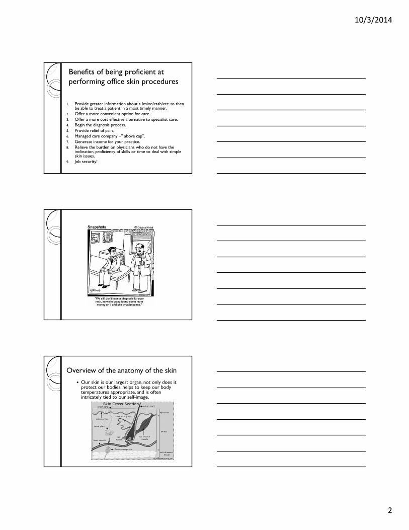
Benefits of being proficient at performing office skin procedures
1. Provide greater information about a lesion/rash/etc. to then be able to treat a patient in a most timely manner.
2. Offer a more convenient option for care.3. Offer a more cost effective alternative to specialist care.4. Begin the diagnosis process.5. Provide relief of pain. 6. Managed care company –” above cap”. 7. Generate income for your practice.8. Relieve the burden on physicians who do not have the
inclination, proficiency of skills or time to deal with simple skin issues.
9. Job security!
Overview of the anatomy of the skin
Our skin is our largest organ, not only does it protect our bodies, helps to keep our body temperatures appropriate, and is often intricately tied to our self-image.
10/3/2014
3
What can happen to skin?
Rashes Viral infections Bacterial infections Fungal infections Parasitic infections Pigmentation disorders Tumors/cancers Trauma Other conditions
Dermatologic Examination
Perform complete cutaneous exam including scalp, nails, between toes, etc.
Identify primary lesions Identify secondary lesions Identify pattern of cutaneous involvement Trunk vs. extremities Involvement of mucous membranes Involvement of palms/soles DOCUMENT
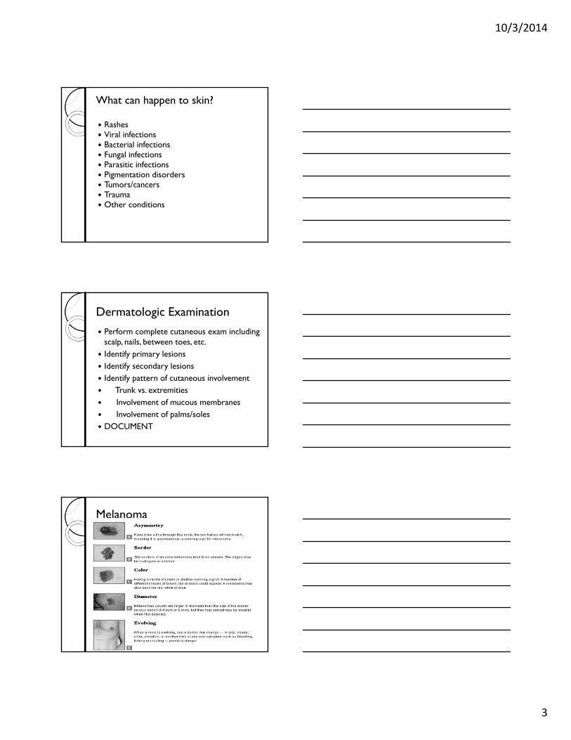
Melanoma
10/3/2014
4
What is it that you see?
Skin lesion guide
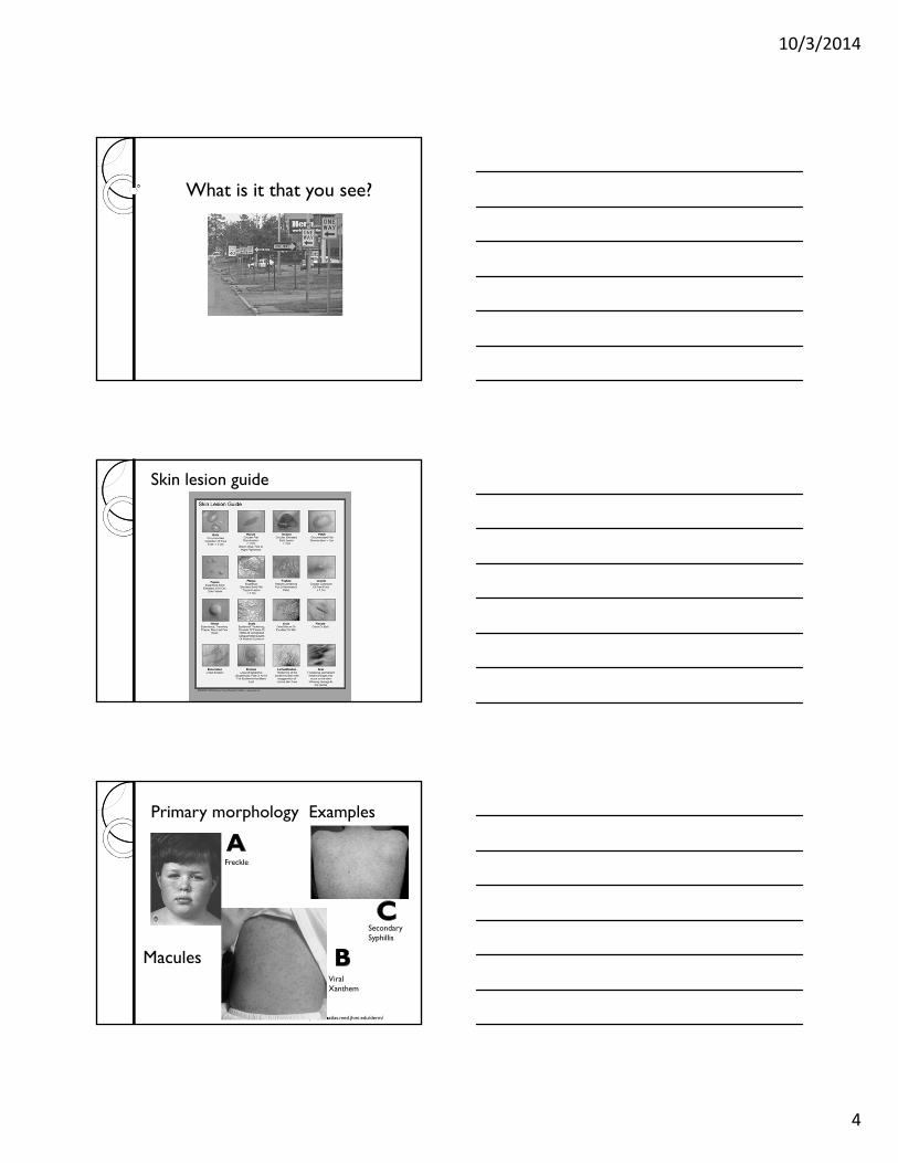
Primary morphology Examples
http://dermatlas.med.jhmi.edu/derm/
Freckle
Viral Xanthem
Secondary Syphillis
Macules
10/3/2014
5
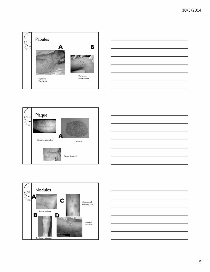
Papules
Erythema Multiforme
Molluscum contagiousum
Plaque
Granuloma AnnularePsoriasis
Atopic dermatitis
Nodules
Sarcoid nodules
Erythema noduosum
Cutaneous T cell lymphoma
Pruirigo nodularis
10/3/2014
6
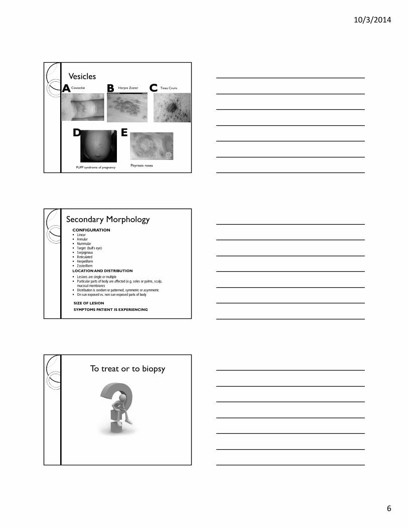
VesiclesCoxsackie Herpes Zoster Tinea Cruris
PUPP syndrome of pregnancy Pityriasis rosea
Secondary MorphologyCONFIGURATION Linear Annular Nummular Target (bull’s eye) Serpiginous Reticulated Herpetiform ZosteriformLOCATION AND DISTRIBUTION
Lesions are single or multiple Particular parts of body are affected (e.g. soles or palms, scalp,
mucosal membranes Distribution is random or patterned, symmetric or asymmetric On sun exposed vs. non sun exposed parts of body
SIZE OF LESION
SYMPTOMS PATIENT IS EXPERIENCING
To treat or to biopsy
10/3/2014
7
Basic Steps
Informed consent Anesthesia Prep area Remove lesion Control bleeding Close wound Post procedure
instructions
Skin biopsy: consideration
Biopsy does not substitute for good clinical skills
Biopsy promptly Risks by patient, location
o Bleedingo Infectiono Nerve damageo Scar formationo Damage to underlying structureso Failure to make diagnosiso Lesion recurrenceo Pain
Patrick C. Alguire, MDmand Bbarbara Mathes MD Skin Biopsy Techniqeus for the Internist J. Gen Intern Med 1998 January 13 (1) 46-54
Skin Biopsy: Indications
DiagnosisAll suspected neoplastic lesions
All bullous disordersTo clarify a diagnosis when a limited number of
entities under consideration
• TherapyRemoval of irritating, benign lesion
Removal of precancerous or cancerous lesion

10/3/2014
8
Skin Biopsy: Contraindications
Relative o Lesion of face (especially eyelids, nose)o Use of anticoagulant ?
o Bleeding disordero Infection at the site
Absoluteo Pigmented lesion should never be shaved
How can you determine what the rash is? Depends upon your comfort level What are the treatment options? What tools do you have to assist you??
- Punch Biopsy
- scraping /KOH
Skin Biopsy: Site Selection
Non-bullous lesions Choose lesions with most advanced inflammatory change
1-4 mm lesions: biopsy center
> 4 mm biopsy edge, thickest part, or most discolored
Bullous lesions Choose lesions 24-48 hrs. old (more specific pathology)
Avoid lesions with secondary changes
Attempt to remove entire vesicles
Biopsy bullae at their edge
10/3/2014
9
Skin biopsy : what site?
Areas to avoid if possibleo Cosmetically important areaso Deltoid and chest (hypertrophic scarring area)
o Skin over tibia (delayed healing esp. in diabetics)o Areas where incidence of secondary infection is higher
– groin, axilla
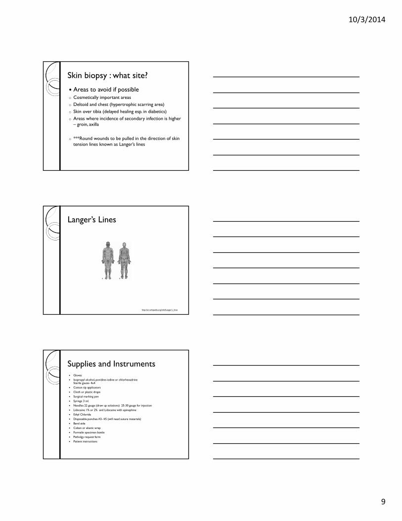
o ***Round wounds to be pulled in the direction of skin tension lines known as Langer’s lines
Langer’s Lines
http://en.wikipedia.org/wiki/Langer's_lines
Supplies and Instruments Gloves
Isopropyl alcohol, povidine-iodine or chlorhexadrineSterile gauze- 4x4
Cotton tip applicators
Cloth or plastic drape
Surgical marking pen
Syringe 3 ml.
Needles 22 gauge (draw up solutions) 25-30 gauge for injection
Lidocaine 1% or 2% and Lidocaine with epinephine
Ethyl Chloride
Disposable punches #3- #5 (will need suture materials)
Band aide
Coban or elastic wrap
Formalin specimen bottle
Patholgy request form
Patient instructions
10/3/2014
10
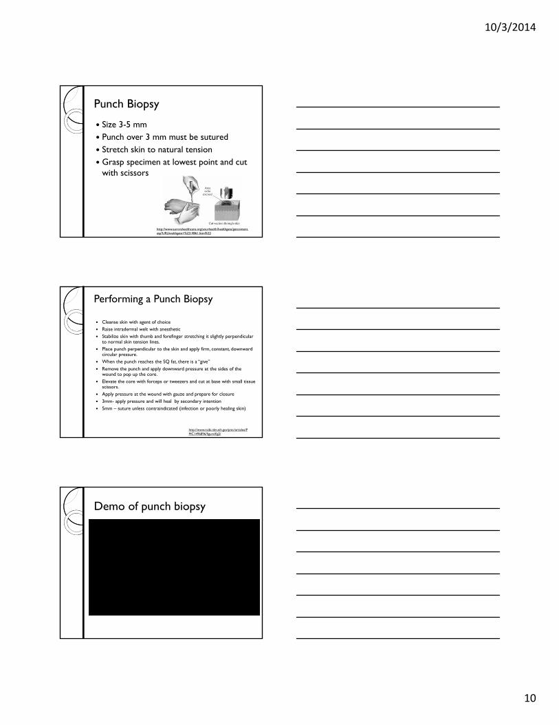
Punch Biopsy
Size 3-5 mm Punch over 3 mm must be sutured Stretch skin to natural tension Grasp specimen at lowest point and cut
with scissors
http://www.aurorahealthcare.org/yourhealth/healthgate/getcontent.asp?URLhealthgate=%2214861.html%22
Performing a Punch Biopsy
Cleanse skin with agent of choice Raise intradermal welt with anesthetic Stabilize skin with thumb and forefinger stretching it slightly perpendicular
to normal skin tension lines. Place punch perpendicular to the skin and apply firm, constant, downward
circular pressure. When the punch reaches the SQ fat, there is a “give” Remove the punch and apply downward pressure at the sides of the
wound to pop up the core. Elevate the core with forceps or tweezers and cut at base with small tissue
scissors. Apply pressure at the wound with gauze and prepare for closure 3mm- apply pressure and will heal by secondary intention 5mm – suture unless contraindicated (infection or poorly healing skin)
http://www.ncibi.nlm.nih.gov/pmc/articles/PMC1496896/figure/fig2/
Demo of punch biopsy
10/3/2014
11
Shave Biopsy DO NOT use for pigmented lesions, if an unsuspected melanoma is
removed, it cannot be properly staged.
A depressed scar, the size of the initial lesion is likely to occur
Quick, do not require sutures
Consider for seborrheic or acinitic keratoses, skin tags, warts, superficial basal or squamos carcinomas.
Raise the lesion with a wheal of injected anesthetic.
Lesion is stabilized at base with tweezers or toothed forceps, then cut at base.
Stop bleeding, electric cautery, silver nitrate, Monsel’s
Provide instructions for post procedure care
Innovative Shave Bx Technique
Skin biopsy- excision Assure you have all descriptors of the lesion
Decide on direction of incision, mark where you are going to incise or have a firm picture in your mind
Cleanse area, shave if necessary
Use topical anesthesia spray if needed, otherwise proceed with injection
Incise from apex to apex, then incise again deeper and at angle towards center of lesion.
Carefully raise lesion and cut away from skin.
Remove and place in formalin preservative container for lab.
Apply pressure to limit bleeding, assure skin will approximate, apply benzoine, steri strips, and tegaderm.
10/3/2014
12
Eliptical Excision
Sending specimen to pathology
Make sure your specimens are clearly labeled with patient name, date, and location
If multiple specimens label with LETTER and location
Letter and location should correspond to what is on requisition
Include lesion or rash, characteristics of each.
Rash- areas of body involved, pruritic?, duration, primary and secondary morphology, how sample was obtained
Lesion – location, size, characteristics, how lesion was obtained (punch, shave, excision, etc.)
Acrochordon Skin tags ROOT OF THE WORD Acquired. Look like a small piece of soft, hanging skin. Harmless growths Not associated with any major medical conditions Increased weight, heredity, large breasts Typical location: base of neck, underarms, eyelids, groin
folds, and under breasts May be as small as a flattened pinhead size bump, but can
be as large as a large grape or even a fig (5 cm) Invariably benign Do not become cancerous if left untreated. Can mimic seborrhic keratosis, moles, warts, cysts, milia,
neurobifromas and nevus lipomatosus. Skin tags may become irritated and red from bleeding or black
from twisting.
10/3/2014
13
Skin tags continuedWays to remove skin
tags Tie off at narrow base
with a piece of dental floss or string.
Freeze tag with liquid nitrogen
Burn tag using electric cautery
Remove tag with scissors, with or without anesthesia
Furuncles, CarbunclesandSebaceous cysts
One of the most rewarding “make someone quick” experiences in primary care!
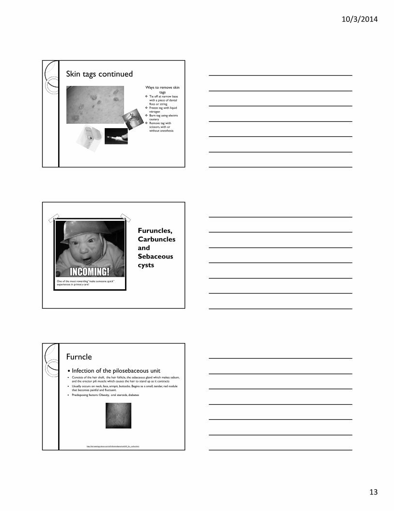
Furncle
Infection of the pilosebaceous unit Consists of the hair shaft, the hair follicle, the sebaceous gland which makes sebum,
and the erector pili muscle which causes the hair to stand up as it contracts
Usually occurs on neck, face, armpit, buttocks. Begins as a small, tender, red nodule that becomes painful and fluctuant.
Predisposing factors: Obesity, oral steroids, diabetes
http://dermatology.about.com/od/infectionbacteria/a/foll_fur_carbun.htm
10/3/2014
14
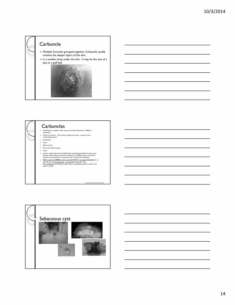
Carbuncle Multiple furuncles grouped together. Carbuncle usually
involves the deeper layers of the skin. Is a swollen lump under the skin. It may be the size of a
pea or a golf ball.
Carbuncles Assemble your supplies. Wear a gown and mask if desired (or if MRSA is
suspected)
Prepare the patient – offer options, explain procedure , reassure, assure comfortable position
Anesthetize
Incise
Obtain culture
Pack with iodoform gauze
Cover
Instruct patient that choices initially often made without benefit of culture, and therefore often default to choices covering for CA-MRSA. Culture of purulent material recommended for any patient who is treated with antibiotics.
Mild-moderate (MRSA, choice provide 80-92% coverage):TMP-SMX DS 1-2 pills PO twice daily, doxycycline or minocycline 100mg PO twice daily, clindamycin 300-450mg PO q8h--follow susceptibility profile of culture and adjust if needed.
http://www.freemd.com/carbuncle/treatment.htm
Sebaceous cyst
10/3/2014
15
How to incise and drain cysts Universal precautions Materials:Skin cleansing materials – alcohol, povidine-iodine or chlorhexadrineLocal anesthetic – 1% Lidocaine (epi if not a digit or single vessel blood supply)5-10 ml syringe 25 or 30 gauge needleScalpel (11 blade or 15 blade)Small curved hemostat Normal saline single use or with sterile bowl Large syringe with splash guard Swabs for bacterial cultureWound packing material Scissor Gauze – large amount 1-2 inchersTape Obtain informed consent – risk/benefit
Procedure
Take time to prepare your environment
Apply skin cleanser
Anesthetize top of wound
Incise the abscess
Disrupt loculations
Obtain culture
Identify any deep tracts that extend into surrounding tissue
Gently irrigate with normal saline
Pack wound
Cover with gauze and tape
ANTIBIOTICS?? (know your regional management guidelines)
I&D of Sebaceous cyst
10/3/2014
16
Excision of the actual cyst
Warts
How to tackle a wart?
Duct tape? Creams/topicals Cryoprocedure Shave/Cautery Destruction Bleomycin (refer to dermatologist or
podiatrist who does this in your area)
10/3/2014
17
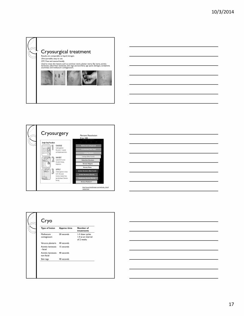
Cryosurgical treatment Results are comparable to liquid nitrogen. Ultra-portable, easy to use. CFC Free and ozone-friendly Used to treat skin lesions such as common warts, plantar warts, flat warts, acinitic keratoses, seborrheic karatoses, skin tags (acrocordon), age spots (lentigo), condyloma acuminata and molluscum contagiousum.
Cryosurgery
http://www.histofreezer.com/intl/why_histofreezer.html
Percent Resolution 0 to 100
CryoType of lesion Approx time Number of
treatments
Molloscumcontagiosum
20 seconds 1-3 thaw cycles 1-4 at an interval of 2 weeks
Verucca plantaris 40 seconds
Acinitic keratosis - facial
15 seconds
Acinitic keratosis-non facial
40 seconds
Skin tags 40 seconds
10/3/2014
18
Billing for Skin Procedures Diagnosis code= ICD 9 codes
Must be coded to highest level of specificity
078.10 Viral warts, unspecified
078.12 plantar wart 216.___ Benign neoplasm of skin of ____
E.g. 216.6 skin of upper limb including shoulder
238.2 Neoplasm of uncertain behavior of skin
701.9 Unspecified hypertrophic and atrophic conditions of skin
702.11 Inflammed seborrhiec keratosis 706.2 Sebacceous cyst
708.9 Unspecified Urticaria
CPT code Size Method Number
CPT codes 11200 Removal of skin tags, multiple fibrocutaneous tags, any area up to and
including 15 lesions
11201 Same as above each additional 10 lesions
113___ Shaving of epidermal or dermal lesion ….identify location and size
11301 Shaving of single lesion trunk, arms or legs, 0.5 cm or less
11307 Shaving of single lesion scalp, neck, hands, feet, genitalia; lesion 1.1-2.0cm
• 114___ Excision benign lesion
11401 Excision, including margins, trunk, arms, legs, .6-1.0cm
11406 Excision, including margins of trunk, arms , legs over 4.0 cm.
• 11000 Punch biopsy of one area
• 11101 Add for additional punch
• 17000 Cryo of one lesion, not a wart
17003 Cryo of non-wart lesions 2-14
Now that you have the report…
10/3/2014
19
Pathology Report
Read report carefully If further excision is recommended follow
through on this If malignant melanoma refer to plastic
surgeon immediately Consult if needed If you are not sure, collaborate and refer
if needed!