slp-76 (total) tr-fret assay kit
TRANSCRIPT
www.caymanchem.comCustomer Service 800.364.9897Technical Support 888.526.53511180 E. Ellsworth Rd · Ann Arbor, MI · USA
SLP-76 (Total) TR-FRET Assay Kit
Powered by Bioauxilium’s THUNDER™ TR-FRET Technology
Item No. 500223
3GENERAL INFORMATION
TABLE OF CONTENTS GENERAL INFORMATION 3 Materials Supplied
3 Safety Data4 Precautions4 Before You Start5 If You Have Problems5 Storage and Stability6 THUNDERTM General Information7 Materials Needed but Not Supplied
INTRODUCTION 8 Background8 About This Assay9 Principle Of This Assay
PRE-ASSAY PREPARATION 10 Assay Optimization11 Reagent Preparation14 TR-FRET Plate Reader Settings
ASSAY PROTOCOL 15 Workflow 16 Assay Summary 17 Performing the Assay: 2-plate (Transfer) Protocol
24 Performing the Assay: 1-plate (All in One Well) Protocol
ANALYSIS 29 Calculations30 Performance Characteristics
RESOURCES 32 Troubleshooting
34 References
35 Notes
35 Warranty and Limitation of Remedy
GENERAL INFORMATION
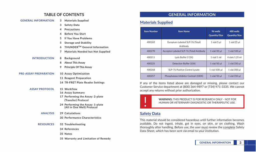
Materials Supplied
Item Number Item Name 96 wells Quantity/Size
480 wells Quantity/Size
400269 Europium-Labeled SLP-76 (Total)
Antibody
1 vial/5 µl 1 vial/25 µl
400270 Acceptor-Labeled SLP-76 (Total) Antibody 1 vial/20 µl 1 vial/100 µl
400211 Lysis Buffer 2 (5X) 1 vial/1 ml 4 vials/1.25 ml
400225 Detection Buffer (10X) 1 vial/50 µl 1 vial/250 µl
400268 SLP-76 Positive Control Lysate 1 vial/100 µl 1 vial/200 µl
400257 Phosphatase Inhibitor Cocktail (100X) 1 vial/50 µl 1 vial/250 µl
If any of the items listed above are damaged or missing, please contact our Customer Service department at (800) 364-9897 or (734) 971-3335. We cannot accept any returns without prior authorization.
! WARNING: THIS PRODUCT IS FOR RESEARCH ONLY - NOT FORHUMAN OR VETERINARY DIAGNOSTIC OR THERAPEUTIC USE.
Safety DataThis material should be considered hazardous until further information becomes available. Do not ingest, inhale, get in eyes, on skin, or on clothing. Wash thoroughly after handling. Before use, the user must review the complete Safety Data Sheet, which has been sent via email to your institution.

4 GENERAL INFORMATION 5GENERAL INFORMATION
PrecautionsPlease read these instructions carefully before beginning this assay.Do not mix or substitute reagents or materials from other kit lots or kits. Kits are quality control tested as a set of components and performance cannot be guaranteed if utilized separately or substituted.We cannot guarantee the performance of the product outside the conditions detailed in this kit booklet.The kits are designed for the detection of endogenous cellular proteins across a wide variety of cell lines. However, until each cell line in particular is tested, the possibility of the presence of undetectable levels of the target protein cannot be excluded.Users should ensure that their cell line has measurable levels of the target protein. Expression levels of signaling proteins in different cell types vary widely. The cell line used for the assay validation of this kit is shown in the figures starting on page 30.
Before You StartPlease note the following:ONLY white plates should be used for TR-FRET.DO NOT modify the assay protocol or volumes.DO optimize the cell density, serum starvation (optional), and stimulation or inhibition parameters.ALWAYS use the included positive control lysate for every assay.
If You Have ProblemsTechnical Service Contact Information
Phone: 888-526-5351 (USA and Canada only) or 734-975-3888Email: [email protected]
In order for our staff to assist you quickly and efficiently, please be ready to supply the lot number of the kit (found on the outside of the box).
Storage and StabilityThis kit will perform as specified if stored as directed at -80°C and used before the expiration date indicated on the outside of the box.
6 GENERAL INFORMATION 7GENERAL INFORMATION
THUNDERTM General InformationTHUNDER™ TR-FRET Cell Signaling Assay Kits are designed for the semi-quantitative measurement of phosphorylated and/or total (both phosphorylated and unphosphorylated) proteins in cell lysates using homogeneous (no wash) TR-FRET technology. The kits are compatible with both adherent and suspended cells.THUNDER™ TR-FRET Cell Signaling Assay Kits are based on Bioauxilium’s enhanced proprietary time-resolved Förster resonance energy transfer (TR-FRET) technology. THUNDER™ assays can be read on most commercially available TR-FRET-compatible plate readers (a list of suitable TR-FRET readers can be found at www.Bioauxilium.com). TR-FRET-based assays are homogeneous because they do not require any washing or separation steps. In addition, the THUNDER™ assays use a standardized, simple, and rapid “add-incubate-measure” protocol with a single step reagent addition. This streamlined assay protocol dramatically decreases hands-on time and provides a powerful alternative to cumbersome, error-prone and time-consuming techniques such as Western blot and ELISA.THUNDER™ TR-FRET Cell Signaling Assay Kits contain the essential reagents necessary to carry out the measurement of signaling proteins in cells, with the exception of the plate(s).
Materials Needed But Not Supplied1. A plate reader equipped with a TR-FRET option2. Adjustable pipettes; multichannel or repeating pipettor recommended3. A source of ultrapure water, with a resistivity of 18.2 MΩ·cm and total
organic carbon (TOC) levels of <10 ppb, is recommended. Pure water - glass-distilled or deionized - may not be acceptable. NOTE: UltraPure Water is available for purchase from Cayman (Item No. 400000).
4. Culture plate: 96-well clear, flat-bottom polystyrene tissue culture-treated plate(s) for culturing cells when using the 2-plate (transfer) assay protocol. NOTE: Do not use this type of plate for the 1-plate (all in one well) assay protocol.
5. Detection plate (96-well plate option): Half-area, 96-well white plate(s) for TR-FRET detection when using the 2-plate (transfer) assay protocol
6. Detection plate (384-well plate option): Low-volume, 384-well white plate(s) for TR-FRET detection when using the 1- or 2-plate assay protocols
7. Adhesive sealing film for plates8. Orbital microplate shaker

8 INTRODUCTION 9INTRODUCTION
Principle Of This AssayThis assay is based on the traditional sandwich immunoassay principle (Figure 1, below). Following cell treatment, cells are lysed with the specific lysis buffer provided in the kit. Then, the target protein in the cell lysates is detected with a pair of fluorophore-labeled antibodies reactive to human samples.
Eu-Total Ab FR-Total Ab
EuropiumEmission 615 nm
TR-FRETEmission 665 nm
Excitation320-340 nm
FRET
PROTEIN
Figure 1. Schematic of the TR-FRET cell signaling assay principle
The first antibody is labeled with a long-lifetime donor fluorophore (a europium chelate; Eu-Total Ab) and the second with a far-red acceptor fluorophore (FR-Total Ab). The binding of the two labeled antibodies to distinct epitopes on the target protein takes place in solution and brings the two dyes into close proximity. Excitation of the donor Eu chelate molecules with a flash lamp (320 or 340 nm) or laser (337 nm) triggers a FRET from the donor to the acceptor molecules, which, in turn, emit a TR-FRET signal at 665 nm. The signal at 665 nm is proportional to the concentration of target protein in the cell lysate. Residual energy from the Eu chelate generates light at 615 nm, which can be used as an internal standard to normalize light emitted at 665 nm.TR-FRET assays exhibit very low background fluorescence levels and high signal-to-background (S/B) ratios. The data can be expressed and analyzed as either the signal at 665 nm or the 665 nm/615 nm ratio. The ratiometric measurement further increases assay reproducibility and robustness.
INTRODUCTION
Background SLP-76 is an adapter protein and a member of the SLP-76 family of adapters that has a role in regulating the immune response downstream of T cell receptors.1 It is composed of an N-terminal region containing three tyrosine phosphorylation motifs, a proline-rich region, and a C-terminal SH2 domain.1,2 SLP-76 is expressed in hematopoietic cells and localized to the cytoplasm but can be translocated to the plasma membrane at the T cell-to-antigen presenting cell (APC) junction following T cell receptor activation and SLP-76 interaction with certain molecules.1 SLP-76 is phosphorylated by the Syk family kinase ZAP-70 in the N-terminal region, which induces interactions with a variety of proteins, including the Tec family kinase Itk. It also associates with the adapter protein GADS and PLCγ1 via the proline-rich region and the adapter protein ADAP and hematopoietic progenitor kinase 1 (HPK1) via the SH2 domain. Phosphorylation of SLP-76 at serine 376 (Ser376) in the SH2 domain by HPK1 induces an interaction with 14-3-3 adapter proteins, followed by ubiquitination and proteolytic degradation of SLP-76, which provides negative feedback to T cell receptor signaling.1,2 Mutations in LCP2, the gene encoding SLP-76, resulting in a loss of SLP-76 protein expression are associated with T and B cell immunodeficiency, impaired platelet aggregation, and severe neutrophil defects.3 SLP-76 is expressed ectopically in human chronic lymphocytic leukemia (CLL) cells, where it mediates B cell signaling, and protein levels of SLP-76 are positively associated with the rate of disease progression.4
About This AssayThis SLP-76 (Total) TR-FRET Assay Kit uses a homogeneous TR-FRET assay method amenable to rapid measurement of total protein levels in cells. This SLP-76 (Total) TR-FRET Assay Kit is suitable for screening a large number of samples. The signal is stable at room temperature for at least 24 hours, affording flexibility in read times. The amount of reagents provided is sufficient for testing either 96 or 480 total protein wells, depending on the size of the kit.
10 PRE-ASSAY PREPARATION 11PRE-ASSAY PREPARATION
PRE-ASSAY PREPARATION
Assay OptimizationA critical step in performing any cell-based assay is the optimization of cell culture and treatment conditions. The following protocol assumes that both the cell number and treatment conditions have been previously optimized, as these key parameters often vary for each cell line. It is, therefore, strongly recommended to optimize these parameters in order to maximize the assay signal and ensure optimum performance with a high S/B ratio.
Cell number, serum starvation (optional), and stimulation or inhibition time (at either room temperature or 37°C) should be optimized for each cell line and target protein. Cell numbers that are too high or too low can negatively influence the activation of intracellular signaling pathways. Cell seeding densities of 40,000-80,000 cells/well for adherent cells or 100,000-200,000 cells/well for suspended cells are generally acceptable for most cell lines. Of note, the optimal length of time for stimulation can vary widely among cell lines from a few minutes to more than one hour. As such, a time-course study is strongly recommended to determine the optimal stimulation time, ideally at both room temperature and 37°C, since incubation temperature has an effect on the kinetics of target protein stimulation. Additional assay development guidelines are available on Bioauxilium’s website (www.Bioauxilium.com).
Reagent PreparationThe instructions described below are for testing the entire number of wells in each kit. Adjust volumes accordingly when testing fewer wells.Bring all reagents to room temperature prior to use.Centrifuge all tubes before use to improve recovery of content (2,000 x g, 10-15 seconds). Mix the lysis and detection buffers and the positive control lysate by vortexing gently before use. Do NOT vortex the antibodies.Use ultrapure water (18 MΩ·cm) to dilute the lysis and detection buffers.NOTE: It is recommended to test all samples and controls at least in duplicate.NOTE: ALWAYS include a positive control using the positive control lysate provided.1. Supplemented Lysis Buffer
Supplemented Lysis Buffer 2 (1X) for the 2-Plate (Transfer) Assay Protocol with Adherent Cells: The supplemented Lysis Buffer 2 (1X) is designed for use in the 2-plate (transfer) assay protocol using adherent cells (see page 17). Each well requires 50 µl of supplemented Lysis Buffer 2 (1X). Dilute the Lysis Buffer 2 (5X) (Item No. 400211) with ultrapure water and add the Phosphatase Inhibitor Cocktail (100X) (Item No. 400257), which contains sodium fluoride (NaF), sodium orthovanadate (Na3VO4), and glycerophosphate at 100, 200, and 200 mM, respectively, to final NaF, Na3VO4, and glycerophosphate concentrations of 1, 2, and 2 mM, respectively. NOTE: It is mandatory to supplement Lysis Buffer 2 with the Phosphatase Inhibitor Cocktail (100X). Store unused Lysis Buffer 2 (1X) at 4°C; it will be stable for approximately two days.
OR
12 PRE-ASSAY PREPARATION 13PRE-ASSAY PREPARATION
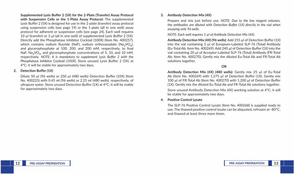
Supplemented Lysis Buffer 2 (5X) for the 2-Plate (Transfer) Assay Protocol with Suspension Cells or the 1-Plate Assay Protocol: The supplemented Lysis Buffer 2 (5X) is designed for use in the 2-plate (transfer) assay protocol using suspension cells (see page 19) or the 1-plate (all in one well) assay protocol for adherent or suspension cells (see page 24). Each well requires 10 µl (transfer) or 3 µl (all in one well) of supplemented Lysis Buffer 2 (5X). Directly add the Phosphatase Inhibitor Cocktail (100X) (Item No. 400257), which contains sodium fluoride (NaF), sodium orthovanadate (Na3VO4), and glycerophosphate at 100, 200, and 200 mM, respectively, to final NaF, Na3VO4, and glycerophosphate concentrations of 5, 10, and 10 mM, respectively. NOTE: It is mandatory to supplement Lysis Buffer 2 with the Phosphatase Inhibitor Cocktail (100X). Store unused Lysis Buffer 2 (5X) at 4°C; it will be stable for approximately two days.
2. Detection Buffer (1X)Dilute 50 µl (96 wells) or 250 µl (480 wells) Detection Buffer (10X) (Item No. 400225) with 0.45 ml (96 wells) or 2.25 ml (480 wells), respectively, of ultrapure water. Store unused Detection Buffer (1X) at 4°C; it will be stable for approximately two days.
3. Antibody Detection Mix (4X)Prepare and mix just before use. NOTE: Due to the low reagent volumes, the antibodies are diluted with Detection Buffer (1X) directly in the vial when assaying only 96 wells.NOTE: Each well requires 5 µl of Antibody Detection Mix (4X).Antibody Detection Mix (4X) (96 wells): Add 255 µl of Detection Buffer (1X) into the vial containing 5 µl of Europium-Labeled SLP-76 (Total) Antibody (Eu-Total Ab; Item No. 400269). Add 240 µl of Detection Buffer (1X) into the vial containing 20 µl of Acceptor-Labeled SLP-76 (Total) Antibody (FR-Total Ab; Item No. 400270). Gently mix the diluted Eu-Total Ab and FR-Total Ab solutions together.
ORAntibody Detection Mix (4X) (480 wells): Gently mix 25 µl of Eu-Total Ab (Item No. 400269) with 1,275 µl of Detection Buffer (1X). Gently mix 100 µl of FR-Total Ab (Item No. 400270) with 1,200 µl of Detection Buffer (1X). Gently mix the diluted Eu-Total Ab and FR-Total Ab solutions together. Store unused Antibody Detection Mix (4X) working solution at 4°C; it will be stable for approximately two days.
4. Positive Control LysateThe SLP-76 Positive Control Lysate (Item No. 400268) is supplied ready to use. The thawed positive control lysate can be aliquoted, refrozen at -80°C, and thawed at least three more times.

15ASSAY PROTOCOL14 PRE-ASSAY PREPARATION
TR-FRET Plate Reader Settings We recommend reading the TR-FRET assays at two wavelengths, detecting both the emission from the Eu chelate donor fluorophore at 615 nm and the acceptor fluorophore at 665 nm. Table 1, below, provides instrument settings to be used as guidelines.
TR-FRET-compatible Plate Reader
Parameter Flash lamp excitation Laser excitation
Excitation filter 320 nm (or 340 nm) N/A
Emission filter 615 nm (or 620 nm) 615 nm (or 620 nm)
Delay time 90 µs 50 µs
Flash energy level 100% or High 100%
Number of flashes 100 20
Window (integration time) 300 µs 100 µs
Table 1. Recommended TR-FRET plate reader settings
ASSAY PROTOCOL
WorkflowThe THUNDER™ TR-FRET Cell Signaling Assay workflow consists of 3 simple steps.
CELLTREATMENT5 MINUTES TO OVERNIGHT
· Seed cells
· Treat cells (stimulator and/or inhibitor)
STEP 1PROTEIN
DETECTION60 MINUTES TO OVERNIGHT
· Transfer to detection plate, if applicable
· Add antibody mix and incubate
· Read plate (TR-FRET)
STEP 3STEP 2CELLLYSIS
15 TO 60 MINUTES
· Lyse cells with the specific kit’s lysis buffer
Figure 2. Assay workflow
16 ASSAY PROTOCOL 17ASSAY PROTOCOL
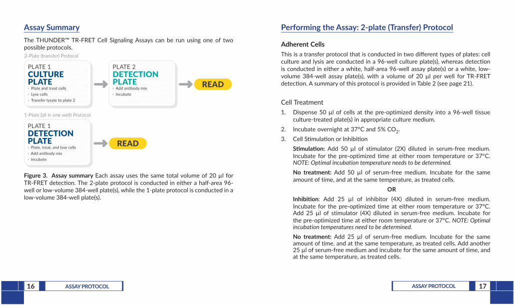
Assay SummaryThe THUNDER™ TR-FRET Cell Signaling Assays can be run using one of two possible protocols.
PLATE 1CULTUREPLATE· Plate and treat cells· Lyse cells· Transfer lysate to plate 2
PLATE 2DETECTION PLATE· Add antibody mix· Incubate
READ
2-Plate (transfer) Protocol
PLATE 1DETECTION PLATE · Plate, treat, and lyse cells· Add antibody mix· Incubate
READ
1-Plate (all in one well) Protocol
Figure 3. Assay summary Each assay uses the same total volume of 20 µl for TR-FRET detection. The 2-plate protocol is conducted in either a half-area 96-well or low-volume 384-well plate(s), while the 1-plate protocol is conducted in a low-volume 384-well plate(s).
Performing the Assay: 2-plate (Transfer) Protocol
Adherent CellsThis is a transfer protocol that is conducted in two different types of plates: cell culture and lysis are conducted in a 96-well culture plate(s), whereas detection is conducted in either a white, half-area 96-well assay plate(s) or a white, low-volume 384-well assay plate(s), with a volume of 20 µl per well for TR-FRET detection. A summary of this protocol is provided in Table 2 (see page 21).
Cell Treatment1. Dispense 50 µl of cells at the pre-optimized density into a 96-well tissue
culture-treated plate(s) in appropriate culture medium.2. Incubate overnight at 37°C and 5% CO2.3. Cell Stimulation or Inhibition
Stimulation: Add 50 µl of stimulator (2X) diluted in serum-free medium. Incubate for the pre-optimized time at either room temperature or 37°C. NOTE: Optimal incubation temperature needs to be determined.No treatment: Add 50 µl of serum-free medium. Incubate for the same amount of time, and at the same temperature, as treated cells.
ORInhibition: Add 25 µl of inhibitor (4X) diluted in serum-free medium. Incubate for the pre-optimized time at either room temperature or 37°C. Add 25 µl of stimulator (4X) diluted in serum-free medium. Incubate for the pre-optimized time at either room temperature or 37°C. NOTE: Optimal incubation temperatures need to be determined.No treatment: Add 25 µl of serum-free medium. Incubate for the same amount of time, and at the same temperature, as treated cells. Add another 25 µl of serum-free medium and incubate for the same amount of time, and at the same temperature, as treated cells.
18 ASSAY PROTOCOL 19ASSAY PROTOCOL
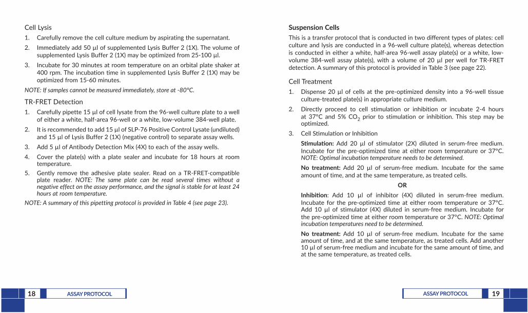
Cell Lysis1. Carefully remove the cell culture medium by aspirating the supernatant.2. Immediately add 50 µl of supplemented Lysis Buffer 2 (1X). The volume of
supplemented Lysis Buffer 2 (1X) may be optimized from 25-100 µl.3. Incubate for 30 minutes at room temperature on an orbital plate shaker at
400 rpm. The incubation time in supplemented Lysis Buffer 2 (1X) may be optimized from 15-60 minutes.
NOTE: If samples cannot be measured immediately, store at -80°C.
TR-FRET Detection1. Carefully pipette 15 µl of cell lysate from the 96-well culture plate to a well
of either a white, half-area 96-well or a white, low-volume 384-well plate.2. It is recommended to add 15 µl of SLP-76 Positive Control Lysate (undiluted)
and 15 µl of Lysis Buffer 2 (1X) (negative control) to separate assay wells.3. Add 5 µl of Antibody Detection Mix (4X) to each of the assay wells.4. Cover the plate(s) with a plate sealer and incubate for 18 hours at room
temperature. 5. Gently remove the adhesive plate sealer. Read on a TR-FRET-compatible
plate reader. NOTE: The same plate can be read several times without a negative effect on the assay performance, and the signal is stable for at least 24 hours at room temperature.
NOTE: A summary of this pipetting protocol is provided in Table 4 (see page 23).
Suspension CellsThis is a transfer protocol that is conducted in two different types of plates: cell culture and lysis are conducted in a 96-well culture plate(s), whereas detection is conducted in either a white, half-area 96-well assay plate(s) or a white, low-volume 384-well assay plate(s), with a volume of 20 µl per well for TR-FRET detection. A summary of this protocol is provided in Table 3 (see page 22).
Cell Treatment1. Dispense 20 µl of cells at the pre-optimized density into a 96-well tissue
culture-treated plate(s) in appropriate culture medium.2. Directly proceed to cell stimulation or inhibition or incubate 2-4 hours
at 37°C and 5% CO2 prior to stimulation or inhibition. This step may be optimized.
3. Cell Stimulation or InhibitionStimulation: Add 20 µl of stimulator (2X) diluted in serum-free medium. Incubate for the pre-optimized time at either room temperature or 37°C. NOTE: Optimal incubation temperature needs to be determined.No treatment: Add 20 µl of serum-free medium. Incubate for the same amount of time, and at the same temperature, as treated cells.
ORInhibition: Add 10 µl of inhibitor (4X) diluted in serum-free medium. Incubate for the pre-optimized time at either room temperature or 37°C. Add 10 µl of stimulator (4X) diluted in serum-free medium. Incubate for the pre-optimized time at either room temperature or 37°C. NOTE: Optimal incubation temperatures need to be determined.No treatment: Add 10 µl of serum-free medium. Incubate for the same amount of time, and at the same temperature, as treated cells. Add another 10 µl of serum-free medium and incubate for the same amount of time, and at the same temperature, as treated cells.

20 ASSAY PROTOCOL 21ASSAY PROTOCOL
Cell Lysis1. Add 10 µl supplemented Lysis Buffer 2 (5X).2. Incubate for 30 minutes at room temperature on an orbital plate shaker at
400 rpm. The incubation time in supplemented Lysis Buffer 2 (5X) may be optimized from 15-60 minutes.
NOTE: If samples cannot be measured immediately, store at -80°C.
TR-FRET DetectionFollowing cell lysis, proceed to the TR-FRET detection step as described for the standard 2-plate (transfer) protocol for adherent cells (see page 18).
2-Plate (Transfer) Assay Summary
Step Adherent Cells
Cell Treatment Stimulation No Treatment Inhibition No Treatment
50 µl cells 50 µl cells 50 µl cells 50 µl cells
Incubate cells overnight
50 µl stimulator
(2X)
50 µl serum-free
medium
25 µl inhibitor
(4X)25 µl serum-free
medium
Incubate for pre-optimized time
25 µl stimulator
(4X)
25 µl serum-free
medium
Incubate for pre-optimized time
Cell Lysis Remove media
50 µl supplemented Lysis Buffer 2 (1X)*
Incubate 30 minutes on an orbital shaker
Protein Detection 15 µl lysate
5 µl Antibody Detection Mix (4X)
Cover and incubate 18 hours
Read TR-FRET signal
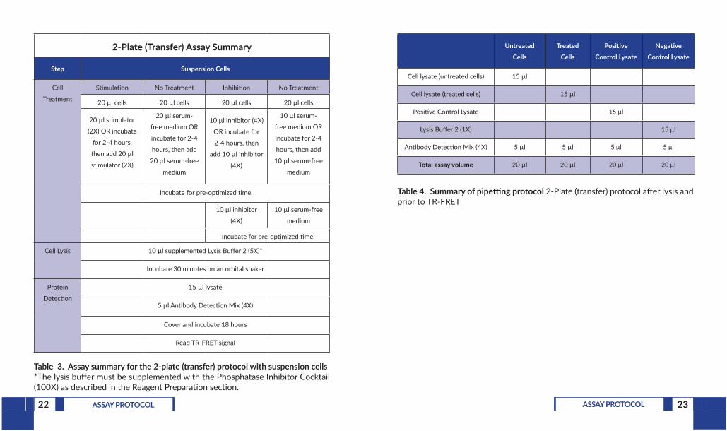
Table 2. Assay summary for the 2-plate (transfer) protocol with adherent cells*The lysis buffer must be supplemented with the Phosphatase Inhibitor Cocktail (100X) as described in the Reagent Preparation section.
22 ASSAY PROTOCOL 23ASSAY PROTOCOL
Untreated Cells
Treated Cells
Positive Control Lysate
Negative Control Lysate
Cell lysate (untreated cells) 15 µl
Cell lysate (treated cells) 15 µl
Positive Control Lysate 15 µl
Lysis Buffer 2 (1X) 15 µl
Antibody Detection Mix (4X) 5 µl 5 µl 5 µl 5 µl
Total assay volume 20 µl 20 µl 20 µl 20 µl
Table 4. Summary of pipetting protocol 2-Plate (transfer) protocol after lysis and prior to TR-FRET
2-Plate (Transfer) Assay Summary
Step Suspension Cells
Cell
Treatment
Stimulation No Treatment Inhibition No Treatment
20 µl cells 20 µl cells 20 µl cells 20 µl cells
20 µl stimulator
(2X) OR incubate
for 2-4 hours,
then add 20 µl
stimulator (2X)
20 µl serum-
free medium OR
incubate for 2-4
hours, then add
20 µl serum-free
medium
10 µl inhibitor (4X)
OR incubate for
2-4 hours, then
add 10 µl inhibitor
(4X)
10 µl serum-
free medium OR
incubate for 2-4
hours, then add
10 µl serum-free
medium
Incubate for pre-optimized time
10 µl inhibitor
(4X)
10 µl serum-free
medium
Incubate for pre-optimized time
Cell Lysis 10 µl supplemented Lysis Buffer 2 (5X)*
Incubate 30 minutes on an orbital shaker
Protein
Detection
15 µl lysate
5 µl Antibody Detection Mix (4X)
Cover and incubate 18 hours
Read TR-FRET signal
Table 3. Assay summary for the 2-plate (transfer) protocol with suspension cells*The lysis buffer must be supplemented with the Phosphatase Inhibitor Cocktail (100X) as described in the Reagent Preparation section.

24 ASSAY PROTOCOL 25ASSAY PROTOCOL
Performing the Assay: 1-plate (All in One Well) Protocol
Adherent and Suspension CellsThis is an all-in-one-well protocol. No transfer step is needed. Conduct the assay in a white, low-volume 384-well assay plate(s) with a total assay volume of 20 µl per well. A summary of this protocol is provided in Tables 5-7 (see pages 26-28).
Cell Treatment1. Dispense 8 µl of cells at the pre-optimized density in serum-free medium
into a white, low-volume 384-well assay plate(s).2. Cell Stimulation or Inhibition
Stimulation: Add 4 µl of stimulator (3X) diluted in serum-free medium. Incubate for the pre-optimized time at either room temperature or 37°C. NOTE: Optimal incubation temperature needs to be determined.No treatment: Add 4 µl of serum-free medium. Incubate for the same amount of time, and at the same temperature, as treated cells.
ORInhibition: Add 2 µl of inhibitor (6X) diluted in serum-free medium. Incubate for the pre-optimized time at either room temperature or 37°C. Add 2 µl of stimulator (6X) diluted in serum-free medium. Incubate for the pre-optimized time at either room temperature or 37°C. NOTE: Optimal incubation temperatures need to be determined.No treatment: Add 2 µl of serum-free medium. Incubate for the same amount of time, and at the same temperature, as treated cells. Add another 2 µl of serum-free medium and incubate for the same amount of time, and at the same temperature, as treated cells.
Cell Lysis1. Add 3 µl of supplemented Lysis Buffer 2 (5X).2. Incubate for 30 minutes at room temperature on an orbital plate shaker at
400 rpm. The incubation time in supplemented Lysis Buffer 2 (5X) may be optimized from 15-60 minutes.
NOTE: If samples cannot be measured immediately, store at -80°C.
TR-FRET Detection1. Add 15 µl of SLP-76 Positive Control Lysate (undiluted) and 15 µl of Lysis
Buffer 2 (1X) (negative control) to separate assay wells.2. Add 5 µl of Antibody Detection Mix (4X) prepared in Detection Buffer (1X)
to each of the assay wells.3. Cover the plate(s) with a plate sealer and incubate for 18 hours at room
temperature.4. Gently remove the adhesive plate sealer. Read on a TR-FRET-compatible
plate reader. NOTE: The same plate can be read several times without a negative effect on the assay performance, and the signal is stable for at least 24 hours at room temperature.
26 ASSAY PROTOCOL 27ASSAY PROTOCOL
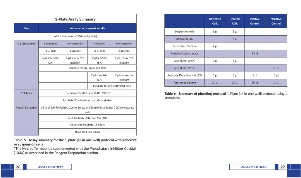
1-Plate Assay Summary
Step Adherent or suspension cells
White, low-volume 384-well plate(s)
Cell Treatment Stimulation No treatment Inhibition No treatment
8 µl cells 8 µl cells 8 µl cells 8 µl cells
4 µl stimulator
(3X)
4 µl serum-free
medium
2 µl inhibitor
(6X)
2 µl serum-free
medium
Incubate for pre-optimized time
2 µl stimulator
(6X)
2 µl serum-free
medium
Incubate for pre-optimized time
Cell Lysis 3 µl supplemented Lysis Buffer 2 (5X)*
Incubate 30 minutes on an orbital shaker
Protein Detection 15 µl of SLP-76 Positive Control Lysate and 15 µl of Lysis Buffer 2 (1X) to separate
wells
5 µl Antibody Detection Mix (4X)
Cover and incubate 18 hours
Read TR-FRET signal
Table 5. Assay summary for the 1-plate (all in one well) protocol with adherent or suspension cells*The lysis buffer must be supplemented with the Phosphatase Inhibitor Cocktail (100X) as described in the Reagent Preparation section.
Untreated Cells
Treated Cells
Positive Control
Negative Control
Suspension cells 8 µl 8 µl
Stimulator (3X) 4 µl
Serum-free Medium 4 µl
Positive Control Lysate 15 µl
Lysis Buffer 2 (5X) 3 µl 3 µl
Lysis Buffer 2 (1X) 15 µl
Antibody Detection Mix (4X) 5 µl 5 µl 5 µl 5 µl
Total assay volume 20 µl 20 µl 20 µl 20 µl
Table 6. Summary of pipetting protocol 1-Plate (all in one well) protocol using a stimulator

29ANALYSIS28 ASSAY PROTOCOL
Untreated Cells
Treated Cells
Positive Control
Negative Control
Suspension cells 8 µl 8 µl
Inhibitor (6X) 2 µl
Stimulator (6X) 2 µl
Serum-free Medium 4 µl
Positive Control Lysate 15 µl
Lysis Buffer 2 (5X) 3 µl 3 µl
Lysis Buffer 2 (1X) 15 µl
Antibody Detection Mix (4X) 5 µl 5 µl 5 µl 5 µl
Total assay volume 20 µl 20 µl 20 µl 20 µl
Table 7. Summary of pipetting protocol 1-Plate (all in one well) protocol using an inhibitor
ANALYSIS
Calculations1. TR-FRET data are typically calculated and presented ratiometrically using
the following formula:
[(665 nm/615 nm) x 1,000]
2. Calculate the TR-FRET ratio for each well.3. TR-FRET assays are homogeneous; do not subtract average negative control
data (no lysate) from any other well readings.4. For concentration-response curves, analyze data according to a
nonlinear regression using the four-parameter logistic equation (sigmodal dose-response curve with variable slope) and a 1/Y2 data weighting.
5. Assay quality control: The undiluted SLP-76 Positive Control Lysate must generate an S/B ratio of at least 2 when compared to the negative control (Lysis Buffer 2 (1X) only). If this is not the case, your reader is not compatible with THUNDER™ TR-FRET Cell Signaling Assay Kits.
NOTE: The positive control lysate is provided as a control reagent, not for conducting a standard curve.

30 ANALYSIS 31ANALYSIS
Performance Characteristics
Representative DataData shown here are examples of data typically generated with the SLP-76 (Total) TR-FRET Assay Kit. The TR-FRET signal was recorded at 665 and 615 nm (EnVision®; lamp excitation) using the recommended plate reader settings. Note that both the TR-FRET ratios and S/B ratios will vary from one TR-FRET-compatible reader to another. In addition, note that excitation with a laser (337 nm) generates higher counts and, usually, higher S/B ratios.
Log [H2O2] (M)
-5-∞ -4 -3 -2 -1
EC50 = 3 mM
S/B = 6.3
TR
-FR
ET
Rat
io(6
65/6
15 X
1,0
00)
70
60
50
40
30
20
10
0
Total
Phospho
Figure 4. Stimulation of SLP-76 phosphorylation at Ser376 in Jurkat cells Jurkat cells were seeded at 400,000 cells/well in triplicate and incubated with serial dilutions of H2O2 for 15 minutes at RT.
Dilution S/B
Not Diluted 4.9
1:2 4.8
1:4 4.1
1:8 3.3
1:16 2.5
SLP-76 (Total)
ND 1:2 1:4 1:8 1:16 Buffer
Dilutions
TR
-FR
ET
Rat
io(6
65/6
15 X
1,0
00)
40
30
20
10
0
Figure 5. Jurkat control cell lysate titration (QC Test) for phospho-Ser376 SLP-76 and total SLP-76 The SLP-76 (Total) and SLP-76 (Phospho-Ser376) TR-FRET Assay Kit is routinely quality control tested using Jurkat cell lysates treated with H2O2. Jurkat cells were cultured in a T175 flask, centrifuged and resuspended at 20 million cells/ml, and stimulated with 30 mM of H2O2 for 15 min at RT. Following cell lysis using Lysis Buffer 2 (5X) at a final dilution of 1X, lysates were serially diluted with Lysis Buffer 2 (1X) and tested in triplicate in separate wells for total SLP-76 and phospho-Ser376-SLP-76. NOTE: Due to the high sensitivity of the Total kit components, lysates from the T175 flask required at least a 1:2 predilution in order to be within the dynamic assay range.

32 RESOURCES 33RESOURCES
RESOURCES
Troubleshooting
Problem Possible Causes Recommended Solutions
Assay S/B ratio is <2 for the positive control lysate versus the negative control (i.e. Lysis Buffer (1X) alone)
Plate reader and/or settings not suitable for TR-FRET assays
Use of low-quality water for reagent preparation
Use of black plates
Plate read with the adhesive plate sealer
Use a filter-based instrument to read the plate(s).
Ensure the correct excitation and emission filters and mirror module have been used.
Use recommended instrument settings. Optimize the delay time, measurement window, and number of flashes.
Only use ultrapure water for preparation of the Lysis and Detection Buffers.
Only use white plates.
The plate sealer MUST be removed before reading the plate(s).
Problem Possible Causes Recommended Solutions
Low S/B ratio in the cellular experiment
Suboptimal cell culture and/or treatment conditions
Use of a different lysis buffer than the one included in the kit
Lack of phosphatase inhibitors in the lysis buffer
Use of low-quality water for reagent preparation
Use of black plates
Use the positive control lysate to determine whether the poor signal comes from the kit reagents or from the cellular experimental conditions used in the assay.
Optimize cell culture conditions. Too high OR low cell numbers can affect basal and maximal activation.
Ensure the cell passage number is not too high OR low and that cells are behaving as expected (i.e. doubling time, viability).
The lysis buffer MUST be supplemented with the Phosphatase Inhibitor Cocktail (100X) (final concentrations depend on the use of 1X or 5X lysis buffer). Additional phosphatase inhibitors and/or protease inhibitors are typically NOT required.
The assay S/B ratio might be increased by decreasing the volume of lysis buffer used to lyse the cells to 25 µl to increase the target protein concentration in the lysate.
Only use ultrapure water.
Only use white, opaque plates.

34 RESOURCES 35RESOURCES
References1. Wu, J.N. and Koretzky, G.A. The SLP-76 family of adapter proteins. Semin.
Immunol. 16(6), 379-393 (2004).2. Soini, L., Leysen, S., Davis, J., et al. The 14-3-3/SLP76 protein-protein
interaction in T-cell receptor signalling: A structural and biophysical characterization. FEBS Lett. 595(3), 404-414 (2021).
3. Lev, A., Lee, Y.N., Sun, G., et al. Inherited SLP76 deficiency in human causes severe combined immunodeficiency, neutrophil and platelet defects. J. Exp. Biol. 218(3), e20201062 (2020).
4. Dezorella, N., Katz, B.-Z., Shapiro, M. S., et al. SLP76 integrates into the B-cell receptor signaling cascade in chronic lymphocytic leukemia cells and is associated with an aggressive disease course. Haematologica 101(12), 1553-1562 (2016).
NOTES
Warranty and Limitation of RemedyBuyer agrees to purchase the material subject to Cayman’s Terms and Conditions.Complete Terms and Conditions including Warranty and Limitation of Liability information can be found on our website.This document is copyrighted. All rights are reserved. This document may not, in whole or part, be copied, photocopied, reproduced, translated, or reduced to any electronic medium or machine-readable form without prior consent, in writing, from Cayman Chemical Company.©11/01/2021, Cayman Chemical Company, Ann Arbor, MI, All rights reserved. Printed in U.S.A.