skin piks
DESCRIPTION
DERMATRANSCRIPT

THEME weird skin stuff
498 Reprinted from AusTRAliAn FAMily PHysiciAn Vol. 38, No. 7, July 2009
Dermatologic complaints are a common reason for presentation to a general practitioner. in such cases, one needs to determine if the complaint may be a manifestation of a more serious underlying systemic disease. Disorders of the every organ system may cause skin symptoms and signs, some of which are due to treatment of these conditions. it is beyond the scope of this review to cover all potential skin manifestations of systemic disease. This article highlights the more common, classic and important manifestations in three different groups:• ‘When to look further’–wheredermatologicpresentations
require further assessment to exclude underlying systemic disease, and guide appropriate management
• ‘What to look for’–wherecertainsystemicdiseaseshaveclassic cutaneous findings
• ‘Whatnottomiss’–wherespecificcutaneoussignsmightbethe initial presentation of an underlying malignancy.
WhentolookfurtherGeneralised pruritusPruritus or itch is a common complaint of dermatologic disease. Generalised pruritus in the absence of a rash requires investigation and exclusion of an underlying systemic disorder (Table 1).1,2 Many patients will develop excoriations, and with time some may develop prurigo nodules and it is important to distinguish these from an underlying primary skin disease such as scabies or eczema (Figure 1). In some patients, addressing xerosis (dry skin) with simple measures including soap substitutes and emollients will reduce the itch. In others, it is clear that the pruritus developed in a temporal relationship to the commencement of a new medication, and cessation or substitution will result in resolution of the symptom. In patients where the pruritus persists, a thorough clinical history, examination and pruritus screen is necessary to exclude an underlying systemic disorder. An approach to a patient who presents with generalised pruritus is shown in Figure 2. Management is targeted at
Adriene lee BSc(Med), MBBS(Hons), FACD, is visiting dermatologist, St Vincent's Hospital and Monash Medical Centre, and Lecturer, Department of General Practice, Monash University, Victoria. [email protected]
skin manifestations of systemic diseaseBackgroundDermatologic complaints are a common reason for presentation to a general practitioner. In some cases, one needs to determine if the complaint may be a manifestation of a more serious underlying systemic disease.
ObjectiveThis article aims to highlight common dermatologic presentations where further assessment is needed to exclude an underlying systemic disease, to discuss classic cutaneous features of specific systemic diseases, and to outline rare cutaneous paraneoplastic syndromes.
DiscussionSkin manifestations of systemic disease are wide, varied, specific and nonspecific. Generalised pruritus and cutaneous vasculitis are more common cutaneous presentations where an underlying systemic disease may be present and will influence management. In certain chronic diseases such as connective tissue disease and chronic liver disease, there are characteristic cutaneous findings. Internal malignancies such as multiple myeloma may present with distinctive cutaneous findings, which need to be recognised to institute a search for the underlying neoplasm. The skin has the potential to provide a window into the patient and aid in the diagnosis of diseases of all organ systems.
Reprinted from AusTRAliAn FAMily PHysiciAn Vol. 38, No. 7, July 2009 499
the underlying systemic disorder if found. Other therapeutic options reported to be of benefit include antihistamines, doxepin, selective serotonin reuptake inhibitors and mirtazepine.1,2
Practice tips• Generalisedpruritusintheabsenceofrashanddryskinmayhavean
underlying systemic cause• Treatmentofthecauseisnecessary,inadditiontosymptomatic
measures.
Erythema nodosum
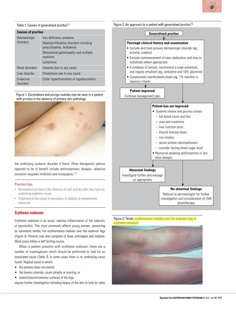
Erythema nodosum is an acute, reactive inflammation of the subcutis, or panniculitis. This most commonly affects young women, presenting as symmetric tender, hot erythematous nodules over the extensor legs (Figure 3). Patients may also complain of fever, arthralgias and malaise. Most cases follow a self limiting course. When a patient presents with erythema nodosum, there are a number of investigations which should be performed to look for an associated cause (Table 2). In some cases there is no underlying cause found. Atypical cases in which:• theprocessdoesnotresolve• thelesionsulcerate,causeatrophyorscarring,or• extendbeyondextensorsurfacesofthelegsrequire further investigation including biopsy of the skin to look for other
Table 1. Causes of generalised pruritus1,2
causes of pruritus
Haematologic disorders
Iron deficiency anaemiaMyeloproliferative disorders including polycythaemia, leukaemiaMonoclonal gammopathy and multiple myelomaLymphoma
Renal disorders Uraemia due to any cause
Liver disorder Cholestasis due to any cause
Endocrine disorders
Either hyperthyroidism or hypothyroidism
Figure 1. Excoriations and prurigo nodules may be seen in a patient with pruritus in the absence of primary skin pathology
Figure 2. An approach to a patient with generalised pruritus1,2
Generalised pruritus
Thorough clinical history and examination•Excludeandtreatprimarydermatologicdisorder(eg.
eczema, scabies)•Excludecommencementofnewmedicationandstopor
substitute where appropriate•Ifevidenceofxerosis,recommendasoapsubstitute,
and regular emollient (eg. sorbolene and 10% glycerine)•Symptomaticmentholatedcream(eg.1%mentholin
aqueous cream)
Patient improved Continue management plan
Abnormal findingsInvestigate further and manage
as appropriate
no abnormal findingsReferral to dermatologist for further
investigation and consideration of UVB phototherapy
Figure 3. Tender erythematous nodules over the extensor legs in erythema nodosum
Patient has not improved• Systemsreviewandpruritusscreen – full blood count and film – urea and creatinine – liver function tests – thyroid function tests – iron studies – serum protein electrophoresis – consider fasting blood sugar level•Nocturnalsedatingantihistamineorlow
dose doxepin

skin manifestations of systemic diseaseTHEME
500 Reprinted from AusTRAliAn FAMily PHysiciAn Vol. 38, No. 7, July 2009
presents with a cutaneous vasculitis, it is important to look for any evidence of systemic vasculitis, which has more serious implications. Small vessel vasculitis affects the arterioles, capillaries and venules and classically presents as palpable purpuric papules and plaques (Figure 4). This is the most common form of cutaneous vasculitis, typically affecting the lower legs and dependent areas. There are a large number of causes reported (Table 3).4,5
Medium vessel vasculitis is much less common and often associated with a systemic vasculitis and connective tissue disorder including systemic lupus erythematosus (SLE), Wegener granulomatosis,
causes of panniculitis. These cases should be referred for specialist assessment. Management is supportive, including bed rest, the use of support stockings, and nonsteroidal anti-inflammatory drugs. In more severe cases, systemic corticosteroids may be needed.3
Practice tips• Inallcases,considerandexcludeanunderlyingsystemicdisorder,in
particular sarcoidosis• Atypicalclinicalpresentationsrequireaskinbiopsytolookforother
causes of panniculitis• Treatmentisbothsupportiveandaimedattheunderlyingdisorder.
cutaneous vasculitis
Vasculitis refers to an inflammation of the blood vessels, which can affect small, medium or large vessels. Both small and medium vessel vasculitis may present with cutaneous findings. In any patient who
Figure4.Multiplepalpablepurpuricpapulesandplaques–acaseofa florid small vessel cutaneous vasculitis
Table 2. Causes and investigation of erythema nodosum3
causes of erythema nodosum Approach and investigations
infections •Bacterial,eg.Streptococcus,Yersinia,Salmonella,
Campylobacter•Viral,eg.EpsteinBarrvirus•Mycobacterial
•Throatswab,antistreptolysinOtitre(ASOT),anti-doublestrandedDNAantibodies•Serologyandstoolcultureswhereappropriate•Serologywhereappropriate•Investigateifclinicalsuspicion–chestX-ray,Mantoux,Quantiferongold
Sarcoidosis ChestX-ray,serumangiotensinconvertingenzymeinhibitor(ACEI),calciumandreferral if abnormal
Inflammatory bowel disease History and examination
MalignancyLeukaemia, lymphoma (rare)Postradiotherapy
Full blood count and film
Pregnancy History, ßHCG level
Behcet syndrome History of oral and genital aphthous ulcers and examination
Drugs Recent commencement in particular oral contraceptive pill, tetracycline antibiotics, sulphur based drugs, bromides and iodides
Table 3. Causes of cutaneous vasculitis5,6
infectionsBacterial
ViralMycobacterial
•Streptococcal,meningococcal,urinarytractinfections
•HepatitisBandC,HIV•Tuberculosis
connective tissue disorders
•SLEandrelatedconditions•Rheumatoidarthritis•Systemicsclerosis,Sjogrensyndrome•Dermatomyositis•Mediumvesselvasculitides(Wegener
granulomatosis, polyarteritis nodosa, Churg-Strauss syndrome)
Malignancy •Haematologic – myeloproliferative – lymphoma – monoclonal gammopathy – multiple myeloma
Drugs Including antibiotics, antihypertensives
idiopathic Henoch-Schonlein purpura
skin manifestations of systemic disease THEME
Reprinted from AusTRAliAn FAMily PHysiciAn Vol. 38, No. 7, July 2009 501
ofHSP,whichcanalsoaffectthejoints,bowelandkidneys,andneedslong term monitoring of renal function, as involvement may develop years after the episode of cutaneous vasculitis.6 A systemically unwell, septic patient should be treated as meningococcaemia until proven otherwise.
Practice tips• Ifthepatientissepticandunwell,excludemeningococcaemia• Itisimportanttolookforanyevidenceofsystemicvasculitis,in
particular renal involvement• Treatmentisdirectedattheunderlyingcause.
Whattolookforchronic liver disease
Chronic liver disease is associated with a number of cutaneous manifestations. Some of these occur in any patient, while others are more specific to the nature or cause of the liver disease. Any patient with chronic liver impairment may have multiple spider naevi, palmar erythema, an acquired ichthyosis or macular purpura in association withacoagulopathy.Cholestasismaybeassociatedwith jaundiceandgeneralised pruritus, which can result in secondary changes such as excoriations and prurigo nodules (Figure 1). Hepatitis C infection has been associated with a range of cutaneous manifestations (Table 5, Figure 4, 6).7,8
connective tissue disorders
Connective tissue disorders often have classic cutaneous findings, some of which are disease specific. Recognising these signs will help to differentiate between the different diseases, although overlap between them can be seen. The different specific and nonspecific cutaneous signs are detailed in Table 6, and illustrated in Figures 7–10.9–12
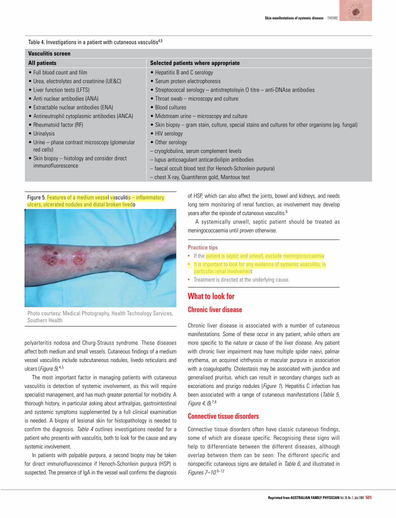
polyarteritis nodosa and Churg-Strauss syndrome. These diseases affect both medium and small vessels. Cutaneous findings of a medium vessel vasculitis include subcutaneous nodules, livedo reticularis and ulcers (Figure 5).4,5
The most important factor in managing patients with cutaneous vasculitis is detection of systemic involvement, as this will require specialist management, and has much greater potential for morbidity. A thorough history, in particular asking about arthralgias, gastrointestinal and systemic symptoms supplemented by a full clinical examination is needed. A biopsy of lesional skin for histopathology is needed to confirm the diagnosis. Table 4 outlines investigations needed for a patient who presents with vasculitis, both to look for the cause and any systemic involvement. In patients with palpable purpura, a second biopsy may be taken for direct immunofluorescence if Henoch-Schonlein purpura (HSP) is suspected. The presence of IgA in the vessel wall confirms the diagnosis
Figure5.Featuresofamediumvesselvasculitis–inflammatoryulcers, ulcerated nodules and distal broken livedo
Photo courtesy: Medical Photography, Health Technology Services, Southern Health
Table 4. Investigations in a patient with cutaneous vasculitis4,5
Vasculitis screen
All patients selected patients where appropriate
•Fullbloodcountandfilm•Urea,electrolytesandcreatinine(UE&C)•Liverfunctiontests(LFTS)•Antinuclearantibodies(ANA)•Extractablenuclearantibodies(ENA)•Antineutrophilcytoplasmicantibodies(ANCA)•Rheumatoidfactor(RF)•Urinalysis•Urine–phasecontrastmicroscopy(glomerular
red cells)•Skinbiopsy–histologyandconsiderdirect
immunofluorescence
•HepatitisBandCserology•Serumproteinelectrophoresis•Streptococcalserology–antistreptolsyinOtitre–anti-DNAseantibodies•Throatswab–microscopyandculture•Bloodcultures•Midstreamurine–microscopyandculture•Skinbiopsy–gramstain,culture,specialstainsandculturesforotherorganisms(eg.fungal)•HIVserology•Otherserology– cryoglobulins, serum complement levels– lupus anticoagulant anticardiolipin antibodies– faecal occult blood test (for Henoch-Schonlein purpura)–chestX-ray,Quantiferongold,Mantouxtest

skin manifestations of systemic disease THEME
Reprinted from AusTRAliAn FAMily PHysiciAn Vol. 38, No. 7, July 2009 503
Table 6. Classic and more common cutaneous findings in connective tissue disorders9–12
connective tissue disease cutaneous manifestationlupus erythematosusSystemic lupus erythematosusSubacute cutaneous lupus erythematosus
Discoid lupus erythematosus
•Malarerythema(Figure7),lupushairs•Annular/psoriasiformrashaffectingarmsand‘V’chest
(Figure 8)•Welldemarcatedplaqueswithadherentscalecausing
a scarring or a scarring alopecia (Figure 9)Dermatomyositis •Heliotroperash
•Gottronpapulesandsign(violaceousmaculesandpapules over interphalangeal and metacarpophalangeal joints)
•Macularviolaceousorpoikilodermatousrashovershoulders and hips
•Mechanicshands(hyperkeratosisonulnarborderoffingers)
•Calcinosissystemic sclerosis, cREsT syndrome (Figure 10)
•Calcinosis•Raynaudphenomenon•Sclerodactyly•Telangiectasia•Digitalinfarcts
Rheumatoid arthritis •Rheumatoidnodules•Linearsubcutaneousbands•Rheumatoidneutrophilicdermatitis
Other cutaneous changes of connective tissue disease
•Photosensitivity•Vasculitisandulcers•Nailfoldchanges(periungualerythema,ragged
cuticles, nail fold telangectasia)•Pyodermagangrenosum
Figure 9. Scarring, hypopigmented and hyperpigmented erythematous welldemarcatedplaquesinsunexposed areas of discoid lupus erythematosus
Figure 8. Psoriasiform rash of subacute cutaneous lupus erythematosus affecting the ‘V’ of the chest
Figure6.Lichenplanus–violaceousflattoppedpapuleshavebeenreported in hepatitis C infection
Table 5. Reported cutaneous manifestations of hepatitis C infection7,8
Cutaneous vasculitis – uritcarial, leucocytoclastic, cryoglobulinaemic
Polyarteritis nodosa
Porphyria cutanea tarda
Lichen planus
Necrolyticacralerythema
Photo courtesy: Medical Photography, Health Technology Services, Southern Health
Figure 7. Malar erythema of systemic lupus erythematosus

skin manifestations of systemic diseaseTHEME
504 Reprinted from AusTRAliAn FAMily PHysiciAn Vol. 38, No. 7, July 2009
If a patient presents with any constellation of the cutaneous changes consistent with a connective tissue disease, then it is important to fully investigate to:• confirm thediagnosisof theconnective tissuediseaseand to
distinguish between them using serology including anti nuclear antibodies,extractablenuclearantibodies,doublestrandedDNA
• look for any evidence of systemic disease – full blood countand film, urea, electrolytes and creatinine, liver function tests, urinalysis, and where appropriate, lupus anticoagulant, anticardiolipin antibody.
In cases of suspected dermatomyositis appropriate investigations include: creatinine kinase, biopsy, magnetic resonance imaging of affected muscle or electromyogram and, in adult cases, malignancy screen. A biopsy of the skin may show histologic features specific to SLE, dermatomyositis and scleroderma, and direct immunofluorescence can be used to look for a lupus band. Treatment of the cutaneous changes requires strict
photoprotection and the use of topical corticosteroids. In some cases, systemic treatment is needed. Corticosteroids, hydroxychloroquine, methotrexate, azathioprine, mycophenolate, cyclosporine, cyclophosphamide, intravenous immunoglobulin, and more recently, rituximab, have all been used.13 Patients with a suspected connective tissue disease should be assessed by a specialist physician.
Practice tips• Usetheclinicopathologiccorrelationofsymptoms,signs,serology
and histology to confirm the diagnosis and differentiate between connective tissue disorders
• Alwayslookfortheextentofsystemicinvolvement• Patientswilloftenneedamultidisciplinaryapproachtomanagement.
Figure 12. Pyoderma gangrenosum may be associated with connective tissue disease, inflammatory bowel disease and certain malignancies
Table 7. Cutaneous manifestations of internal malginancies14–22
cutaneous feature clinical findings Associated malignancyAcanthosis nigricans (malignant) (Figure 11)
Velvety hyperpigmented thickening extending beyond the flexures and neck to involve lips, palms
Intra-abdominal adenocarcinoma, lung carcinoma, lymphoreticular malginancies
Acrokeratosis paraneoplastica(Bazex syndrome)
Psoriasiform dermatitis with nail dystrophy affecting hands, feet, ears, nose
Squamous cell carcinoma of upper aerodigestive tract
Erythema gyratum repens Wood grain pattern annular, scaling erythema Lung carcinomaNecrolyticmigratoryerythema Eroded erythematous annular polycylic eruption
affecting intertriginous areasGlucagonoma
Sweet syndrome Plum coloured nodules affecting head, neck and dorsae hands
Can be associated with leukaemia, lymphoma, multiple myeloma
Pyoderma gangrenosum especially bullous variant (Figure 12)
Painful inflammatory ulcers with raised violaceous edge and overhanging borders; associated pathergy
Can be associated with leukaemia, lymphoma, multiple myeloma
Paraneoplastic pemphigus Bullous,erosivemucosal+/-cutaneouseruption Haematologic malignancies, thymomaNecrobioticxanthogranuloma(Figure13) Purpuric yellow plaques in periorbital and flexural areas Monoclonalgammopathy/multiplemyelomaDiffuse plane xanthomas Yellow-orangemaculesandplaques Monoclonalgammopathy/multiplemyelomaScleromyxoedema Scleroderma-like thickening of skin associated with skin
coloured or erythematous papular infiltrateMonoclonalgammopathy/multiplemyeloma
Primary systemic amyloidosis Macroglossia, purpura especially periorbital and infiltrated papules
Monoclonalgammopathy/multiplemyeloma
Figure 10. Cutaneous calcinosis, sclerodactyly and telangiectasia of CREST syndrome. Cutaneous calcinosis may also be seen in systemic sclerosis and dermatomyositis
Figure 11. Marked velvety papillomatous thickening of the neck extending onto the face and lips seen in malignant acanthosis nigricans

skin manifestations of systemic disease THEME
Reprinted from AusTRAliAn FAMily PHysiciAn Vol. 38, No. 7, July 2009 505
WhatnottomissThis section has been included to highlight cutaneous paraneoplastic syndromes. Certain malignancies, in particular haematological malignancies, may often present with specific cutaneous features (Table 7). Multiple myeloma, in particular, has been associated with a number of cutaneous syndromes.14–22 The presence of these paraneoplastic syndromes and cutaneous features should institute a search for the malignancy. The lack of recognition of these features may result in a delay in diagnosis and treatment.
Conflict of interest: none declared.
References1. Greaves MW. Itch in systemic disease: Therapeutic options. Dermatol Ther
2005;18:323–7.2. WardJR,BernhardJD.Willan’sitchandothercausesofpruritusintheelderly.IntJ
Dermatol 2005;44:267–73.3. RequenaL,YusES.Erythemanodosum.DermatolClin2008;26:425–38.4. Fiorentino DF. Cutaneous vasculitis. J Am Acad Dermatol 2003;48:311–40.5. Chen KR, Carlson JA. Clinical approach to cutaneous vasculitis. Am J Clin Dermatol
2008;9:71–92.6. Dillon MJ. Henoch-Schonlein purpura: Recent advances. Clin Exp Rheumatol
2007;25:s66-8.7. Galossi A, Guarisco R, Bellis L, et al. Extrahepatic manifestations of chronic HCV
infection. J Gastrointestin Liver Dis 2007;16:65–73.8. AbdallahMA,GhozziMY,MonibHA,etal.Necrolyticacralerythema:Acutaneous
sign of hepatitis C virus infection. J Am Acad Dermatol 2005;53:247–51.9. D’CruzDP.Systemiclupuserythematosus.BMJ2006;332:890–4.10. Callen JP. Dermatomyositis. Lancet 2000;355:53–7.11. BachmeyerC,Tillie-LeblondLI,LacertA,etal.‘Mechanicshands’:Amisleadingsign
of the antisynthetase syndrome. Br J Dermatol 2007;156:192–4.12. Yamamoto T. Cutaneous manifestations associated with rheumatoid arthritis.
Rheumatol Int 2009; Feb 26 (Epub ahead of print).13. QuainRD,WerthVP.Managementofcutaneousdermatomyositis:currenttherapeutic
options. Am J Clin Dermaol 2006;7:341–51.14. Kleyn CE, Lai-Cheong E, Bell HK. Cutaneous manifestations of internal malignancy:
Diagnosis and management. Am J Clin Dermatol 2006;7:71–84.15. Boyce S, Harper J. Paraneoplastic dermatoses. Dermatol Clin 2002;20:523–32.16. Gill D, Fergin P, Kelly J. Bullous lesions in Bazex syndrome and successful treatment
with oral psoralen phototherapy. Aus J Dermatol 2001;42:278–80.17. BoydAS,NeldnerKH,MenterA.Erythemagyratumrepens:Aparaneoplasticerup-
tion. J Am Acad Dermatol 1992;26:757–62.18. Van Beek A, de Haas ER, Van Vloten WA, et al. The glucagonoma syndrome and
necrolytic migratory erythema: A clinical review. Eur J Endocrinol 2004:151;531–7.19. Wallach D, Vignon-Pennamen MD. From acute febrile neutrophilic dermatosis
to neutrophilic disease: Forty years of clinical research. J Am Acad Dermatol 2006;55:1066–71.
20. Bayer-Garner IB, Smoller BR. The spectrum of cutaneous disease in multiple myeloma. J Am Acad Dermatol 2003;48:497–507.
CORRESPONDENCE [email protected]
Figure13.Purpuricplaqueoftheperiorbitalarea–necrobioticxanthogranuloma