complicated skin & skin structure infections
TRANSCRIPT
SHOCK
…and the Trauma Victim
JP PretoriusDepartment of Surgery & SICUSteve Biko Academic Hospital.
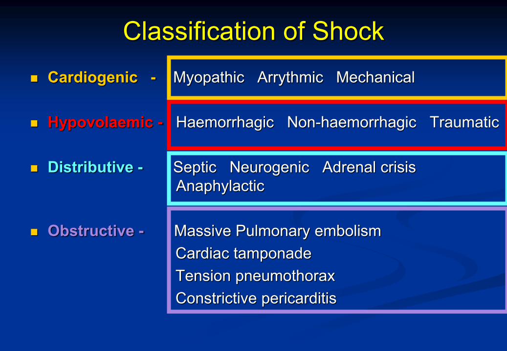
Classification of Shock Cardiogenic - Myopathic Arrythmic Mechanical
Hypovolaemic - Haemorrhagic Non-haemorrhagic Traumatic
Distributive - Septic Neurogenic Adrenal crisisAnaphylactic
Obstructive - Massive Pulmonary embolismCardiac tamponadeTension pneumothoraxConstrictive pericarditis
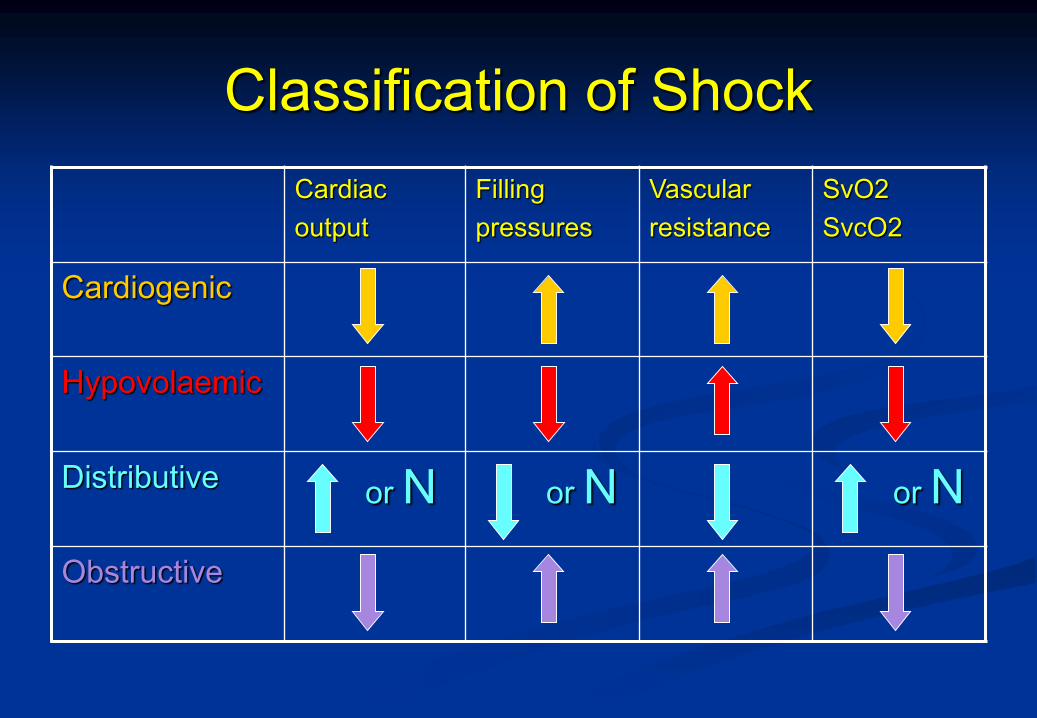
Classification of ShockCardiacoutput
Filling pressures
Vascularresistance
SvO2SvcO2
Cardiogenic
Hypovolaemic
Distributive or N or N or NObstructive
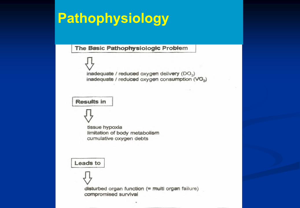
Pathophysiology
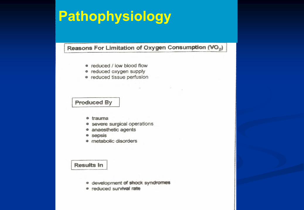
Pathophysiology

Case Report – Mr B. Sethole
22 year old man
Sustained GSW in peripheral town
Taken to local hospital
Patient in shock 90/60
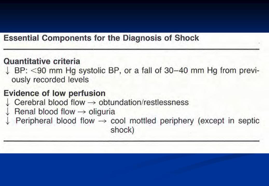
? Definition of shock
Hemothorax diagnosed
ICD placed – drained 2 litres blood
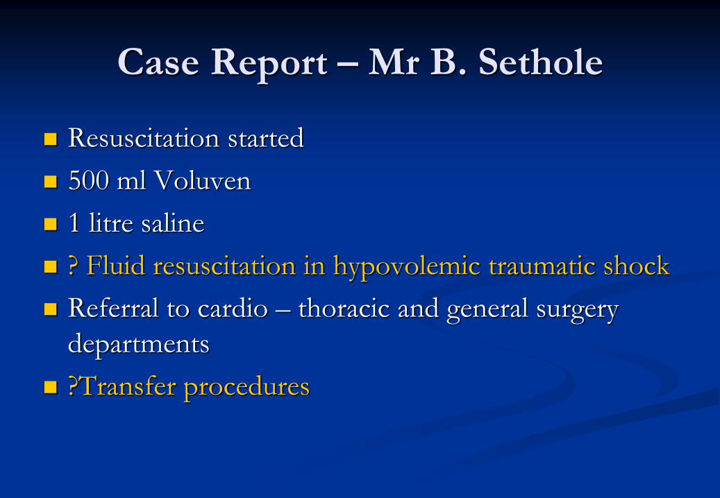
Resuscitation started
500 ml Voluven
1 litre saline
? Fluid resuscitation in hypovolemic traumatic shock
Referral to cardio – thoracic and general surgery
departments
?Transfer procedures
Case Report – Mr B. Sethole
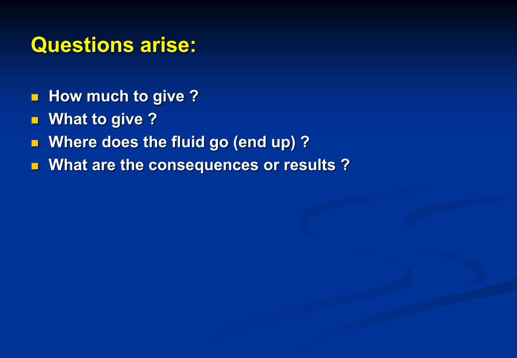
Questions arise:
How much to give ? What to give ? Where does the fluid go (end up) ? What are the consequences or results ?
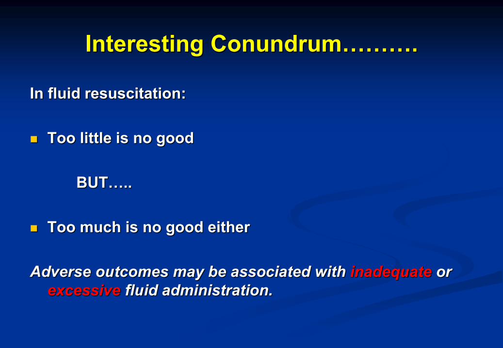
Interesting Conundrum……….
In fluid resuscitation:
Too little is no good
BUT…..
Too much is no good either
Adverse outcomes may be associated with inadequate or excessive fluid administration.
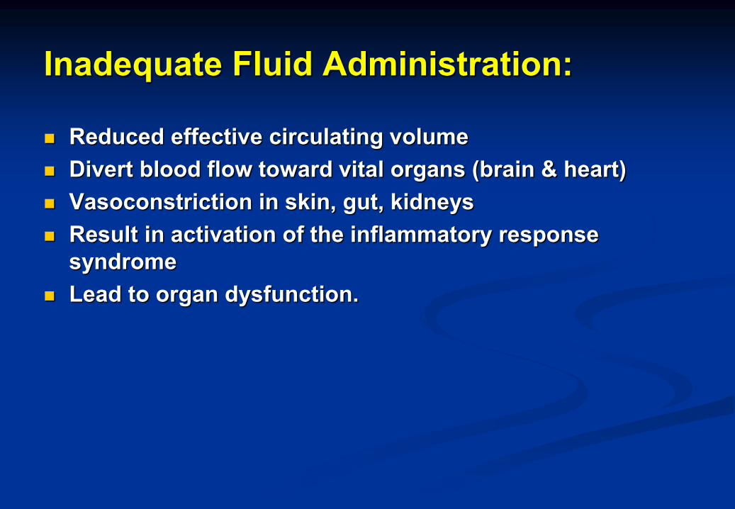
Inadequate Fluid Administration:
Reduced effective circulating volume Divert blood flow toward vital organs (brain & heart) Vasoconstriction in skin, gut, kidneys Result in activation of the inflammatory response
syndrome Lead to organ dysfunction.
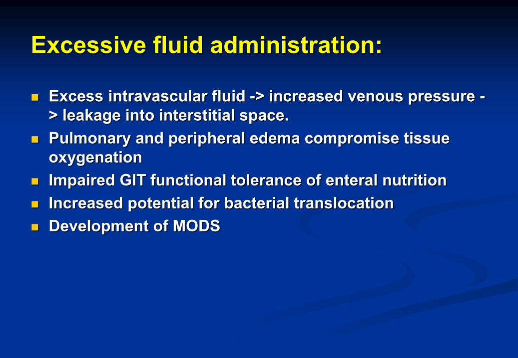
Excessive fluid administration:
Excess intravascular fluid -> increased venous pressure -> leakage into interstitial space.
Pulmonary and peripheral edema compromise tissue oxygenation
Impaired GIT functional tolerance of enteral nutrition Increased potential for bacterial translocation Development of MODS
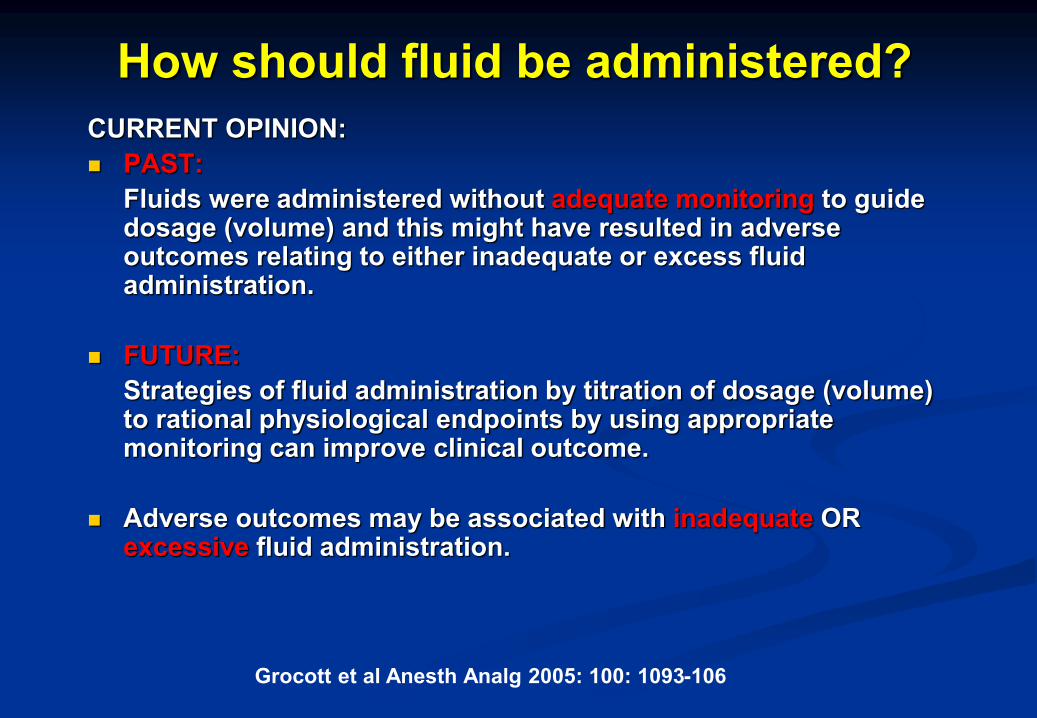
How should fluid be administered?CURRENT OPINION: PAST:
Fluids were administered without adequate monitoring to guide dosage (volume) and this might have resulted in adverse outcomes relating to either inadequate or excess fluid administration.
FUTURE:Strategies of fluid administration by titration of dosage (volume) to rational physiological endpoints by using appropriate monitoring can improve clinical outcome.
Adverse outcomes may be associated with inadequate OR excessive fluid administration.
Grocott et al Anesth Analg 2005: 100: 1093-106
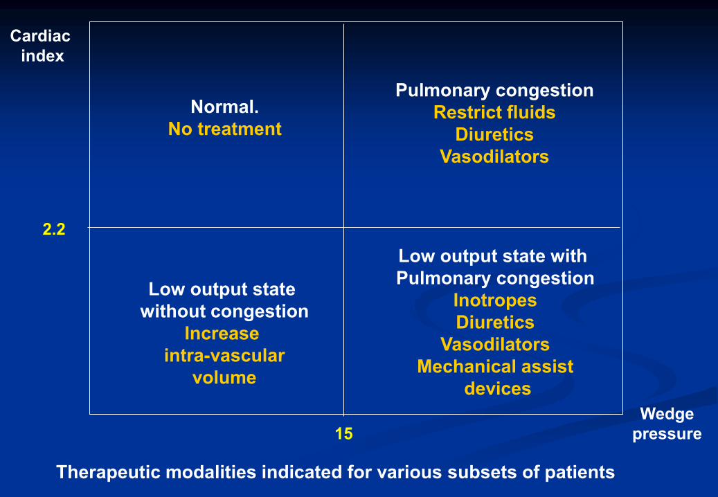
Normal.No treatment
Pulmonary congestionRestrict fluids
DiureticsVasodilators
Low output state without congestion
Increase intra-vascular
volume
Low output state with Pulmonary congestion
InotropesDiuretics
VasodilatorsMechanical assist
devices
Cardiac index
2.2
15Wedge
pressure
Therapeutic modalities indicated for various subsets of patients
How should fluid be administered?
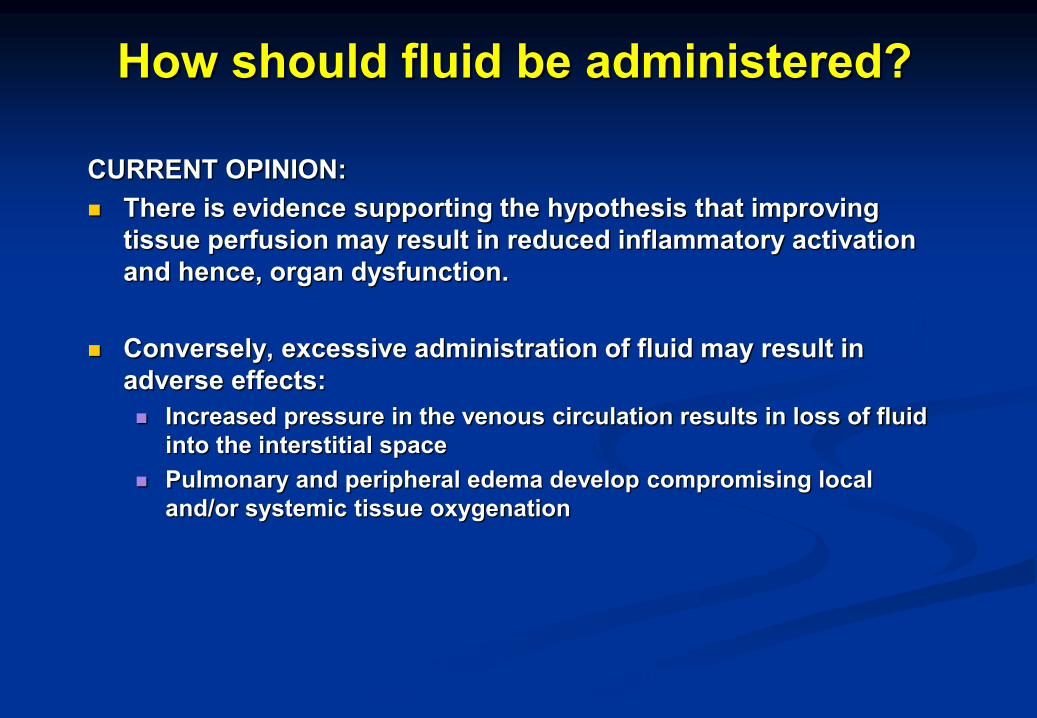
CURRENT OPINION: There is evidence supporting the hypothesis that improving
tissue perfusion may result in reduced inflammatory activation and hence, organ dysfunction.
Conversely, excessive administration of fluid may result in adverse effects: Increased pressure in the venous circulation results in loss of fluid
into the interstitial space Pulmonary and peripheral edema develop compromising local
and/or systemic tissue oxygenation
Grocott et al Anesth Analg 2005: 100: 1093-106
How should fluid be administered?
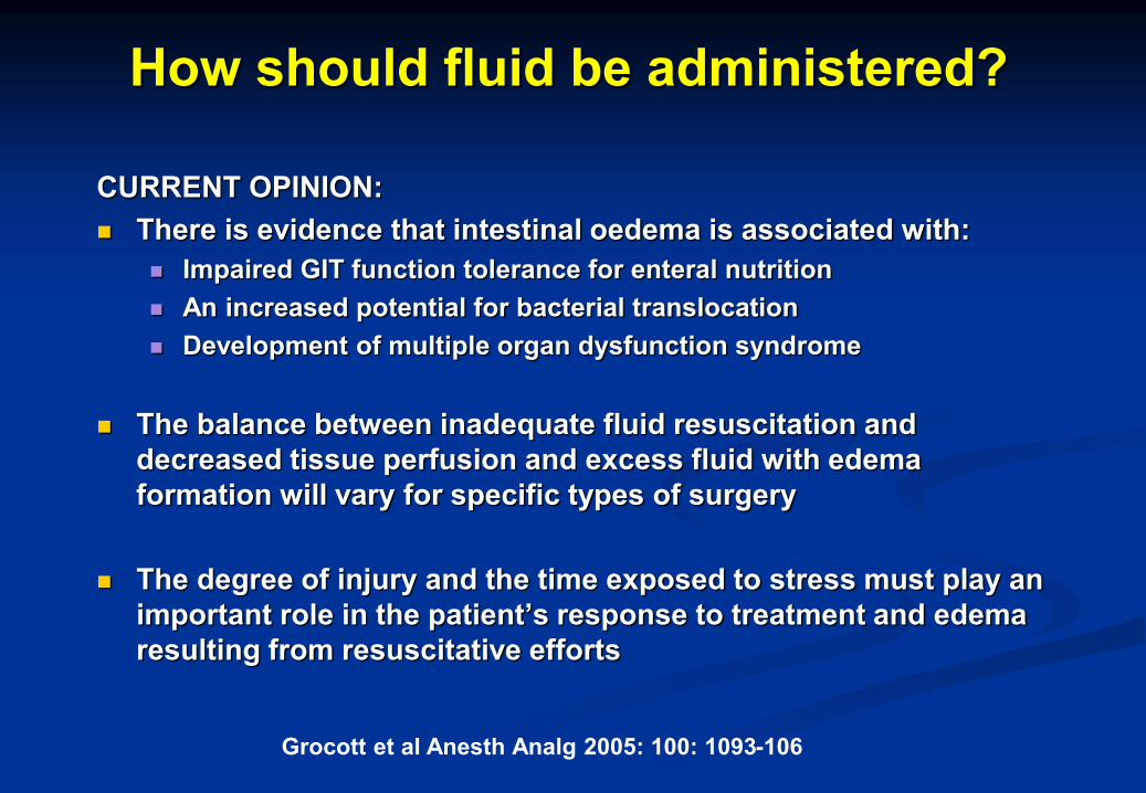
CURRENT OPINION: There is evidence that intestinal oedema is associated with:
Impaired GIT function tolerance for enteral nutrition An increased potential for bacterial translocation Development of multiple organ dysfunction syndrome
The balance between inadequate fluid resuscitation and decreased tissue perfusion and excess fluid with edema formation will vary for specific types of surgery
The degree of injury and the time exposed to stress must play an important role in the patient’s response to treatment and edema resulting from resuscitative efforts
Grocott et al Anesth Analg 2005: 100: 1093-106
How should fluid be administered?
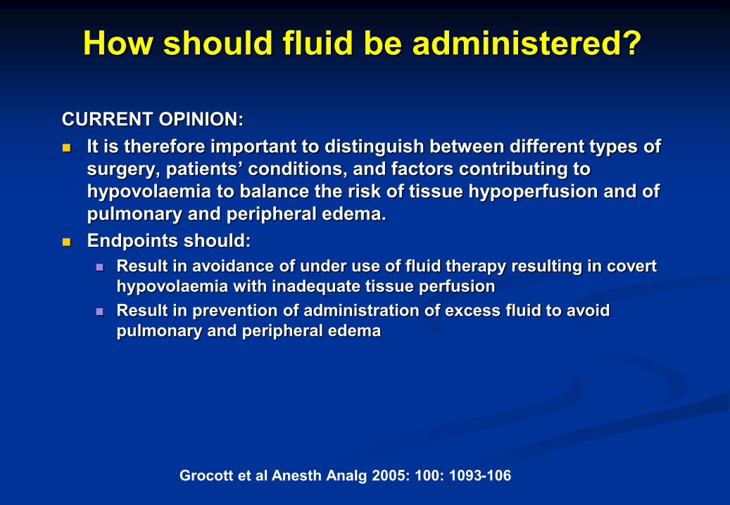
CURRENT OPINION: It is therefore important to distinguish between different types of
surgery, patients’ conditions, and factors contributing to hypovolaemia to balance the risk of tissue hypoperfusion and of pulmonary and peripheral edema.
Endpoints should: Result in avoidance of under use of fluid therapy resulting in covert
hypovolaemia with inadequate tissue perfusion Result in prevention of administration of excess fluid to avoid
pulmonary and peripheral edema
Grocott et al Anesth Analg 2005: 100: 1093-106

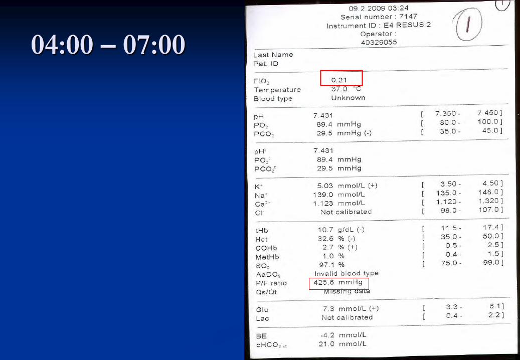
04:00 – 07:00
On arrival BP 126/68 Pulse 102
ICD in situ
GSW – entrance T5 Left posterior axillary line
- exit T6 Left mid clavicular line
? Surgical assessment of this scenario
? Pre – op investigation and preparation
04:00 – 07:00
04:00 – 07:00
Equal good air entry bilaterally
Still bleeding from ICD – 400ml
? Interpretation
Gastric contents in ICD
Abdomen – board - like rigidity
X – ray confirmed: both lungs expanded
? Antibiotic therapy
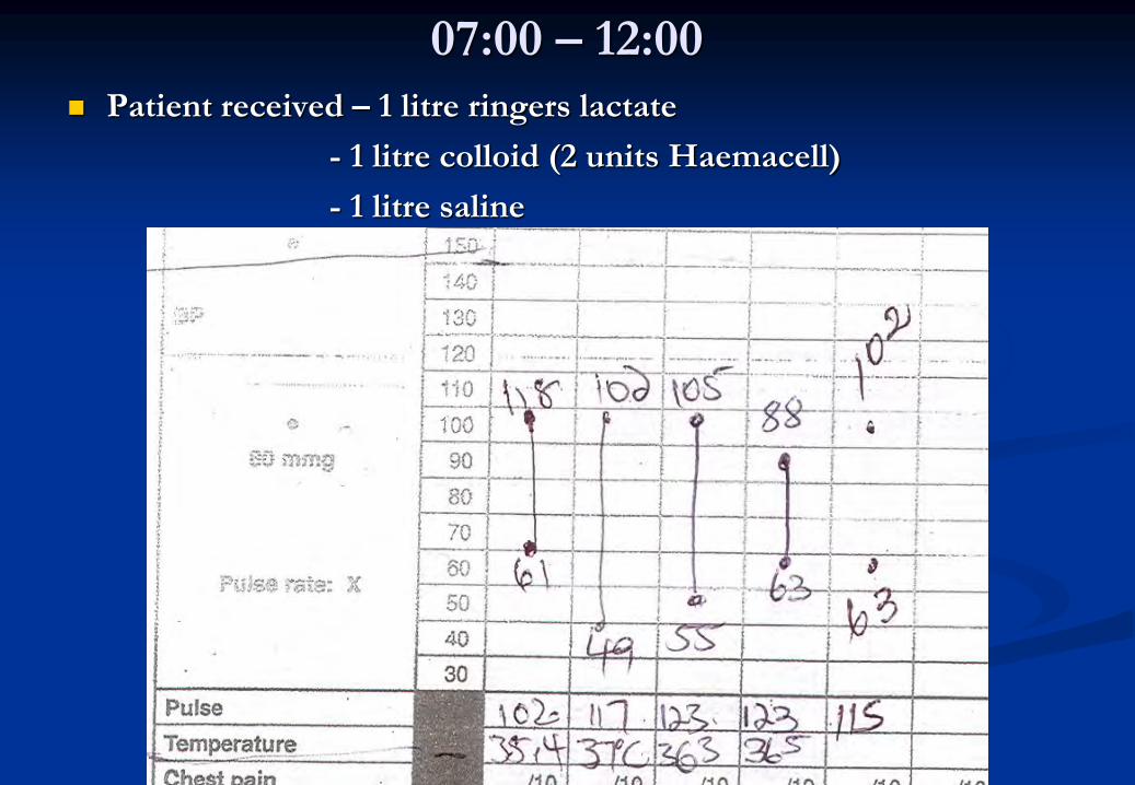
07:00 – 12:00
Patient received – 1 litre ringers lactate
- 1 litre colloid (2 units Haemacell)
- 1 litre saline
07:00- 12:00
4 Units blood ordered - not yet given
? Blood component therapy in trauma
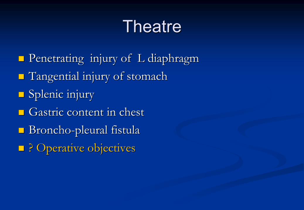
Theatre
Penetrating injury of L diaphragm
Tangential injury of stomach
Splenic injury
Gastric content in chest
Broncho-pleural fistula
? Operative objectives
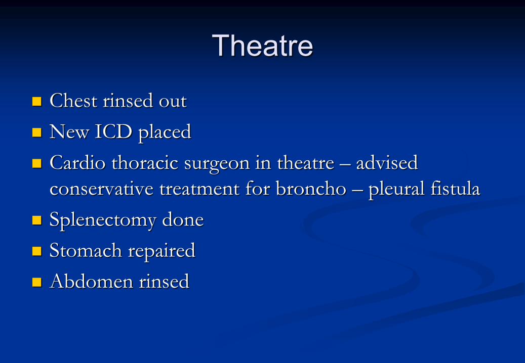
Theatre
Chest rinsed out
New ICD placed
Cardio thoracic surgeon in theatre – advised
conservative treatment for broncho – pleural fistula
Splenectomy done
Stomach repaired
Abdomen rinsed
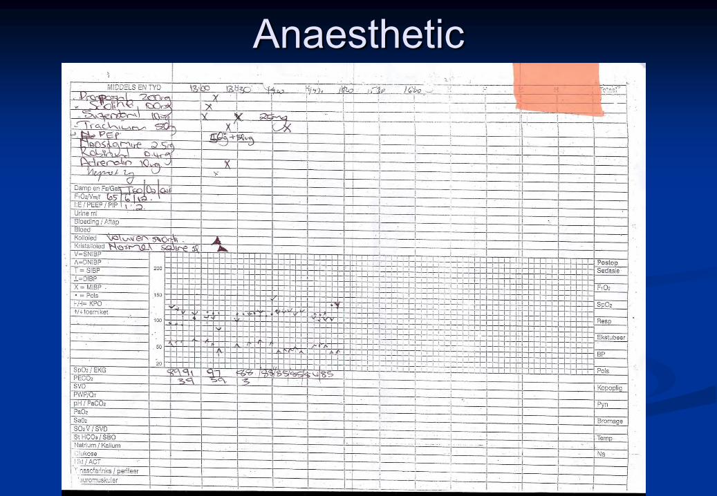
Anaesthetic
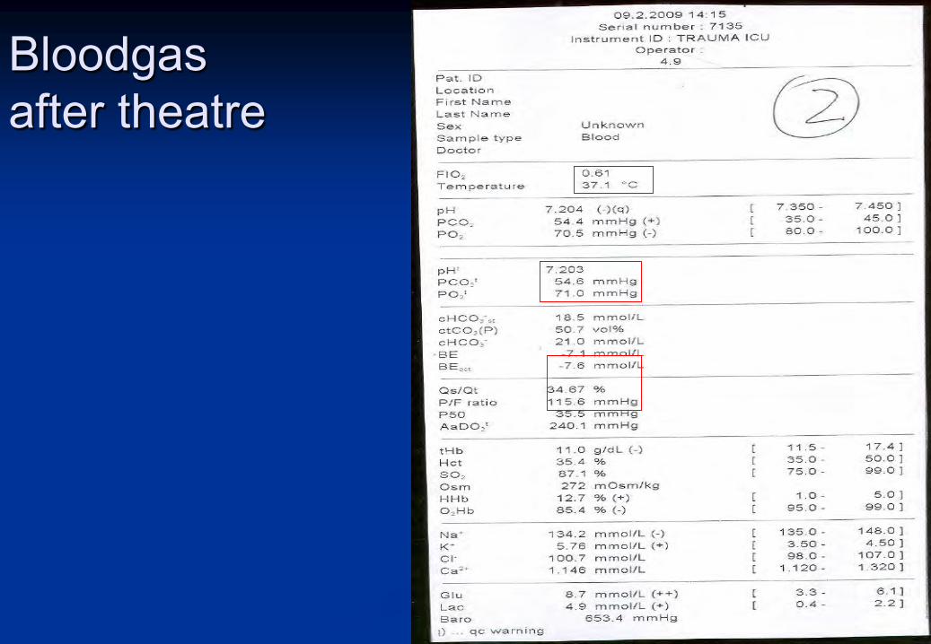
Bloodgas after theatre

ICU
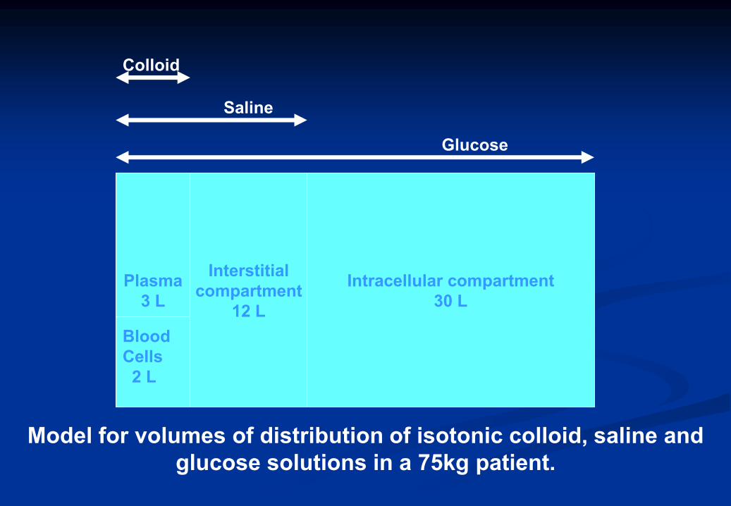
Intracellular compartment30 L
Interstitialcompartment
12 L
Plasma3 L
BloodCells2 L
Glucose
Saline
Colloid
Model for volumes of distribution of isotonic colloid, saline and glucose solutions in a 75kg patient.
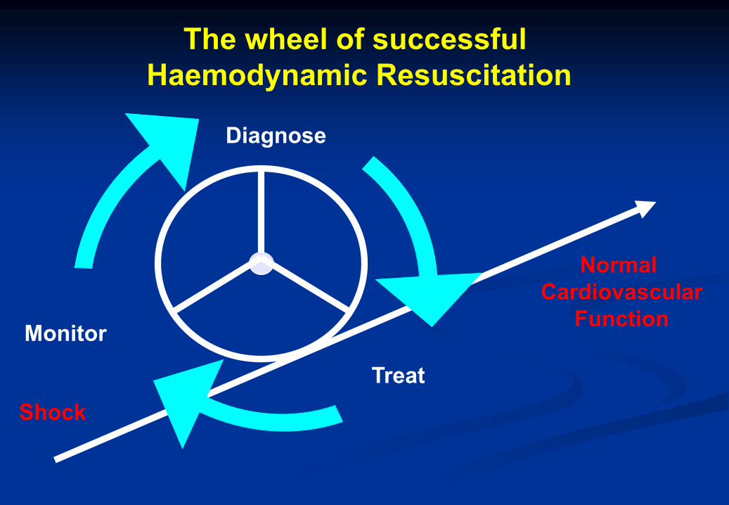
The wheel of successful Haemodynamic Resuscitation
Shock
Normal Cardiovascular
Function
Diagnose
TreatMonitorMonitor
TreatShock

Monitoring

Monitoring
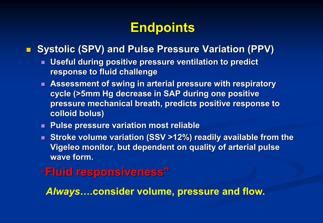
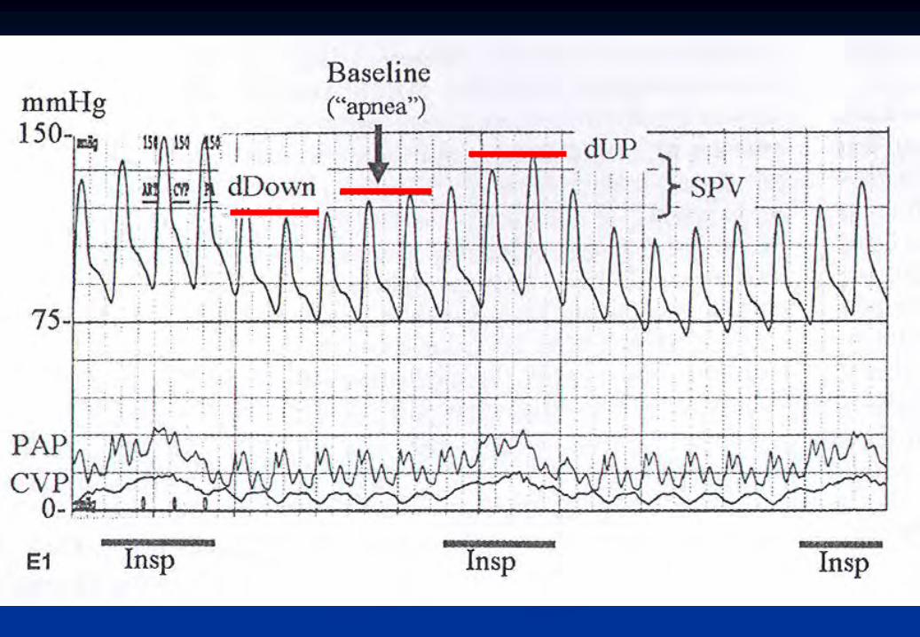
Endpoints Systolic (SPV) and Pulse Pressure Variation (PPV)
Useful during positive pressure ventilation to predict response to fluid challenge
Assessment of swing in arterial pressure with respiratory cycle (>5mm Hg decrease in SAP during one positive pressure mechanical breath, predicts positive response to colloid bolus)
Pulse pressure variation most reliable Stroke volume variation (SSV >12%) readily available from the
Vigeleo monitor, but dependent on quality of arterial pulse wave form.
“Fluid responsiveness”Always….consider volume, pressure and flow.
Endpoints The Fluid Challenge
Observe the response of CVP or PAOP to a fluid challenge(A bolus of 200ml colloid over 15 min) CVP stays the same or decreases: covert hypovolaemia A sustained increase > 3mm Hg – at the limit of vascular
compliance
Consider volume, flow and pressure

Endpoints
The Fluid Challenge and Measurement of Blood Flow Esophageal Doppler Colloid bolusses Maximizing stroke volume rather than targeting a specific
figure Avoids inappropriate excessive fluid infusion
Consider volume, flow and pressure
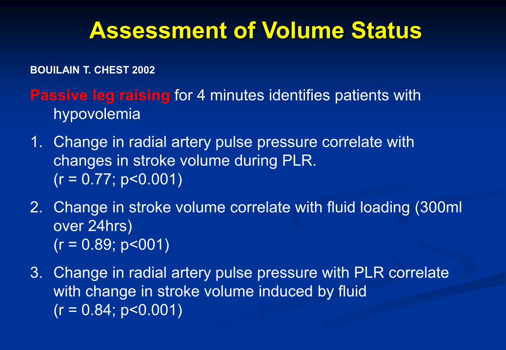
Assessment of Volume StatusBOUILAIN T. CHEST 2002
Passive leg raising for 4 minutes identifies patients with hypovolemia
1. Change in radial artery pulse pressure correlate with changes in stroke volume during PLR. (r = 0.77; p<0.001)
2. Change in stroke volume correlate with fluid loading (300ml over 24hrs) (r = 0.89; p<001)
3. Change in radial artery pulse pressure with PLR correlate with change in stroke volume induced by fluid (r = 0.84; p<0.001)
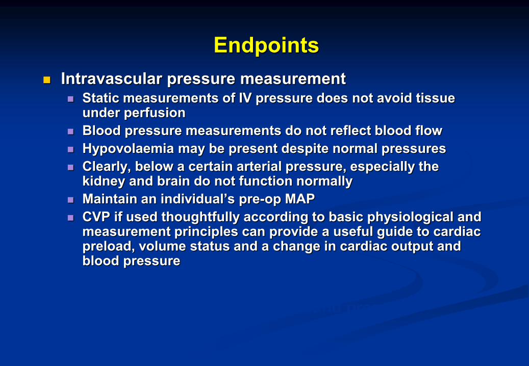
Endpoints Intravascular pressure measurement
Static measurements of IV pressure does not avoid tissue under perfusion
Blood pressure measurements do not reflect blood flow Hypovolaemia may be present despite normal pressures Clearly, below a certain arterial pressure, especially the
kidney and brain do not function normally Maintain an individual’s pre-op MAP CVP if used thoughtfully according to basic physiological and
measurement principles can provide a useful guide to cardiac preload, volume status and a change in cardiac output and blood pressure
Consider volume, flow and pressure
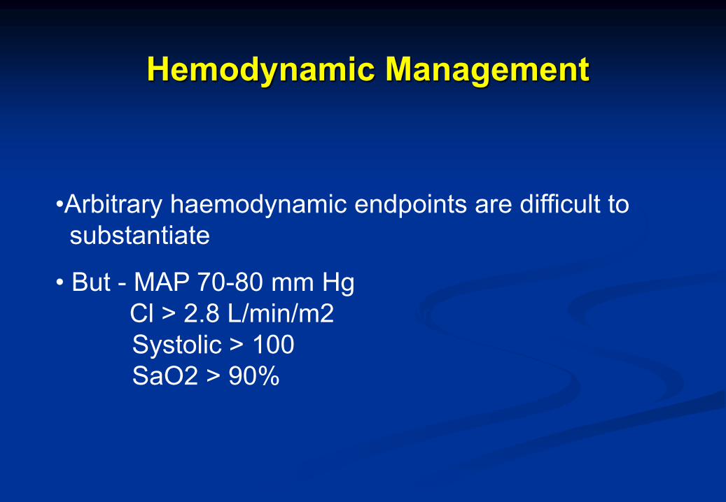
•Arbitrary haemodynamic endpoints are difficult to substantiate
• But - MAP 70-80 mm HgCl > 2.8 L/min/m2Systolic > 100SaO2 > 90%
is reasonable
Hemodynamic Management
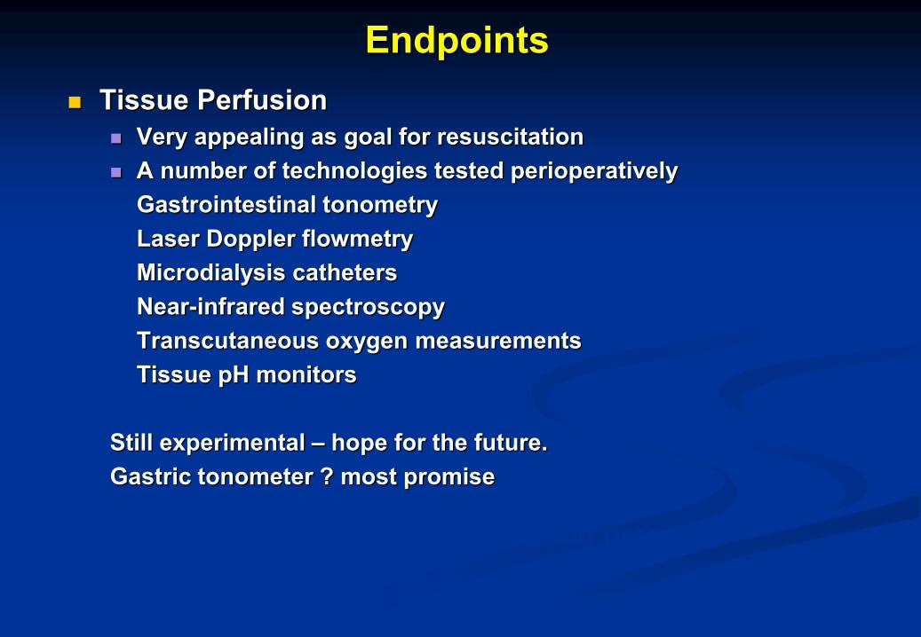
Endpoints Tissue Perfusion
Very appealing as goal for resuscitation A number of technologies tested perioperatively
Gastrointestinal tonometry Laser Doppler flowmetryMicrodialysis cathetersNear-infrared spectroscopyTranscutaneous oxygen measurementsTissue pH monitors
Still experimental – hope for the future. Gastric tonometer ? most promise
Consider volume, flow and pressure
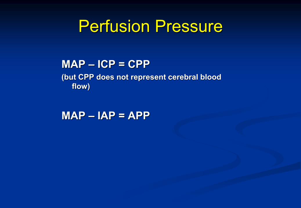
Perfusion Pressure
MAP – ICP = CPP(but CPP does not represent cerebral blood
flow)
MAP – IAP = APP
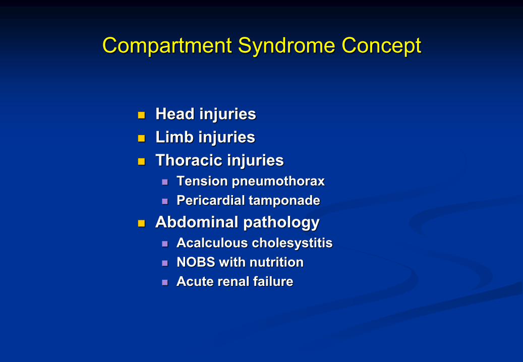
Compartment Syndrome Concept
Head injuries Limb injuries Thoracic injuries
Tension pneumothorax Pericardial tamponade
Abdominal pathology Acalculous cholesystitis NOBS with nutrition Acute renal failure

TOTAL FLUID MANAGEMENT
TFM
MAINTENANCE RESUSCITATION REPLACEMENT
1. Indication: Daily requirements Hypovolaemia Abnormal or continuing
losses.
2. Intention: According to a formula based
on body mass
“Aggressively” according to
endpoints
Collect drainage for 4 hours,
replace % during next 4 hours,
while collecting again
3. Infusion rate: Continuously per 24 hours
= 24 equal doses
Bolus Continuously according to
losses.
4. Type of fluid: Maintenance:
Maintelyte 5%,
Electrolyte No2 10%
Sustenance 5%
Volume expander:
Ringers Lactate (Modified),
PlasmalyteB,
Saline,
Colloids
According to fluid lost;
Rehydration,
5% Dextrose in water,
0,45% NaCl,
0,9% NaCl,
Ringers Lactate
5. Monitor Serum and urine electrolytes
and osmol.
Fluid balance chart.
Central haemodynamics,
Urine flow,
Stroke Volume Variation,
SvO2, Lactate, pH, BE
Serum and urine
electrolytes and osmol
INDICATIONS FOR FLUID AND ELECTROLYTE THERAPY IN SURGICAL PATIENTS
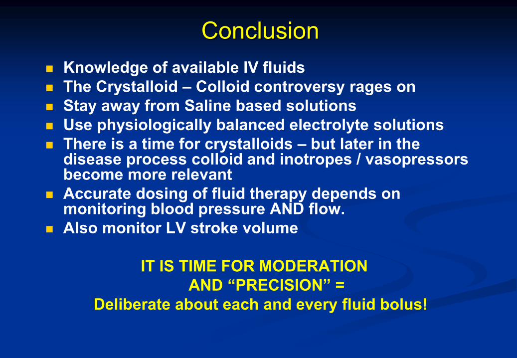
Conclusion Knowledge of available IV fluids The Crystalloid – Colloid controversy rages on Stay away from Saline based solutions Use physiologically balanced electrolyte solutions There is a time for crystalloids – but later in the
disease process colloid and inotropes / vasopressors become more relevant
Accurate dosing of fluid therapy depends on monitoring blood pressure AND flow.
Also monitor LV stroke volume
IT IS TIME FOR MODERATIONAND “PRECISION” =
Deliberate about each and every fluid bolus!

Thank you for your attention