single locking compression plate fixation of extra …...original article single locking...
TRANSCRIPT

ORIGINAL ARTICLE
Single locking compression plate fixation of extra-articular distalhumeral fractures
Malhar N. Kumar • M. R. Ravishankar •
Ravikiran Manur
Received: 19 December 2013 / Accepted: 27 September 2014 / Published online: 19 October 2014
� The Author(s) 2014. This article is published with open access at Springerlink.com
Abstract
Background Earlier literature on fixation of distal third
humeral fractures describes the use of elaboratemodification
of existing implants, custom-made implants and dual plating.
These modifications have the disadvantages of limitations of
hardware availability and cost as well as longer surgical
exposure to accommodate the plates. The aim of this study
was to assess the effectiveness of osteosynthesis of extra-
articular diaphyseal fractures of the distal third of the
humerus using a single 4.5-mm locking compression plate
(LCP) with two-screw purchase in the distal fragment.
Materials and methods We performed internal fixation of
distal third extra-articular humeral fractures in 22 adult
patients using 2–3 lag screws neutralized with a single 4.5-
mm locking compression plate with only two screws in the
distal fragment. The mean follow-up period was approxi-
mately 1.6 years.
Results Fractures united in all 22 patients with minimal
complications. The mean time to union of fracture was
13 weeks.TheMayo elbow score and theDASHscoreswere in
the excellent andgoodcategory in all patients at final follow-up.
Conclusions Our study showed that it is possible to
obtain excellent outcomes in distal third fractures using
only a single 4.5-mm LCP with two-screw (4-cortices)
purchase in the distal fragment. The disadvantages inherent
in the previous methods can be avoided with the use of the
present technique. This technique obviates the need for the
use of customized distal humeral implants and modified
implants in most patients.
Level of evidence Level IV.
Keywords Distal humerus fractures � Lockingcompression plate � Plate osteosynthesis of humerus �Metaphyseal fractures of humerus
Introduction
Fractures of the distal third of the humerus are challenging
injuries due to their peri-articular location, small size of the
distal bone fragments, and the osteopenic quality of the
bone in older adults. Methods of management of distal
humerus fractures include conservative management using
plaster cast immobilization or functional bracing, plate
osteosynthesis and intra-medullary nailing [1–4]. Stewart
et al. proposed that fractures of the distal-third humerus
shaft should not be treated by hanging cast because angu-
lation is difficult to control [1]. Sarmiento et al. treated 85
extra-articular comminuted distal-third humeral fractures
with a functional brace. The nonunion rate in their series
was 4 % and the malunion rate was 16 % (varus angulation
in the majority). A decrease in the range of motion at the
elbow and shoulder was another significant problem in
their series [2]. Jawa et al. compared the use of functional
bracing and plate fixation for extra-articular distal-third
diaphyseal fractures of the humerus. They concluded that
for extra-articular distal-third diaphyseal humeral fractures,
surgical treatment achieves more predictable alignment and
potentially quicker return of function but risks iatrogenic
nerve injury and infection and the need for reoperation [3].
It is difficult to manage extra-articular distal humerus
fractures with locking intra-medullary nails. The flat cross
section of the distal humerus with a narrow medullary
canal makes it difficult to insert intra-medullary nails and
increases liability for comminution of the distal fragment
during nail insertion. The short distal fragment makes it
M. N. Kumar (&) � M. R. Ravishankar � R. Manur
HOSMAT Hospital, 45, McGrath Road, Bangalore 560025,
India
e-mail: [email protected]
123
J Orthopaed Traumatol (2015) 16:99–104
DOI 10.1007/s10195-014-0325-8

difficult to achieve stable fixation with distal interlocking.
Radial nerve injury, if present, cannot be addressed without
a separate incision. Plate osteosynthesis has distinct
advantages in the distal humerus, and compression plating
has been established as a successful modality for the sur-
gical treatment of humerus fractures [4].
Recommendations for improving stability of plate fixa-
tion include plate thickness of[3.5 mm (a large-fragment
plate) formost adults, and at least four screw holes in both the
proximal and distal fragments [5]. However, adhering to
these principles becomes difficult in distal humeral shaft
fractures, especially those around the metaphyseal transition
zone between the shaft and the supracondylar ridges. Fixa-
tion with three or four screws in the distal fragment is diffi-
cult as longer plates tend to impinge on the olecranon fossa.
Livani et al. reported the use of percutaneous osteo-
synthesis in a small series of six patients with distal
humerus fractures with preoperative radial nerve palsy [6].
They used a dynamic compression plate for fixation (with
two screws on either side of the fracture). The purpose of
our study was to assess the effectiveness of a contoured
standard 4.5-mm locking compression plate (LCP), with
the use of only two screws in the distal fragment, in the
management of distal-third fractures of the humerus. Inter-
fragmentary screws were used wherever possible and the
plate was used as a neutralization plate. If this method is
effective in achieving fracture union with minimal rates of
complications, it offers the advantages of the use of a
standard and easily available implant, avoiding fixation
beyond the olecranon fossa and avoiding extension of the
incision beyond the elbow crease.
Materials and methods
A prospective study was conducted between October 2011
and December 2012. Permission was obtained from the
hospital ethics committee prior to commencing the study.
Informed written consent was obtained from the patients
prior to the study. The patient cohort consisted of 22
patients with distal-third diaphyseal humerus fractures. All
adult patients with closed extra-articular fractures of the
distal third of the humerus were included in this study.
Patients with open fractures, pathological fractures, frac-
tures with articular or intercondylar extension, floating
elbow injury and children with distal humerus fractures
were excluded from the study. The mean age of the patients
was 32.6 years (21–58 years); almost 60 % of the patients
were aged 21–30 years. 14 fractures were on the left side
and 8 on the right side. The predominant mode of injury
was road traffic accident (17 patients). In three patients, the
fracture was the result of a fall and in two patients, the
fracture was due to assault. All patients were operated on
within 48 h of injury. The fractures were classified based
on the anteroposterior and lateral radiographs. The OTA
classification is shown in Table 1. An LCP was chosen
(even though the bone quality of our patients was good due
to the younger age) to ensure reliable fixation with only
two screws distally.
All patients were treated with LCP fixation with a pos-
terior midline triceps-splitting approach. The patient was
positioned in the lateral decubitus position with the elbow
flexed over a well-padded radiolucent bolster. The incision
stopped short of the tip of the olecranon. The triceps was
split in the midline until the apex of the olecranon fossa, and
no dissection was performed distally. The triceps was not
reflected from the medial or lateral supracondylar ridges.
The radial nerve was dissected in the region of the spiral
groove, traced until the junction of the middle and distal
thirds of the humerus. The fracture was stabilized using two
or three 3.5-mm lag screws, and a 4.5-mm LCP was used as
a neutralization plate (Figs. 1, 2, 3). Two lag screws were
used in 14 patients with short oblique fracture patterns.
Three lag screws were used in the remaining eight patients
with a long oblique/spiral fracture pattern. The plate was
contoured intra-operatively to match the dorsal surface of
the humerus accurately. Bending was performed at the site
of the dynamic compression hole. Following plate fixation,
the radial nerve was repositioned superficial to the plate and
the wound was closed in layers. A long arm slab was
applied for 3 weeks following the operation for pain relief.
Physiotherapy including active assisted range of motion
exercises was started 1 week post-operatively. Between 1
and 3 weeks following the operation, an active and gentle
passive range of motion exercises was performed twice a
week under the direct supervision of the surgeon (the slab
was removed and reapplied). From the fourth week
onwards, patients were allowed to perform active and gentle
passive exercises on their own with the aid of the physio-
therapist. Lifting of light weights was permitted only after
complete radiological union was seen at the fracture site.
Patients were followed up clinically and radiologically
every 6 weeks until fracture union. Functional outcome
Table I Orthopaedic Trauma Association (OTA) classification of
fractures
OTA fracture subtype No. of patients Percentage
12A1.3 5 22.7
12A2.3 2 9.1
12B1.3 7 31.8
12B2.3 5 22.7
12B3.3 1 4.5
12C1.3 2 9.1
Total 22 100
100 J Orthopaed Traumatol (2015) 16:99–104
123

was measured by the ‘Mayo Elbow Performance Index’
(MEPI) and the ‘Disabilities of Arm, Shoulder and Hand’
(DASH) questionnaire at final follow-up. The MEPI is one
of the most commonly used physician-based elbow rating
systems [7]. This index consists of four parts—pain (with a
maximum score of 45 points), ulnohumeral motion (20
points), stability (10 points) and the ability to perform five
functional tasks (25 points). The DASH questionnaire is a
standardized questionnaire which evaluates impairments
and activity limitations, as well as participation restrictions
for both leisure activities and work [8]. It includes ques-
tions about symptoms and disabilities of upper limb (30
items). Statistical analysis was performed using SPSS 20.0
software [version 17 Chicago, IL: SPSS, Inc.; 2008].
Fig. 1 a Anteroposterior and
lateral views showing an
oblique fracture with medial
butterfly fragment. b Immediate
post-operative radiographs
showing fixation with two lag
screws and pre-contoured LCP
with two locking screws in the
distal fragment
Fig. 2 a Anteroposterior and
lateral views showing a
transverse fracture of the distal
third of the humerus. b Post-
operative anteroposterior and
lateral radiographs showing
sound union at 5 months
following fixation with LCP
J Orthopaed Traumatol (2015) 16:99–104 101
123
Results
The mean duration of surgery was 110 ± 15.3 min
(90–150 min). Average blood loss was 155 ± 25.5 ml
(130–240 ml), measured using the surgical swab weighing
technique. The mean duration of follow-up was
15.3 ± 1.3 months (14–17 months), with a minimum fol-
low-up period of 14 months. Radiological union was evi-
dent by an average of 13.5 ± 1.46 weeks (10–17 weeks).
Complications were found in 2 of the 22 patients—one
patient had signs of early myositis ossificans and another
patient had a broken lag screw (the fracture had clinically
and radiologically united). There were three patients with
pre-operative radial nerve palsy, diagnosed in the emer-
gency department; operative findings showed radial nerve
contusion in all the three patients. There were no post-
operative/iatrogenic radial nerve palsies. The three patients
with pre-operative radial nerve palsy recovered within a
mean period of 5 months. All patients had full range of
shoulder and elbow motion, except one patient who had
loss of extension of 10 degrees in the elbow.16 out of 22
patients (72.7 %) in our series had excellent scores and six
(27.3 %) had good scores on the MEPI scoring system. The
mean DASH score in our series was 14.3 (SD ± 8.3). The
DASH score was\15 in 16 out of 22 patients (5.0–14.20)
and between 15 and 30 in six patients (15.80–29.20).
Discussion
At present, there is a paucity of literature on the manage-
ment of distal-third diaphyseal fractures of the humerus.
The current study deals with lower metaphyseal fractures
of the humerus (extra-articular distal humerus fractures)
treated using a 4.5-mm LCP (contoured intra-operatively)
as a neutralization plate with 3.5-mm lag screws, using the
posterior triceps-splitting approach. Various modifications
of plate osteosynthesis have been introduced. These
include the use of a modified lateral tibial head buttress
plate, custom-made ‘hybrid’ locking plates, double recon-
struction plates and anterior plating of the distal humerus
[9–11]. Each of these methods has its own disadvantages
both in surgical techniques as well as in the choice of
implants. Levy et al. [9] modified the Synthes� Lateral
Tibial Head Buttress Plate for use at the distal humerus. An
ipsilateral Lateral Tibial Head Buttress Plate was modified
using a high-speed rotary diamond-cutting tool to remove
the posterior hole of the proximal expanded section of the
plate. The resulting sharp edges were rounded off with a
diamond-cutting wheel. The plate was then bent so that the
bend in the proximal section of the plate was reversed. This
resulted in a 4.5-mm limited contact dynamic compression
plate (LC-DCP) with a distal angular offset of approxi-
mately 22� that allowed the modified plate to be placed on
the lateral column of the distal humerus. The authors
reported good results in their series. The problem with this
approach is the necessity for elaborate modification of an
existing design or the necessity for bulk production of such
a modified design.
Spitzer et al. [10] used a custom-made ‘hybrid’ locking
plate for difficult fractures of the meta-diaphyseal humeral
shaft. This was a special plate prepared for use by the
author with 4.5-mm locking holes at one end and a cluster
of 3.5-mm locking holes at the other end (distal).The out-
come was excellent in their series; however, this approach
also involves modification of existing designs and their
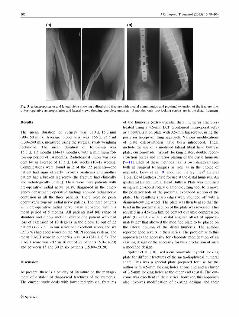
Fig. 3 a Anteroposterior and lateral views showing a distal-third fracture with medial comminution and proximal extension of the fracture line.
b Post-operative anteroposterior and lateral views showing complete union at 4.5 months; only two locking screws are in the distal fragment
102 J Orthopaed Traumatol (2015) 16:99–104
123

bulk production for universal use. Zhiquan et al. [11]
treated 13 distal third humeral shaft fractures with mini-
mally invasive percutaneous osteosynthesis (MIPO).
Fractures were reduced by closed means and fixed with a
long narrow 4.5-mm dynamic compression plate intro-
duced through two small incisions away from the fracture
site. The plate was fixed on the anterior aspect of the
humerus under fluoroscopy guidance. The radial nerve was
not exposed during this procedure. They reported that the
fractures united with a mean healing time of 16.2 weeks, a
little longer than the reported time of 9–12 weeks in pos-
terior open plating of the humerus. Disadvantages of this
approach are that the radial nerve is not visualized directly
during the exposure and, biomechanically, the posterior
surface of the humerus is considered better for plate
application especially of distal-third fractures. Schatzker
and Tile listed four reasons for plating the distal humerus
posteriorly—the posterior surface of the distal humerus
provides a flat surface suitable for plating; placement of the
most distal screws from a posterior approach allows direct
visualization and avoids the antecubital fossa; posterior
placement allows for the plate to extend distally permitting
additional screw placement; and the posterior approach
provides the option of double plating [12]. Livani et al. [6]
obtained good results following minimally invasive per-
cutaneous DCP fixation of distal humerus fractures in six
patients with radial nerve palsy. We chose an LCP due to
the presence of significant comminution in many of our
patients. The open surgical approach that we used required
more soft tissue stripping which made stable fixation
mandatory. Since the majority of our patients had no radial
palsy pre-operatively, nerve exploration and protection
required an open approach.
Prasarn et al. [13] treated extra-articular fractures of the
distal third of the humerus with dual plates from a single
posterior midline incision (2.7- and 3.5-mm pelvic recon-
struction plates). The average time to union was
11.5 weeks and the mean elbow flexion/extension arc was
4�–131�. Possible disadvantages of this approach are the
necessity to reflect the triceps to accommodate plate
application on the lateral column, and the need for using
two plates to secure reduction. The 2.7- and 3.5-mm plates
used in this series tend to be less strong than 4.5-mm
compression plates.
Advantages of our technique are that fracture stabiliza-
tion is achieved with a single 4.5-mm LCP without any
modification of the implant except for slight contouring.
The posterior approach dissection was limited up to the
olecranon fossa hence avoiding triceps fibrosis/elbow
stiffness as it was not necessary to expose the lateral col-
umn until the distal tip. Use of a 4.5-mm LCP obviates the
need for double plating and simplifies the procedure.
Contouring allows the plate to match the posterior surface
of the humerus and prevents the tip of the plate from rising
above the humerus just proximal to the olecranon fossa.
Secondly, it minimizes stress on the skin and soft tissues
overlying the plate [14]. Bending was performed at the
level of the dynamic hole in the plate as recommended by
Smith et al. [15]. Since the distal fixation relies on only two
screws, quality of bone is important and the technique is
best avoided in elderly patients with poor bone quality and
in highly comminuted fractures. Our results were excellent
in terms of fracture union as well as elbow and shoulder
range of motion. We had two complications, namely
breakage of a lag screw in one patient and early myositis
ossificans in the second patient; however, the patients were
not seriously affected and the quality of the results did not
suffer due to these complications. Our post-operative pro-
tocol consisted of immobilization of the elbow in a long
arm slab for 3 weeks. However, the slab was removed
every week and the elbow was mobilized under the direct
supervision of the surgeon. This subsequently proved to be
helpful in the early recovery of range of movement. There
is no need for elaborate modification of existing implants
and no need for the use of custom-made implants. It can be
argued that use of only two screws in the distal fragment
might compromise the stability of fixation. It has been
shown by Hak et al. [16] that two locking screws per
segment are sufficient and the addition of a third screw in
the locked plate construct did not add to the mechanical
stability in axial loading, bending, or torsion. It is possible
to insert at least two locking screws in the distal fragment
in the vast majority of distal humeral fractures.
We conclude that the use of one or two lag screws along
with a single posteriorly placed 4.5-mm contoured locking
compression plate having at least two locking screws in the
distal fragment provides sufficient rigid fixation in distal
metaphyseal fractures of the humerus. The dissection does
not extend beyond the apex of the olecranon fossa. The
implant stops well short of the olecranon fossa. Excellent
results can be achieved in these fractures without the use of
dual plating and without the need for expensive customized
implants or elaborately modified implants. Careful patient
selection is important for this technique and indiscriminate
use of single-plate fixation should be avoided. Physiolog-
ically, young patients with good bone quality and good
motivation for post-operative physiotherapy are suitable
for this technique. Patients with open fractures, highly
comminuted fractures, fractures with intercondylar exten-
sions and pathological fractures are not suitable for this
type of fixation.
Conflict of interest Authors have not received any funding for this
study.
Ethical standards Written informed consent was obtained from the
patients prior to enrolling them in the study. The study was approved
J Orthopaed Traumatol (2015) 16:99–104 103
123

by hospital ethics committee and performed in accordance with the
ethical standards of the 1964 Declaration of Helsinki as revised in
2000.
Open Access This article is distributed under the terms of the
Creative Commons Attribution License which permits any use, dis-
tribution, and reproduction in any medium, provided the original
author(s) and the source are credited.
References
1. Stewart MJ, Hundley JM, Tennessee M (1955) Fractures of the
humerus-A comparative study in methods of treatment. J Bone
Joint Surg Am 37-A(4):11
2. Sarmiento A, Horowitch A et al (1990) Functional bracing for
comminuted extra-articular fractures of the distal third of the
humerus. J Bone Joint Surg Br 72(2):283–287
3. Ring D, Harris M, Doornberg J, McCarty P, Jawa A (2006) Extra-
articular distal-third diaphyseal fractures of the humerus. A
comparison of functional bracing and plate fixation. J Bone Joint
Surg Am 88-A:2343–2347
4. McKee MD (2006) Fractures of the shaft of the humerus. In:
Bucholz RW, Heckman JD, Court-Brown CM (eds) Rockwood
and green’s fractures in adults. Lippincott Williams & Wilkins,
Philadelphia, pp 1117–1159
5. Archdeacon MT, Cannada LK, Herscovici D, Anglen JO (2008)
Avoiding complications in the treatment of humeral fractures.
J Bone Joint Surg Am 90-A:1580–1589
6. Livani B, Belangero WD, Castro de Medeiros R (2006) Fractures
of the distal third of the humerus with palsy of the radial nerve:
management using minimally-invasive percutaneous plate oste-
osynthesis. J Bone Joint Surg Br 88(12):1625–1628
7. Morrey BF, An KN, Chao EYS (1993) Functional evaluation of
the elbow. In: Morrey BF (ed) The elbow and its disorders, 2nd
edn. W. B. Saunders, Philadelphia, pp 86–89
8. Atroshi I, Gummesson I, Andersson B, Dahlgren E, Johansson A
(2000) The disabilities of the arm, shoulder and hand (DASH)
outcome questionnaire. Acta Orthop Scand 71:613–618
9. Levy JC, Kalandiak SP, Hutson JJ, Zych G (2005) An alternative
method of osteosynthesis for distal humeral shaft fractures.
J Orthop Trauma 19:43–47
10. Spitzer AB, Davidovitch RI, Egol KA (2009) Use of a ‘‘hybrid’’
locking plate for complex metaphyseal fractures and non unions
about the humerus. Injury 40:240–244
11. Zhiquan A, Bingfang Z, Yeming W, Chi Z, Peiyan H (2007)
Minimally invasive plating osteosynthesis (MIPO) of middle and
distal third humeral shaft fractures. J Orthop Trauma 21:628–633
12. Schatzker J, Tile M (1996) The rationale of operative fracture
care, 2nd edn. Springer, Toronto, pp 83–94
13. Prasarn ML, Ahn J, Paul O, Morris EM, Kalandiak SP, Helfet
DL, Lorich DG (2011) Dual plating for fractures of the distal
third of the humeral shaft. J Orthop Trauma 25:57–63
14. Sudkamp NP, Niemeyer P (2006) Principles and clinical appli-
cation of the locking compression plate (LCP). Acta Chir Orthop
Traumatol Cech 73:221–228
15. Smith WR, Ziran BH, Anglen JO, Stahel PF (2007) Locking
plates: tips and tricks. J Bone Joint Surg Am 89A:2298–2307
16. Hak DJ, Althausen P, Hazelwood SJ (2010) Locked plate fixation
of osteoporotic humeral shaft fractures: are two locking screws
per segment enough? J Orthop Trauma 24:207–211
104 J Orthopaed Traumatol (2015) 16:99–104
123