a comparative study between locking compression …
TRANSCRIPT

“A COMPARATIVE STUDY BETWEEN LOCKING
COMPRESSION PLATE AND NON LOCKING
COMPRESSION PLATE IN THE TREATMENT OF
INTRAARTICULAR CALCANEAL FRACTURE”
DISSERTATION SUBMITTED FOR
MASTER OF SURGERY DEGREE EXAMINATION
(BRANCH – II, ORTHOPAEDIC SURGERY)
MARCH-2015
THE TAMILNADU
DR.M.G.R MEDICAL UNIVERSITY
CHENNAI. TAMILNADU

CERTIFICATE
This is to certify that the work entitled “A comparative study
between locking compression plate and nonlocking compression plate in
the treatment of intrarticular calcaneal fracture” which is being
submitted for M.S. ORTHOPAEDICS, is a bonafide work of
DR.S. ESWARAPANDI, post graduate student in the Department of
Orthopaedics, Tirunelveli Medical College, Tirunelveli.
DEAN
Tirunelveli Medical College,
Tirunelveli.

CERTIFICATE
This is to certify that the work entitled “A comparative study
between locking compression plate and nonlocking compression plate in
the treatment of intrarticular calcaneal fracture” which is being
submitted for M.S. ORTHOPAEDICS, is a bonafide work of
DR.S. ESWARAPANDI, post graduate student in the Department of
Orthopaedics , Tirunelveli Medical College, Tirunelveli.
He has completed the necessary period of stay in the department and
has fulfiled the conditions required for submission of this thesis according to
the university regulations.The study was undertaken by candidate himself
and observations recorded have been periodically checked by us.
Recommended and forwarded
Prof. Dr. Elangovan Chellapa
M.S. Ortho, D. ortho,
Professor and Head,
Department Of Orthopaedics,
Tirunelveli Medical College,
Tirunelveli.


DECLARATION
I Dr.S.Eswarapandi, solemnly declare that the dissertation titled “A
COMPARATIVE STUDY OF LOCKING COMPRESSION PLATE
AND NONLOCKING COMORESSION PLATE IN THE
TREATMENT OF INTRAARTICULAR FRACTURES OF
CALCANEUM” has been prepared by me. This is submitted to “The
Tamilnadu Dr. M.G.R. Medical University, Chennai”, in partial
fulfilment of the regulations for the award of MS degree Orthopaedics.
Place : Tirunelveli Dr. S.ESWARAPANDI
Date

ACKNOWLEDGEMENT
At the very outset I would like to thank Dr. L.D. ThulasiRam M.S
the Dean, Tirunelveli Medical College for permitting me to carry out this
study in this hospital.
I am greatly indebted to my beloved chief, Prof . Dr. Elangovan
Chellapa MS Ortho, D.Ortho, Professor and Head of the Department of
Orthopaedic Surgery, Tirunelveli Medical College for his invaluable help,
encouragement and guidance rendered to me in preparing this dissertation.
I am grateful to Prof. Dr. Manikandan MS. Ortho in guiding me to
prepare this dissertation.
I am most indebted and take immense pleasure in expressing my deep
sense of gratitude to Prof. Dr. Thanikaimani MS Ortho,
Prof. Dr. N. Thanappan MS.Ortho for their easy accessibility and timely
suggestion which enabled me to bring out this dissertation.
I also take this oppourtunity to thank Dr. Sureshkumar,
Dr. Maheshwaran, Dr.Senthil Kumar, Dr. Arogya Ammayan,
Dr. Babu Aloy, Dr. Abraham James, Dr. Abul Kasim,
Dr. Chandrasekar, Dr. Sundarapandian Assistant Professors, Department
Of Orthopaedics, Tirunelveli medical college, for their timely help and
guidance given to me during all stages of the study.
Last but not least, I express my gratitude to the patients for their
kind co-operation.

A COMPARATIVE STUDY BETWEEN LOCKING COMPRESSION
PLATE AND NONLOCKING COMPRESSION PLATE IN THE
TREATMENT OF INTRAARTICULAR CALCANEAL FRACTURE
ABSTRACT
Background:
In a prospective study we analysed intra-articular calcaneal fracture
treatment by comparing results and complications related to frature
stabilization with locking and nonlocking compression plates.
Materials and Methods:
Our study 23 osteosynthesis(20 patients) of intra articular calcaneal
fracture using the standard extensile lateral approach from July 2012 to July
2014.10 operation using locking plate,10 operation using nonlocking
compression plate.In the Sanders type 4 fractures,reconstruction of the
calcaneal shape was attempted .The patients were evaluated by the AOFAS
Ankle Hindfoot Score.
Results:
Wound healing complication,late subtler arthritic changes high in
nonlocking compression plate.preoperative size of Bohlers,Gissanes angle
correlated with post operativ and one year follow up clinical and radiological
results in both groups.There were no late complications in locking
compression plate group.All the late complication occur in the nonlocking
compression group.The overall results according to the AOFASankle
Hindfoot Score were LCP group better than the NONLOCKING group.
Conclusion:
Open reaction and internal fixation of intraarticular calcaneal fractures has
become a standard surgical method.Fewer complications and better results
related to treatment with locking compression plates confirmed in comparison
to nonlocking ones were noted for all Sanders type of intraarticular calcaneal
fractures

CONTENTS
TITLE PAGE NO:
1. Introduction 1
2. Aim of the Study 3
3. Materials and Methods 42
4. Results 61
5. Statistical Analysis 64
6. Discussion 96
7. Limitations 99
8. Conclusion 100
9. Bibliography
ANNEXURES:
A. Master chart
B. Proforma
C. AOFAS - Ankle Hind Foot Score
E. Anti Plagiarism certificate
F. Ethical committee approval certificate.

1
INTRODUCTION
The calcaneum or Os Calcis is a unique bone, with a unique
mechanism of fracture. The most frequently fractured bone of the foot is the
calcaneum. Calcaneal fractures commonly occur in young active male and
industrial workers9. The burden to the society is significant with the
socioeconomic impact being substantial in the terms of days away from
work and recreation.
Till the end of 19th century calcaneal fractures were treated
nonoperatively with rest and limb elevation8.
The advent of advanced imaging modalities that kindled desire of
restoring the distorted anatomy of this bone. The operative treatment of
calcaneal fractures includes percutaneous k-wire, percutaneous screw, non-
locking compression plate and locking compression plate.
The controversy between the operative and nonoperative interventions
remains ongoing subsequent analysis and other related publictions has
pushed the pendulum towards the surgical option114.
More high level research is needed towards determining exactly which
operative exposures, techniques, instrumentations and other parameters are
ideal. The advent of CT scan provides information regarding size and
number of fracture fragments, sustentaculum displacement relative to the
supromedial fragment, posterior facet congruity, lateral malleolus

2
impingement on the tuberosity22 and hence a newer classification system
with prognostic significance.
In our study we have tried to compare the outcome between the
locking compression plate and nonlocking compression plate in the
treatment of intrarticular calcaneal fractures admitted during the period from
july 2012 to july 2014 at Tirunelveli medical college hospital, Tirunelveli.

3
AIM
To compare the clinical, radiological and functional outcome between
the locking compression plate and nonlocking compression plate in the
treatment of intra articular calacaneal fractures.

4
EPIDEMIOLOGY
Calcaneal fractures represent only 2% of all fractures of adult
population6 and 75% fractures of foot and the extraarticular fractures
represent upto 60% of calcaneal fracture in children and their incident has
been reported to be 25% to 40% of adult calcaneal fractures9.
ANATOMICAL CONSIDERATIONS
The Os Calcis is described as an irregular bone ,predominantly
cancellous in structure and enveloped in the a shell of thin cortical bone. It
represents the largest bone of the seven tarsal bones.
Calcaneum possess six surfaces and articulate with two bones talus
and cuboid. The anterior surface articulate with the cuboid, its saddle in
shaped.
Anterior Surface
It is the smallest surface. It is obliquely placed and saddle shaped. Its
articular facets articulates with cub
Posterior Surface
The Posterior surface is divided into three parts, the proximal area
which is separated from tendo Achilles by a bursa and fibro fatty tissue, the
middle part which gives attachment to the Achilles tendon and the distal part
which is the subcutaneous weight bearing surface.

5
SUPERIOR SURFACE (FIG 1)
The superior surface articulates with the talus. The superior surface is
divided into posterior, middle and anterior facets. Posterior facet is inclined
to the saggital plane at 45 degree and the middle facet articulate with the
talar head and neck and it lies on the sustentaculum tali. Anterior facet
located over the anterior end of the superior surface and articulates with
anterior talar facet and it rests on the beak of calacaneum. Between the
posterior and middle facets lies the calcaneal sulcus which together with the
similar groove on the talus forms the sinus tarsi. Sinus tarsi lodges the artery
of the sinus tarsi and the intraosseous talocalcaneal ligament. Anterior to the
anterior facet the bifurcate y ligament is attached and lateral to it is the
origin of extensor digitorum brevis.
The posterior subtalar joint which is the primary weight bearer is
located in the middle of the superior surface. It is supported by the strong
compression trabeculae under the surface of compression trabeculae called
the thalamic portion of the bone. Posterior subtalar joint is saddle shaped
joint and supports the eversion inversion of the foot.

6
Fig 1:Superior Surface
Fig 2: Lateral Surface
Lateral Surface (fig 2).
This surface is subcutaneous and flat and palpable beneath under
lateral malleolus. An elevation located 2cm distal to lateral malleolus is
peroneal tubercle. Peroneal tubercle serves as an insertion for peroneal
retinaculum. Calcaneal tubercle is above and behind the trohlea it gives
attachment to calcaneofibular ligament.peroneus longus and brevis tendon is

7
separated by the peroneal trochlea to which the inferior peroneal retinaculum
is attached
Medial Surface (Fig 3)
On the anterosuperior aspect of its medial surface is larger and
stronger, sustentaculum tali groove for tibialis posterior tendon and flexor
hallusis longus, is inferior to the sustentaculum tali. The flexor accessories
medial head is inserted to the distal to this groove. The intraosseous
ligamen,plantar calcaneo navicular ligament,anterior fibers of the deltoid
ligament and the medial talocalcaneal ligament holds the talus medial to the
calcaneum
Fig 3: Medial Surface
8
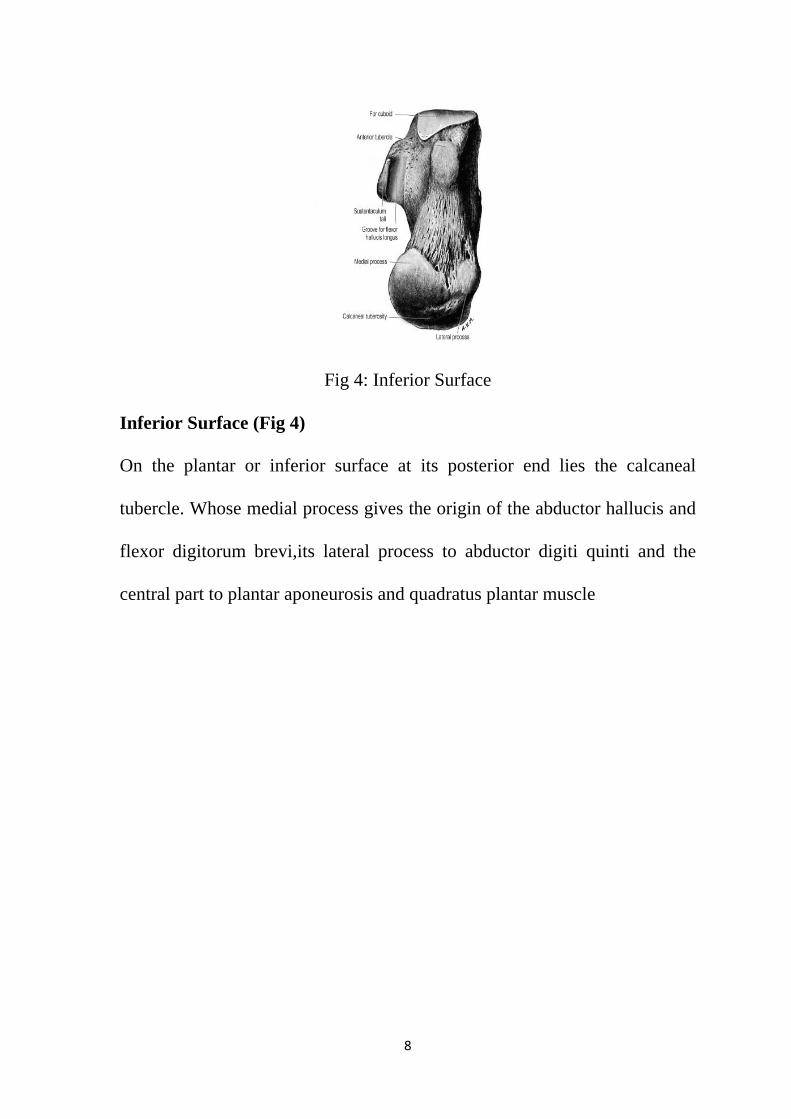
Fig 4: Inferior Surface
Inferior Surface (Fig 4)
On the plantar or inferior surface at its posterior end lies the calcaneal
tubercle. Whose medial process gives the origin of the abductor hallucis and
flexor digitorum brevi,its lateral process to abductor digiti quinti and the
central part to plantar aponeurosis and quadratus plantar muscle

9
SOFT TISSUE RELATIONS
Fig 5 .Soft tissue in relation to the medial surface of the hind foot
The interosseous talocalcaneal and cervical ligaments and medial root of
inferior extensor retinaculum are attached to the calcaneal sulcus. The
nonarticular area in the distal part of the posterior facet gives attachment to
the extensor digitorum brevis, inferior extensor retinaculum and bifurcated y
ligament6.
The sustentaculum gives attachment to flexor retinaculum. The
plantar calcaneo ligament is attached to the medial margin9.
The long plantar ligament orginates between the medial and lateral
process. It extends to the tubercle distally. The lateral head of the flexor
accessories is attached to the lateral margin of long plantar ligament.

10
BLOOD SUPPLY9
Fig 6. Blood supply of foot showing calcaneal supply

11
Avascular necrosis of the calcaneum is rare compared to that of the
talus due to profuse muscular, tendinious, ligamentous attachments.
Vascular anastomosis from
1. Posterior tibial artery gives branches to medial and lateral plantar
arteries.
2. Anterior tibial artery gives branches to medial and lateral malleolar
arteries.
3. Perforating branch of the peroneal artery.
NERVE SUPPLY
The branches of tibial,sural and deep peroneal nerves innervates the
calcaneum6.
OSSIFICATION
Calcaneum ossifies from one primary during the third month of
intrauterine life. One secondary center which appears at six to eight years ,at
14- 16 years secondary centre fuses6.

12
MECHANISM OF INJURY
Calcaneal fractures with intraarticular involvement and displacement
can occur due to high velocity injury which usually is due to fall from
height. In this mode of injury the patient’s weight is concentrated on the
heels on axial loading. Other mode of injuries like high velocity injuries are
due to motor vehicle accidents. Axial load injuries are associated with spine
and pelvic injuries. The pattern of communition and the location of the
fracture lines are dependant on the position of foot at the time of impact, the
forces of impact and bone quality.
According to Carl et al two primary fracture lines were consistently
observed. One line divided the calcaneum to medial and lateral portion. The
other fracture line divided the calcaneum into anterior and posterior portions,
starting laterally from angle of Gissane running medially. These two fracture
lines resulted in a variety of tongue and joint depression type fractures

13
Fig 7.1: The primary fracture line.
Fig 7.2: Mechanism Of Injury.A-C Joint depression type. D-F Tongue type

14
CLASSIFICATION
Calcaneal fractures initially classification based on conventional
radiographs but posed difficulties in planning treatment. Nowadays CT
based classifications, treatment has improved.
ESSEX-LOPRESETI (1952)
Fig 8: Essex- Lopresti Classification. Line 1 represents primary shear
fracture, Line 2 –secondary compression fracture giving the tongue
fragment, Line 3- secondary compression fracture of the joint depression
type.

15
TONGUE TYPE FRACTURE: the articular fragment remain attached to
the tuberosity fragment.
JOINT DEPRESSION TYPE: The articular fragment was separate from
the adjacent tuberosity.
SHOEUR AND REMY (1975) : based on the number of articular bony
fragments.
FIRST DEGREE: undisplaced shear type fractures with separation of joint
surfaces .
SECOND DEGREE: includes secondary fracture lines and a minimum of
three fragments , of which two should include the articular surface.
THIRD DEGREE: Highly communited fractures.
CROSBY AND FITZGIBBONS
TYPE I –Undisplaced
TYPE II - Displaced
TYPE III - Communited

16
SANDERS (CT Based)
This classification refers to intra-articular fractures and was developed
in 1993 after following 120 patients for a minimum of 1 year. It utilizes the
axial and coronal hindfoot CT cuts. The planes of reconstruction are the
semicoronal(perpendicular to posterior facet ) and the axial ( parallel to the
sole of foot). The cut showing the widest part of the undersurface of the
posterior facet of the talus is taken into consideration and this is divided into
three equal columns by two lines ( A & B). Another line C that is drawn
from the medial edge of the posterior facet of the talus, divides the
corresponding calcaneus from the sustentaculum. Thus the entire calcaneus
is divided into four segments : lateral, central, medial and sustentaculum
segments. Four types of fractures are then described according to the number
and location of the fracture fragments(fig 9).
TYPE I– Undisplaced fractures with less than 2mm displacement regardless
of fracture lines
TYPE II- Two part fractures of the posterior facet. Based on the location of
the primary fracture line.It is further divided into three types IIA, IIB, IIC.
TYPE III- Three part fractures of the posterior facet. Usually centrally
depressed fragment further its divided into three types based on the primary
fracture line. Types IIIAB, IIIAC, IIIBC.

17
TYPE IV – Highly communited 4 parts articular fragment. Often had more
than four articular fragments
Universally Sander classification is most commonly used. In our study we
have followed sanders for planning our treatment.
Fig9: Sander’s classification.

18
RADIOLOGICAL ANATOMY
Calcaneum is an important bone as it transmits the body weight to the
ground and also helping the calf muscle action by creating a strong lever.
Traction trabeculae extending from the inferior cortex of calcaneum
converges over the compression trabeculae supporting the posterior and
anterior articular facets.
The area under the thalamic segment of the bone with relatively sparse
trabeculae called neutral triangle (refer fig 10). This area is considered to be
of little significance in the pathological anatomy of fractures. But when the
posterior subtalar joint is depressed the vacuous neutral triangle might fail to
support the articular surface even after it has been elevated to its original
state. The adjuvant is in favour of grafting in the region in the depressing
fractures affecting this area.
Fig 10:The Neutral Triangle

19
Lateral view of the radiograph of the calcaneum is used to identify
two important angles one is bohler angle otherwise called tuber angle of
bohler(refer fig 11) usually between 20 to 40 degree, formed by two lines
first line starts from the highest point on the anterior process of the
calcaneum to highest point on the posterior facet. The second lines tangential
to the superior edge of the tuberosity. Decreases in this angle it indicates the
weight bearing surface of the calcaneum has collapsed and shifting the body
weight anteriorly
Fig 11: Bohler’s angle.

20
Second angle, cruciate angle of gissane (refer fig 12) is seen directly
inferior to the lateral process of the talus it is represented by two strong
cortical struts that extend laterally and form an obtuse angle. The first strut
extend along the lateral border of the posterior facet, the second extends
anteriorly to the beak of the calcaneum. Normal gissane angle 90 to 110
degree.
Fig 12: The Gissane’s Angle.
McLaughln pointed out that reduction or reversal of this angle
indicates only the degree of the proximal displacement of the tuberosity and
therefore the angle can be decreased in both the extraarticular, intraarticular
fractures thus limiting its usefulness.

21
RADIOGRAPHIC EVALUATION:
The initial evaluationof the fracture should be done with plain
radiographs.This includes anteroposterior, lateral and Harris axial views.If
articular involvement is diagnosed CT scanning should be performed. Many
special radiographic views like Mortise view, Broden view and Anthonson’s
oblique view were described.
The lateral view confirms the diagnosis and classifies fractures
according to Essex-Lopresti into joint depression type or tongue type.
The Harris axial view of the heel visualizes the joint surface, loss of height,
increase in width and angulation of the tuberosity fragment. Axial view is
taken with ankle dorsiflexed by means of a bandage held by the patient and
the x-ray tube tilted 45 degrees from the foot withits axis parallel to the
posterior compartment of the joint.
Fig 13 –Axial Harris view

22
Fig 14 – AXIAL VIEW
Broden’s view is a reproducible means of demonstrating the posterior facet
on radiographs.It is obtained with foot in neutral position and leg internally
rotated 30-40 degrees.The x-ray beam is centered over the lateral malleolus
and radiographs made with the tube angled 40,30,20,10 degrees
cephalad.The 10 degree view shows the posterior portion of the facet and 40
degrees shows the anterior portion.Intraopertive fluoroscopic visualization of
Broden projection is helpful in verifying the reduction of posterior facet.

23
Fig 16.BRODEN’S VIEW - 100
Fig 17.BRODEN’S VIEW- 200

24
Fig 18.BRODEN’S VIEW- 400
Fig 19: Broden’s view

25
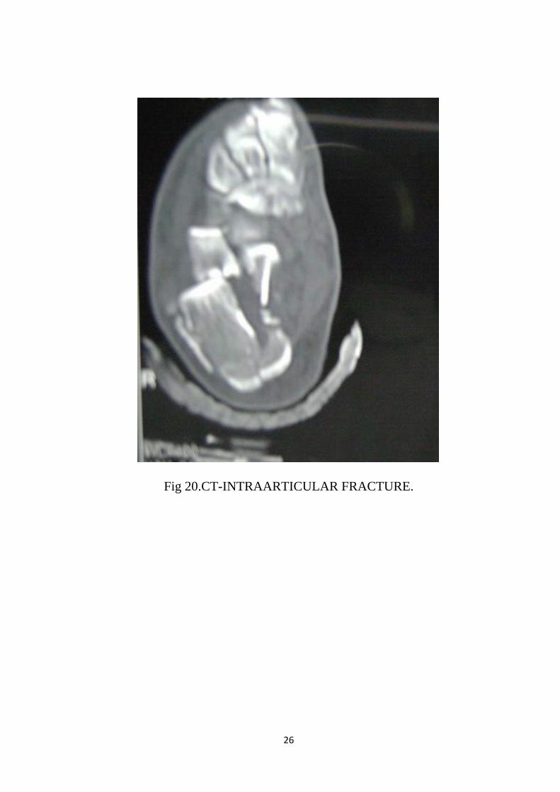
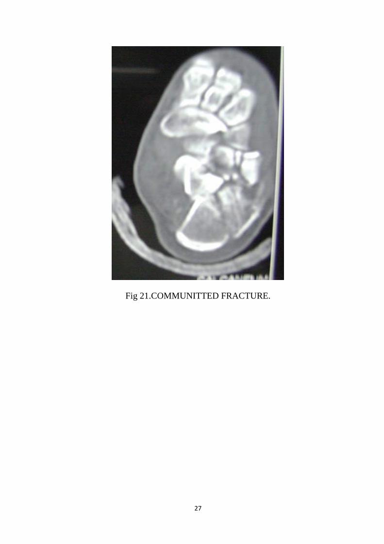
CT SCAN
CT scanning provides excellent information regarding the number of
fractures on the posterior facet and its location. It offers data for
diagnosis,classification and treatment.CT images ( Fig 20,21) should be
obtained in the axial, 30 degree semicoronal, and saggittal planes with three-
dimensional reconstruction.
Axial views gives information about calcaneocuboid joint, the
anteroinferior aspect of the posterior facet, and the sustentaculum. Coronal
views shows the posterior facet, sustentaculum,shape of the heel overall.
Saggittal views shows the posterior facet, the calcaneal tuberosity and the
anterior process. Though three dimensional CT scanning is an interesting
modality the definition of articular surface is not sufficient for pre-operative
planning.
CT evaluation has allowed classification systems to offer prognostic
significance. By reducing the need for multiple views of the heel, it reduces
the radiation dose and discomfort to the patient.
In our series we have used the anteroposterior, lateral and Harris axial
views along with CT imaging for classification and preoperative planning.
26
Fig 20.CT-INTRAARTICULAR FRACTURE.
27
Fig 21.COMMUNITTED FRACTURE.

28
TREATMENT OPTIONS
CONSERVATIVE TREATMENT
SURGICAL MANAGEMENT
A. CONSERVATIVE TREATMENT
INDICATIONS:
1. Undisplaced or minimally displaced extraarticular fractures
2. Undisplaced intraarticular fractures.
3. Anterior process fractures with less than 25% involvement of the
calcaneocuboid articulation.
4. Fracture in patients with other comorbid conditions which are
prohibiting the surgery.
5. Elderly patients who are household ambulator
6. Chronic smokers, uncontrolled diabetes and peripheral vascular
diseases.
The nonoperative treatment consists of initial rest, ice, compression,
elevation regime followed by a supportive splint. After the swelling subside
the prefabricated fracture boot, with the ankle locked in the neutral flexion to
prevent the equinus contracture. Apply the elastic compression stocking to
minimise the dependant edema. Early subtalar and ankle joint range of
motion exercise are initiated. Weight bearing restriction are maintained for
upto 10 to 12 weeks until radiographic union is confirmed. After three
months gradual progressing weight bearing is started as tolerated.

29
A. SURGICAL MANAGEMENT:
1. Minimally invasive technique
2. Open reduction internal fixation with plate osteosynthesis
3. Ligamentotaxis
4. Primary arthrodesis
1. MINIMALLY INVASIVE METHOD
INDICATION :
a. Communitted fracture or impending soft tissue compromise
b. Peripheral vascular disease.
c. Heavy smoker
d. Uncontrolled diabetes
IMPLANT :
a. K-wire
b. Cancellous screw
TECHNIQUE
Small incision over the fracture fragments, reduced by Essex-
Lopressti manoeuvre, this manoeuvre entails indirect reduction of the
fracture by leveraging with a K-wire/ Steinman pin or a Schantz screw
inserted into the posterior tuberosity fragment like a joystick. The
manoeuvre has its role in tongue type fractures, particularly , Sander’s type
IIC, in which this fragments contains the whole posterior facet, displaced in

30
entirely. However , it can also be used in type IIB as well as more complex
fractures, with or without other maneuver.
ADVANTAGE
a. Postoperative wound complication is less.
b. Tongue type fractures can be managed.
DISADVANTAGE
a. In case of communited fractures, subtalar congruity couldnot be
obtained.
2. PLATE OSTEOSYNTHESIS
Open reduction internal fixation with plate osteosynthesis is the
standard operative treatment for intraarticular calcaneal fractures.
APPROACH: Extensile Lateral Approach
Advantage of the approach is excellent exposure of the subtalar joint
and anatomical reduction of the fracture fragment under vision.
IMPLANT :
A. Nonlocking compression plate with 3.5mm cancellous screw
B. Locking compression plate with locking compression screw
These can also be used :-
a. One third tubular plate
b. Cervical h plate
c. 3.5mm recon plate
d. Recon y plate
e. Tentacle calcaneal plate

31
ADVANTAGE
a. Early mobilization
b. Prevent the post operative collapse
3. LIGAMENTOTAXIS
Reduced fracture fragments by FORGON principles of
ligamentotaxis.
Skeletal traction over the three bony points( tuberosity of calcaneum, talus
and cuboid )
Under fluoroscopy control depress the fracture fragments elevated
percutaneously by k wires and fixed with lag screw.
ADVANTAGE
a. Re-establishing the calcaneal morphology
b. Talocalcaneal relationship resulting in restoration of ankle and
transverse dorsal joint range of motion
4. PRIMARY ARTHRODESIS
Only indication is Sander’s Type IV fractures
TECHNIQUE
Posterior facet is denuded of its cartilage in both the surface after
adequate reduction of the calcaneal tuberosity and joint surface, then the
graft is placed. The fusion is stabilized by 6.5mm cancellous screw, screw
directed from axially through the OsCalcis into talar dome. Upto three
months nonweight bearing walking.

32
BONE GRAFTS AND SUBSTITUTES
Pioneer of the bone graft in calcaneal fractures PALMER.
Graft is being used to fill the defect and maintain the neutral angle
with rotation even upto 90 degree relation to the subtalar joint when axial
load depress the posterior facet.
120 cases operated by Sander’s Et al17 without grafting not even single
case developed in postoperative collapse. Overall the decision to use bone
grafts and substitutes based on the
a. Patient status
b. Functional needs
c. Fracture pattern
d. Type of fixation
e. Available materials
TIME OF SURGERY
The most commonly accepted course is to wait until the swelling
resolves and the blisters are reepithelized before proceding with the surgery.
The great deal of swelling of foot and ankle with blistering of skin.
Soft tissue damage aggravated by surgery and a difficult postoperative
wound closure due to skin edema, so increase the risk of skin necrosis and
infection. These are the factors, operative treatment undertaken within three
weeks of fracture within early consolidation, if after 3 weeks fracture
fragment reduction will be difficult.

33
Resolve of the swelling is marked by the “ WRINKLETEST “ it
presents as skin wrinkles over the lateral aspect of foot and ankle with
dorsiflexion and eversion.
Fig 22: The Wrinkle Test

34
VARIOUS SURGICAL APPROACHES9
Fig 23 Extensile Lateral approach Vs Sinus Tarsi approach
1- Sural nerve, 2- superficial peroneal nerve, 3- sinus tarsi approach, 4-
extensile lateral approach

35
CALCANEAL OPERATIVE APPROACHES
1. The Palmer approach
2. Sinus tarsi approach
3. Small lateral approach
4. Extensile sinus tarsi approach
5. Geel and Flemister approach
6. Posterior plantar approach
7. Straight lateral subtalar approach
8. Extended lateral transcalcaneal approach
9. Sustentatcular approach
10. Medial approach
11. Carr’s modification of medial approach
12. Gallie ‘s approach
13. Cincinatti University approach
14. Combined medial and lateral approach
15. Ollier approach

36
EXTENSILE LATERAL APPROACH
Described by FERNANDEZ
Standard approach for surgical fixation of displaced intraarticular
calcaneal fractures.
It provides excellent exposure to subtalar joint, anterior process,
lateral wall and calcaneal tuberosity. Adequate visualisation of these
structures facilitates the anatomic reduction of articular surface and the
restoration of normal calcaneal morphology.
These are the primary goal of surgical treatment
LOCKING COMPRESSION PLATE
With the advent of locking plate concept by Wagner and A.O group,
the concept was extended for use in foot and ankle. The plate is applied
through the lateral extensile approach and fragment screw provide bicortical
and (or) unicortical fixation. The threaded locking holes offer a fixed angle
construct to buttress the articular surfaces of the calacaneum and permit
multiple points of fixation to buttress small fragments. The locking holes
provide 15 degree of angulation when using 2.7 mm cortical screws and 5
degree of angulation when using 3.5 mm cortical system. This locking
calcaneal plate is side specific, made of titanium and comes in two sizes of
69mm and 76mm variation of this concept have been marketed by different
implant manufactures. The calcaneal locking plate seems to fulfil all the
requisites desired in calcaneal fracture fixation. It has specific proven

37
advantages with better hold in bone, lower profile, versatility in screw
placement, avoiding or minimizing the need for graft or bone substitutes and
allowing for earlier weight bearing. This is evidenced by the study of Race et
al19.
COMPLICATIONS
Divided into early complications and late complications
EARLY COMPLICATIONS
1. Primary healing with superficial wound infection
2. Deep wound complication with osteomyelitis
3. Compartment syndrome
4. Sural nerve injury
5. Implant related
LATE COMPLICATIONS
1. Arthritis
2. Malunion
3. Nonunion
4. Nonreduction in tuberosity
5. Heel exostosis
6. Reflex sympathetic dystrophy
WOUND INFECTION
Superficial wound infection is the most common complication.
Treated with conservatively using saline dressing and oral antibiotic.

38
In cases of deep infection repeated wound debridement is tried and free
tissue transfer is attempted if needed. Implant removal in case of diffuse
osteomyelitis is present.
SURAL NERVE INJURY
Operative treatment associated with nerve injury mostly with sural
nerve presents with neuroma or loss of sensation in the affected region. But
in extensile lateral approach this would be avoided.
COMPARTMENT SYNDROME
The literature document an incident 1-17% of patients who develop
compartment syndrome following calcaneal fracture.But Indian literature
does not report cases of compartment syndrome after calcaneal fractures 9.
ARTHRITIS
Subtalar arthritis is the most common form of arthritis seen in
calcaneal fractures. Calcaneocuboid joint arthritis can also occur in calcaneal
fractures.
It was diagnosed by intrarticular injection of local anaesthetics which
relieves the pain.
Treatment conservative measures like footwear modification,
NSAIDs, walking aids. If these measures are not relieving the pain, subtalar
arthrodesis is next line of management.

39
MALREDUCON OF TUBEROSITY
Malposition can occur in both locking compression plate and
nonlocking compression plate resulting in varus angulation of hindfoot.
When the patient stands on toes the fibular fragment become more
pronounced and the treatment consists of lateral shoe wedges or corrective
osteotomy.
HEEL EXOSTOSIS
Plantar aspect of the heel develop painful bony prominence due to
fracture healing. Treatment consist of the use of heel pads or operative
removal. In 1921 COTTON first described the operative management of
heel exostosis by avoiding a plantar incision7.
REFLEX SYMPATHETIC DYSTROPHY (COMPLEX REGIONAL
PAIN SYNDROME)
There is two types CRPS Type I – not limited to the single peripheral
nerve distribution, but associated with edema, changes in the skin blood
flow.
CRPSType II – more commonly associated with open surgery or open
injury in lateral side affecting sural nerve9. In case of medial side injuries
tibial nerve is affected. Diagnosis is based mainly on clinical examination.
Treatment – physiotherapy, analgesics, antidepressant, sympathetic
blockade.

40
CALCANEAL MALUNION
In 1921, COTTON identified calcaneal malunion in heel. This
malunion would produce lateral wall abutment, peroneal impingement and
heel malalignment7. In case of heel malalignment extraarticular osteotomy is
done giving good results.
CLASSIFICATION OF CALCANEAL MALUNION ( STEPHEN’S
AND SANDER’S CLASSIFICATION)9
TypeI – Large lateral wall exostosis with or without marginal subtalar
arthrosis.
TypeII – Lateral wall exostosis combined with subtalar arthritis involving
the whole of subtalar joint.
TypeIII- Type II + Malpositioned heel either in varus or valgus.
TYPE I PERONEAL TENOLYSIS,
LATERAL EXOSTECTOMY
TYPE II TENOLYSIS,
EXOSTECTOMY,SUBTALAR
ARTHRODESIS
TYPE III ABOVE MEASURES WITH
ADDITIONAL OSTEOTOMY OF
THE CALCANEAL BODY.

41
Fig 24: Stephens and Sanders classification of calcaneal Malunion

42
MATERIALS AND METHODS
In our institution 20 patients with 23 fractures with displaced intra-
articular calcaneal fractures were selected for this prospective study. The
study period is from July 2012 to July 2014. Out of these patients 3 patients
had bilateral intra-articular calcaneal fractures. 17 fractures occurred in men
and 3 fractures occurred in women.
10 patients were treated with Locking Compression Plate and 10
patients were treated with non-locking compression plate based on our
inclusion and exclusion criteria .Only sanders II, III, and IV part fractures
were included in the study.
MODE OF INJURY:
Fall from height : 11
Road traffic accidents : 9
ASSOCIATED INJURIES:
Out of 20 patients 5 patients had associated injuries, 3 patients had
spine injuries without neurological defecit.one patient had Intertrochontric
fracture another patient had subtrochantric fracture.
The locking compression plate group had 3 cases associated with
other injuries. one case a fracture subtrochanter Seinsheimer’s classification
type IIB for whom Proximal Femoral Nail was performed and 2 cases of

43
spine fractures one being a D12 anterior wedge compression fracture and
other L2 wedge compression fracture,both patients without neurological
defecit.
The NonLocking Compression plate group had 2 associated injuries.
Of these 1 patient had spine injuries (L1Anterior Wedge Compression
fracture) without neurological deficit. One patient had intertrochanteric
fracture treated with Dyanamic Hip Screw.
No other systemic injuries were associated with these fractures.
INCLUSION CRITERIA:
1. Age group between 20-60 years
2. Non diabetic or under strict glycemic control.
3. Sander’s type II-IV.
4. Closed fractures, compound fracture grade I.
5. Dislocated calcaneal fracture ( posterior articular facet stepoff>2mm,
significant shortening, loss of height, widening of calcaneum ( decrease the
Bohler’s angle and increase the Gissane angle, valgus deviation >10 degree ,
varus deviation >5degree. )
6. Presenting within 3 weeks of injury.
7. If associated with other fractures.

44
EXCLUSION CRITERIA:
1.Age <20- >60 years.
2. Severe uncontrolled diabetis mellitus/severe medical problems/severe soft
tissue injury.
3. Sander’s type I
4. Grade II & III compound fracture
5. Associated vertebral fracture with neurological deficit.
6. Non-reducible dislocation of the calcaneum which needs subtalar
arthrodesis.
7. >21 days of injury.
AGE AND SEX DISTRIBUTION:
10 patients were operated with Locking Compression Plate of which 9
were male and one female. The Non-locking compression plate group which
consists of 8 patients, out of these 2 fractures occurred in female.
PRE-OPERATIVE ASSESSMENT:
All patients were evaluated using the Computerised Tomography (CT)
method and classified using SANDERS classification.
X-RAYS:
1.Standard ANTEROPOSTERIOR AND LATERAL VIEWS
AXIAL VIEW{HARRIS VIEW}
CT : Axial and coronal views

45
ANTIEDEMA MEASURES like :-
• Elevation of Limb
• Posterior Plaster splint
• Antibiotics
• Anti-inflammatory drugs
• Daily dressing in cases of abrasion.
IMPLANTS REQUIRED:
1. Calcaneal Locking compression Plate with locking compression screw
( 4mm cancellous locking compression screw )
2. Calcaneal nonlocking compression plate with 3.5mm cancellous
screw.
Fig 25a: Non-locking plates and 3.5mm cancellous screws.

46
Fig 25b: Locking plates and screws

47
BONE GRAFT SUBSTITUTES:
G Bone
Fig26: Filling the void with ‘G bone’.
Synthetic granules are of synthetic calcium hydroxyappatite in low
crystalline form. It is a mixture of hydroxyappatite, tricalcium phosphate and
other form of calcium such as calcium carbonate and bicalcium phosphate.
The body absorbs it fast.
G-bone derived from bovine bone which has been sintered at a very
high temperature of +5000C. It does not carry any risk of transmission of
diseases.
TIMING OF SURGERY:
All patients were operated within three weeks of injury, ranging from
10-19 days from the time of fracture, if the soft tissue condition was
satisfactory. The mean duration was 14 days. In one patient, calcaneum was

48
operated 7 days later, after the fixation of intertrochanteric fracture. In
another case subtrochanteric fracture was operated, PFN placed and after 6
days calcaneal fracture was treated.
ANAESTHESIA AND POSITION:
Surgery was done in standard radiolucent table with patient in lateral
position . Spinal anaesthesia was used in all patients. Image intensifier was
placed opposite to the surgical site for free movement. Pneumatic tourniquet
inflated to 300 mm of Hg was used in all patients to minimize blood loss and
to achieve a clear operative field for visualization of subtalar joint and
accurate reduction of posterior facet.
SURGICAL TECHNIQUE:
An extensile lateral approach was used in all patients. The land
marks are lateral malleolus, calcaneocuboid joint and base of fifth
metatarsal. Incision made in a right angled fashion with the vertical limb
started 4 cm above the lateral malleolus between fibula and tendoachilles
and extended downwards till the skin colour between the lateral ankle and
sole changed. The angle of incision should be obtuse to prevent skin
necrosis. The horizontal limb is extended distally up to the calcaneo cuboid
joint.(fig 27&28).

49
Fig 27 &28: Skin incision using extensile lateral approach.
The wound is then dissected deep onto the periosteum. Once the bone
is reached over the lateral wall of the calcaneum, a full thickness flap is thus
raised along with the periosteum by sharp dissection until subtalar joint is
exposed.

50
The sural nerve lies approximately 1.5cm anterior to the insertion of
Achilles tendon is cautiously retracted and raised along with the flap.
It crosses the line of incision at its proximal and distal part. Retractors
should be avoided as it tears the skin from subcutaneous layer, which leads
to postoperative skin necrosis.
The flap is retracted by means of two or three k wires placed either
over talus, lateral malleolus and cuboid until the subtalar joint and
calcaneocuboid is visualized (fig29). The peroneal tendons are dissected
from the peroneal tubercle and subluxated over the lateral malleolus and held
with k wire over lateral malleolus.
Fig 29: Retraction by k wires

51
REDUCTION TECHNIQUE:
The lateral wall of the calcaneum is elevated with a curved osteotome.
The depressed posterior facet lies within the neutral triangle .The articular
fragment is elevated by a small periosteal elevator or by an osteotome (fig
30 & 31).
Fig30&31: lateral wall opened by a curved osteotome.

52
Fig 32 & 33: Reduction maneuver
At this stage reduction of the tuberosity is done if in varus angulation.
A schanz screw or a 3 mm k wire is passed from the posteroinferior corner
of the calcaneal tuberosity. Reduction can be aided by a periosteal elevators
or osteotome placed through the void beneath the articular surface and the
tuberosity is pulled plantarward and distracted medially. This corrects the

53
varus angulation and provisionally fixed with axially directed 2.5mm k
wires
The posterior facet reduction is done under direct vision. In cases with
severe crush injuries the articular surface may be rotated to 90-180 degrees
and lies within the void in the neutral triangle, we elevated the fragment
using cervical spine inter-body spreader gently and held in position by
means of k wires (fig35). Intra-operative radiographic assessment of
Bohler’s and Gissane’s angle is done.
Fig 35: Reduction of posterior facet using cervical spine inter-body
spreader

54
Fig 36: Intra-operative images
The reduction of posterior facet results is a large void which is filled
with bone graft or bone graft substitutes and impacted. Graft impaction
prevents postoperative collapse of the posterior facet.

55
Fig37&38: Defect filled with autograft or G bone.
The lateral wall remnant is then placed and the low profile calcaneal
plate is plate is positioned. The plate is secured by 4mm cancellous screws

56
over the anterior process, posterior tuberosity and the thalamic portion which
lies beneath the facet.
Fig 39&40: Fixation with calcaneal plate.
Bending of the plate is not done as the plate is low profile; it contours
with placement of screws. We used suction drain in all cases passing through
the tip of the vertical limb. The flap is sutured using ‘0’ vicryl from the apex
to proximal and distal ends and temporarily clamped and tied in uniform
tension. The skin is sutured with ‘3’o silk. A compression bandage is applied

57
and tourniquet released. The limb is immobilized in a posterior plaster splint
in 90 degrees.
FIG41&42: wound closure

58
POSTOPERATIVE PROTOCOL:
All patients were immobilized with below knee posterior plaster splint
and limb elevated. Drain is removed after 48 hrs and first EOT done on 3rd or
4th day under sterile conditions. We maintained operated foot in below knee
slab until suture removal which is done at 18th to 21st postoperative days.
Patients with serous discharges or signs of skin necrosis had a dressing
change at 9th or 10th day and further dressings as per skin conditions. Range
of motion exercise were started in patients with satisfactory wound, after
suture removal.
In case of non-locking compression plate, all patients were kept on
posterior plaster splint with non-weight bearing until 10 weeks and partial
weight bearing upto 30% body weight is allowed until 12 weeks. Full weight
bearing is allowed after 12 weeks.
In case of locking compression plate this will be two weeks earlier. In
bilateral calcaneal fractures two weeks later.

59
Fig 43: After suture removal.
FOLLOW UP PROTOCOL
All patients were regularly followed up once a month for three
months, after that three months interval followup was maintained. At
discharge, end of 3 months,6 month and after one year following x-rays were
taken; standard anteroposterior, lateral, and axial views.
In all the patients following radiological parameters were assessed:-
• Bohler’s angle
• Gissan’s angle
• Calcaneal height
• Calcaneal width
Functional evaluation was done us AOFAS-Ankle Hind Foot scale.
All operated cases were followed for minimum period of one year.

60
1 YEAR FOLLOWUP( Fig 44)
Fig 44

61
RESULTS
Our study includes 20 patients with 23 fractures.10 patients[GROUP
A] were operated with Locking Compression plate osteosynthesis and 10
patients[GROUP B] underwent non-locking compression plate treatment.
The mean age group of all patients in locking compression plate group was
31.3years (21-40 }and in the non-locking compression plate group was 29.4
years (21-50). The age group 31-40 dominates the series accounting for 60%
among LCP and 40% of non-locking compression cases.
Bilateral fractures accounts for 3 patients (15%) were operated LCP.
Left calcaneum constitutes 40% and right calcaneum constitutes 60% in the
remaining patients.
Calcaneal fractures caused by fall from height includes 11 fractures
(55%) and by Road Traffic Accident includes 9 fractures (45%) overall. In
the LCP group 6 fractures (60%) are caused by fall from height and 4
fractures (40%) by RTA. In the non-LCP group 5 fractures (50%) are caused
by fall from height and 5 fractures (50%) by RTA. .
Among the fractures , LCP group sanders II was 3 fractures (30%),
type III was 3 fractures (30%) and type IV was 4 fractures (40%) . The
NLCP group has sanders type II in 5 fractures (50%), type III in 4 fractures
(40%) and type IV in one fractures (10%). Sanders type IV dominated in the
LCP group and type II in the NLCP group.
The average time from injury to surgery was 14(range 10-19) days.
An Extensile lateral approach is used in all patients.All patients were grafted

62
with autogenous graft in 4 patients(20%) and G-bone in 16cases(80%)
fracture union achieved in all patients.
The mean preoperative Bohler’s angle is 12.3(8-16 degrees) whereas
the mean postoperative Bohler;s angle at the time of discharge 28.6degree
,one year follow up is 28.2(24-32 degrees). Preoperative Gissane’s angle
averages 133.4(128-141 degrees) and postoperative Gissane’s angle at the
time of diccharge 118.6degree,one year followup averages 119.88 degrees
(116-124 degrees). The Bohler’s angle and Gissane’s angle was maintained
and there was insignificant postoperative collapse of the angles after LCP
group.
The non-LCP group had a mean Bohler’s angle of 14.7 degrees (10-18
degrees) which had a decrease during the follow up period.At the time of
discharge28.3degree,one year followup 20.7degree and the Gissane’s angle
is 131.2(128-136 degrees),at the time of discharge 117.3degree and mean
during follow up is 126.6 (120-136 degrees).There was a followup period
decrease in the Bohler’s angle and a increase in the Gissane’s angle in the
non LCP group.
In our study,at end of one year radiological parameters which includes
the Bohlers angle, Gissane angle among the LCP group when compared with
non-LCP group is statistically significant. Even though calcaneal height and
width better in LCP group compared to Non-LCP group but statistically
insignificant.

63
Fracture union was achieved in all patients (100%). At the end of one
year followup both LCP and NLCP had decrease in calcaneal height and
width when compared to at the time of discharge.But this measures NLCP
group compared to LCP group was statistically insignificant.
The mean range of movements at the end of one year includes
dorsiflexion 17.94 degrees, plantarflexion 25.8degrees which is
72%and74.2% of normal among the LCP group and dorsiflexion of 16.88
degrees and plantar flexion 24.6degrees which corresponds to 70% and 74%
of the normal among the non-LCP group.
The average subtalar range of movements is inversion 12.4 degrees
and eversion 9.4 degrees in the operated foot and inversion of 11.2 degrees
and eversion of 9.1 degrees in the non LCP foot.
The AOFAS – Ankle Hindfoot Score at the end of one year is
excellent in 4 cases(40%),good in 3 cases(30%),fair in 1 cases(10%) and
poor in 2 cases(20%) among the LCP group whereas among the non-LCP
group excellent score in two patient (20%),good in 2 patients(20%),fair in 2
patients(20%) and poor in 4 patients(40%).
The individual fracture patterns, based on Sander’s classification.LCP
group is better than non operative group with respect to AOFAS- ANKLE
HINDFOOT SCORE, when compared is statistically insignificant.
The average score is 82.70% among the patients who underwent LCP and
73.46% among the patients treated operatively.

64
STATISTICAL ANALYSIS
TABLE: 1
DISTRIBUTION OF AGE BETWEEN LCP AND NLCP GROUP
S.N
O
AGE(IN YEARS)
LCP
(N=10) NON LCP(N=10)
N % N %
1.
2.
3.
BELOW 30
31- 40
41AND ABOVE
3
6
1
30
60
10
6
4
0
60
40
-
TOTAL 10 100 10 100
From the above table it is very clear that majority of the LCP group
cases [60%] belongs to the age group of 31-40 years. Similarly majority of
the non-LCP [60%] also belongs to the age group of below 30 years.

65
TABLE: 2
DISTRIBUTION OF FRACTURES BASED ON SANDERS
CLASSIFICATION
S.N
O
SANDERS LCP NON LCP TOTAL
N % N % N %
1.
2.
3.
TYPE II
TYPE III
TYPE IV
3
3
4
30
30
40
5
4
1
50
40
10
8
7
5
40
35
25
TOTAL 10 100 15 10 20 100
Among the fractures LCP group sanders II was 3 fractures (30%), type
III was 3 fractures (30%) and type IV was 4 fractures (40%). The NLCP
group has sanders type II in 5 fractures (50%), type III in 4 fractures (40%)
and type IV in 1 fractures (40%). Sanders type IV dominated in the LCP
group and type II in the NLCP group.

66
TABLE: 3
COMPARISON OF AOFAS – ANKLE HINDFOOT SCORE S.
NO
ANKLE
HINDFOOT
SCORE
LCP
NON
OPERATIVE
STATISTICS
N % N %
1.
2.
3.
4.
EXCELLENT
GOOD
FAIR
POOR
4
3
1
2
40
30
10
20
2
2
2
4
20
20
20
40
CHI-SQUARE
1.867
The AOFAS - Ankle Hind Foot Score at the end of one year is
excellent in 4 cases(40%),good in 3 cases(30%),fair in 1 case(10%) and poor
in 2 cases(20%) among the LCP group whereas among the non-LCP group
excellent score in 2 patients (20%),good in 2 patients(20%),fair in 2
patients(20%) and poor in 4 patients(40%).The difference between the LCP
and NonLCP cases with respect to AOFAS Hind Foot Score is statistically
not significant. since the ‘t’ (1.867)value and is insignificant at P value>
0.05 level.

67
TABLE: 4
TABLE SHOWING COMPARISION OF AOFAS-ANKLE HIND
FOOT SCORE BETWEEN LCP AND NLCP CASES.
S.N
O
VARIABLE
LCP
(N=10)
NON LCP
(N=10) ‘t’ VALUE
mean SD mean SD
1. AOFAS- ANKLE
HINDFOOT SCORE 82.53 14.88 73.47 17.18 1.599
The above table shows comparison between LCP and non-LCP group
by ‘t’ test which shows that it is statistically insignificant.

68
TABLE: 5
COMPARISION OF AOFAS-ANKLE HIND FOOT SCORE BASED
ON INDIVIDUAL FRACTURE PATTERNS.
S. N
O SANDERS VARIABLES N MEAN SD
‘t’
VALUE ‘p’VALUE
1 TYPE II
LCP 3 80.6667 61.60 1.372*
Not
Significant
0.219
NONLCP 5 61.60 22.70
2. TYPE III LCP 6 64.33 22.50 0.557
Not
Significant
0.602 NON LCP 6 57.25 11.18
3. TYPE IV
LCP 7 62.00 21.10 1.017
Not
Significant
0.384
NON LCP 1 38.00 24.698
P.<0.05
The individual fracture patterns,based on Sander’s classification.LCP
group is better than Non- LCP group with respect to AOFAS- ANKLE
HINDFOOT SCORE, when compared is statistically insignificant(P>0.05)
69
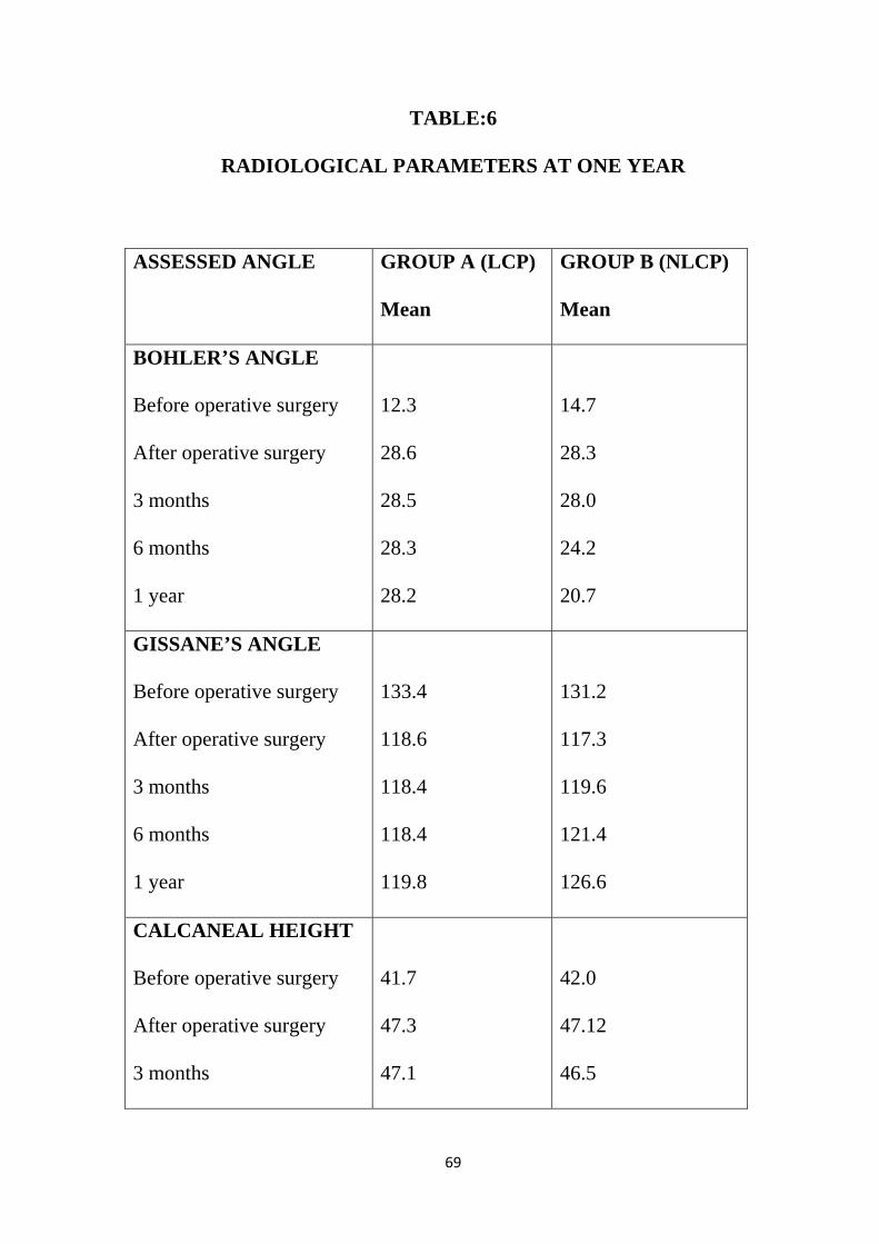
TABLE:6
RADIOLOGICAL PARAMETERS AT ONE YEAR
ASSESSED ANGLE GROUP A (LCP)
Mean
GROUP B (NLCP)
Mean
BOHLER’S ANGLE
Before operative surgery
After operative surgery
3 months
6 months
1 year
12.3
28.6
28.5
28.3
28.2
14.7
28.3
28.0
24.2
20.7
GISSANE’S ANGLE
Before operative surgery
After operative surgery
3 months
6 months
1 year
133.4
118.6
118.4
118.4
119.8
131.2
117.3
119.6
121.4
126.6
CALCANEAL HEIGHT
Before operative surgery
After operative surgery
3 months
41.7
47.3
47.1
42.0
47.12
46.5

70
6 months
1 year
47.1
47.0
46.2
46.2
CALCANEAL WIDTH
Before operative surgery
After operative surgery
3 months
6 months
1 year
55.4
49.1
49.3
49.3
49.7
55.1
49.3
49.7
50.1
50.9

71
TABLE 7
TABLE SHOWING COMPARISON OF RADIOLOGICAL
PARAMETERS
S.N
O
VARIABLES N SD ‘t’
VALUE
‘p’
VALUE
1.
FOLLOW UP
BOHLER
LCP 10 3.33 8.452* .000*
NON LCP 10 1.69
2. FOLLOW UP
GISSANE
LCP 10 4.477 3.977* .001*
NON LCP 10 5.581
3. FOLLOW UP
HEIGHT
LCP 10 1.716 .115 .909
NON LCP 10 2.097
4. FOLLOW UP
WIDTH
LCP 10 2.945 1.092 0.289
NON LCP 10 3.190
In our series the radiological parameters which includes the Bohler’s
angle, Gissane’s angle among the LCP group when compared with the non-
LCP group is statistically significant P<0.05. Even though calcaneal height
and width better in LCP group compared to NLCP group with respect to
AOFAS – Ankle Hindfoot score, stastically insignificant at p value <0 .05.

72
TABLE-8
COMPARISSION OF VARIOUS STUDY
Our Study RAK, V, IRA D, et al
ZEMAN P, et al
Design Prospective, Randamized
Retrospective Retrospective
Total no of patient
20 76 30
Study period 2012 – 14 2004 – 07 2005 – 07 Time of surgery (days)
14 81 12
Approach (Extensile Lateral)
Yes Yes Yes
Bongraft or bone substitutes
100% 82% Non locking10% Locking
40%
Superficial skin infection LCP Non LCP
10% 20%
3% 17%
21% ---
Deep infection LCP Non LCP
--
10%
--
12%
-- --
Radiological parameters
LCP better maintained than
Non LCP
LCP better than Non LCP
Better LCP
AOFAS-ANKLE HIND FOOTSCORE
LCP better than Non LCP
LCP better than Non LCP
Better LCP
73
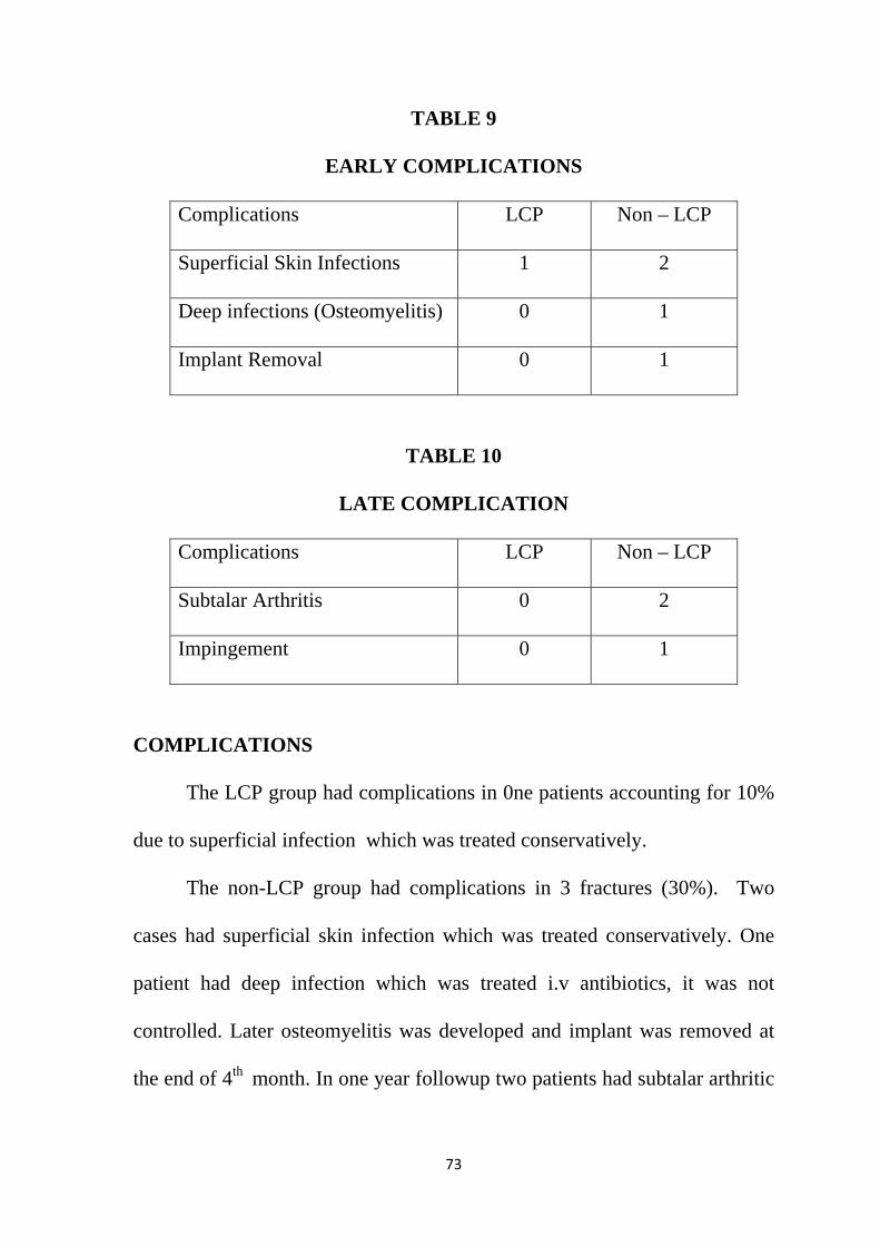
TABLE 9
EARLY COMPLICATIONS
Complications LCP Non – LCP
Superficial Skin Infections 1 2
Deep infections (Osteomyelitis) 0 1
Implant Removal 0 1
TABLE 10
LATE COMPLICATION
Complications LCP Non – LCP
Subtalar Arthritis 0 2
Impingement 0 1
COMPLICATIONS
The LCP group had complications in 0ne patients accounting for 10%
due to superficial infection which was treated conservatively.
The non-LCP group had complications in 3 fractures (30%). Two
cases had superficial skin infection which was treated conservatively. One
patient had deep infection which was treated i.v antibiotics, it was not
controlled. Later osteomyelitis was developed and implant was removed at
the end of 4th month. In one year followup two patients had subtalar arthritic

74
changes present in the followup X-ray. One patient had heel pad pain,
impingement treated by analgesics and foot wear change.
Fig 45: Skin Necrosis
Fig 46: Plate Exposed raw area.

75
SEX RATIO
MODE OF INJURY
0
1
2
3
4
5
6
7
8
9
10
Locking Compression PlateNon- Locking Compression Plate
Male Female
Fall
RTA
0
1
2
3
4
5
6
Locking Compression Plate
Non-Locking Compression Plate
Fall RTA

76
FRACTURES BASED ON SANDER’S CLASSIFICATION
COMPLICATIONS
EARLY COMPLICATION
0
0.5
1
1.5
2
2.5
3
3.5
4
4.5
5
GROUP A (LCP)
GROUP B (NON LCP)
SANDER'S CLASSIFICATION I II III IV
0 0.5 1 1.5 2 2.5
SUPERFICIAL SKIN INFECTION
DEEP INFECTION (OSTEOMYELITIS)
IMPLANT REMOVAL
GROUP B GROUP A

77
0 0.5 1 1.5 2 2.5
SUBTALAR ARTHRITIS
IMPINGEMENT
LATE COMPLICATION
GROUP B GROUP A
0123456
EXCE
LLEN
T
GO
OD
FAIR
POO
R
SAN
DER
'S
TOTA
L N
O O
F CA
SES
EXCE
LLEN
T
GO
OD
FAIR
POO
R
GROUP A GROUP B
AOFAS- ANKLE HINDFOOT SCORE
II III IV
020406080
100120140160
RADIOLOGICAL ANGLES OF 1 YEAR FOLLOWUP
GROUP A GROUP B

78
OPERATIVE CASES WITH LCP -1
PRE OP-XRAY/CT
INTRA OPERATIVE

79
POST OPERATIVE X-RAYS

80
AT THE TIME OF SUTURE REMOVAL

81
MOVEMENTS

82
OPERATIVE CASES FOR LCP -2
PRE-OP

83
INTRAOPERATIVE

84
AFTER 1 YEAR
MOVEMENTS
85
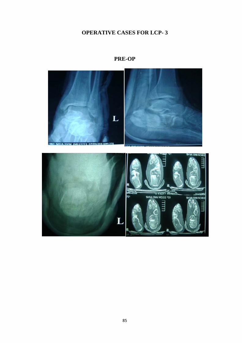
OPERATIVE CASES FOR LCP- 3
PRE-OP

86
INTRA-OP

87
POST-OP X-RAYS
AT 1 YEAR FOLLOWUP

88
MOVEMENTS AND MEASUREMENTS

89
OPERATIVE CASES FOR NLCP - 1
PRE-OP
INTRA-OP
POST-OP

90
AT 6 MONTHS
AT 1 YEAR

91
MOVEMENTS

92
OPERATIVE CASE FOR NLCP- 2
PRE-OP CT & X-RAY
INTRAOPERATIVE

93
POST-OP
AFTER 1 YEAR

94
ARTHRODESIS
AFTER 18 MONTHS

95
MOVEMENTS

96
DISCUSSION
In the last decade open reduction and internal plate fixation of
intraarticular calcaneal fractures has become a standard surgical method with
low complication rate and better quality of life after the surgery1-25.
Our study group were treated with locking compression plate with
locking compression screw and non locking compression plate with 3.5mm
cancellous screw. 80% of the patient were treated with bone graft substitute
‘G’ bone 20% patient treated with autograft. Autograft patient had another
scar incision and chances for wound complication. ‘G’ bone is the better
bone substituent and does not carry any infection and disease, prevent the
long term tuberosity collapse and maintain calcaneal height and width.
Excellent results in Extensile Lateral approach with plate
osteosynthesis it was supported by many studies19,10.Skin related
complication was very less in this approach.
Buckley and meek et al10 in their comparative study of 34 cases stated
that operative treatment yields better outcome provided a anatomical
reduction of subtalar joint is achieved in this approach.
Our study with Muller et al regarding comminution. we confirmed the
benefit of LCP implanting over the NonLCP for all Sander’s type of
intraarticular fractures .The most comminuted fractures in Sanders IV shows

97
excellent and good results with LCP plate osteosynthesis than with non- LCP
treatment.
The most difficult part is to bring the anatomical reduction in Sander’s
Type IV. It is mostly communited and subtalar joint congruity maintained in
intra operative period was difficult19. 4 patients had this type of fracture in
the study 3 treated with LCP, one patient with Non-LCP. No patient had any
skin complications
The number of patients came for postoperative followup in this period
was very low.
We have evaluated the radiological parameters at the end of one year,
Bohler’s angle, Gissane’s angle which has statistically significant ,but
calcaneal height,width which has statistically insignificant results when LCP
group compared with the non LCP group.
Restoration of Bohler’s angle and Gissane’s angle is associated with
excellent results in Locking plate in our study. This fact, proved and verified
by a lot of authors, confirmed the role of Bohler’s angle size as a predictive
factor for subsequent late complications.10,11
The AOFAS- Ankle Hindfoot score which evaluates pain, functional
ability, cosmesis and range of movements LCP group is better than non -
LCP group but statistically insignificant.

98
When comparison is based on individual fracture patterns with respect
to AOFAS Ankle Hindfoot Score, Sanders classification fractures LCP
group is better19 than non –LCP group but it is statistically insignificant.
Sanders type III and severely comminuted type IV fractures LCP
group is better. Our study type IV fractures experiences poor results in
NLCP. This may be due to subtalar restriction and arthritis, soft tissue
impingement and smashed heel pad syndrome19.Since the fractures are
randomised and the NLCP group only one patient had Sander’s Type IV, we
need more number of patients and followup.
In our study, the AOFAS- Ankle Hindfoot score is 80.666 in LCP
group and 71.66 in Non-LCP group.We followed a standard operative
protocol,but done by different surgeons and grafting was done in all cases.

99
LIMITATIONS
Our study is limited by less number of patients and a randomized
Our sample size is small and the mean follow up period is a short and
many studies have shown improvement in functional outcome after
one year of follow up.
More over plate osteosynthesis is done by different surgeons and the
observer is not blinded.

100
CONCLUSION
Open reduction Internal fixation remains the gold standard treatment
for displaced fractures of the calcaneum, since it restores the congruity
of the subtalar joint and restores the normal anatomy of the bone.
Accurate anatomical restoration of the posterior facet is associated
with excellent outcome and it should be the primary aim in reduction
techniques.
CT imaging is an essential in pre-operative evaluation of fractures,
classification and in planning treatment.
The complications associated with wound healing can be overcome by
extensile lateral approach, delayed intervention after subsidence of
edema and meticulous soft tissue handling.
Bone grafting and bone graft substitutes is recommended in all cases
with a void and to prevent post-operative collapse.
To conclude open reduction and internal fixation with LC plate
osteosynthesis provides excellent and statistically significant results
when compared to non-LCP patients were noted for all intra-articular
calcaneal fractures.
Sander’s type IV and severely comminuted intrarticular fractures are
not considered to be the contraindication to surgery.

101
In Sander’s type IV and severely comminuted fractures, though the
outcome LCP group is better than non-LCP group is statistically
insignificant due to short period of follow-up.
LCP group patient early mobilization with weight bearing walking
comparatively in non LCP group patients, implant related
complications was less.
Since the functional outcome of operated patients tends to improve
even after one year, we recommend a longer period of follow up of
these patients for significant results when compared to the non-LCP
group.

BIBLIOGRAPHY
1. Susan Nolshikawa. Campbell’s Operative Orthopaedics. 12th edition. .
P 4139-4157
2. Robert Bucholz W, James Heckman D, Charles M, Court –Brown
Paul Tornetta. Rockwood and Greens Fractures in Adults. Seventh
Edition. Philadelphia: Lippincott Williams & Wilkins; 2010. P
2064-2109
3. Michael W. Chapman, Joseph M. Lane, Roger A. Mann , Richard A.
Marder et al. Chapman’s Orthopaedic Surgery. 3rd edition. Lippincott
Williams & Wilkins 2001. P 2966-2981
4. Wilson J N FRCS, Watson-Jones Fractures and Joint Injuries. Seventh
Edition. Elsevier; 2009.P 1022-1034.
5. Suresh Sivanathan , Eugene Sherry , Patrick Warnke, Mark D Miller .
Mercers Textbook of Orthopaedics and Trauma. Tenth Edition.
Hodder Arnold ; P 398-402
6. Standring S, Ellis H, Healy C.Grays anatomy. The anatomical basis of
clinical practice. Thirty ninth Edition. Elseiver Churchill livingstone
2005.P1513-1514.
7. Sanders R. Current concepts Review. Intra-articular fractures of the
calcaneus: J Bone Joint Surg 2000; 82:225-50.

8. Paul M, Peter R, Hoffmeyer. Fractures of the calcaneum. J Bone Joint
surg Am.2004; 86-B: 1142-45.
9. Mandeep S Dillion Fractures of the calcaneus P25-34, P 39-48,62-72
10. Buckley R, Tough S, McCormack R, et al. Operative compared with
non operative treatment of intra-articular calcaneal fractures: a
prospective, randomized controlled multicenter trial. J Bone Joint surg
Am.2002 Oct; 84 A(10): 1733-1745
11. Buckley RE, Meek RN. Comparison of open versus closed reduction
of intra-articular calcaneal fractures. J Orthop Trauma.1992;6(2):212-
22
12. Brauer AC, Braden J, Michael K, Donaldson C, Buckley R. An
economic evaluation of operative compared with non-operative
treatment of intraarticular calcaneal fractures. J Bone Joint surg
Am.2005 Dec; Vol 87-A,P2741-2749.
13. Gaskil T, Schweitzer K, Nunley J. Comparison of surgical outcome of
intraarticular calcaneal fractures by age. J Bone Joint surg Am. 2010
Dec.Vol.92-A; 2884-2889.
14. Lamglait E, Cronier P, Talha A, Massin P. Internal fixation of
articular calcaneus fractures with AO 3.5mm Reconstruction plate-
Review of 181 cases ; J Bone Joint surg Am. Br 2009; Vol 91-B ,Supp
-79

15. Potter MQ, Nunley JA. Long Term Functional outcomes after
operative treatment for intra-articular fractures of the calcaneus. J
Bone Joint surg Am.2009; Aug91(8):1854-1860
16. Jain V, Kumar R, Mandal DK, Osteosynthesis for intra-articular
calcaneal fractures. J of orthop surg 2007;51(2):P 144-148
17. Sanders R, Fortin P, De Pasquale T, Walling A. Operative treatment
in 120 displaced intra-articular calcaneal fractures. Clin Orthop Rel
Research; May 1993; 290:P 82-95
18. Luc Besse J, Chotel A, Lerat J, Moyen B. Calcaneal intra-articular
fracture osteosynthesis. Clinical and radiological prospective study of
31 cases. Foot and Ankle Surgery12 (2006); P 19-27.
19. Rak V, Ira D, Masek M. Operative treatment of intra-articular
calcaneal fractures with calcaneal plates and its complications. Indian
journal of orthopaedics July-September 2009;vol 43;issue 3 :P271-279
20. Essex-Lopresti P. The mechanism, reduction technique, and results in
fractures of os calcis,1951-1952.Clinical orthopaedic and related
research 1993;290:P 3-16
21. Kitaoka H, Schaap E, Chao E. Displaced intra-articular fractures of
calcaneus treared non-operatively. J BoneJoint surg Am. Oct 1994
;76-A;p1531-1540.

22. Tanaka H K, Laing PW, Functional outcome following operative
fixation of intra-articular calcaneal fractures. The journal of bone and
joint surgery Br 2003 vol 85-B;Supp II 128
23. Zwipp H, Rammelt S, Barthel S. Osteosynthesis for displaced intra-
articular calcaneal fractures. Clinical orthopaedic and related research
1993;290:76-86
24. Yang Y, Zhao H, Zhao J, Yu G. Treatment of displaced intra-articular
calcaneal fractures with or without bone grafts. A systematic review of
literature. Indian journal of orthopaedics. March 2012; vol 46: Issue 2;
P130-137.
25. Mostafa FM, Gamal E, Hassanin E, Abdellatif S. Surgical treatment of
displaced intra-articular calcaneal fracture using a single small lateral
drt6approach. Strat Traum Limb Recon 2010; 5:P87-95.
26. Dewall M, Henderson CE, Mckinley T, Phelps T,Dolan L et al.
Percutaneous reduction and fixation of displaced intra-articular
calcaneal fractures. J orthop Trauma 2010; 22:P466-476.
27. Pendse A, Davethwar RN, Bhatt J, Shivkumar. Outcome after open
reduction and internal fixation of intra-articular fractures of the
calcaneum without the use of bone grafts.Indian journal of
orthopaedics April 2006; vol 40: Number 2:P111-114.

28. Paley D, Hall H. Intra-articular fractures of the calcaneus: a critical
analysis of results and prognostic factors. J Bone Joint Surg Am.
1993; 75A:P342-354.
29. Radnay SC, Clare PM, Sanders R. Subtalar fusion after displaced
intra-articular calcaneal fractures: Does initial operative treatment
matters?. J Bone Joint Surg Am.2009 March;91(3):P541-546.
30. Herscorci J, Widmaier J, Scaduto J, Sanders W. Operative treatment
of calcaneal fractures in elderly patients. J Bone Joint Surg Am.2005;
87(6):P1260-1264.
31. Poeze M, Verbruggen J, Peter RG. The relationship between the
outcome of operatively treated calcaneal fractures and institutional
fracture load. A systematic review of literature. J Bone Joint Surg
Am.May 2008; 90(5):P1013-1021.
32. Richter M, Droste P, Goesling T, Krettek C. Polyaxially-locked plate
screws increase stability of fracture fixation in an experimental model
of calcaneal fracture. J Bone Joint Surg Am.2006; vol-88:P1257-1263.
33. Hospodar P, Guzmann C, Johnson P, Richard U. Treatment of
displaced calcaneal fractures using a minimally invasive sinus tarsi
approach. Trauma update; November 2008:Vol 31.

34. Tomesan T, Biert J, Frolke JPM. Treatment of displaced intra-articular
calcaneal fractures with closed reduction and percutaneus screw
fixation. J Bone Joint Surg Am.May2011; 93(10):P920-928.
35. Daftary A, Hains AH, Baumgaertner M. Fractures of the calcaneus: A
Review with emphasis on CT. Radiographics 2005; 25:1215-1226.
36. Thordarson DB, Krieger LE.Operative vs.Non-operative treatment of
intra-articular calcaneal fractures: a prospective randomized trial.Foot
Ankl Int 1996; 17:2-9.
37. Stephenson JR. Surgical treatment of displaced intra-articular
fractures of the calcaneus. Clin Orthop Relat Res 1993;290;68-75.
38. Song KS, Kang CH, Min BW. Preoperative and postoperative
evaluation of intra-articular calcaneal fractures based on CT. J Orthop
Trauma 1997; 11:435-40.
39. Stephenson JR. Surgical treatment of displaced intra-articular
fractures of the calcaneus with combined medial and lateral approach.
. Clin Orthop Relat Res 290;68-75.1993.
40. Tornetta p. Open reduction and internal fixation of calcaneus using
minifragment plates. J Orthop Trauma 10.63-67, 1993.
41. Zwipp H, Tscherne H. Osteosynthesis of displaced intra-articular
fractures of the calcaneus. Clin Orthop Relat Res 1993; 290:76-86.

42. Redfern DJ, Oliveira ML, Campbell JT, et al. A Biomechanical
Comparison of locking and nonlocking plate for the fixation of
calcaneal fractures. Foot ankle Int. Mar 2006;27(3); 196-201
43. Blake MH, Owen JR, Sanford TS et al, Biomechanical evolution of a
locking and nonlocking reconstruction plate via an osteoporotic
calcaneal fracture model. Foot Ankle Int,2011;32(4) 432-6
44. Staffel K, Booth G, Rohri SM et al, A Comparison of conventional
V/s locking plate in intra-articular calcaneal fractures. A
Biomechanical study in human cadavers. Clin Biomech,2000, 22(1)
100-5
45. Illert J et al, Stability of locking and non locking plates in an
osteoporotic calcaneal fracture model. Foot ankle Int.2011, Mar
32(3),307-13
46. Zeman P, Zeman J, Matejka J, Kondela K. Longterm result of
calcaneal fracture treatment by ORIF using calcaneal locking
compression plate from an extensile lateral approach, A cta chir ortho
traumatol cech 2008 Dec 75(6) 457-64

ANNEXURE: I
ANKLE-HINDFOOT SCALE(100 POINTS TOTAL)
I. PAIN(40 POINTS)
1. None 40
2. Mild,occasional 30
3. Moderate, daily 20
4. Severe almost always present 0
II.FUNCTION: (50 POINTS)
a. ACTIVITY LIMITATIONS, SUPPORT REQUIREMENT
1. No limitations, no support 10
2. No limitation of daily activities, limitation
of recreational activities, no support
07
3. Limited daily and recreational activities,
cane
04
4. Severe limitation of daily and recreational
activities,walker,crutches,wheelchair, brace
0

b. MAXIMUM WALKING DISTANCE,BLOCKS:
1. Greater than 6 5
2. 4—6 4
3. 1-3 2
4. Less than 1 0
c. WALKING SURFACES
1. No difficulty on any surface 5
2. Some difficulty on uneven terrain, stairs, inclines,
ladders
3
3. Severe difficulty on uneven terrain, stairs,
inclines, ladders
0
d. GAIT ABNORMALITY
1. None, slight 8
2. Obvious 4
3. Marked 0

e. SAGITTAL MOTION (FLEXION PLUS EXTENSION)
1. Normal or mild restriction (30
degree or more)
8
2. Moderate restriction (15-19 degree) 4
3. Severe restriction (less than 150) 0
a. HINDFOOT MOTION (INVERSION PLUS EVERSION)
1.
Normal or mild restriction(75%-
100% normal)
6
2. Moderate restriction(25%-
74%normal)
3
3. Marked restriction (less than 25%
normal)
0
b. ANKLE – HINDFOOT STABILITY ( ANTEROPOSTERIOR,
VARUS-VALGUS)
1. Stable 8
2. Definitly unstable 0

b. ALIGNMENT (10 POINTS)
1. Good, plantigrade foot, midfoot well
aligned
15
2. Fair, plantigrade foot, some degree of
midfoot malalignment observed, no
symptoms
8
3. Poor, nonplantigrade foot, severe
malalignment, symptoms
0
ASSESS WOUND HEALING
SUBTALAR ARTHRITIS
CALCANEAL MALPOSITION/ MALUNION
LATERAL/TALOTIBIAL IMPINGEMENT SYNDROME
BONE GRAFT REQUIREMENT

ANNEXURE II:
PROFORMA
NAME: AGE:
SEX:
ADDRESS:
I.P NO: UNIT: DOA: DOS:
WARD:
MODE OF INJURY: SIDE:
ASSOCIATED INJURIES:
SANDERS CLASSIFICATION:
ESSEX LOPRESTI CLASSIFICATION:
PAST MEDICAL HISTORY:
INVESTIGATION:
URINE: ALBUMIN, SUGAR, DEPOSITS
BLOOD: HB, UREA, SUGAR, CREATININE, GROUPING
&TYPING,VCTC
ECG:
X-RAYS: AP,LATERAL VIEW AND HARRIS AXIAL VIEW OF ANKLE
CHEST PA VIEW
CT CALCANEUM: AXIAL, CORONAL WITH 3 D RECONSTRUCTION

INTIAL MANAGEMENT: BK SLAB,LIMB ELEVATION,NSAIDS
OPERATIVE GROUP /NON OPERATIVE GROUP:
SURGERY AND IMPLANTS:
DATE OF SURGERY: TIME INTERVAL:
PLATE AND SCREWS:
OPERATIVE TIME:
COMPLICATIONS:
SKIN NECROSIS:
MALUNION:
ARTHRITIS:
NEUROLOGICAL:
PERONEAL TENDINITIS:
HEEL EXOSTOSIS:

OTHERS: LEFT
RIGHT
Month Ankle
dorsiflexion
Ankle
plantarflexion
Inversion Eversion AOFAS
Ankle HIND
FOOT
SCORE
Month Ankle
dorsiflexion
Ankle
plantarflexion
Inversion Eversion AOFAS
Ankle HIND
FOOT
SCORE

MASTER CHART
LCP CASES
Non-LCP CASES
SAMPLE AGE SEX MODE OF INJURY SANDER'S ASSOCIATED INJURY TIME OF SURGERY (DAPRE OP BOHLER POST OP BOHLER PRE OP GISSANE POST OP GISSANE PREOP HEIGHT POST OP Ht PREOP Width POSTOP Wdth GRAFTING UNION COMPLICAAHF SCALELATE COMFOLLOW PERIOD IMPLANT EXIT1 21 M FALL III 12 13 32 128 120 42 46 54 49 G BONE ** EXCELLENT2 35 M FALL II SPINE 10 15 28 135 122 44 48 57 50 G BONE ** EXCELLENT3 33 M RTA IV 14 12 28 138 124 43 48 60 51 G BONE ** FAIR4 25 M RTA II 17 16 32 141 120 40 48 58 50 BONE GRAFT ** EXCELLENT5 37 M RTA IV 13 13 26 130 118 39 45 51 48 G BONE ** GOOD6 31 F FALL IV 15 12 28 132 122 41 46 53 49 G BONE ** POOR7 36 M RTA III SUBTROCHANTERIC F 10 8 32 134 118 38 45 56 51 BONE GRAFT * SUPERFICI POOR 8 42 M FALL IV 18 10 24 136 116 40 47 61 50 G BONE ** EXCELLENT9 30 M FALL II 19 12 26 128 120 46 48 52 49 G BONE ** GOOD
10 23 M FALL III SPINE 11 11 26 132 118 44 49 52 50 G BONE ** GOOD
SAMPLE AGE SEX MODE OF INJURY SANDER'S ASSOCIATED INJURY TIME OF SURGERY (DAPRE OP BOHLER POST OP BOHLER PRE OP GISSANE POST OP GISSANE PREOP HEIGHT POST OP Ht PREOP Width POSTOP Wdth GRAFTING UNION COMPLICAAHF SCALELATE COMFOLLOW PERIOD IMPLANT EXIT1 25 M FALL II 12 14 22 132 124 44 46 56 52 G BONE ** SUPERFICI EXCELLENT2 34 M FALL II SPINE 16 16 20 130 122 42 48 58 51 G BONE ** POOR SUBTALAR ARTHRITIS3 37 M FALL III 18 18 22 128 128 43 50 54 51 BONE GRAFT ** GOOD4 23 M RTA III 14 17 21 134 126 42 46 60 53 G BONE ** GOOD5 21 F FALL II 13 15 20 130 136 40 46 50 50 G BONE ** POOR6 40 M RTA II INTER TROCHANTERIC 14 16 22 132 120 38 46 61 51 G BONE ** SUPERFICI EXCELLENT7 28 M RTA III 15 13 20 128 122 46 48 56 51 G BONE ** POOR SUBTALAR ARTHRITIS8 26 F FALL II 17 12 20 136 128 44 48 50 50 G BONE * DEEP INFEFAIR PLATE EXPAFTER 4 MONTHS9 29 M RTA III 19 10 18 128 124 39 46 54 50 BONE GRAFT ** OSTEOMY FAIR
10 31 M RTA IV 11 16 22 134 136 42 48 52 50 G BONE ** POOR SUBTALAR ARTHRITIS