section 1, chapter 9 muscular system. muscle is derived from musculus, for “mouse” functions of...
TRANSCRIPT
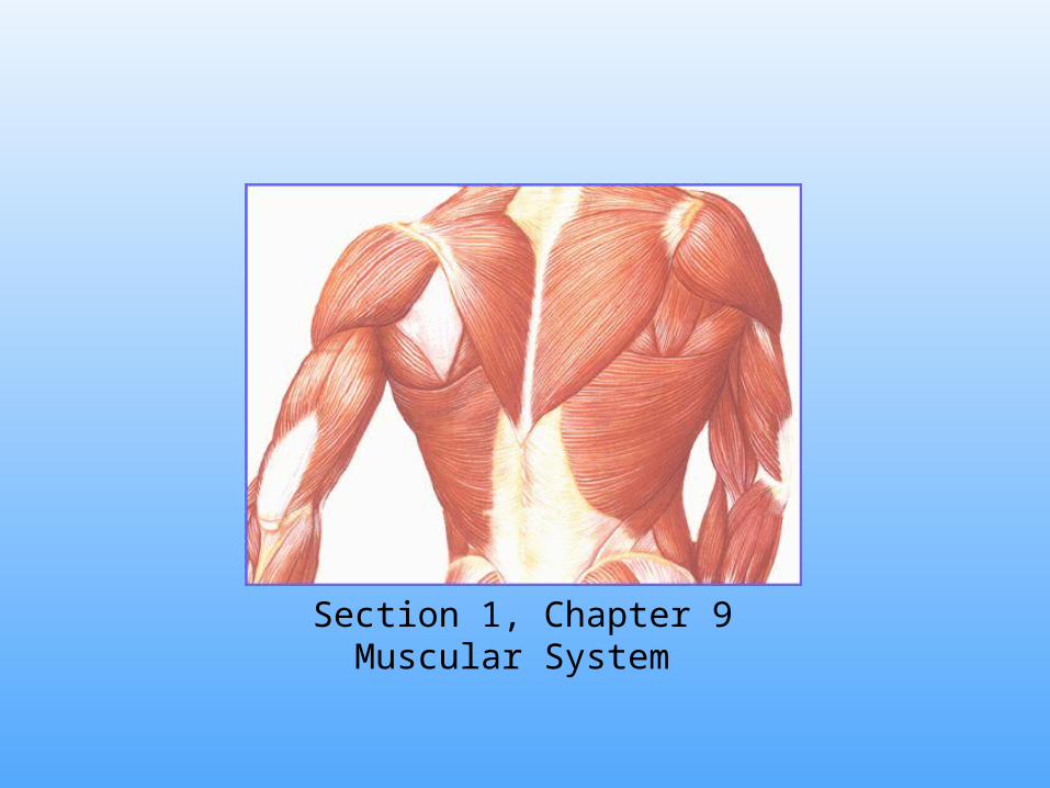
Section 1, Chapter 9Muscular System

Muscle is derived from Musculus, for “Mouse”
Functions of Muscles:1. Body movement 2. Maintain posture3. Produces heat4. Propel substances
through body5. Heartbeat
Types of muscles include:1. Smooth muscle2. Cardiac muscle3. Skeletal muscle
Imagine a mouse running beneath the
skin.

Characteristics of smooth muscles• Involuntary control• Tapered cells with a single, central nucleus• Lack striations
Smooth Muscle
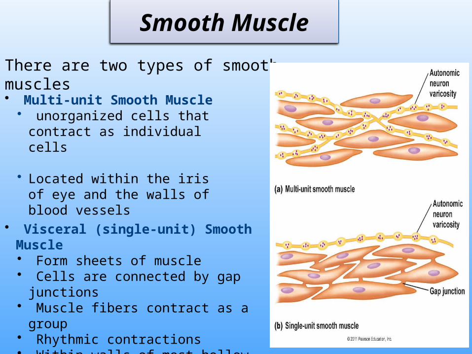
• Visceral (single-unit) Smooth Muscle• Form sheets of muscle• Cells are connected by gap junctions• Muscle fibers contract as a group• Rhythmic contractions• Within walls of most hollow organs
(viscera)
• Multi-unit Smooth Muscle• unorganized cells that contract
as individual cells
• Located within the iris of eye and the walls of blood vessels
There are two types of smooth muscles
Smooth Muscle

Cardiac Muscle
• Located only in the heart
• Striated cells
• Intercalated discs
• Muscle fibers branch
• Muscle fibers contract as a unit
• Self-exciting and rhythmic
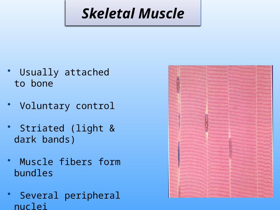
• Usually attached to bone
• Voluntary control
• Striated (light & dark bands)
• Muscle fibers form bundles
• Several peripheral nuclei
Skeletal Muscle

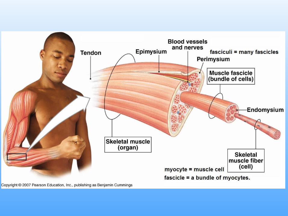
Coverings of Skeletal Muscle
Fascia• Dense connective tissue surrounding skeletal muscles
Tendons• Dense connective tissue that attaches muscle to bones• Continuation of muscle fascia and bone periosteum
Aponeurosis• Broad sheet of connective tissue attaching muscles to
bone, or to other muscles.


Coverings of Skeletal Muscle
Epimysium• Connective tissue that covers the entire muscle
• Lies deep to fascia
Perimysium• Surrounds organized bundles of muscle fibers, called fascicles
Endomysium• Connective tissue that covers individual muscle fibers (cells)

Figure 9.3 Scanning electron micrograph of a fascicle surrounded by its perimysium. Muscle fibers within the fascicle are surrounded by endomysium.
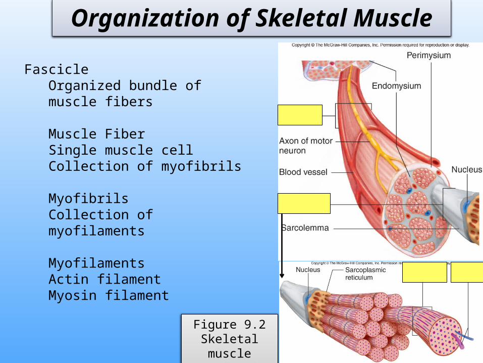
Organization of Skeletal Muscle
Figure 9.2Skeletal muscle
organization
FascicleOrganized bundle of muscle fibers
Muscle FiberSingle muscle cellCollection of myofibrils
MyofibrilsCollection of myofilaments
MyofilamentsActin filamentMyosin filament
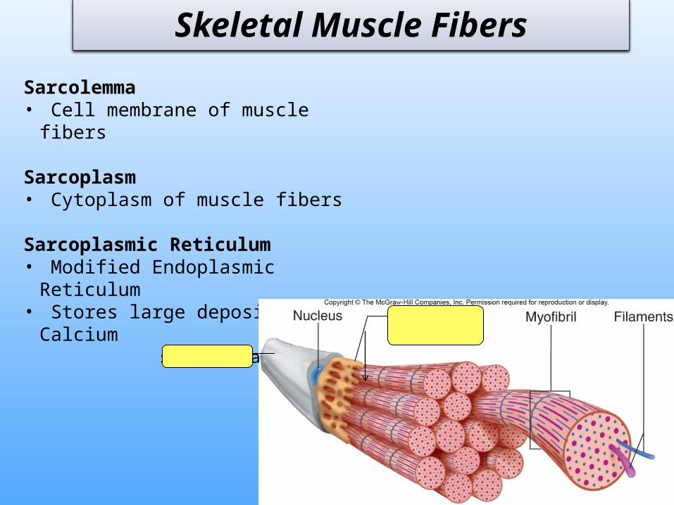
Sarcolemma• Cell membrane of muscle fibers
Sarcoplasm• Cytoplasm of muscle fibers
Sarcoplasmic Reticulum• Modified Endoplasmic Reticulum• Stores large deposits of Calcium
Skeletal Muscle Fibers
sarcolemma
Skeletal Muscle Fibers
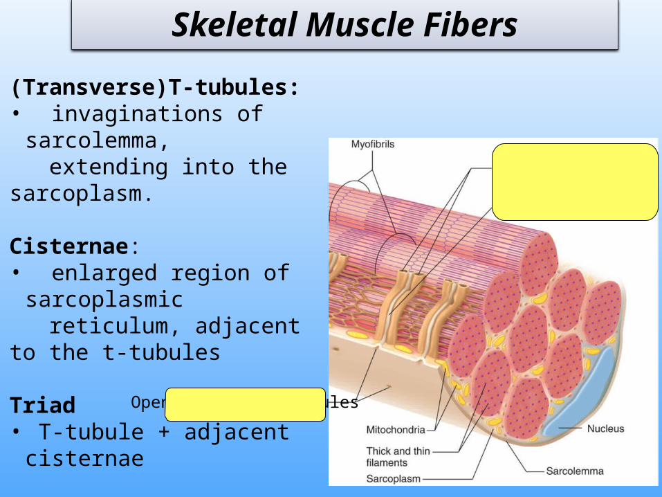
Openings into t-tubules
(Transverse)T-tubules: • invaginations of sarcolemma, extending into the sarcoplasm. Cisternae:• enlarged region of sarcoplasmic reticulum, adjacent to the t-tubules Triad • T-tubule + adjacent cisternae

Myofibrils
Figure 9.4 Organization of actin and myosin filaments
Myofibrils are bundles of actin and myosin filaments.
• Actin – thin filament• Myosin – thick filament
Striations appear from the organization of actin and myosin filaments

SarcomereA sarcomere is the functional unit of
skeletal muscle
• A sarcomere is the area between adjacent Z-lines.
• During a muscle contraction the z-lines move together and the sarcomere shortens.

Sarcomere

Figure 9.5 thin and thick filaments in a sarcomere.
Z Line is the attachment site of actin filaments (center of I bands)
Striations appear from alternate light and dark banding patterns.
I Bands (light band): consists of only actin filaments
A Bands (dark band) : consists of myosin filaments and the overlapping portion of actin filaments

Thick filaments composed of myosin proteins
During muscle contraction the heads on myosin filaments bind to actin filaments forming a Cross-bridge
Thin filamentscomposed of actin proteins
Thin filaments are associated with troponin and tropomyosin proteins
filaments

Cross-BridgesWhen a muscle is at rest, myosin heads are extended in the “cocked” position.
During a contraction, myosin heads bind to actin, forming a cross-bridge and the myosin head pivot forward (Power Stroke) and back (Recovery stroke)
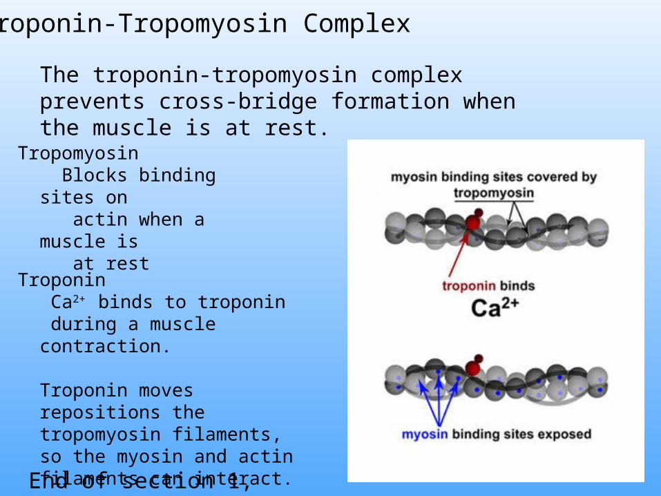
Tropomyosin Blocks binding sites on actin when a muscle is at rest
Troponin Ca2+ binds to troponin during a muscle contraction.
Troponin moves repositions the tropomyosin filaments, so the myosin and actin filaments can interact.
Troponin-Tropomyosin Complex
The troponin-tropomyosin complex prevents cross-bridge formation when the muscle is at rest.
End of section 1, chapter 9
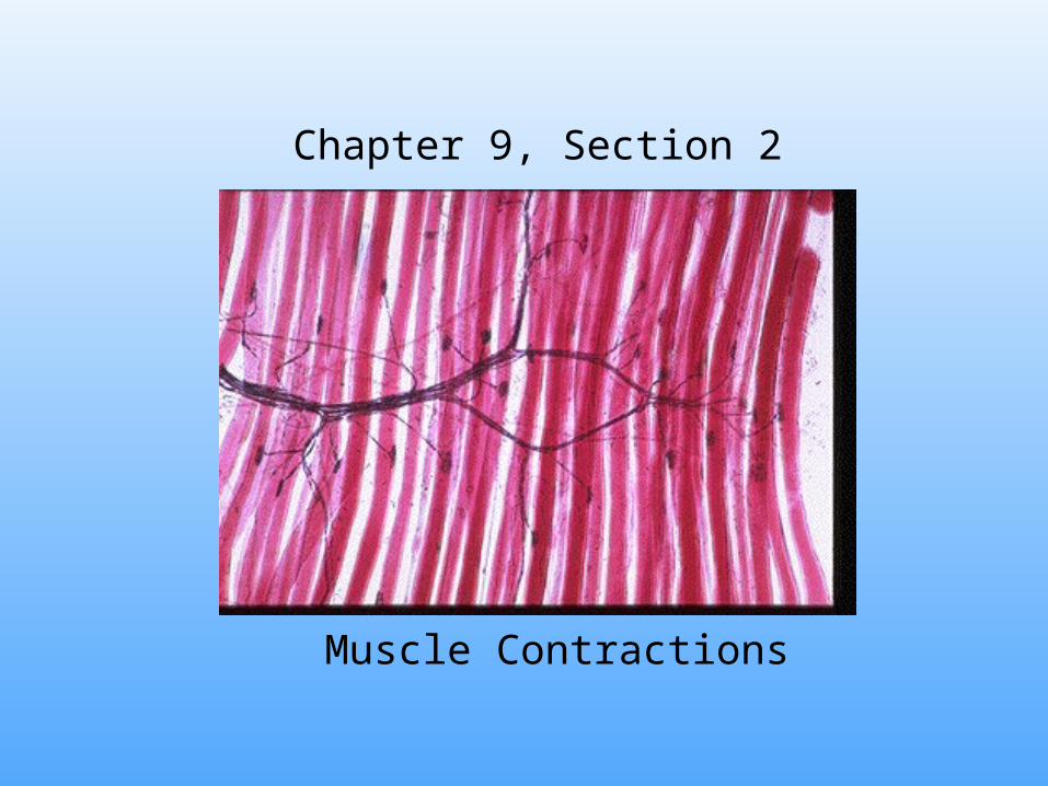
Muscle Contractions
Chapter 9, Section 2
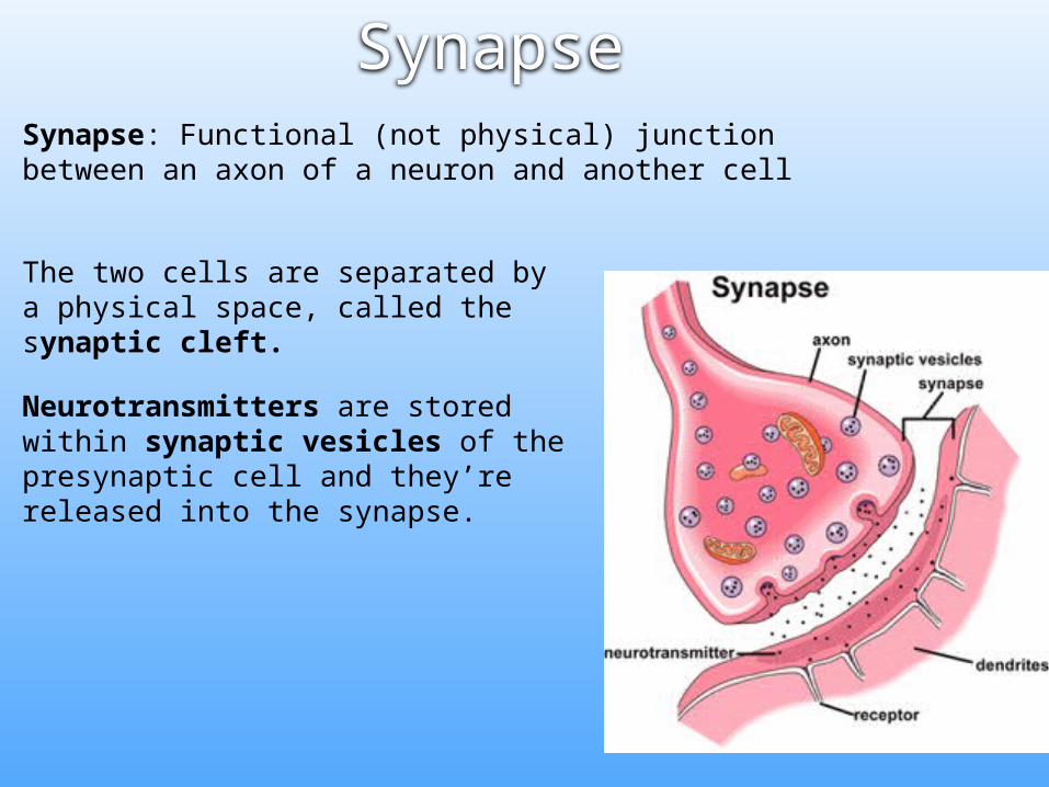
SynapseSynapse: Functional (not physical) junction between an axon of a neuron and another cell
The two cells are separated by a physical space, called the synaptic cleft.
Neurotransmitters are stored within synaptic vesicles of the presynaptic cell and they’re released into the synapse.
Neuromuscular Junction
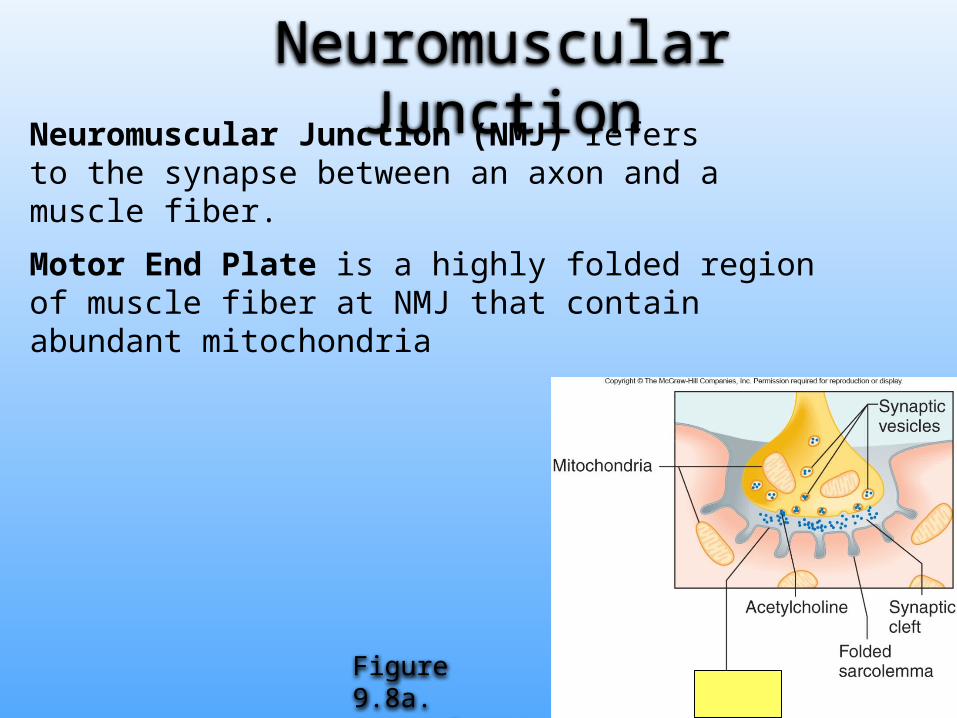
Neuromuscular Junction (NMJ) refers to the synapse between an axon and a muscle fiber.
Motor End Plate is a highly folded region of muscle fiber at NMJ that contain abundant mitochondria
Figure 9.8a. General NMJ
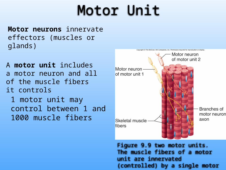
Motor neurons innervate effectors (muscles or glands)
A motor unit includes a motor neuron and all of the muscle fibers it controls
Figure 9.9 two motor units. The muscle fibers of a motor unit are innervated (controlled) by a single motor neuron.
Motor Unit
1 motor unit may control between 1 and 1000 muscle fibers

Stimulus for Contraction
Acetylcholine (ACh) is the only neurotransmitter that initiates skeletal muscle contraction
1. A nerve impulse (Action Potential) reaches axon terminal
2. The impulse opens calcium channels at the axon terminal • Calcium diffuse into axon
3. The calcium triggers the release of ACh from vesicles into synaptic cleft.
Sequence of Actions
Sequence of Actions…Continued
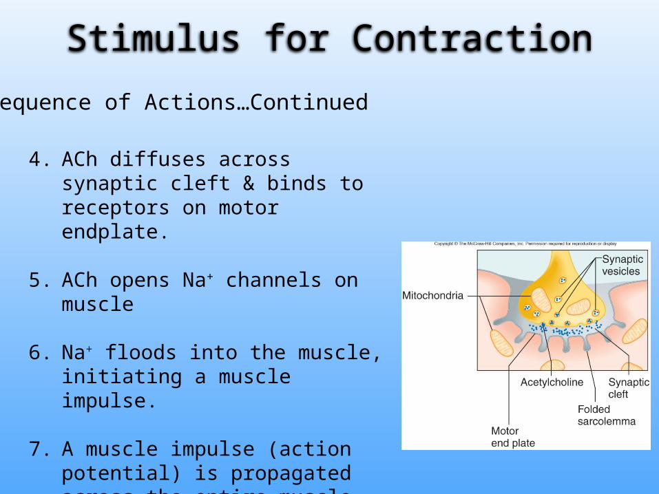
4. ACh diffuses across synaptic cleft & binds to receptors on motor endplate.
5. ACh opens Na+ channels on muscle
6. Na+ floods into the muscle, initiating a muscle impulse.
7. A muscle impulse (action potential) is propagated across the entire muscle.
Stimulus for Contraction
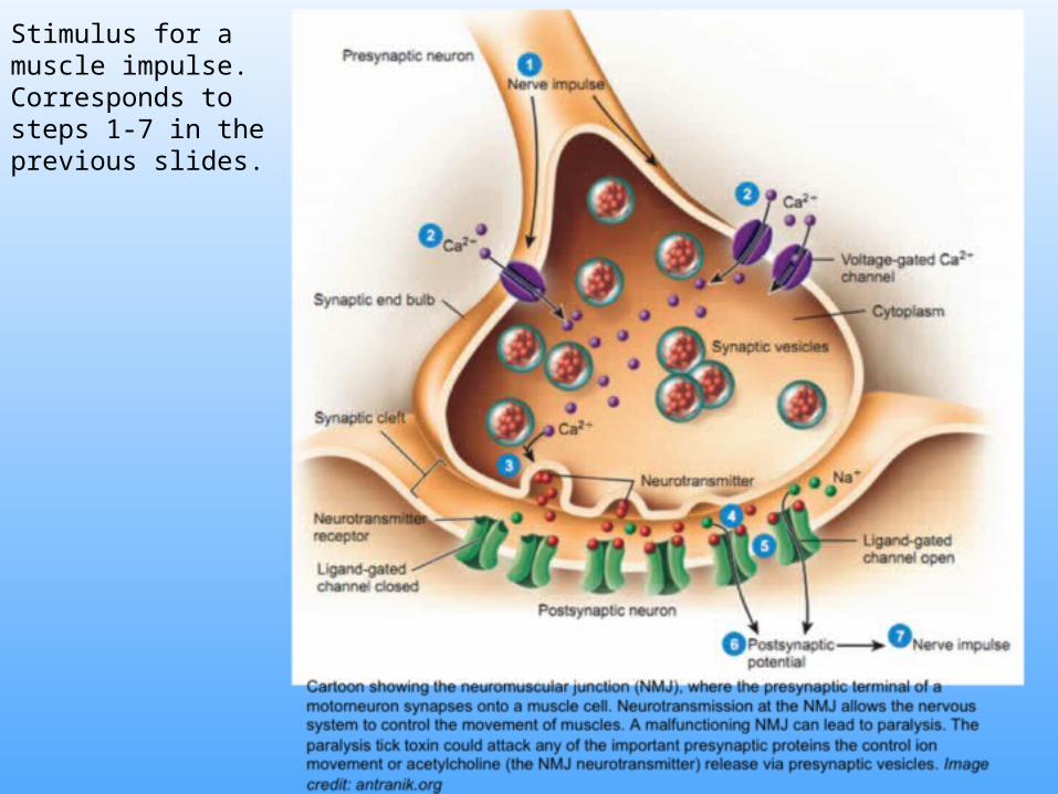
Stimulus for a muscle impulse. Corresponds to steps 1-7 in the previous slides.

The muscle impulse causes the release of calcium from the SR. Calcium binds to troponin and tropomyosin is repositioned exposing the actin filaments.

8. The muscle impulse diffuses across sarcolemma and down the t-tubules into the cisternae of sarcoplasmic reticula.
9. The sarcoplasmic reticula release their calcium supplies into the sarcoplasm.
10. Calcium binds to troponin and the troponin repositions the tropomyosin, so the myosin can bind to actin.
11. Cross-bridge cycling causes the muscle to contract.
Stimulus for contraction continued…

Excitation-Contraction Coupling
Calcium released from sarcoplasmic reticulum binds to troponin.
Troponin moves tropomyosin, exposing actin filaments to myosin cross-bridges.
myosin heads bind to actin, forming a cross bridge and cross-bridge cycling causes the muscle to contract.
End of section 2, chapter 9

Sliding Filament Theory of Contraction
section 3, chapter 9
ivyanatomy.com

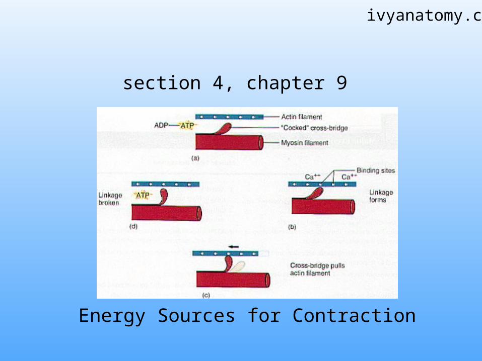
The Sliding Filament Model of Muscle Contraction
Figure 9.11a. Individual sarcomeres shorten as thick and thin filaments slide past one another.
During a muscle contractionThick (myosin) filaments and thin (actin) filaments slide across one another
The filaments do not change lengths
Z-bands move closer together causing the sarcomere to shorten.
I bands appear narrow

Cross Bridge Cycling
1. When a muscle is relaxed, tropmyosin covers the binding sites on actin.
A molecule of ADP and Phosphate remains attached to myosin from the previous contraction.
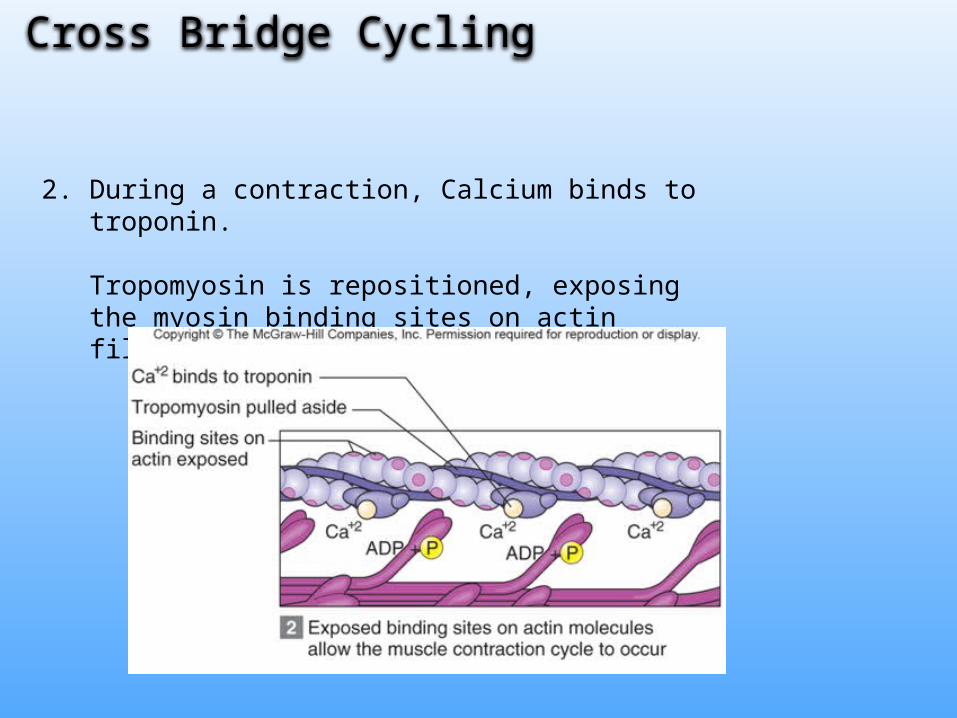
2. During a contraction, Calcium binds to troponin.
Tropomyosin is repositioned, exposing the myosin binding sites on actin filaments
Cross Bridge Cycling

3. Myosin heads bind to actin filaments.
The phosphate is released.
Cross Bridge Cycling

4. Myosin heads spring forward “Power Stroke” pulling the actin filaments.
ADP is released from Myosin
Cross Bridge Cycling
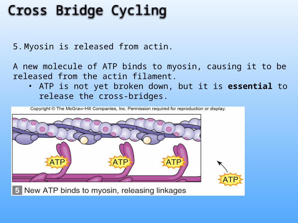
5. Myosin is released from actin.
A new molecule of ATP binds to myosin, causing it to be released from the actin filament.
• ATP is not yet broken down, but it is essential to release the cross-bridges.
Cross Bridge Cycling
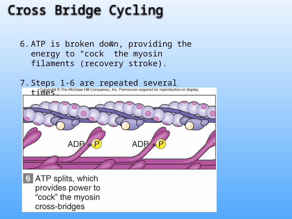
6. ATP is broken down, providing the energy to “cock” the myosin filaments (recovery stroke).
7. Steps 1-6 are repeated several times.
Cross Bridge Cycling

Figure 9.10. The cross-bridge cycle. The cycle continues as long as ATP is present, and nerve impulses release Acetylcholoine.
Watch the You-Tube video “Sliding Filament” to view cross-bridge cycling in action.
Relaxation
When a nerve impulse ceases, two events relax muscle fibers.
1. Acetylcholinesterase breaks down Ach in the synapse.• Prevents continuous stimulation of a muscle fiber.
2. Calcium Pumps (Ca2+ATPase) remove Ca2+ from the sarcoplasm and returns it to the SR.• Without calcium, tropomyosin covers the binding sites on actin
filaments.

Relaxation
Rigor Mortis is a partial contraction of skeletal muscles that occurs a few
hours after death.
• After death calcium leaks into sarcoplasm, triggering the muscle contractions.
• But ATP supplies are diminished after death, so ATP is not available to remove the cross-bridge linkages between actin and myosin.
• muscles do not relax*.
• Contraction is sustained until muscles begin to decompose.
* Notice that ATP is required for muscle relaxation!

End of Chapter 9, Section 3
section 4, chapter 9
Energy Sources for Contraction
ivyanatomy.com

Energy Sources for Contraction
ATP provides the energy to power the interaction between actin & myosin filaments.
• However, ATP is quickly spent and must be replenished
New ATP molecules are synthesized by 1. Hydrolysis of Creatine Phosphate2. Glycolysis (anaerobic respiration)3. Aerobic Respiration

The energy from creatine phosphate hydrolysis cannot be used to directly power muscles. Instead, it’s used to produce new ATP.
Creatine Phosphate
Creatine Phosphate can be hydrolyzed into Creatine, releasing energy that is used to make new ATP.

Creatine Phosphate…continued
When cellular ATP is abundant, creatine phosphate can be replenished by phosphorylating creatine.
Creatine Phosphate provides energy for only about 10 seconds of a high intensity muscle contraction.
Glycolysis
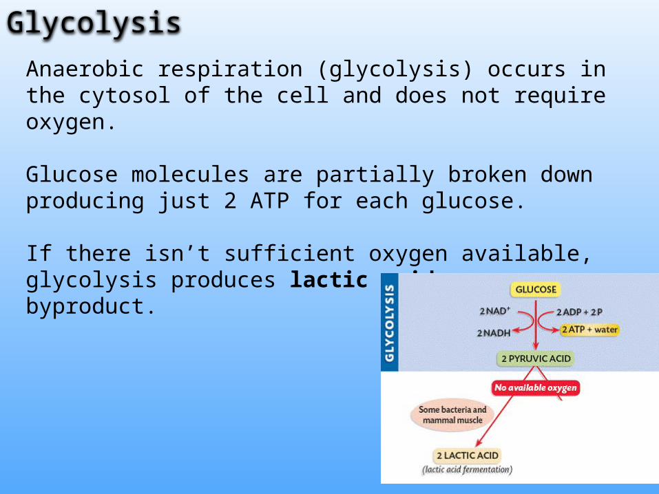
Anaerobic respiration (glycolysis) occurs in the cytosol of the cell and does not require oxygen.
Glucose molecules are partially broken down producing just 2 ATP for each glucose.
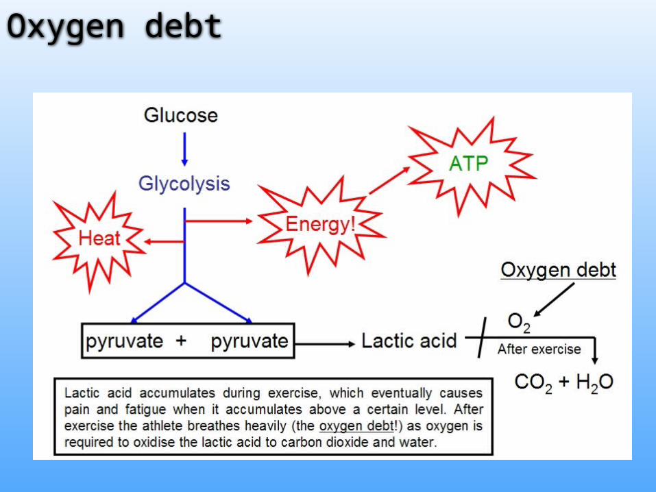
If there isn’t sufficient oxygen available, glycolysis produces lactic acid as a byproduct.

Oxygen debt of glycolysisExercise and strenuous activity depends on anaerobic respiration for ATP supplies.
Oxygen debt is the amount of oxygen needed by liver cells to convert accumulated lactic acid back to glucose.
During exercise anaerobic respiration causes lactic acid to accumulate in the cells.
After exercise, when oxygen is available the O2 is used to convert lactic acid back to glucose in the liver.
Oxygen debt

Aerobic respiration (uses oxygen) occurs in the mitochondria and it includes the citric acid cycle & electron transport chain.
Aerobic respiration is a slower reaction than glycolysis, but it produces the most ATP.
MyoglobinOxygen binding protein (similar to hemoglobin) within
muscles-Provides additional oxygen supply to muscles
Aerobic Respiration

Aerobic RespirationAerobic respiration is used primarily at rest or during light exercise.
Muscles that rely on aerobic respiration have plenty of mitochondria and a good blood supply.
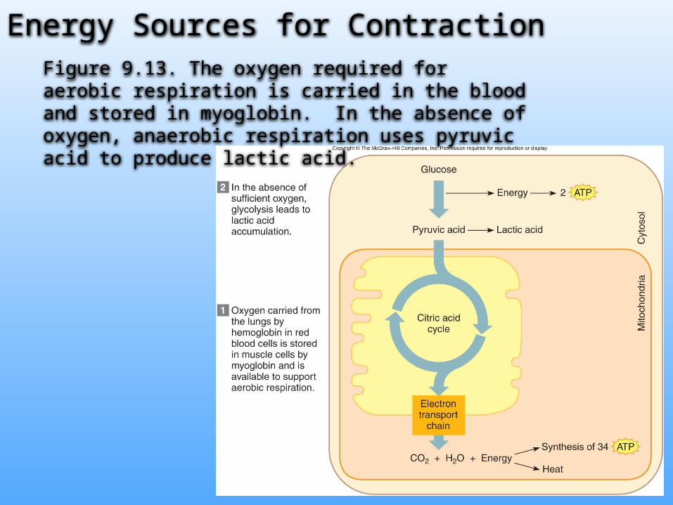
Energy Sources for ContractionFigure 9.13. The oxygen required for aerobic respiration is carried in the blood and stored in myoglobin. In the absence of oxygen, anaerobic respiration uses pyruvic acid to produce lactic acid.
Muscle Fatigue
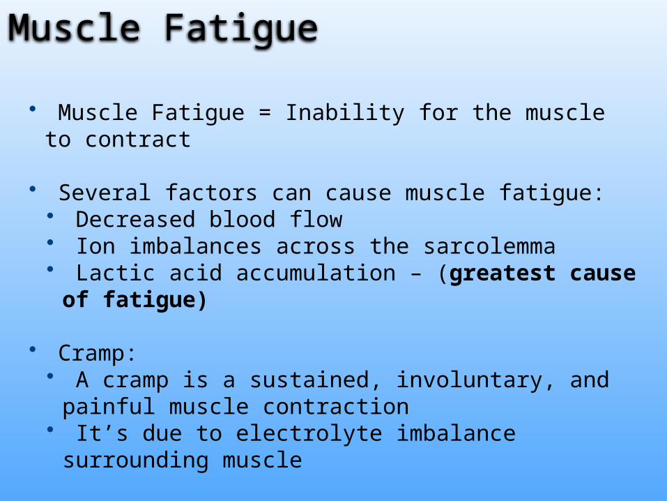
• Muscle Fatigue = Inability for the muscle to contract
• Several factors can cause muscle fatigue:• Decreased blood flow• Ion imbalances across the sarcolemma• Lactic acid accumulation – (greatest cause of fatigue)
• Cramp: • A cramp is a sustained, involuntary, and painful muscle
contraction• It’s due to electrolyte imbalance surrounding muscle
Heat Production• Heat is produced as a by-product of cellular
respiration
• Muscle cells are major source of body heat
• Blood transports heat throughout body core
End of Chapter 9, Section 4

section 5, chapter 9
muscular responses
ivyanatomy.com
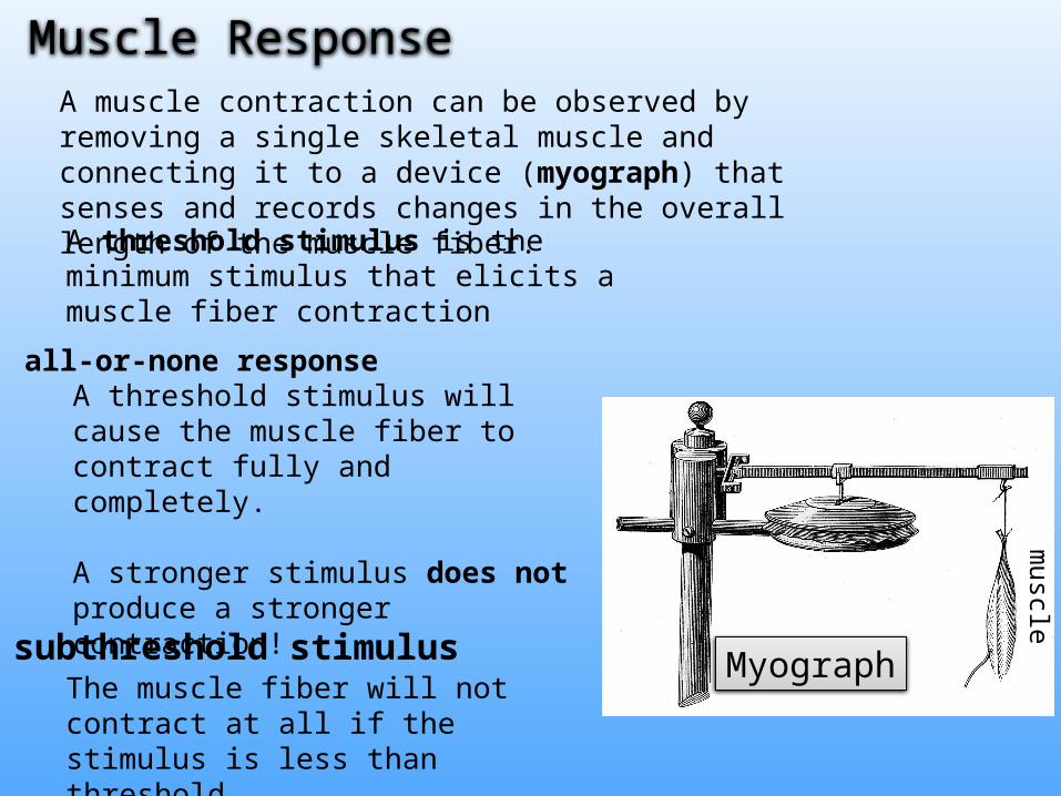
A muscle contraction can be observed by removing a single skeletal muscle and connecting it to a device (myograph) that senses and records changes in the overall length of the muscle fiber.
A threshold stimulus is the minimum stimulus that elicits a muscle fiber contraction
Muscle Responsem
uscle
MyographThe muscle fiber will not contract at all if the stimulus is less than threshold.
all-or-none responseA threshold stimulus will cause the muscle fiber to contract fully and completely.
A stronger stimulus does not produce a stronger contraction!
subthreshold stimulus
Recording of a Muscle Contraction
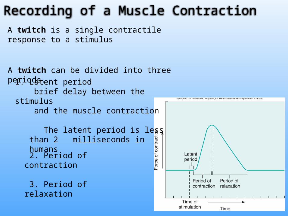
2. Period of contraction
3. Period of relaxation
A twitch is a single contractile response to a stimulus
A twitch can be divided into three periods. 1. Latent period
brief delay between the stimulus and the muscle contraction
The latent period is less than 2 milliseconds in humans

Summation
If the muscle is allowed to relax completely before each stimulus than the muscle will contract with the same force.
If the muscle is stimulated again before it has completely relaxed, then the force of the next contraction increases.
i.e. stimulating the muscle at a rapid frequency increases the force of contraction. This is called summation
Figure 9.17a series of twitches
summationFigure 9.17b

Tetanic Contraction (c) If the muscle is stimulated at a high frequency the contractions fuse together and cannot be distinguished.
A tetanic contraction results in a maximal sustained contraction without relaxation
Summation
Figure 9.17c

all-or-none response
A muscle that is stimulated with threshold potential contracts completely and fully.
A stronger stimulus does not produce a stronger contraction!
Instead, the strength of a muscle is increased by recruitment of additional motor units.
Recruitment of Motor Units

Recruitment of Motor Units
Recall that a motor unit is a motor neuron plus all of the fibers it controls.
• Muscles are composed of many motor units.
• As a general rule, motor units are recruited in order of their size• Small motor units are stimulated with light activities, but additional
motor units are recruited with higher intensity activity.
Recruitment – progressive activation of motor units to
increase the force of a muscle contraction.
As the intensity of stimulation increases, recruitment of motor units continues until all motor units are activated.

Sustained Contractions
The central nervous system can increase thestrength of contractions in 2 ways:
1. Recruitment• Smaller motor units are recruited first, followed by larger motor units.• The result is a sustained contraction of increasing strength.
2. Increased firing rate• A high frequency of action potentials results in summation of the muscle
contractions. • If the frequency is too high, however, it may produce tetanic contractions, in which
case the muscle does not relax.
Muscle tone is produced because some muscles are in a continuous state of partial contraction in response to repeated nerve impulses from the spinal cord.
Types of Contractions
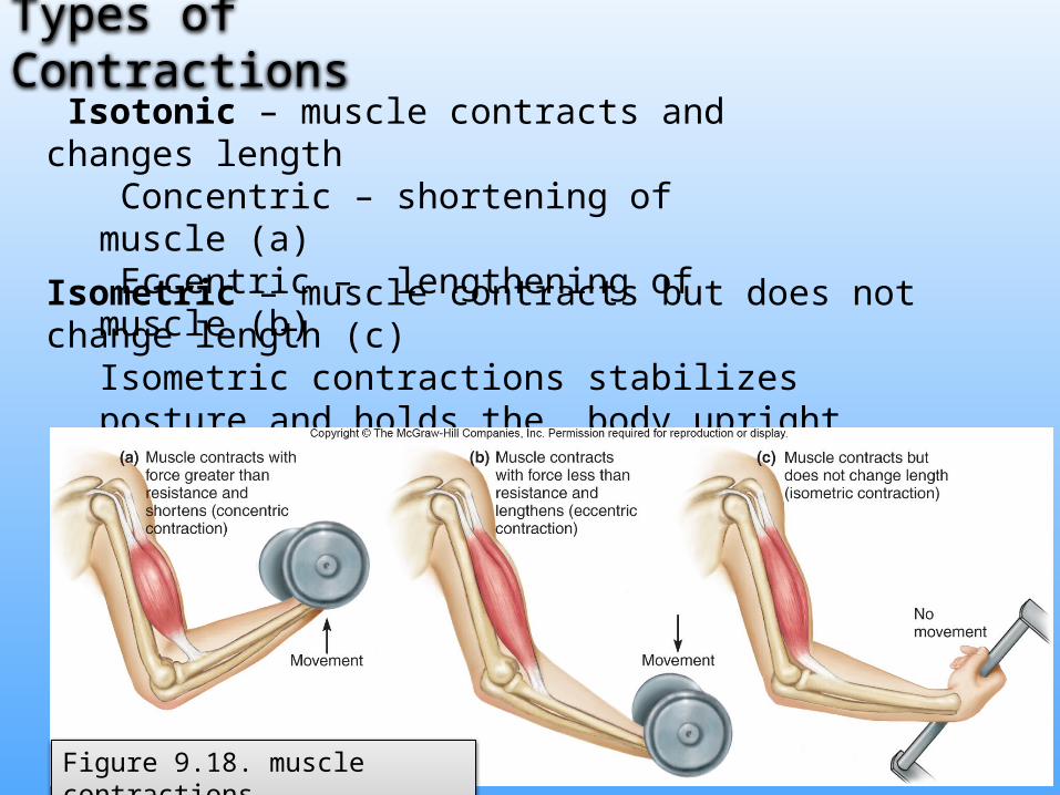
Isotonic – muscle contracts and changes length Concentric – shortening of muscle (a) Eccentric – lengthening of muscle (b)
Isometric – muscle contracts but does not change length (c)Isometric contractions stabilizes posture and holds the body upright
Figure 9.18. muscle contractions

Fast twitch and slow twitch muscle fibers
Fast & Slow twitch refers to the contraction speed, and to whether muscle fibers produce ATP oxidatively (by aerobic respiration) or glycolytically (by glycolysis)
Slow-twitch fibers (Type I)• Always oxidative and resistant to fatigue
• Contain myoglobin for oxygen storage “red fibers”
• Also have many mitochondria for aerobic respiration
• Good blood supply
Slow-twitch fibers (Type I)
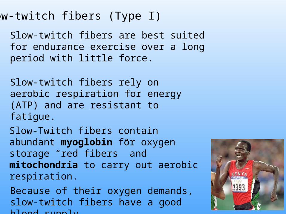
Slow-twitch fibers rely on aerobic respiration for energy (ATP) and are resistant to fatigue.
Slow-Twitch fibers contain abundant myoglobin for oxygen storage “red fibers” and mitochondria to carry out aerobic respiration.
Because of their oxygen demands, slow-twitch fibers have a good blood supply.
Slow-twitch fibers are best suited for endurance exercise over a long period with little force.

Fast twitch muscle fibers – two types
Fast-twitch glycolytic fibers contract rapidly and with great force, but they fatigue quickly.
They are best suited for rapid contractions over a short duration.
Fast-twitch glycolytic fibers (type IIa) contain very little mitochondria and myoglobin and are “white fibers”
Fast twitch muscle fibers – two types
Fast-twitch intermediate or fast oxidative fibers contain intermediate amounts of myoglobin.
They contract rapidly but also have the capacity to generate energy through aerobic respiration.

Migrating birds have abundant slow-twitch fibers for flying long distances, which is why their flesh is dark.
Chickens that can only flap around the barnyard have abundant fast-twitch muscles and mostly white flesh.
End of Chapter 9
Fast twitch and slow twitch muscle fibers