muscles 1. functions of muscular system body movement maintain posture respiration produce body...
TRANSCRIPT

MUSCLES
1

FUNCTIONS OF MUSCULAR SYSTEM
Body movement Maintain posture Respiration Produce body heat Communication Constriction of organs and blood vessels Heartbeat
2

Muscle Types Skeletal: elongated
Moves the skeleton Voluntary striated
Smooth: spindle shaped Found in organs and lining of blood vessels Involuntary no striations
Cardiac: cylindrical shaped involuntary (only responds to direct electrical stimulation) striated 3

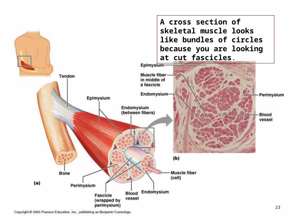
Connective Tissue Sheaths in Skeletal Muscle
Figure 10.1a4

Connective Tissue Sheaths The MUSCLE FASCIA is loose connective tissue on the
outside of the muscle. It creates a slippery surface for muscles to rub against each other. Superficial to the fascia is fat. Deep to the fascia is the EPIMYSIUM, (dense irregular fibrous connective tissue), and which eventually becomes the tendon (which is connected to bone). The epimycium extends into the muscle belly to form compartments called FASICLES. This tissue surrounding the fascicles is now called the PERIMYCIUM. Each fascicle contains MUSCLE FIBERS, which are individual muscle cells, each one surrounded by ENDOMYCIUM. When you eat steak and find it’s stringy, each string is a fascicle.
5
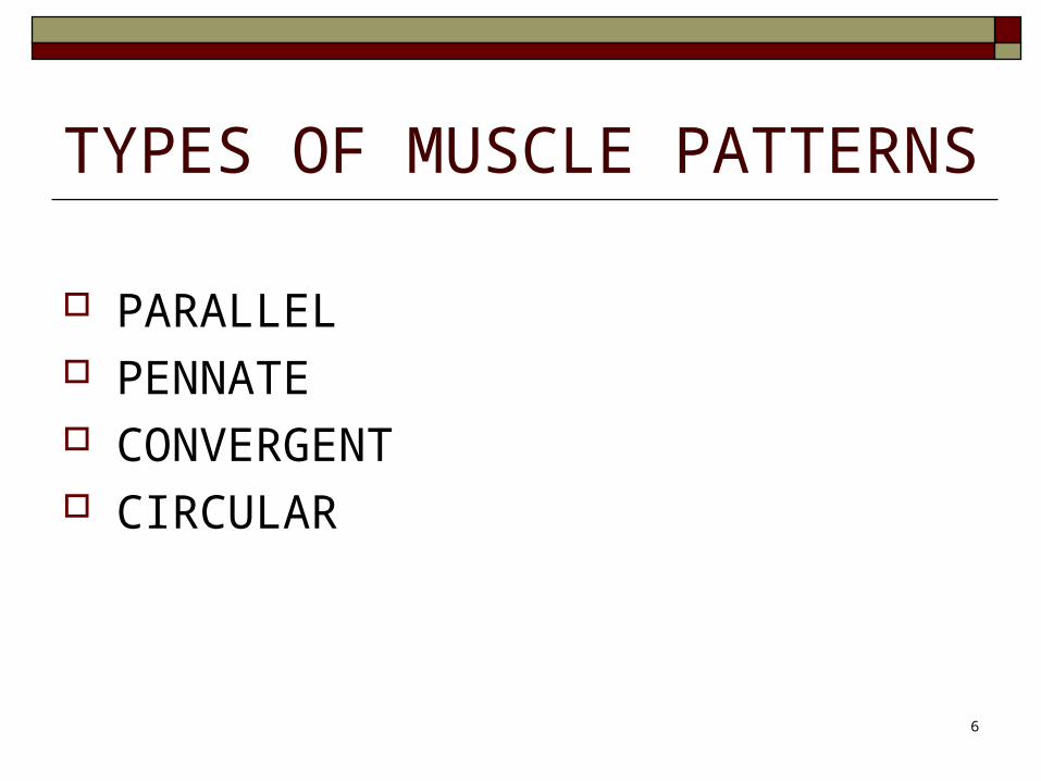
TYPES OF MUSCLE PATTERNS
PARALLEL PENNATE CONVERGENT CIRCULAR
6

PARALLEL MUSCLE The fascicles are parallel. They are long fibers, which can contract to 75% of their length. They contract a long way, but they are relatively weak, because there are relatively few fascicles. E.g. Sternocleidomastoid.
7

Arrangement of Fascicles in
Muscles Figure 11.38

PENNATEPENNATE (means “feather shape”) MUSCLES: three types:
UNIPENNATE; looks like half a feather. The fascicles are short, but there are more of them. They are stronger, but do not have the same length contraction ability of the parallel muscles.
BIPENNATE are fascicles that insert into the tendon from both sides; they are stronger than unipennate (quadriceps).
MULTIPENNATE are the strongest (deltoid). The fascicles are in multiple bundles inserting on one tendon
9

PENNATE
10

11

CONVERGENTCONVERGENT MUSCLE has more fibers
than parallel, but contracts a greater distance than pinnate. E.g. Pectoralis major.
12

13

CIRCULAR MUSCLECIRCULAR MUSCLE (Sphincter) is arranged
in a circle, with a small area of tendon on the sides. It allows closure of the eyes, mouth, etc. They are not very strong, but they don’t need to be.
14

15

TERMS: ORIGIN = The region which usually doesn’t move when the
muscle contracts. Look at the biceps brachii; does the shoulder move when I bend my arm? No; the shoulder = origin.
INSERTION= The point of attachment that moves; bend arm, radial tuberosity = attachment.
AGONIST = The main muscle for a particular action; bend arm, biceps = agonist.
ANTAGONIST = Does the opposite action; bend elbow, antagonist extends. Every muscle in the body has to have an antagonist.
SYNERGIST = The muscle that helps the agonist. There are several muscles that assist when the arm is bent.
16

Muscle Attachments
17

Skeletal Muscle Characteristics Contractility
The ability to shorten with force However, they lengthen passively, by gravity or by the contraction of an
opposing muscle. Excitability
Capacity to respond to a stimulus (nerves) Extensibility
Can be stretched After a contraction, they can be stretched to their normal resting length
and beyond to a limited degree. Elasticity Can recoil to their original resting length after they have been stretched
Has thousands of nuclei per cell, unlike smooth and cardiac muscle 18

SKELETAL MUSCLE They have thousands of nuclei because they start
from many stem cells that fuse together into one skeletal muscle fiber.
Theses are very long fibers (biceps muscle can be 8-10 cm).
19

Skeletal Muscle Myoblasts exist in adults, so muscle heals
well. A muscle cell torn in half can regenerate. There are almost no muscle diseases for this
reason (muscular dystrophy is the main muscle disease).
Muscles Overview Video http://www.youtube.com/watch?v=ren_IQPOhJc
20

Skeletal Muscle: Longitudinal section
In skeletal muscle fibers, there are light and dark stripes called striations, which can be seen under a microscope.
21
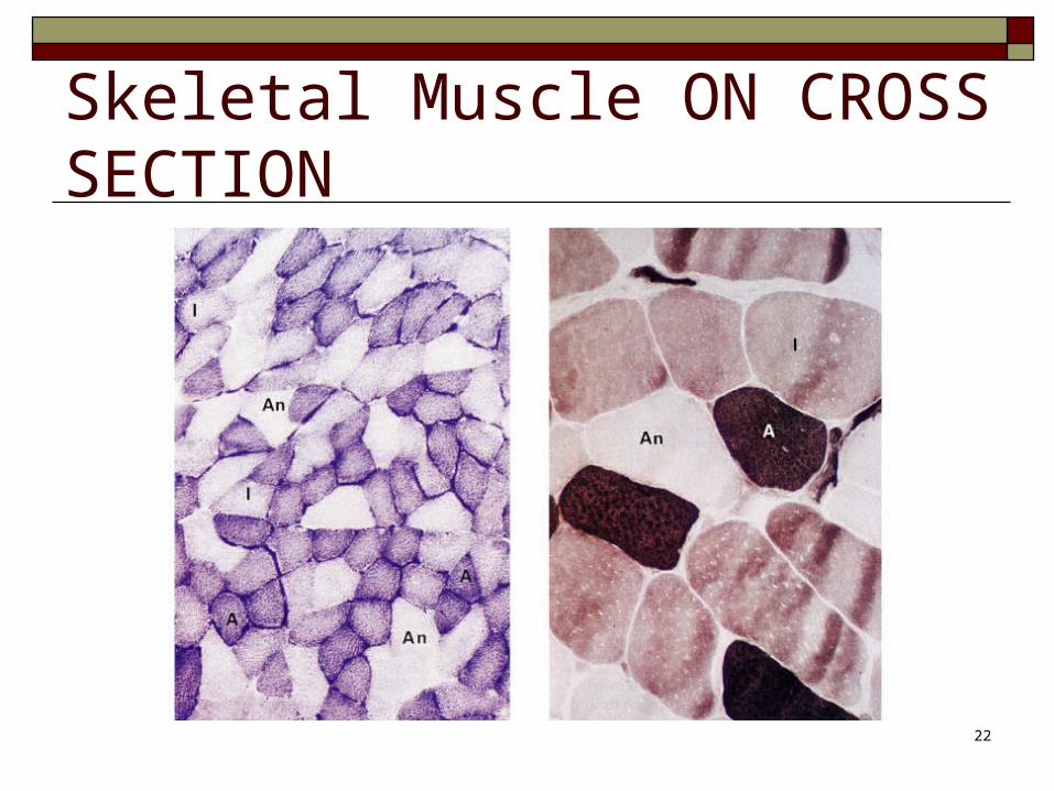
Skeletal Muscle ON CROSS SECTION
22
A cross section of skeletal muscle looks like bundles of circles because you are looking at cut fascicles.
23

Skeletal Muscle The plasma membrane of muscles is called a
SARCOLEMMA. The cytoplasm of muscle cells is called
SARCOPLASM. Muscle cells contain many mitochondria and
other organelles. One type of unusual organelle found only in
muscle cells is called a myofibril. They are packed in bundles and fill up most of the cell.
24

• MUSCLE MYOFIBRILS • Cylindrical organelles found within muscle cells
• Extend from one end of the muscle fiber (muscle cell) to the other
• Contain sarcomeres joined end to end.
• The sarcomeres are made of actin and myosin myofilaments
25

Skeletal Muscle: Longitudinal section
These striations (stripes) are caused by dark and light bands.
The dark band is called an A band. (There is an “A” in dark)
The light band is called an I band. There is an “I” in light)
26
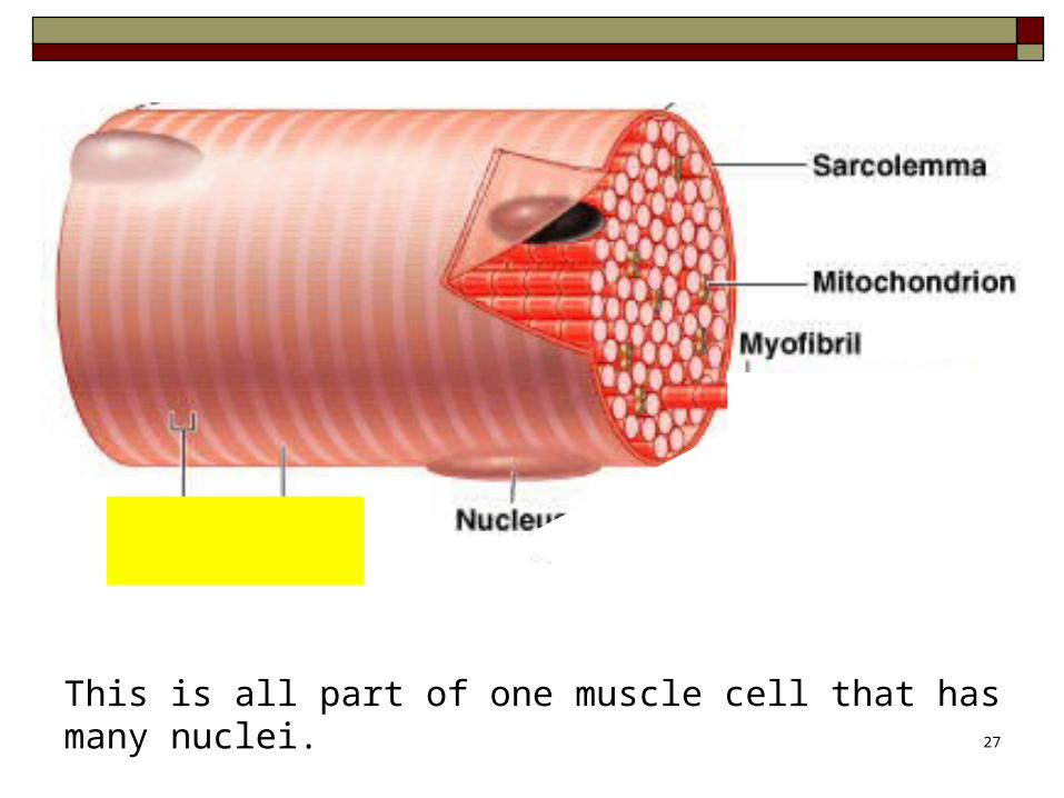
This is all part of one muscle cell that has many nuclei.27

Every dark band + light band is one sarcomere
In the center of each light I band is a Z discOne sarcomere is the area from one Z disc to the next Z disc.So, each sarcomere extends from the middle of one light band to the middle of the next light band. In the center of the dark band is a lighter colored area called the H zone. It is the area of the myosin without heads.
28
SARCOMERES The striations result from the internal structure of SARCOMERES
within the sarcoplasm. The sarcomere is the basic structural and functional unit of
skeletal muscle. The sarcomere is what contracts. Each sarcomere
Extends from one Z disc to the next Z disc Has a light colored H zone in the center (found in the middle of
the dark band, which is in the center of the sarcomere. It is the area of myosin in the center that does not have myosin heads).
Contains parts of two I (light) bands and all of one A (dark) band Contains overlapping actin and myosin myofilaments.
29

Note: the I band consists only of actin myofilaments.The A band consists of both actin and myosin. 30

31

Actin and Myosin Sarcomeres consist of two types of myofilaments
made out of protein: thin (ACTIN) myofilaments
Look like two strands of beads twisted together. Actin myofilaments are attached to the Z disc at one end.
thick (MYOSIN) myofilaments. Both ends of a thick filament are studded with knobs
called myosin heads (look like little golf clubs). Myosin is NOT attached to the Z disc.
Sarcomere model video 1Sarcomere model video 2
32

33
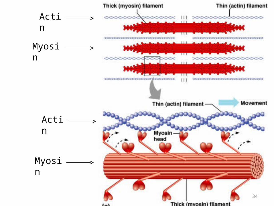
Actin
Myosin
Actin
Myosin
34

Don’t confuse these terms!
MUSCLE FASCICLE: a group of muscle fibers, surrounded by perimysium.
MUSCLE FIBER: a single muscle cell
MYOFIBRIL: a long organelle inside a muscle fiber, contains actin and myosin myofilaments.
MYOFILAMENTS: these are proteins, and there are two types: actin (with troponin and tropomyosin) and myosin. The myofilament is the lowest level of organization that is composed of actin, myosin, troponin, and tropomyosin proteins.
Therefore, a myofilament is part of a myofibril, which is inside a muscle fiber, which is inside a muscle fascicle. 35
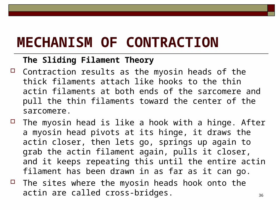
MECHANISM OF CONTRACTIONThe Sliding Filament Theory
Contraction results as the myosin heads of the thick filaments attach like hooks to the thin actin filaments at both ends of the sarcomere and pull the thin filaments toward the center of the sarcomere.
The myosin head is like a hook with a hinge. After a myosin head pivots at its hinge, it draws the actin closer, then lets go, springs up again to grab the actin filament again, pulls it closer, and it keeps repeating this until the entire actin filament has been drawn in as far as it can go.
The sites where the myosin heads hook onto the actin are called cross-bridges.
36

37

Sarcomere Contraction The complete process of contraction of the sarcomere takes
only a fraction of a second. The actin and myosin filaments do not shorten; they
merely slide past each other. The energy required is ATP. The A band (dark stripe) in a sarcomere does not
change length in a contraction. This sliding filament mechanism begins whenever calcium
ions bind to the thin filament. Where does the calcium come from?
38

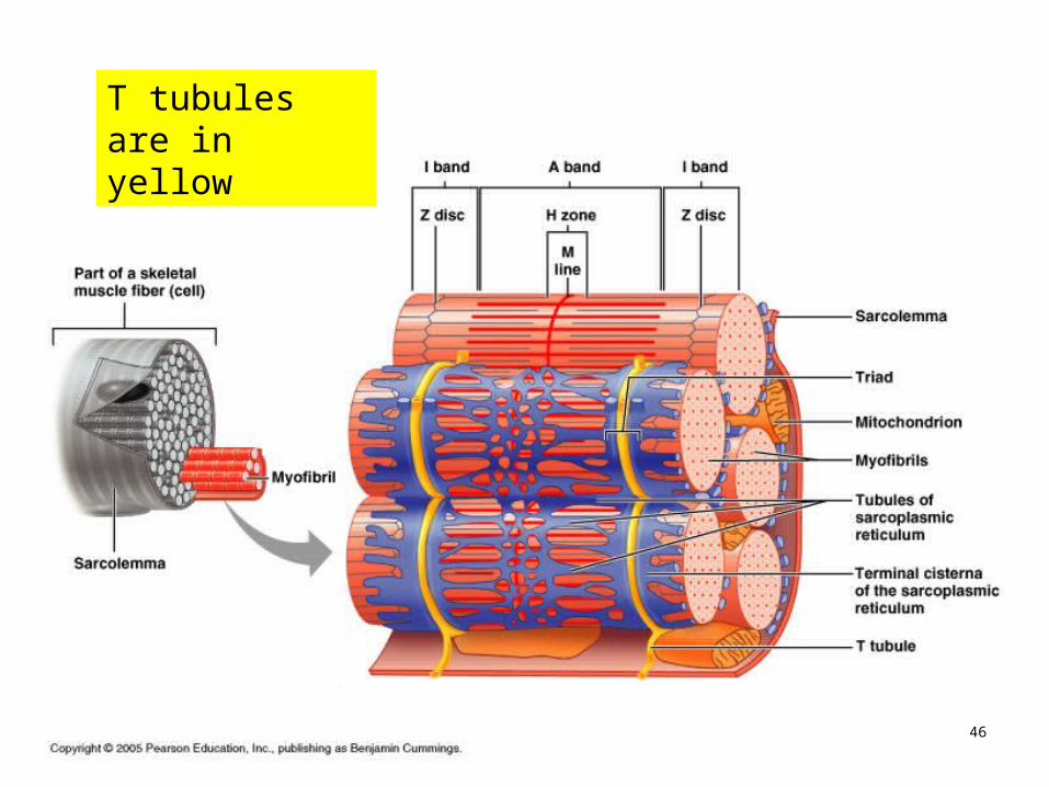
SARCOPLASMIC RETICULUM AND T TUBULES
Within the cytoplasm of all body cells is an endoplasmic reticulum.
The endoplasmic reticulum in muscle cells is called the SACROPLASMIC RETICULUM.
It surrounds each sarcomere like the sleeve of a loosely crocheted sweater.
39

Sarcoplasmic reticulum is in blue
T tubules are in yellow
40
Calcium is needed for muscle contraction The sarcoplasmic reticulum stores a lot of calcium
ions, which are released when the muscle is stimulated to contract.
The calcium diffuses out of the sarcoplasmic reticulum and land on the actin filaments, where they trigger the sliding filament mechanism of contraction.
After the contraction, the calcium ions are pumped back into the sarcoplasmic reticulum for storage.
41
Calcium is needed for muscle contraction
ACTIVE TRANSPORT is required to return the calcium ions to the sarcoplasmic reticulum.
It also requires energy to break the cross-bridge so the myosin head can cock back again, ready to spring onto the next binding site.
Therefore, ATP is used. ATP is used to return calcium to the sarcoplasmic
reticulum ATP is used to cock back the myosin heads
42

ATP is required for contraction ATP attaches to the myosin myofilaments
Provides energy for the movement of the cross bridges
ATP is required for muscle relaxation ATP releases part of its energy as heat.
That is why we get hot when we exercise When we are cold, we shiver (muscle contraction)
to warm up. In order for the mitochondria to produce enough ATP,
it needs oxygen and the sugars that are in storage.43

For contraction to take place, you need a nerve signal and calcium
For skeletal muscle to contract, the synaptic knob of a neuron must first release a chemical called ACETYLCHOLINE onto the region where it sits on the muscle cell, known as the ENDPLATE.
Calcium is also needed for muscle contraction. The nerve signal is called an ACTION
POTENTIAL. It causes a release of calcium from the sarcoplasmic
reticulum, which causes contraction.
44
Muscle Contraction In a muscle fiber, an action potential results in
muscle contraction. How does this happen? The action potential continues to travel along the
sarcolemma (cell membrane of the muscle). Part of this electrical impulse breaks away from
the sarcolemma and travels down the T-tubules, while the rest of the electrical impulse continues longitudinally down the muscle cell to the next sarcomere and T-tubule.
45
T tubules are in yellow
46

T TUBULES T TUBULES (“T” stands for “transverse”)
are continuations of the sarcolemma (cell membrane) which invaginate to the deepest regions of the muscle cell.
Since the T tubules conduct the nerve impulse throughout the muscle cell, all the sarcomeres of that cell contract at the same time.
47

Muscle Contraction The action potential of the nerve goes down the T-
tubules and causes calcium to leak out of the sarcoplasmic reticulum.
The calcium causes the muscle fibers to contract. After a while, the calcium gets pumped back where
it came from, the muscle fibers relax, although it requires gravity or another muscle to pull the sarcomere back to its original length.
How does the calcium cause the muscle fibers to contract?
48

49
TROPOMYOSIN is a single long protein strand like a piece of yarn that winds around the actin filament.
Tropomyosin blocks actin’s attachment site for the myosin head, so the myosin “hook” has nothing to grab onto, thus preventing contraction.
TROPONIN is a globular complex of three proteins, and is found in clumps around the tropomyosin protein.
Troponin is the specific molecule that provides the calcium binding site on actin.
Calcium binds to troponin and causes troponin to move a little, taking the tropomyosin thread with it, so the attachment sites on the actin molecule are now exposed. The myosin heads can now hook into the exposed sites on the actin myofilament.
Both troponin and tropomyosin cover the actin filament when the muscle is relaxed.

This is an illustration of an actin molecule. You can see the thready tropomyosin and the globular troponin proteins wrapping around the double-stranded actin. 50
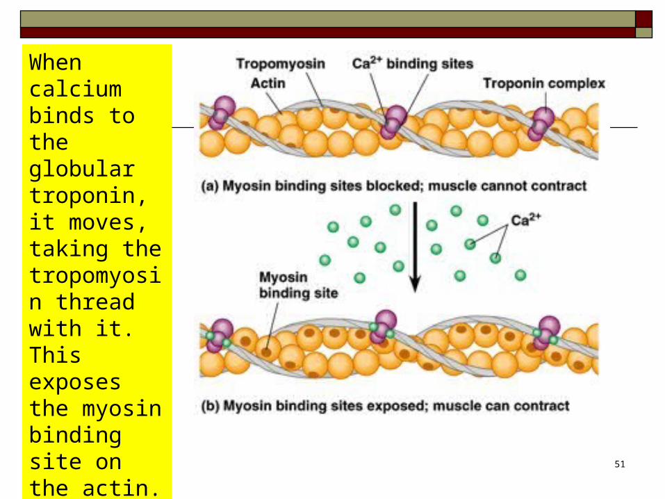
When calcium binds to the globular troponin, it moves, taking the tropomyosin thread with it. This exposes the myosin binding site on the actin.
51

Calcium in muscle contraction When the muscle cell is stimulated to contract by an action
potential, calcium channels open in the sarcoplasmic reticulum and release calcium into the sarcoplasm.
Some of this calcium attaches to troponin, causing a conformational change that moves tropomyosin out of the way so that the myosin heads can attach to actin and produce muscle contraction.
When the calcium gets pumped back where it came from, the tropomyosin protein blocks the myosin head again so it can no longer get its hook into the actin filament, and the muscle will relax.
52

Rigor Mortis A new ATP molecule must bind to the myosin
before the cross-bridge can be release. When ATP is not available after a person dies, the cross-bridges that have formed are not released, causing muscle to become rigid (rigor mortis)
NOTE: Sarcomeres lengthen during muscle relaxation only if gravity or an opposing muscle pulls the sarcomere back to its original length.
53

Muscle Contraction Muscle Contraction http://www.youtube.com/watch?v=CepeYFvqmk4
Sarcoplasmic Reticulum http://www.youtube.com/watch?v=InIha7bCTjM&NR=1
54
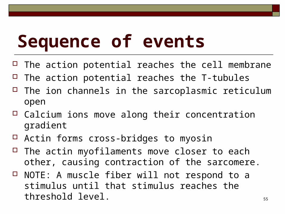
Sequence of events The action potential reaches the cell membrane The action potential reaches the T-tubules The ion channels in the sarcoplasmic reticulum open Calcium ions move along their concentration gradient Actin forms cross-bridges to myosin The actin myofilaments move closer to each other,
causing contraction of the sarcomere. NOTE: A muscle fiber will not respond to a stimulus
until that stimulus reaches the threshold level.55
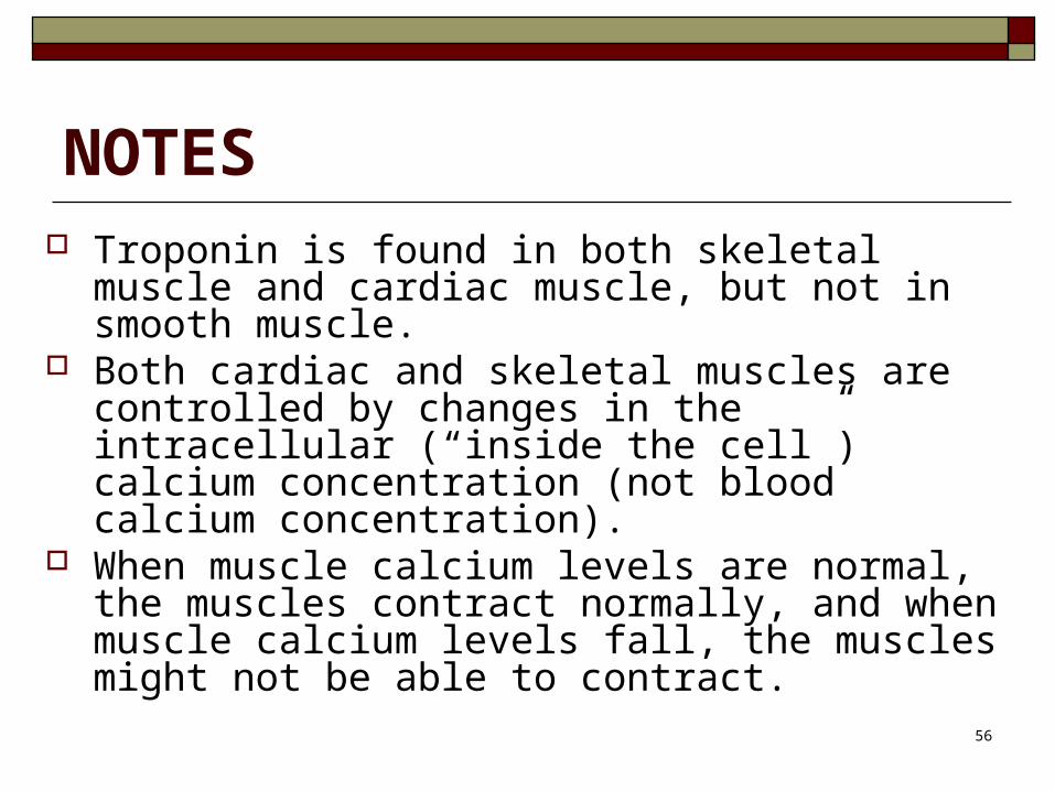
NOTES Troponin is found in both skeletal muscle and
cardiac muscle, but not in smooth muscle. Both cardiac and skeletal muscles are controlled by
changes in the intracellular (“inside the cell”) calcium concentration (not blood calcium concentration).
When muscle calcium levels are normal, the muscles contract normally, and when muscle calcium levels fall, the muscles might not be able to contract.
56

Muscle Contraction
A muscle TWITCH is one single muscle fiber contraction.
It takes 1/20th of a second. How is it that I can pick up and hold a chair if the
fiber only contracts for 1/20th second? There are ten thousand fibers per muscle; each
one contracts at different intervals, so contraction is maintained, just like tug-of-war. One person in ten can drop the rope and get a better grip because the others are maintaining the tension. 57

Motor UnitsA MOTOR UNIT is a single neuron and all of
the muscle fibers on which it synapses.
If one neuron sends a signal, only its muscle fibers contract (the motor unit). This allows for strength variations in lifting a chair vs. an eraser. For full strength, all the motor units contract. For half strength, half of the motor units contract.
58

Motor Units
There are 3 motor units in this diagram; that allows for 3 different levels of contraction. The more motor units there are, the more precisely the muscle can respond.
59

Motor Units A neuron that lands on skeletal muscle is called a motor neuron,
because it moves the body. The action potential continues from one motor neuron in the
brain to the next motor neuron in the spinal cord, which is the one that goes out and lands on the skeletal muscle fibers.
A single motor neuron and all the skeletal muscle fibers it innervates constitute a motor unit.
A muscle in your tongue may only have a few muscle fibers innervated by a neuron to allow for precise movement. However, large thigh muscles may have as many as 1000 muscle fibers per neuron, since precision is not necessary.
60
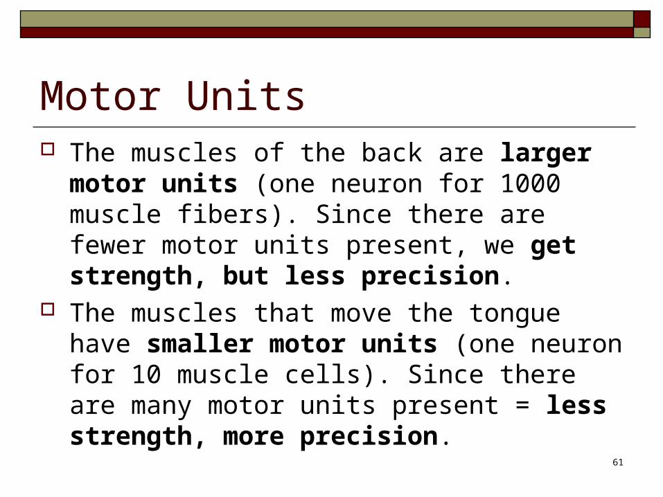
Motor Units The muscles of the back are larger motor
units (one neuron for 1000 muscle fibers). Since there are fewer motor units present, we get strength, but less precision.
The muscles that move the tongue have smaller motor units (one neuron for 10 muscle cells). Since there are many motor units present = less strength, more precision.
61
Physiology of the Neuromuscular Junction VIDEO
https://www.youtube.com/watch?v=hzXVe4RS8-A
62

63

64

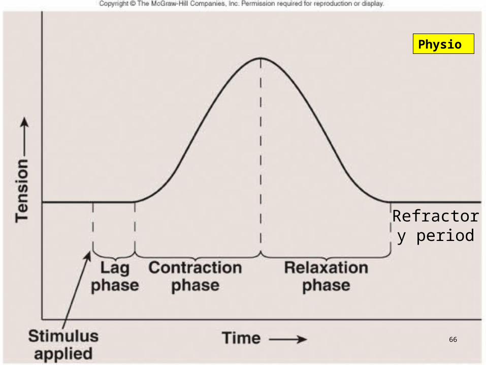
Muscle Twitch Phases
A muscle twitch has three phases The lag phase is the time between the application of a
stimulus and the beginning of contraction. The contraction phase is the time of contraction. The relaxation phase is the time during which the
muscle relaxes.
The refractory period is the time between muscle twitches.
65
Physio
Refractory period
66
Physio
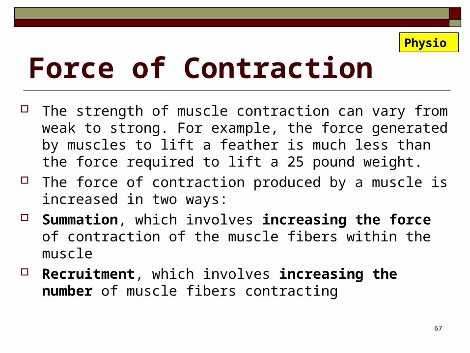
Force of Contraction The strength of muscle contraction can vary from weak to
strong. For example, the force generated by muscles to lift a feather is much less than the force required to lift a 25 pound weight.
The force of contraction produced by a muscle is increased in two ways:
Summation, which involves increasing the force of contraction of the muscle fibers within the muscle
Recruitment, which involves increasing the number of muscle fibers contracting
67
Physio

Summation The force of contraction of individual muscle fibers is increased by rapidly
stimulating them. Stimulus frequency is the number of times a motor neuron is stimulated per
second. When the stimulus frequency is low, there is time for complete relaxation of
muscle fibers between twitches. As stimulation frequency increases, there is not enough time between
contractions for muscles to completely relax. Thus, one contraction summates, or is added onto, a previous contraction.
As a result, the overall force of contraction increases. Tetanus is the condition in which a muscle remains contracted between
stimuli without relaxing.
68
Physio

TETANUS TOXIN A toxin caused by a certain bacteria can cause muscle to remain
contracted (in tetanus). It quickly results in death because the diaphragm and other
respiratory muscles cannot function properly, and the person suffocates.
The bacteria that make this toxin live deep in the soil and cannot survive in air. If you step on something that imbeds soil deeply into your tissues (like a rusty nail), you might contract the bacteria. You will need a tetanus shot before the toxins accumulate. 69

Recruitment In recruitment, the strength of contraction of the muscle is
increased by increasing the number of motor units stimulated. When only a few motor units are stimulated, a small force of
contraction is produced, because only a small number of muscle fibers are contracting.
As the number of motor units stimulated increases, more muscle fibers are stimulated to contract, and the force of contraction increases.
Maximum force of contraction is produced in a given muscle when all the motor units of that muscle are stimulated, or recruited.
70
Physio

Recruitment Motor unit recruitment allows muscles to have slow,
smooth sustained contractions so our movements are not jerky.
If all the motor units in a muscle could be stimulated simultaneously, a quick, jerky motion would occur.
Because the motor units are recruited gradually so that some are stimulated and held in tetanus while additional motor units are recruited, slow, smooth, sustained contractions occur.
71
Physio

Types of Muscle ContractionsMuscle contractions are classified as either isometric or isotonic. Most muscle contractions are a combination.
Isometric (equal distance) tension increases during contraction length of the muscle does not change Example is when you push against a wall or try to pick up an object
that is too heavy to lift
Isotonic (equal tension) tension is generally constant during contraction
Although in one type of isotonic contraction, the tension increases Length of the muscle changes (either increases or decreases). Example is when you lift a weight.
72
Physio

Concentric and Eccentric Contractions
Two types of isotonic contractions: CONCENTRIC CONTRACTIONS are isotonic contractions
in which the muscle tension increases as the muscle shortens. Most movements performed by muscle contractions are of this type.
ECCENTRIC CONTRACTIONS are isotonic contractions in which tension is maintained as the muscle lengthens. An example is when a person lets down a heavy weight slowly. Substantial force is produced in the muscles and injuries can occur from repetitive eccentric contractions, such as in the hamstring muscles when a person runs downhill.
73
Physio

Muscle Tone Even when muscles are relaxed, some of their fibers
are still contracting, giving the muscle some tone. Therefore, the normal state of a muscle, with some
contraction, is called muscle tone. This is important in posture so you can stand upright but mostly relaxed.
Muscle tone refers to the constant tension produced by muscles of the body over long periods of time. It is responsible for keeping the back and legs straight, the head held in an upright position, and the abdomen from bulging. it declines during REM sleep. 74

Motor Neurons A neuron (nerve cell) that innervates (supplies)
skeletal muscle is called a motor neuron (causes the body to move). There are 2 motor neurons involved in this task.
The Upper Motor Neuron has its cell body in the brain, and its axon (like a stem) lands on the cell body of the Lower Motor Neuron, which is in the spinal cord.
The axon of the Lower Motor Neuron leaves the spinal cord and innervates the muscle.
75
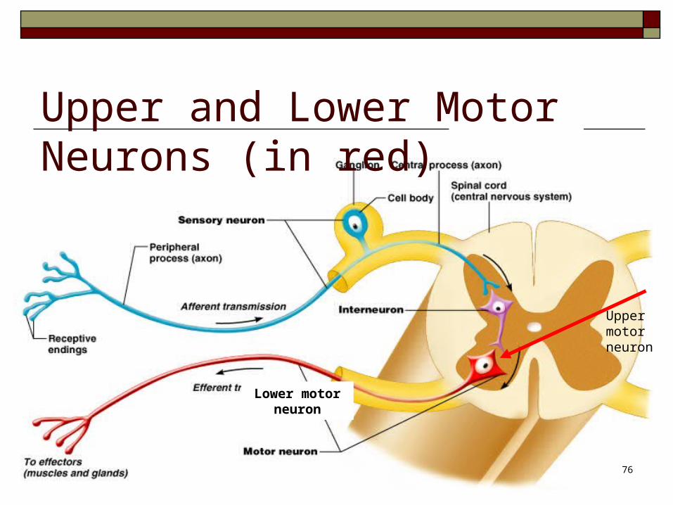
Upper and Lower Motor Neurons (in red)
Upper motor neuron
Lower motor neuron
76

Muscle Tone Hypertonia
Can present clinically as either spasticity or rigidity. Seen in upper motor neuron diseases, such as multiple sclerosis or cerebral palsy.
Hypotonia Seen in lower motor neuron diseases (spinal cord
damage and ALS/Lou Gehrig Disease) Can present clinically as muscle flaccidity, where the
limbs appear floppy, stretch reflex responses are decreased, and the limb’s resistance to passive movement is also decreased.
77

Muscle Tone Upper Extremity Tone, normal VIDEO Upper Extremity Tone, abnormal VIDEO Lower Extremity Tone, normal VIDEO Lower Extremity Tone, abnormal VIDEO
78
Muscle Spasticity: Hypertonia Clinically spasticity is defined as velocity dependent
resistance to stretch. Passively moving (the doctor does the movement) the
patient’s elbow or foot quickly will elicit spastic twitches, but passively moving elbow or foot slowly is normal.
It mostly occurs from upper motor neuron lesions (scar, tumor, or other damage), but it can also present in multiple sclerosis, which is an autoimmune condition. Can also be seen in cerebral palsy (lack of oxygen at birth).
79

Muscle Spasticity There is a difference in cause of two of the most
common spasticity conditions, spastic diplegia (cerebral palsy) and multiple sclerosis.
In spastic diplegia, the upper motor neuron lesion arises often as a result of neonatal asphyxia (lack of oxygen in a newborn), while in conditions like multiple sclerosis, spasticity is from multiple sclerosis, which is an autoimmune destruction of the myelin sheaths around nerve endings.
80

Muscle Spasticity Causes include
Spastic diplegia (Cerebral palsy) Multiple sclerosis Spinal cord injury Stroke
Test for clonus to see if spasticity is present.81
Muscle Clonus Clonus (from the Greek for "violent, confused motion") is a
series of involuntary muscular contractions initiated by a reflex. Clonus is a sign of certain neurological conditions, and is
particularly associated with upper motor neuron lesions such as in spastic diplegia, multiple sclerosis, stroke, spinal cord damage.
Clonus is most commonly tested for in the ankles, where it is tested by rapidly dorsiflexing the foot. If the foot then jerks 5 times or more, clonus is present. A positive clonus test means the patient has spasticity, usually due to an UMN disorder.
VIDEO: Ankle clonus http://www.youtube.com/watch?v=iWEJIVO85TI
82

Muscle Rigidity Unlike spasticity, rigidity is velocity-
independent resistance to passive stretch. There is uniform increased tone whether the
elbow is passively moved quickly or slowly.
83

Muscle Fasciculations These are small, local, involuntary muscle
contractions. Fasciculations have a variety of causes, the
majority of which are benign, but can also be due to disease of the lower motor neurons.
Fasciculations VIDEO Tremor VIDEO
84

Muscle Fasciculations Benign causes of fasciculations include:
Magnesium deficiency Diarrhea Overexertion Inadequate intake from diet (almonds are a good source
of magnesium)
Dehydration Fatigue A small neuron dying can also cause fasciculations.
85

Muscle Fasciculations They can also be caused by long-term use of:
Benadryl (antihistamine) Dramamine (for nausea and motion sickness). Caffeine Sudafed (for allergies) Asthma medicines ADD medicines
86

Muscle Fasciculations More serious conditions causing
fasciculations include Fibromyalgia Myasthenia Gravis Lyme Disease Rabies
87

Hyperreflexia We talked about hypertonia; some people have
hyperreflexia. The most common cause of exaggerated reflexes is spinal
cord injuries (upper motor neuron diseases). Other causes include
Medication Stimulants Hyperthyroidism Electrolyte imbalance Severe brain trauma.
88

Hyporeflexia This means diminished or absent reflexes. The most common cause is lower motor
neuron diseases.
89

Muscle Strength and Coordination Upper Extremity Strength, normal VIDEO Upper Extremity Strength, abnormal VIDEO Lower Extremity Strength, normal VIDEO Lower Extremity Strength, abnormal VIDEO Strength Evaluation using squats VIDEO Hand coordination using Rapid Alternating
Movements (RAM) VIDEO
90

Muscle Contractures Muscle contractures can occur from paralysis,
muscular atrophy (immobilization from a cast), muscular dystrophy, and chronic spastic conditions like cerebral palsy.
Fundamentally, the muscle and its tendons shorten, resulting in reduced flexibility.
Muscle contractures in tendons are caused from the fibrinogen leaking out of the fibroblasts, which turn the elastic fibers into inelastic fibers.
Most treatments involve surgery, so physical therapy efforts focus on prevention of contractures.
91

Energy Requirements of Muscle What fuel does a car use?
Gasoline What fuel does a candle use?
Wax What fuel do humans use?
Oxygen? NO
Sugars? NO
ATP YES
92

ATP Where do we get ATP? We can make a little ATP in the cytoplasm of our cells, but not
enough to live on. Most of our ATP is made by the mitochondria inside our cells. Mitochondria are like little protozoa (animals) that live in our
cells. Each cell has hundreds of them. Muscle cells have thousands of them.
What is their fuel? Oxygen and glucose THAT is why we need to inhale oxygen and consume
sugars….to feed our mitochondria so they can make ATP for us! 93

Energy Requirements For Muscle Contraction In order for the muscle mitochondria to produce
enough ATP, they need oxygen (for their own aerobic respiration) and sugars that are in storage.
Mitochondria can only perform aerobic respiration. What can we do to make ATP if our muscle cells run
out of oxygen? Start performing anaerobic respiration. We can do this ourselves in the cytoplasm of our cells.
94
Making ATP by Aerobic RespirationAerobic cellular respiration Breaks down glucose to produce ATP Takes place in the mitochondria Requires oxygen Waste products are CO2 and H2O (we exhale them) The good thing about making ATP from our mitochondria
is that we can make a LOT of it. The bad things are that it takes longer to make it, and it
requires oxygen, and a muscle cell may have used up all the oxygen during a sprinting run.
95

Making ATP by Anaerobic RespirationAnaerobic cellular respiration Breaks down glucose to produce ATP Takes place in the cytoplasm Does not require oxygen Waste product is lactic acid The good thing about making ATP this way is that we
can make it FAST. The bad thing is that it does not make much ATP,
and we deplete the reserves quickly.96

Lactic Acid The waste product of aerobic respiration is carbon
dioxide and water. These are not a problem…we eliminate them by exhaling.
The waste product of anaerobic respiration is lactic acid, which can irritate muscle fibers, causing muscle pain (stitch in your side) and muscle cramps.
We deactivate lactic acid by adding oxygen to it. Therefore, breathing heavily adds the oxygen to our system to deactivate lactic acid, and the muscle pains go away.
97

ATP and Creatine Phosphate What do we do when we run out of ATP? Muscle fibers cannot stockpile ATP in preparation for future
periods of activity. However, they can store another high energy molecule called
creatine phosphate, which is the storage form of ATP. Creatine phosphate is made from the excess ATP that we
accumulate when we are resting. During short periods of intense exercise, the small reserves
of ATP existing in a cell are used first. Then creatine phosphate is broken down to produce ATP.
98

Aerobic vs. Anaerobic Respiration
Why does sprinting require anaerobic respiration? We use up all of the ATP faster than we can make it.
When we run out of ATP, we break down creatine phosphate to make more ATP.
When we run out of ATP and creatine phosphate, we start using anaerobic respiration to make more ATP.
When we run out of glucose, or too much lactic acid is built up, we have to stop and rest.
Anaerobic metabolism is ultimately limited by depletion of glucose and buildup of lactic acid within the muscle fiber.
99

Sprint Runners Why do sprint runners tire out during the last part of a
fast run? Sprinting is an anaerobic activity…the oxygen
requirement is quickly exceeded, so the muscle has to use anaerobic respiration to continue to contract. This requires a lot of glucose and also results in a buildup of lactic acid.
Once the sprint-runner has used up the available glucose, or has produced too much lactic acid, the muscles fatigue.
Sprint Runners http://www.youtube.com/watch?v=MTn1v5TGK_w100
Oxygen Debt Anaerobic respiration produces lactic acid, which
causes the painful cramps because it creates an oxygen debt.
The amount of oxygen needed to replenish the supply following aerobic demand is called the oxygen debt.
When you continue to breathe heavily after exercising, it means you have an oxygen debt.
Muscles can do without oxygen for a while pretty well, unlike the brain.
To pay back a minor oxygen debt, you just have to breathe heavily for a while. 101

Oxygen Debt
This heavy breathing brings in oxygen, which is used to convert lactic acid to glucose, replenish the depleted ATP and creatine phosphate stores in the muscle fibers, and to replenish oxygen stores in the lung, blood, and muscles.
After the oxygen debt has been paid back, breathing returns to normal.
People who are in good physical condition can carry out both aerobic and anaerobic activities efficiently, and do not suffer from an oxygen debt for very long.102

Myoglobin Myoglobin is an iron- and oxygen-binding protein found in
the muscle tissue of mammals. It is related to hemoglobin, which is the iron- and oxygen-
binding protein in blood, specifically in the red blood cells. The only time myoglobin is found in the bloodstream is when
it is released following muscle injury. It is an abnormal finding, and can be diagnostically relevant when found in blood.
103

Myoglobin Myoglobin binds to oxygen more strongly than
hemoglobin. It acts as an oxygen-storage molecule and delivers
the oxygen to cells when needed. High concentrations of myoglobin in muscle cells
allow organisms to hold their breaths longer. Diving mammals such as whales and seals have
muscles with particularly high myoglobin levels.
104

Myoglobin Myoglobin forms pigments responsible for making meat red. The color
that meat takes is partly determined by the oxidation states of the iron atom in myoglobin.
When meat is raw, the iron atom is in the +2 oxidation state (Fe+2). Meat cooked well done is brown because the iron atom is now in the +3
oxidation state (Fe+3), having lost an electron. Under some conditions, meat can also remain pink all through cooking,
despite being heated to high temperatures. If meat has been exposed to nitrites, it will remain pink because the iron atom is bound to NO, nitric oxide (e.g., corned beef or cured hams). Grilled meats can also take on a pink "smoke ring" that comes from the iron binding to a molecule of carbon monoxide.
105
Myoglobin Rhabdomyolysis is the condition when myoglobin is
released from damaged muscle tissue. Released myoglobin is filtered by the kidneys, but it
damages them, so it can lead to renal failure. High blood levels may indicate the person is having
a heart attack, or it could just be a muscle injury. Therefore, CK, cTnT, ECG, and clinical signs
should be taken into account to make the diagnosis of a heart attack.
106

Creatine kinase (CK) CK is the enzyme used to get ATP out of
storage (ATP is stored as creatine). High blood levels of CK may indicate
myocardial infarction (heart attack), rhabdomyolysis (severe muscle breakdown), muscular dystrophy, the autoimmune myositides, or acute renal failure.
107

Troponin (cTnT) Troponin levels in the blood can be used as a
test of several different heart disorders, including myocardial infarction.
Troponin-I is highly specific for cardiac muscle necrosis. Serum levels rise 4-8 hrs after onset of chest pains, peak at 12-16 hrs and return to baseline within 5-9 days.
108

EXERCISE There are many physiological benefits of
exercise:
1. Improved muscular strength, endurance, flexibility
2. Improved cardio-respiratory endurance
3. Increased bone density and strength
4. Relief from depression
5. Increased HDLs109

Hypertrophy Weight training and other exercises can cause muscles to hypertrophy
(enlarge). This occurs as more myofilaments and myofibrils are produced inside a myofiber (muscle cell), causing the muscle cell to enlarge. The number of mitochondria also increases, causing additional enlargement.
However, you don’t grow new muscle cells. The number of cells in a skeletal muscle remains relatively constant following birth. Myoblast stem cells don’t grow into new muscle cells; they just patch up damaged cells.
Hypertrophy because: Increase in number of myofibrils inside a muscle cell Which causes the increase in size of individual muscle cells (myofibers)
Muscle hypertrophy is greater in males due to the hormone testosterone.
110

111

Hypertrophy Eating protein does not automatically increase muscle. The
average person only needs three ounces of protein per day, six ounces if you work out.
Two ounces is the size of a deck of playing cards. Three ounces is like one mini hamburger. Most people eat too much meat.
Fun Fact: -You use 200 muscles to take one step.
112

How much meat per day? Most people need around 0.8 grams of protein per day per 2.2
pounds of body weight, according to registered dietitian Reed Mangels.
Consuming around 46 grams of protein per day if you're female and 56 grams per day if you're male will meet your protein needs, whether you get your protein from meat or from plant sources.
A 3-ounce portion of meat contains 21 to 24 grams of protein. Two 3-ounce meat servings per day would supply all your
protein needs, but this much meat could include a large amount of saturated fat, which could increase your risk of heart disease.
113

Fun Facts A muscle cell is about as thin as a hair. Muscles make up 1/3 of your body weight. There are 655 muscles in the body…that is three
muscles per bone! We all have the same number of muscle cells. Muscles can generate 40 pounds per square inch. If all the muscles in the body were able to
contract at once, they could pull 25 tons!114

Fun Facts Myths About Women’s Body Building http://bodybuilding.about.com/od/womensfitnesstopics/a/womenmyths.htm
115

Atrophy Lack of use causes muscle ATROPHY. This happens quickly.
Astronauts can lose 40% of their muscle in two weeks! It is regained quickly, too.
Atrophy is a decrease in muscle size because of the decrease in myofilaments within muscle fiber.
Casting a broken limb also leads to temporary atrophy. In 3 weeks, they need a fresh cast because it is too loose.
Severe atrophy involves the permanent loss of skeletal muscle fiber and the replacement of those fibers by connective tissue.
Damage to the nervous system, or a severed motor nerve can cause atrophy. The muscle becomes flaccid (having no tone) .
More info on Muscle Atrophy116

Muscular Dystrophy
This refers to a group of inherited muscle disorders in which skeletal, cardiac, and smooth muscle tissue degenerates and the person experiences progressive weakness and other symptoms, including heart problems.
The muscle is replacement by fat and other connective tissue.
117

Muscular Dystrophy
MUSCULAR DYSTROPHY
This is a genetic lack of a protein called DISTROPHIN. It causes the muscle tissue to harden, inhibiting contraction, causing progressive paralysis.
Duchenne muscular dystrophy is more common in males.
118


Research on Muscular Dystrophy Two new types of stem cells have been found
that can seek out injured muscle tissue and replace the damaged cells.
Researchers in Italy used stem cells from blood vessels to repair muscle in mice with muscular dystrophy.
Canadian scientists found that stem cells from damaged muscle give rise to new muscle fibers.
121
Muscle Problems Tendonitis is an inflammation of the tendon or its
attachment point. It usually occurs from overuse of the muscle to which the tendon is attached.
A strain is a tear in a muscle. Remember, a sprain is a tear in a ligament.
A muscle strain will heal faster than a torn ligament because muscles have good blood supply and ligaments do not.
122
Treatment for Injuries: RICE Rest Ice
20 minutes on, 20 minutes off for 3 days Ice pack or frozen bag of peas!
Compression Ace wrap from distal to proximal Don’t leave any openings while wrapping
Elevation Above the heart 123

Treatment for Injuries
Ice for the first 72 hours (NO heat!) Anti-inflammatory medicines
Ibuprofin, 600 mg TID (3x a day) Over the counter (OTC) pills are 200 mg
Heat and massage as needed after third day. Can try a muscle stimulator too…works pretty
well!
124
Muscle Spasms Muscle spasms/cramps are sudden and involuntary muscle
contractions. They are painful, spastic contractions that are usually caused from overexertion. Lactic acid builds up and irritates the overused muscles, causing inflammation. If the muscle remains in spasm for longer than a few minutes, might need heat and massage to increase circulation.
Avoid spasms by stretching before and after activities. For people with frequent low back spasms throughout the day, a
portable muscle stimulator that clips to the belt will help a great deal.
125

Muscle stimulator to relieve muscle spasms or to prevent muscle atrophy in casts$59
http://www.medicalproductsonline.org/meprondi75mu.html 126

127
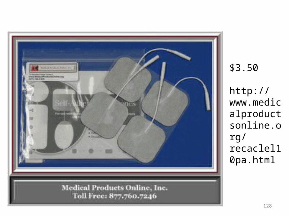
$3.50
http://www.medicalproductsonline.org/recaclel10pa.html
128

Fibromyalgia (muscle and tissue pain)
Common disorder in adults, especially women Painful muscles, debilitating fatigue, sleep
disturbance, and joint stiffness Many trigger points: painful lumps in muscles Treatment includes anti-inflammatory medicines,
physical therapy, acupuncture, and exercise. Muscle stimulators help
129

Fibromyalgia The American College of Rheumatology
guidelines that require a minimum of 11 out of 18 specific tender points for a fibromyalgia diagnosis.
Fewer than 11 tender points may also indicate fibromyalgia, particularly if you also have severe fatigue and widespread pain that has lasted more than three months.
130

Fibromyalgia points of tenderness
131

Fibromyalgia There is a blood test to help diagnose the
condition. The FM/a test identifies markers produced by
immune system blood cells in people with fibromyalgia.
132
Fibromyalgia Treatment There is no cure for fibromyalgia, and people
with the condition usually have it for life. However, it is not likely to get worse as you
age and it does not damage muscles, tendons, or ligaments.
Many people are able to reduce their symptoms with a combination of exercise, medication, physical therapy, and relaxation.
133
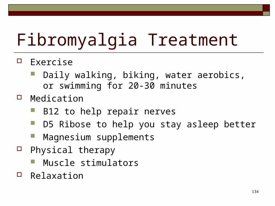
Fibromyalgia Treatment Exercise
Daily walking, biking, water aerobics, or swimming for 20-30 minutes
Medication B12 to help repair nerves D5 Ribose to help you stay asleep better Magnesium supplements
Physical therapy Muscle stimulators
Relaxation134

Ganglion Cysts Ganglion cysts arise as outpouchings from fluid filled
areas such as the fluid around tendon sheaths. When the fluid, called synovial fluid, leaks out from these
spaces, it can become a cystic structure. Treatment is to drain the fluid with a needle, but the fluid
can be jelly-like and difficult to remove, and they frequently grow back.
If conservative treatments fail to correct the cyst, an operation can be done to excise the cyst.
Then you do a surgery to scoop out the whole cyst, find the stalk and tie it off.
135

• Ganglion cyst
136

Baker’s Cyst A Baker's Cyst, or popliteal cyst, is a collection of
fluid in the back of the knee joint. A Baker's cyst is usually a symptom of another
problem, or it may be an incidental finding with no significant meaning.
Most often in adults the Baker's cyst is found in conditions where there is chronic swelling or fluid accumulation in the knee joint.
These conditions include knee arthritis, meniscus injuries, and ligamentous injuries. 137

Baker’s Cyst
Treatment of a Baker's cyst that is the result of a problem within the knee consists of treating the underlying problem. These treatments may include anti-inflammatory medications and cortisone injections.
138

• Baker’s Cyst
139

What is the Rotator Cuff? Rotator Cuff Injury http://www.youtube.com/watch?v=-tx2SqWz3BY
How do rotator cuff injuries occur? http://www.youtube.com/watch?v=t6FCBBijROo
What is an MRI? http://www.youtube.com/watch?v=H0adTNhzGxU
How does a CT scan work? http://www.youtube.com/watch?v=81PeTqmtzjk
140
AGING With aging, fibrous connective tissue
replaces some muscle fibers, causing decreased strength.
As people age, the number of muscle fibers decreases, and new ones cannot be added.
141

FUN FACTS ABOUT STRENGTH
The strongest humans can lift about 3 times their own body weight, but the average gorilla can lift 10 times its own body weight! Gorillas can lift 4,600 pounds. By the way, they don’t drink water. They get it by eating 50 pounds of plants a day.
The rhino beetle can carry 800 times its own weight.
And, pound for pound, the African Crowned Eagle can carry more than a
cargo plane, because it can fly carrying up to 4 times it's own weight.
Something that would keep a cargo plane grounded.
But the strongest creature is the ant. If you had the strength of an ant, you
could lift over your head and carry 6,600 pounds.
VIDEO: Skeletal Muscles 3 mins
142

FUN FACTS If we could jump like a locust, we could jump 300 feet. Locusts have massive muscles in their
thighs and it has elastic bands in its knees that are like stretchy springs that store energy. The tendons in our fingers store enough energy for us to snap our fingers.
At 60 mph, the cheetah is fast, but the basilisk lizard runs so fast that it can walk on water and the ostrich is just about the fastest animal on two legs.
What’s the fastest animal on Earth? The tiger beetle, which can run up to 171 times its size in one second. Despite its famous reputation, the cheetah would have to run 770 kilometres per hour just to catch up with it.
What’s the slowest animal in the world? The sloth. It is a species that moves just five times faster than a snail!
The penguin burns twice as much energy as any other animal when walking. This is due to the fact that its legs are very short, and so it must expend a lot of effort in order to get moving.
Elephants can't jump, not even with the help of hurricane-force winds. It is too heavy to lift all four legs at the same time.
The flea, however, can jump up to 200 times its own height. This is equivalent to a man jumping the Empire State Building in New York.
144
Smooth and Cardiac muscle
145

SMOOTH MUSCLE CELLS
These are found around internal organs (intestines, uterus, blood vessels).
They are involuntary and not striated. When smooth muscle contracts around the
intestines, the movement is called PERISTALSIS.
146

SMOOTH MUSCLE CELLS
• Smooth muscle cells are small and spindle shaped, usually with one nucleus per cell.
• They contain less actin and myosin, and the microfilaments are not organized into sarcomeres.
• As a result, smooth muscle cells are not striated.• They contract more slowly and do not develop an
oxygen debt. • Smooth muscle cells can spontaneously generate
action potentials that cause the cell to contract. 147

SMOOTH MUSCLE CELLS
• Smooth muscle is not under voluntary control, whereas skeletal muscle is voluntary.
• Some hormones in the digestive system can stimulate smooth muscles to contract.
• They have specialized cell to cell contacts that allow the action potential to spread to all of the smooth muscle cells in a given tissue.
• This allows them to function as a unit and contract at the same time.
148

Characteristics of smooth muscle There are no distinct sarcomeres They contract more slowly than skeletal muscle…their twitch
time is very long = several seconds It doesn’t get tired (“I’m too tired to urinate!”) They contract in response to neurons as well as hormones and
changes in local environment (amount of oxygen, lactic acid, etc).
They may be autorhythmic (self-exciting); they can contract spontaneously without being stimulated (like cardiac muscle).
They do not develop oxygen debt.
149

150

Smooth Muscle
151
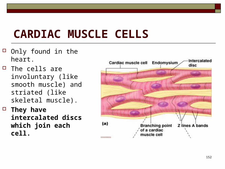
CARDIAC MUSCLE CELLS Only found in the heart. The cells are
involuntary (like smooth muscle) and striated (like skeletal muscle).
They have intercalated discs which join each cell.
152

Cardiac Muscle
153

Cardiac Muscle Cardiac cells are long, striated, and branching,
with one nucleus per cell. The actin and myosin myofilaments are
organized into sarcomeres, but not as uniformly as in skeletal muscle.
As a result, cardiac muscle cells are striated, but not as distinctly as skeletal muscle.
Cardiac muscle is involuntary and does not fatigue. 154

Cardiac Muscle
Cardiac muscle cells are connected to one another by intercalated discs which facilitate action potential conduction between themselves.
This allows them to function as a unit and they all contract together.
Contraction of cardiac cells is influenced by hormones, such as epinephrine.
155

Cardiac Muscle
As one cell contracts, the action potential goes through all the cells, and they all contract as a unit. That’s why the heart contracts all at once.
It has an intrinsic beat. The cells contract on their own, without a signal.
Even if you chop a heart up, each piece will beat by itself!
156

SummarySkeletalmuscle
Smooth muscle
Cardiac muscle
Number of nuclei per cell
Many One One
Involuntary or
voluntary?Voluntary Involuntary Involuntary
Striated or
non-striatedStriated Non-striated Striated
Whereis it
found?Inserts
onto bones
Intestines, blood vessels,
other organs Myocardium of heart

MUSCLE BEEF CUTAdductors Top (inside) roundBiceps femoris Bottom (outside) roundBrachialis Shoulder roseDeltoid Outside chuck (chuck)Gastrocnemius Round heelGluteus medius Top sirloinGracili Inside round capInfraspinatus Top bladeInternal Oblique Sirloin butt Latissimus dorsi Ribeye; Loin eyePectoralis major BrisketPectoralis minor BrisketPsoas major Tenderloin; filet mignonQuadriceps femoris Knuckle; Sirloin tipRectus abdominis FlankSemimembranosus Top (inside) roundSemitendinosus Eye of roundSerratus anterior Boneless short ribsSupraspinatus Chuck tenderTensor fascia latae Tri-tipTeres major Shoulder TenderTrapezius Outside chuckTriceps brachii Ranch Cut

Muscle Musician
• http://vimeo.com/47875656