scientific basis for swedish occupational standards xx
TRANSCRIPT

arbete och hälsa vetenskaplig skriftserieISBN 91–7045–545–7 ISSN 0346–7821 http://www.niwl.se/ah/
1999:26
Scientific Basis for Swedish OccupationalStandards XX
Ed. Johan MonteliusCriteria Group for Occupational StandardsNational Institute for Working LifeS-112 79 STOCKHOLM, Sweden
Translation:Frances Van Sant
National Institute for Working Life

ARBETE OCH HÄLSAEditor-in Chief: Staffan MarklundCo-Editors: Mikael Bergenheim, Anders Kjellberg, BirgittaMeding, Gunnar Rosén and Ewa Wigaeus Hjelm
© National Institute for Working Life & authors 1999National Institute for Working Life,112 79 Stockholm, Sweden
ISBN 91–7045–545–7ISSN 0346-7821http://www.niwl.se/ah/Printed at CM Gruppen
National Institute for Working LifeThe National Institute for Working Life is Sweden’snational centre for work life research, development andtraining.The labour market, occupational safety and health, andwork organisation are our main fields of activity. Thecreation and use of knowledge through learning, in-formation and documentation are important to theInstitute, as is international co-operation. The Institute iscollaborating with interested parties in various deve-lopment projects.The areas in which the Institute is active include:• labour market and labour law,• work organisation,• musculoskeletal disorders,• chemical substances and allergens, noise and
electromagnetic fields,• the psychosocial problems and strain-related disorders
in modern working life.
Preface
The Criteria Group of the Swedish National Institute for Working Life (NIWL) has thetask of gathering and evaluating data which can be used as a scientific basis for theproposal of occupational exposure limits given by the National Board of OccupationalSafety and Health (NBOSH). In most cases a scientific basis is written on request fromthe NBOSH. The Criteria Group shall not propose a numerical occupational exposurelimit value but, as far as possible, give a dose-response/dose-effect relationship and thecritical effect of occupational exposure.
In searching of the literature several data bases are used, such as RTECS, Toxline,Medline, Cancerlit, Nioshtic and Riskline. Also information in existing criteria documentsis used, e.g. documents from WHO, EU, US NIOSH, the Dutch Expert Committee forOccupational Standards (DECOS) and the Nordic Expert Group. In some cases criteriadocuments are produced within the Criteria Group, often in collaboration with DECOS orUS NIOSH.
Evaluations are made of all relevant published original papers found in the searches. Insome cases information from handbooks and reports from e.g. US NIOSH and US EPAis used. A draft consensus report is written by the secretariat or by a scientist appointedby the secretariat. The author of the draft is indicated under Contents. A qualifiedevaluation is made of the information in the references. In some cases the information canbe omitted if some criteria are not fulfilled. In some cases such information is included inthe report but with a comment why the data are not included in the evaluation. Afterdiscussion in the Criteria Group the drafts are approved and accepted as a consensusreport from the group. They are sent to NBOSH.
This is the 20th volume which is published and it contains consensus reports approvedby the Criteria Group during the period July 1998 to June 1999. Previously publishedconsensus reports are listed in the Appendix (p 111).
Johan Högberg Johan MonteliusChairman Secretary

The Criteria Group has the following membership (as of June, 1999)
Olav Axelson Dept Environ Occup MedicineUniversity Hospital, Linköping
Sven Bergström Swedish Trade Union Confederation
Christer Edling Dept Environ Occup MedicineUniversity Hospital, Uppsala
Lars Erik Folkesson Swedish Metal Workers' Union
Lars Hagmar Dept Environ Occup MedicineUniversity Hospital, Lund
Johan Högberg chairman Toxicology and Risk assessmentNIWL
Anders Iregren Toxicology and Risk assessmentNIWL
Gunnar Johanson v. chairman Toxicology and Risk assessmentNIWL
Bengt Järvholm Dept Environ Occup MedicineUniversity Hospital, Umeå
Kjell Larsson Respiratory health and Climate,NIWL
Ulf Lavenius Swedish Factory Workers' Union
Carola Lidén Dept Environ Occup DermatologyKarolinska Hospital, Stockholm
Johan Montelius secretary Toxicology and Risk assessmentNIWL
Bengt Olof Persson observer Medical Unit, NBOSH
Bengt Sjögren Toxicology and Risk assessmentNIWL
Harri Vainio Dept of Environmental MedicineKarolinska Institutet
Kerstin Wahlberg observer Chemical Unit, NBOSH
Arne Wennberg International SecretariateNIWL
Olof Vesterberg Respiratory health and Climate,NIWL
Contents
Consensus report for:
Cyanamide 1Draft: Ulla Stenius, Institute of Environmental Medicine,Karolinska Institutet/NIWL
Phosphorus trichloride, Phosphorus pentachloride, Phosphoryl chloride 7Draft: Birgitta Lindell, Toxicology and Risk assessment, NIWL
Glutaraldehyde 15Draft: Per Lundberg, Toxicology and Risk assessment, NIWL
Methyl tertiary-butyl ether 22Draft: Annsofi Nihlén, Toxicology and Risk assessment, NIWL
Dimethyl adipate, - glutarate, - succinate 39Draft: Birgitta Lindell, Toxicology and Risk assessment, NIWL
Trifluoroethane, Pentafluoroethane 48Draft: Birgitta Lindell, Toxicology and Risk assessment, NIWL
Calcium oxide and Calcium hydroxide, 54Draft: Håkan Löfstedt, Dept of Occupational and EnvironmentalMedicine, Örebro Medical Centre Hospital, Örebro
Cyclohexanone 62Draft: Jill Järnberg, Toxicology and Risk assessment, NIWL
Lactate esters 75Draft: Per Lundberg, Toxicology and Risk assessment, NIWL
Ethylene glycol monomethyl ether + Acetate 83Draft: Gunnar Johanson, Toxicology and Risk assessment, NIWL
Thiourea 97Draft: Margareta Warholm, Institute of Environmental Medicine,Karolinska Institutet/NIWL
Summary 110
Sammanfattning (in Swedish) 110
Appendix: Consensus reports in this and previous volumes 111

1
Consensus Report for Cyanamide
September 30, 1998
Physical and chemical data. Uses
CAS No.: 420-04-2Synonyms: amidocyanogen, carbimide, hydrogen cyanamide,
carbodiimideFormula: CH2N2
Structure: H2NC=NMolecular weight: 42.04Melting point: 45 – 46 °CBoiling point: 127 °CDensity: 1.28 g/mlFlash point: 141 °CConversion factors: 1 ppm = 1.72 mg/m3
1 mg/m3 = 0.58 ppm
Cyanamide at room temperature is a crystalline substance that absorbs moisturefrom the air and forms a damp solid or a solution. No odor threshold has beenreported. Cyanamide is soluble in water (78 g/100 ml), alcohol and ether, but itssolubility in benzene is low.
Cyanamide is used in chemical syntheses, in fertilizers, and as a biocide. It is alsoused in ore refining and in the wood processing and rubber industries. Cyanamidecan also be formed by hydrolysis of calcium cyanamide, a substance which hassimilar uses. Cyanamide and its salts have also been used medicinally in treatmentof alcoholics, since cyanamide inhibits aldehyde dehydrogenase. It is no longerregistered as a medicine. No information on air concentrations was found in theliterature.
Uptake, biotransformation, excretion
Cyanamide can be absorbed via the digestive tract and skin. It is metabolized toacetyl cyanamide, mostly in the liver, by acetyl-S-CoA-dependent N-acetyl-transferase (26). It may also be metabolized in a reaction induced by catalase (8). Inone study, six volunteers were given cyanamide orally (0.25 mg/kg body weight):40% of the dose was excreted in urine as acetylcyanamide during the next 48 hours,most of this within the first 12 hours. A 1-ml dose of a 1% cyanamide solution(0.25 mg/kg) was applied to skin (4 x 4 cm) and left for 6 hours: 7.7% of the dosewas excreted in urine as acetylcyanamide (17).

2
Most cyanamide is excreted in urine. When animals were given 14C-labeledcyanamide (8 mg/kg intraperitoneally for rats, 1.6 mg/kg intravenously and orallyfor dogs and rabbits) nearly all the radioactivity was detected in urine, and theprimary metabolite was found to be N-acetylcyanamide (11, 26). In a study withrats and dogs, the highest plasma concentration was reached in the rats 30 minutesafter oral administration of 4 mg/kg. After intravenous administration of 1.4 mg/kg,the half time in plasma was 30 to 61 minutes for both species (21). Indications ofnon-linear metabolism have been observed in rats under steady-state conditions(0.005 – 32 mg/kg i.p. at 45-minute intervals). Clearance and first passagemetabolism were not constant between the doses, probably because the biotrans-formation capacity of the liver had been saturated (23).
Toxic effects
Human dataThe effects of cyanamide on the liver have been studied in conjunction withcyanamide treatment of alcoholics. One study describes liver biopsies from 37patients who had been treated with cyanamide for from 2 months to 7 years, withdaily doses ranging from 45 to 180 mg. In addition to the liver changes seen withalcoholism, there were structural changes such as fibrosis and changes in connec-tive tissue in all biopsies, as well as a particular type of inclusions in hepatocytes(Lafora-like inclusion bodies consisting of lipid vesicles, glycogen and traces ofdegenerated organelles) (19). Elevated blood levels of liver enzymes (ALAT,ASAT) induced by cyanamide were also detected in this study. The histologicalpicture resembled that of cirrhosis, and the authors concluded that the longer thetreatment, the greater the changes. Other studies have also shown that cyanamideused to treat alcoholism induces inclusion bodies in hepatic cells (2, 19, 30, 31,32).
Two cases of skin sensitization from occupational contact with cyanamide havebeen described (5, 6). One man who was sensitized by working for 1.5 years withmedicines that contained cyanamide had a positive reaction to the substance in apatch test (0.1% in water; 48 hours) (6). Sensitization to cyanamide was reported tobe rare. The other case report describes sensitization in a chemist whose workincluded some contact with cyanamide. He had a positive reaction to a 0.01%cyanamide solution in a patch test (5). Seven cases of cyanamide-induced skineruptions are reported in a study from 1977. The patients had been treated with oraldoses of a 1% cyanamide solution, 7 ml daily for from 1 to 4 months (14). After 10days to 3 months of the treatment, all of them had skin disorders: 6 had scalingdermatitis and one had lichen planus-like eruptions. The authors concluded that thistype of reaction may have been a common but ignored problem. One case reportdescribes granulocytopenia and skin sensitization in a man who was treated with100 mg cyanamide during a 3-week period (1). The symptoms disappeared afterthe treatment was broken off.
3
There is a study on endocrine function (thyroidea, testes) in 21 persons whoworked in a calcium cyanamide production plant and 9 controls (18). One of thereasons it was undertaken was that testicular atrophy had been observed in male ratsexperimentally exposed to cyanamide (28). N-Acetylcyanamide content in urine atthe end of the workday was used as a measure of exposure, and clearly showedexposure in the 21 workers. There were no observed differences in endocrinefunction (testosterone, follicle-stimulating hormone, luteinizing hormone) betweenexposed persons and controls.
Animal dataThe LD50 for rats has been calculated to be 125 mg/kg body weight for oral admini-stration (3) and 56 mg/kg for intravenous administration (13). Cyanamide causesskin irritation (13) and severe eye irritation in rabbits (100 mg dropped in the eye)(7).
In several in vivo studies, cyanamide has been shown to inhibit alcoholdehydrogenase activity. Cyanamide treatment (2 mg/kg, i.p. 1 hour) of ratsinhibited alcohol dehydrogenase and increased the toxicity of alcohol (24).Intraperitoneal doses of 0.35 mg/kg repeated at 45-minute intervals suppressedalcohol dehydrogenase activity completely (23). Elevated acetaldehyde levelsfollowing alcohol exposure were seen in rats pre-treated with cyanamide(0.7 mg/kg, p. o.) 45 minutes before the exposure. Elevated alcohol levels in bloodhave also been related to pre-treatment with cyanamide (10 mg/kg, p.o.) (12, 25).
Reduced body weight and elevated levels of monoamines in the brain werereported in rats after oral or intravenous administration of cyanamide (8 mg/kg,20 weeks or more) (22). Catalase activity in various organs of rats has been shownto diminish at dose levels greater than 1.3 mg/kg (i.p., maximum after 1 hour) (9).Doses exceeding 10 mg/kg ( i.p., 4 hours) increased the level of circulating ketonebodies in rats (10).
Mutagenicity
In a study with Salmonella typhimurium (strains TA98, TA100, TA1535, TA1537,TA1538) and E coli, cyanamide caused no increase in mutation frequency, eitherwith or without metabolic activation (4). Cyanamide was not clastogenic in a micro-nucleus test with mice (16). Elevated frequencies of mitotic gene conversion andnon-disjunction were seen in Aspergillus nidulans (29). No increase of DNA stringbreaks was seen in hepatocytes exposed to cyanamide in vitro (27).
Carcinogenicity
Cancer incidence and mortality were mapped in a cohort of 790 workers in a plantproducing calcium carbide. No increase in cancer was seen among the 117 workerswho had worked with cyanamide/dicyandiamide production for at least 18 monthsduring 1953 – 1970 (15). Exposures are not reported. The National Cancer Institutein the United States has tested calcium cyanamide (which hydrolyzes to cyanamide)

4
for carcinogenic effect in a two-year study (20) with rats and mice. No carcinogeniceffect was observed.
Teratogenicity
In a two-generation reproduction/fertility study, male rats were given cyanamide inoral doses of 2 to 25 mg/kg daily for 70 days before mating, and females for 15days prior to or during gestation. The females in the highest dose group (25 mg/kg)had lower body weights, fewer corpora lutea, fewer implanted embryos and smallerlitters. Males in the highest dose group had bilateral testicular atrophy and lowerfertility. There were no observed effects on the F1 generation. The NOEL in thisstudy was 7 mg/kg (28).
Table 1. Effects of cyanamide on experimental animals.
Exposuremg/kg
Time Species Effect Ref.
125 p.o. Rat LD50 3
56 i.v. Rat LD50 13
25 p.o. daily for 70 days (prior tomating)
Rat (males) Testicular atrophy, reduced fertility 28
25 p.o. daily for 15 days (before orduring gestation)
Rat (females) Lower body weight, fewer corporalutea and implanted embryos,smaller litters
28
10 i.p. 4 hours Rat Elevated levels of ketone bodies 10
8 p.o. 20 weeks Rat Lower body weight, elevated levelsof monoamines in brain
22
2 i.p. 1 hour Rat Inhibited alcohol dehydrogenaseactivity, increased alcohol toxicity
24
1.3 i.p. 1 hour Rat Inhibited catalase activity 9
0.7 p.o. 45 minutes Rat Elevated acetaldehyde levels 12, 25
0.35 p.o. 45-minute intervals Rat Suppressed alcohol dehydrogenaseactivity
23
(p.o = oral; i.v. = intravenous; i.p. = intraperitoneal)

5
Dose-effect/dose-response relationships
There are no data on which to base a dose-effect or dose-response relationship foroccupational exposure to cyanamide. The dose-response and dose-effect relation-ships observed in animal experiments are summarized in Table 1.
Conclusions
Cyanamide inhibits alcohol dehydrogenase when used medicinally. There are nodata on which to base a critical effect for occupational exposure. Cyanamide can beskin sensitizing to humans and has been shown to irritate the eyes of rabbits.
References
1. Ajima M, Usuki K, Igarashi A et al. Cyanamide-induced granulocytopenia. Intern Med1997;36:640-642.
2. Bruguera M, Parés A, Heredia D, Rodés J. Cyanamide hepatotoxicity. Incidence andclinicopathological features. Liver 1987;7:216-222.
3. Budavari S, ed. The Merck Index. An Encyclopedia of Chemicals and Drugs. 11th ed. RahwayNew Jersey, USA: Merck & Co, Inc. 1989:418.
4. Cadena A, Arso J, Valles J M, Llagostera M, Vericat J A. Evaluation of the possiblemutagenicity of cyanamide by the Ames and Devoret tests. Boll Chim Farm 1984;123:75-83.
5. Calnan C D. Cyanamide. Contact Dermatitis Newsletter 1970;7:150.
6. Conde-Salazar L, Guimaraens D, Romero L, Harto A. Allergic contact dermatitis tocyanamide (carbodiimide). Contact Dermatitis 1981;6:329-330.
7. Deichmann W B. In Toxicology of Drugs and Chemicals. New York: Academic Press,1969:190.
8. DeMaster E G, Shirota F N, Nagasawa H T. Catalase mediated conversion of cyanamide to aninhibitor of aldehyde dehydrogenase. Alcohol 1985;2:117-121.
9. DeMaster E G, Redfern B, Shirota F N, Nagasawa H T. Differential inhibition of rat tissuecatalase by cyanamide. Biochem Pharmacol 1986;35:2081-2085.
10. DeMaster E G, Stevens J M. Acute effect of the aldehyde dehydrogenase inhibitors,disulfirame, pargyline and cyanamide, on circulating ketone body levels in the rat. BiochemPharmacol 1988;37:229-234.
11. Dietrich R A, Troxell P A, Worth W, Erwin G V. Inhibition of aldehyde dehydrogenase inbrain and liver by cyanamide. Biochem Pharmacol 1976;25:2733-2737.
12. Garcia de Torres G, Römer K G, Torres Alanis O, Freundt K J. Blood acetaldehyde levels inalcohol-dosed rats after treatment with ANIT, ANTU, dithiocarbamate derivatives orcyanamide. Drug Chem Toxicol 1983;6:317-328.
13. Izmerov N F. Toxicometric Parameters of Industrial Toxic Chemicals Under Single Exposure.Moscow: Centre of International Projects, GKNT 1982;40.
14. Kawana S. Drug eruption induced by cyanamide (carbimide): A clinical and histopathologicstudy of 7 patients. Dermatology 1997;195:30-34.
15. Kjuus H, Andersen A, Langård S. Incidence of cancer among workers producing calciumcarbide. Br J Ind Med 1986;43:237-242.
16. Menargues A, Obach R, Valles J M. An evaluation of the mutagenic potential of cyanamideusing the micronucleus test. Mutat Res 1984;136:127-129.

6
17. Mertschenk B, Bornemann W, Filser J G, von Meyer L, Rust U, Schneider J-C, GloxhuberC. Urinary excretion of acetyl-cyanamide in rat and human after oral and dermal application ofhydrogen cyanamide. Arch Toxicol 1991;65:268-272.
18. Mertschenk B, Bornemann W, Pickardt C R, Rust U, Schneider J-C, Gloxhuber C.Examinations on endocrine functions in employees from a calcium cyanamide productionplant. Zbl Arbeitsmed 1993;43:254-258.
19. Moreno A, Vazquez J J, Ruizdel Arbol L, Guillen F J, Colina F. Structural hepatic changesassociated with cyanamide treatment: Cholangiolar proliferation, fibrosis and cirrhosis. Liver1984;4:15-21.
20. National Cancer Institute. Bioassay of Calcium Cyanamide for Possible Carcinogenicity.Maryland: National Cancer Institute. Technical Report Series No. 163, 1979.
21, Obach R, Colom H, Arso J, Peraire C, Prunonosa J. Pharmacokinetics of cyanamide in dogand rat. J Pharm Pharmacol 1989;41:624-627.
22. Obach R, Menargues A, Vallés J, Vallés J M, Garcia-Sevilla J A. Effects of cyanamide onbody weight and brain monoamines and metabolites in rats. Eur J Pharmacol 1986;127:225-231.
23. Piera J P, Obach R, Sagrista M L, Bozal J. Inhibition of rat hepatic mitochondrial aldehydedehydrogenase isozymes by repeated cyanamide administration: Pharmacokinetic-pharmacodynamic relationships. Biopharm Drug Disp 1993;14:419-428.
24. Rikans L E. The oxidation of acrolein by rat liver aldehyde dehydrogenases. Relation to allylalcohol hepatotoxicity. Drug Metabol Dispos 1987;15:356-362.
25. Römer K G, Torres Alanis O, Garcia de Torres G, Freundt K J. Delayed ethanol eliminationfrom rat blood after treatment with thiram, tetramethylthiuram monosulfide, ziram orcyanamide. Bull Environ Contam Toxicol 1984;32:537-542.
26. Shirota F N, Nagasawa H T, Kwon C H, DeMaster E G. N-Acetylcyanamide, the majorurinary metabolite of cyanamide in rat, rabbit, dog and man. Drug Metabol Dispos1984;12:337-344.
27. Sina J F, Bean C L, Dysart G R, Taylor V I, Bradley M O. Evaluation of the alkalineelution/rat hepatocyte assay as a predictor of carcinogenic/mutagenic potential. Mutat Res1983;113:357-391.
28. Valles J, Obach R, Menargues A, Valles J M, Rives A. A two-generation reproduction-fertility study of cyanamide in the rat. Pharmacol Toxicol 1987;61:20-25.
29. Vallini G, Pera A, de Bertoldi M. Genotoxic effects of some agricultural pesticides in vitrotested with Aspergillus nidulans. Environ Pollution 1983;30:39-58.
30. Vázquez J J, Cervera S. Cyanamide-induced liver injury in alcoholics. Lancet 1980;1:361-362.
31. Vázquez J J, Guillen F J, Zozaya J, Lahoz M. Cyanamide-induced liver injury. A predictablelesion. Liver 1983;3:225-230.
32. Yokoyama A, Sato S, Maruyama K et al. Cyanamide-associated alcoholic liver disease: Asequential histological evaluation. Alcoholism 1995;19:1307-1311.

7
Consensus Report for PhosphorusChlorides
September 30, 1998
This report treats phosphorus trichloride, phosphorus pentachloride and phosphorylchloride.
Chemical and physical data. Uses
phosphorus trichlorideCAS No: 7719-12-2Synonyms: phosphorus(III)chloride, trichlorophosphineFormula: PCl3
Molecular weight: 137.33Boiling point: 76 °CMelting point: - 112°CVapor pressure: 12.7 kPa (20°C)Conversion factors: 1 ppm = 5.70 mg/m3 (20°C)
1 mg/m3 = 0.175 ppm (20°C)
phosphorus pentachlorideCAS No.: 10026-13-8Synonyms: phosphorus(V)perchloride, pentachlorophosphoraneFormula: PCl5
Molecular weight: 208.24Boiling point: sublimates at 160°C *Melting point: 167 °C (three-phase equilibrium) *Conversion factors: 1 ppm = 8.64 mg/m3 (20°C)
1 mg/m3 = 0.116 ppm (20°C)
_______________________
* From Reference 14. Other sources give other boiling and melting points.
phosphoryl chlorideCAS No: 10025-87-3Synonyms: phosphoryl trichloride, trichlorophosphine oxide,
phosphorus oxychloride, phosphorus oxytrichlorideFormula: POCl3
Molecular weight: 153.33Boiling point: 105.5°CMelting point: 1 °CVapor pressure: 3.6 kPa (20°C)Conversion factors: 1 ppm = 6.36 mg/m3 (20°C)
1 mg/m3 = 0.157 ppm (20°C)
8
Phosphorus trichloride at room temperature is a clear liquid that steams in damp air:it hydrolyzes, emitting heat, to phosphorous acid and hydrochloric acid.Phosphorus pentachloride at room temperature is a steaming, yellowish or white togreenish-white solid. It hydrolyzes initially to hydrochloric acid and phosphorylchloride, and in a second step the phosphoryl chloride (a clear, steaming liquid)hydrolyzes, producing heat, phosphoric acid and more hydrochloric acid.Phosphorus trichloride, phosphorus pentachloride and phosphoryl chloride all threehave a sharp, penetrating odor (1, 2, 3, 12, 13, 17, 20).
Phosphorus trichloride is used mostly as an intermediate in the production ofpesticides, surfactants, softeners, gasoline additives and pigments. Phosphoruspentachloride is used as a thickener. Phosphoryl chloride is used in the production ofsofteners and gasoline additives and also in the production of hydraulic fluids and fireretardants. All three substances are used as chlorinators and catalysts (1, 2, 3).
Uptake, biotransformation, excretion
No information was found in the literature.
Toxic effects
Human dataVapor/dust (including hydrolysis products) of all three substances are irritating/cor-rosive to eyes and respiratory passages, but information on exposure levels isusually not given (5, 10, 11, 19, 22, 26, 27, 29). Phosphorus trichloride andphosphorus pentachloride are reported to be strongly irritating to mucousmembranes (4, 29). Phosphoryl chloride has been reported to strongly affect boththe upper and lower respiratory passages and to be more likely to have delayedeffects on respiratory passages than phosphorus trichloride (10, 26, 29). Exposureto phosphorus trichloride and its hydrolysis products has also resulted in skinirritation (17, 27). Occupational exposure to phosphorus chlorides has beenreported to etch the teeth (21), but the type of exposure was not described.
Effects other than local irritation/ulceration have also been reported to result fromexposure to phosphorus chlorides. One study (27) reports nausea, vomiting,headache and transient elevation of lactate dehydrogenase levels in serum in severalpersons acutely exposed to phosphorus trichloride and its hydrolysis products.Another study (5) reports dizziness and severe headache in a person who hadinhaled phosphorus pentachloride vapor for a few seconds. Brief exposure (in somecases only a few seconds) to phosphoryl chloride vapor has caused dizziness,nausea, vomiting and effects on the heart (5, 10, 11). In a few cases, enlarged liver,albuminuria and anemia have also been reported after exposure to phosphorylchloride vapor (22), but it is not clear whether these effects were the result of theexposure.
Only a few studies report both exposure levels and the symptoms of exposedpersons. One study (Dadej, 1962; reviewed in Reference 17) describes effects onsome people who were exposed to phosphorus trichloride and its hydrolysis

9
products by an explosion. Three workers who were exposed for from a fewseconds up to half a minute or so, and who died within 24 hours, had severe skinburns, ulcerated eyes, inflamed bronchi and pulmonary edema. Ulcerated eyes,respiratory passages and skin were also seen in one surviving worker who wasexposed for several seconds. Concentrations during the first 120 seconds wereroughly estimated to have been about 36,800 mg/m3 phosphorus trichloride,116,300 mg/m3 hydrochloric acid, and 62,500 mg/m3 phosphorous acid.
A report from NIOSH (25) states that, of 37 workers exposed to phosphorustrichloride and phosphoryl chloride, about 65% (24/37) suffered acute respiratorysymptoms such as breathing difficulty or chest tightness at least once a month. Only5% (1/22) of non-exposed persons reported these symptoms. Lung function tests,however, revealed no significant differences between the two groups. Air concen-trations were measured with personal monitors for two days, and were below thedetection limits for phosphorus trichloride and phosphoryl chloride in nearly allcases. There was one exposure to 5.7 mg/m3 phosphorus trichloride (1 hour) andone to 4.2 mg/m3 phosphoryl chloride (25 minutes). A significant differencebetween exposed and unexposed workers is also reported in a follow-up medicalstudy made two years later (16). Half (13 of 26 persons) of the exposed workershad periods of acute breathing difficulty, tightness in the chest and breathlessness(5 of them regarded the symptoms as work-related), whereas none of the un-exposed workers (11 persons) reported these symptoms.
Two studies from Italy (23, 24) contain exposure data and describe respectivelyeffects on 23 workers exposed to phosphorus trichloride and 20 workers exposedto phosphorus oxychloride. Air concentrations varied considerably in both cases.They were reported to be 10 – 20 mg/m3 most of the time, but could occasionallyexceed 150 mg/m3 (phosphorus trichloride) or 70 mg/m3 (phosphorus oxy-chloride). The reports contain no information on hydrolysis products. Photophobia,stinging in eyes and throat, chest tightness, coughing and rapid breathing werereported in a few subjects within 2 to 6 hours of exposure, and some of themsubsequently developed bronchitis. In other cases it took 4 or 5 days or up to 8weeks for symptoms to appear, and then in the form of slight throat irritation,conjunctivitis, coughing, shortness of breath and asthmatic bronchitis. Emphysemawas also mentioned. The studies have the form of multiple case reports, and there isno collation or analysis of the findings. The diagnoses are based only on clinicalobservations and x-rays: there is no mention of medical history or smoking habits,for example. All this gives rise to some uncertainty in assessing the results, and it istherefore impossible to draw any definite conclusions from these studies.
A sketchily reported Russian study (21) with volunteers gives irritationthresholds of 4 mg/m3 for phosphorus trichloride, 10 mg/m3 for phosphoruspentachloride and 1 mg/m3 for phosphoryl chloride. Since no details on the resultsor the design of the experiment are given, however, the information can not besatisfactorily assessed.
Skin burns have been reported in persons splashed with phosphorus trichlorideor phosphorus pentachloride (concentrations not given) (9, 19).
10
Animal dataThe LC50 values for exposure to vapor/aerosol of phosphorus trichloride reported indifferent studies range from 226 to > 2582 mg/m3. Reported LC50 values forphosphoryl chloride range from 71 to 330 mg/m3 (18, 21, 28). The reported LC50
value for phosphorus pentachloride is 205 mg/m3 (21). The LD50 values for oraladministration to rats are 18 – 550 mg/kg for phosphorus trichloride, 600 mg/kg forphosphorus pentachloride, and 380 mg/kg for phosphoryl chloride (17, 18, 21).No LD50 information was found for skin application, but an LDLo of 1260 mg/kghas been reported for application of phosphorus trichloride to the skin of rabbits(24 hours) (18). Phosphorus trichloride applied to the skin of rabbits is reported tocause burns (one reference reports that the substance was undiluted) (17, 18).Phosphorus pentachloride (concentration not reported) and phosphoryl chloride(pure substance) have also been reported to cause burns when applied to the skin ofrabbits (17, 18). Application of phosphorus trichloride (concentration not reported)or phosphoryl chloride (pure substance) in liquid form has also been shown tocause severe damage to the eyes of experimental animals (17, 18).
During 4-hour exposures made to determine the LC50 for phosphorus trichlorideand phosphoryl chloride, experimental animals (rats and guinea pigs) showedagitation, indications of irritation, porphyrin secretion around the eyes and laboredbreathing (28). The exposure to phosphorus trichloride also caused severe erosionof the nostrils and paws as well as kidney damage (nephrosis). The exposure tophosphoryl chloride caused irritation in the trachea, bronchi and lungs. The deathsoccurred within 10 days after exposure to phosphorus trichloride and within48 hours after exposure to phosphoryl chloride. The study reports that about 40%of the phosphorus trichloride and 15% of the phosphoryl chloride were hydrolyzed(28).
Effects on eyes and respiratory passages were reported in a study in whichrabbits and cats (one of each per group) were exposed by inhalation to concen-trations of phosphorus trichloride ranging from 4 mg/m3 to 3870 mg/m3 for 3 to10 hours (6). There was a large difference in sensitivity between the two species.Sneezing, coughing, salivation, nasal secretion and reduced respiratory rates wereseen in the cats at exposure as low as 4 – 5 mg/m3, and effects on the eyes werenoted at air concentrations of 13 – 20 mg/m3 or above. Histological examination(cats) revealed liquid in the lungs (13 – 20 mg/m3). Exposed rabbits – two animalsexposed to air concentrations of 13 – 20 mg/m3 and 13 – 27 mg/m3 respectively –became somewhat restless, had greatly reduced respiration rates, slight symptomsof irritation and/or slight nasal secretion (see Table 1).A Russian study (21) reports that the threshold value for irritation of respiratorypassages (rats) was 5 mg/m3 for phosphorus trichloride, 8 mg/m3 for phosphoruspentachloride and 1 mg/m3 for phosphoryl chloride. The study also reports that theeffect was more pronounced for phosphorus trichloride (clouding of the cornea,sores around the mouth and nose, pronounced irritation of respiratory passages)than for the other phosphorus chlorides. The same study reports that “dystrophicchanges,” particularly in the liver, kidneys and nervous system, were observed after

11
single exposures to high (not reported) air concentrations of phosphorus chlorides,and that exposure to 10 mg/m3 for 4 hours caused a reduction of pH in blood andurine. Pronounced morphological changes, most notably in respiratory passages,kidneys, liver, bone tissue (osteoporosis) and brain (degenerative changes in nervecells), as well as cytogenetic effects (see below), were also observed after 4 monthsof exposure to 1.34 mg/m3 phosphoryl chloride, and irritation of mucous mem-branes in airways and elevated kidney weights were noted at 0.48 mg/m3. Since thereport gives no details on the design of the experiment, controls etc., this informa-tion can not be evaluated.
Mutagenicity, carcinogenicity, reproduction toxicity
No mutagenic effects were observed when phosphorus trichloride was tested onbacteria in vitro (15). A Russian study (21), which can not be adequately assessed(see above), reports mutagenic and cytostatic effects in rats (bone marrow) afterchronic exposure to 1.34 mg/m3 phosphoryl chloride but no significant changesafter exposure to 0.48 mg/m3. Phosphoryl chloride (air concentration not given)was also reported to affect the motility of sperm but to have no effect on spermato-genesis.
Dose-effect/dose-response relationships
There are few reliable measurements of air concentrations of these phosphoruschlorides in work environments. Two Italian studies report symptoms of eyeirritation with exposure to phosphorus trichloride and airway irritation withexposure to phosphoryl chloride. Air concentrations varied, but in both caseswere reported to be around 10 – 20 mg/m3 most of the time.
Effects on experimental animals exposed by inhalation to phosphorus trichlorideare summarized in Table 1.
Dose-dependent effects on respiratory passages were observed in cats at airconcentrations of 4 – 5 mg/m3 phosphorus trichloride or higher, and dose-dependent effects on eyes at air concentrations of 13 – 20 mg/m3 or higher.
Conclusions
The critical effect of exposure to phosphorus trichloride, phosphorus pentachlorideand phosphoryl chloride is irritation of respiratory passages. Due to their chemicalcharacteristics, these three substances can also irritate/ulcerate eyes and skin.
12
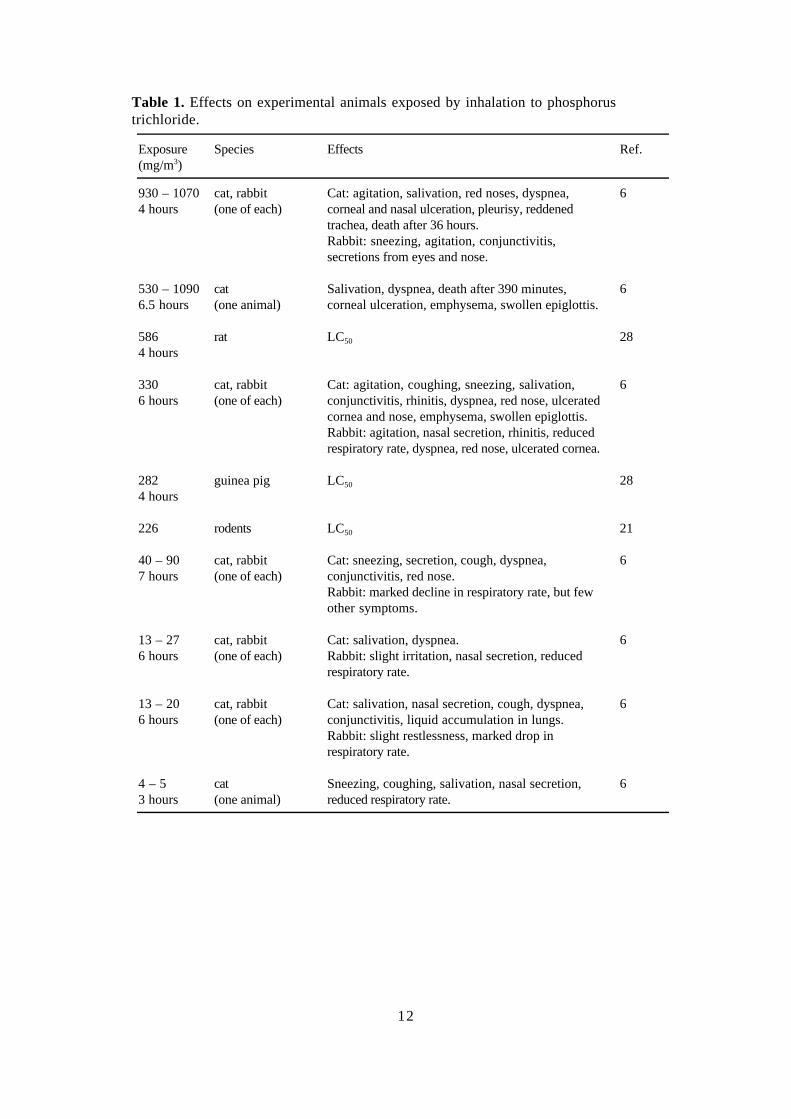
Table 1. Effects on experimental animals exposed by inhalation to phosphorustrichloride.
Exposure(mg/m3)
Species Effects Ref.
930 – 10704 hours
cat, rabbit(one of each)
Cat: agitation, salivation, red noses, dyspnea,corneal and nasal ulceration, pleurisy, reddenedtrachea, death after 36 hours.Rabbit: sneezing, agitation, conjunctivitis,secretions from eyes and nose.
6
530 – 10906.5 hours
cat(one animal)
Salivation, dyspnea, death after 390 minutes,corneal ulceration, emphysema, swollen epiglottis.
6
5864 hours
rat LC50 28
3306 hours
cat, rabbit(one of each)
Cat: agitation, coughing, sneezing, salivation,conjunctivitis, rhinitis, dyspnea, red nose, ulceratedcornea and nose, emphysema, swollen epiglottis.Rabbit: agitation, nasal secretion, rhinitis, reducedrespiratory rate, dyspnea, red nose, ulcerated cornea.
6
2824 hours
guinea pig LC50 28
226 rodents LC50 21
40 – 907 hours
cat, rabbit(one of each)
Cat: sneezing, secretion, cough, dyspnea,conjunctivitis, red nose.Rabbit: marked decline in respiratory rate, but fewother symptoms.
6
13 – 276 hours
cat, rabbit(one of each)
Cat: salivation, dyspnea.Rabbit: slight irritation, nasal secretion, reducedrespiratory rate.
6
13 – 206 hours
cat, rabbit(one of each)
Cat: salivation, nasal secretion, cough, dyspnea,conjunctivitis, liquid accumulation in lungs.Rabbit: slight restlessness, marked drop inrespiratory rate.
6
4 – 53 hours
cat(one animal)
Sneezing, coughing, salivation, nasal secretion,reduced respiratory rate.
6
13
References
1. ACGIH. Phosphorus oxychloride. Documentation of the Threshold Limit Values andBiological Exposure Indices, 6th ed. Cincinnati, Ohio: American Conference of GovernmentalIndustrial Hygienists Inc. 1991:1255-1256.
2. ACGIH. Phosphorus pentachloride. Documentation of the Threshold Limit Values andBiological Exposure Indices, 6th ed. Cincinnati, Ohio: American Conference of GovernmentalIndustrial Hygienists Inc. 1991:1257-1258.
3. ACGIH. Phosphorus trichloride. Documentation of the Threshold Limit Values andBiological Exposure Indices, 6th ed. Cincinnati, Ohio: American Conference of GovernmentalIndustrial Hygienists Inc. 1991:1261-1262.
4. Beliles R P, Beliles E M. Phosphorus, selenium, tellurium, and sulfur. In Clayton G D,Clayton F E, eds. Patty’s Industrial Hygiene and Toxicology, 4th ed. New York: John Wiley& Sons, 1993:789-791.
5. Buess H, Lerner R. Über Asthma bronchiale und asthmoide Bronchitis in der chemischenIndustrie. Z Präventivmed 1956;2:59-74.
6. Butjagin P W. Experimentelle Studien über den Einfluss technisch und hygienisch wichtigerGase und Dämpfe auf den Organismus. Arch f Hygiene 1904;49:307-335.
7. DFG (Deutsche Forschungsgemeinschaft). Toxikologisch-arbeitsmedizinische Begründungenvon MAK-Werten. Phosphoroxidchlorid. Weinheim: Verlag Chemie, 1984:10 pages.
8. DFG (Deutsche Forschungsgemeinschaft). Toxikologisch-arbeitsmedizinische Begründungenvon MAK-Werten. Phosphortrichlorid. Weinheim: Verlag Chemie, 1984:9 pages.
9. Eldad A, Chaouat M, Weinberg A, Neuman A, Ben Meir P, Rotem M, Wexler M R.Phosphorous pentachloride chemical burn – a slowly healing injury. Burns 1992;18:340-341.
10. Floret E. Späterer Tod nach akuter Phosphoroxychloridvergiftung. Zbl Gewerbehyg1929;6:282-283.
11. Herzog H, Pletscher A. Die Wirkung von industriellen Reizgasen auf dieBronchialschleimhaut des Menschen. Schweiz Med Wochschr 1955;20:477-481.
12. Hägg G. Allmän och oorganisk kemi, 5th ed. Stockholm: Almqvist & Wiksell, 1963:526.
13. Kirk-Othmer. Encyclopedia of Chemical Technology, 2nd ed. Vol 15. New York: John Wiley& Sons, 1968:305-308.
14. Lide D R, Frederikse H P R. CRC Handbook of Chemistry and Physics. New York: CRCPress Inc. 1995-1996:4–75, 4–76.
15. McMahon R E, Cline J C, Thompson C Z. Assay of 855 test chemicals in ten tester strainsusing a new modification of the Ames test for bacterial mutagens. Cancer Res 1979;39:682-693.
16. Moody P. Health Hazard Evaluation Report HETA 81-089-965. PB83-161190. FMC Corp.Nitro, West Virginia; NIOSH, Cincinnati, Ohio 1981.
17. Payne M P, Shillaker R O, Wilson A J. Toxicity Review 30. Phosphoric acid, phosphoruspentoxide, phosphorus oxychloride, phosphorus pentachloride, phosphorus pentasulphide.Sudbury, Suffolk, UK: Health and Safety Executive, 1993.
18. Randall D J, Robinson E C. Acute toxicologic evaluation of phosphorus trichloride. AcuteToxic Data 1990;1:71-72.
19. Reinl W. Über gewerbliche Vergiftungen durch Phosphorverbindungen (Phosphorchloride,Phosphorwasserstoff und organische Phosphorsäureester). Arch f Toxikol 1956;16:158-181.
20. Riess G, Niermann H, Mayer D. Phosphor-Verbindungen, anorganische, sonstige. InUllmans Encyklopädie der technischen Chemie, vol 18. Weinheim: Verlag Chemie,1979:365-368.

14
21. Roshchin A V, Molodkina N N. Chloro compounds of phosphorus as industrial hazards.J Hyg Epidemiol Microbiol Immunol 1977;21:387-394.
22. Rumpf Th. Über Vergiftung durch Phosphoroxychlorid. Med Klin 1908;4:1367-1369.
23. Sassi C. L’intossicazione professionale da tricloruro di fosforo. Med Lav 1952;43:298-306.
24. Sassi C. L’intossicazione professionale da ossicloruro di fosforo. Med Lav 1954;45:171-177.
25. Tharr D G, Singal M. Health Hazard Evaluation Determination Report HE 78-90-739. PB81-170920. FMC Corporation, Nitro, West Virginia; NIOSH, Cincinnati, Ohio 1980.
26. Vaubel W. Hygienische Fürsorge für Betriebsbeamte und Arbeiter. Chem Ztg 1903;76:921.
27. Wason S, Gomolin I, Gross P, Mariam S, Lovejoy F H. Phosphorus trichloride toxicity.Am J Med 1984;77:1039-1042.
28. Weeks M H, Nelson P, Musselman P, Yevich P P, Jacobson K H, Oberst F W. Acute vaportoxicity of phosphorus oxychloride, phosphorus trichloride and methyl phosphonic dichloride.Am Ind Hyg Assoc J 1964;5:470-475.
29. Weichardt H. Gewerbliche Vergiftungen durch Phosphorchloride. Chem Ztg 1957;81:421-423.

15
Consensus Report for Glutaraldehyde
September 30, 1998
This report is based primarily on a criteria document produced jointly by the NordicExpert Group and the Dutch Expert Committee (3).
Chemical and physical data. Uses
CAS No.: 111-30-8Name: glutaraldehydeSynonyms: glutaral, pentanedial,
glutardialdehyde, 1,5-pentanedialFormula: CHO-(CH2)3-CHOMolecular weight: 100.13Boiling point: 188 °CFreezing point: - 14 °CVapor pressure: 0.00016 kPa (20% solution)
0.002 kPa (50% solution)Saturation concentration: 6.6 mg/m3 (1.6 ppm) (20% solution)
82 mg/m3 (20 ppm) (50% solution)Distribution coefficient: log Po/w = 0.01Conversion factors: 1 mg/m3 = 0.25 ppm
1 ppm = 4.0 mg/m3
Glutaraldehyde at room temperature is a colorless, oily liquid with a sharp odor.The reported odor threshold is 0.04 ppm (2, 3). Glutaraldehyde is soluble in water,ethanol, benzene, ether and other organic solvents. It can react violently with strongoxidants. An aqueous solution of glutaraldehyde has a pH of 3 – 4.
Glutaraldehyde is marketed in aqueous solutions of 1%, 2%, 25% or 50%. Thesolutions may contain alkalis added to raise their pH to 7.5 – 8.5 (activated solu-tions). Glutaraldehyde has a wide range of uses: as a disinfectant and sterilizer inhospitals, in embalming, as a fixative in electron microscopy, as a slimicide in thepaper industry etc.
Glutaraldehyde was monitored in hospitals in England: concentrations rangedfrom 0.003 to 0.17 mg/m3 (19). In Denmark, concentrations of 0.25 to 0.5 mg/m3
were measured in surgery wards (28). In a Swedish study, the highest concentra-tion – 0.57 mg/m3 – was associated with sterilization of gastroscopes. The averageof 16 measurements made around this task was 0.05 mg/m3 (25).

16
Uptake, biotransformation, excretion
In an in vitro study, skin from rats, mice, rabbits, guinea pigs and humans wasexposed to a 1,5-14C-labeled glutaraldehyde solution for 6 hours. Concentrationswere 0.75% or 7.5%. Between 0.5 and 0.7% of the solution was absorbedthrough/into the skin (11). Human stratum corneum and epidermis were exposed invitro to 450 µl of a 10% glutaraldehyde solution for 1 hour. Penetration through thestratum corneum ranged from 3.3% (skin from the back) to 12% (skin from thestomach), with large individual variations. Penetration through epidermis was about4% of the applied amount (27). When glutaraldehyde (0.75 or 7.5%) was left onthe skin for 24 hours, the amount absorbed was calculated to be 4 – 9% for rats,and 33 – 53% for rabbits (3, 23).
Biotransformation of glutaraldehyde involves oxidation, decarboxylation andhydroxylation. Oxidation to glutaric acid, bonding to coenzyme A and breakdownto acetate yields the end product carbon dioxide. In hepatic and renal tissue of rats invitro, glutaraldehyde is oxidated (probably in the mitochondria) to CO2 (26). Dosesof 0.2 ml of a 0.075 or 0.75% solution of 14C-labeled gutaraldehyde were injectedinto the caudal vein of rats, and up to 80% of the radioactivity was found in CO2
during the next 4 hours. Within 3 days 90% of the radioactivity had left the body(3, 23).
Toxic effects
Human dataGlutaraldehyde solutions can cause skin irritation, the severity of which depends onthe strength of the solution and the duration of the contact. Inhalation of low levelsof glutaraldehyde – less than 0.8 mg/m3 – has been reported to cause irritation ofnose and throat as well as nausea and headache (4). A special effect is the bleedingin mucous membranes of the intestine that may be caused by endoscopes sterilizedin glutaraldehyde (8).
Of 167 nurses in endoscopy units, 65% had complaints of eye irritation, skinirritation, headaches, coughing and nasal congestion. In those cases in whichmeasurements of glutaraldehyde concentrations are reported, they are below 0.2ppm (0.8 mg/m3) (5). There are several reported cases of sensitization caused byglutaraldehyde (3). Repeated or prolonged contact with glutaraldehyde ordisinfectants containing glutaraldehyde has caused dryness, redness, eczema,cracking and sensitization of the skin (3). In a multi-center study of patients patch-tested at dermatology clinics in Germany over a 5-year period (1990 – 1994), it isreported that the number of patients sensitized to glutaraldehyde increased markedlyduring the study period (29). In a follow-up report (30) covering the years 1992 –1995, 1194 women working in health care were tested: 10% had a positiveresponse, compared with 2.6% of about 4000 patients who did not work in healthcare. Dental nurses were found to have the highest risk of skin sensitization (30).
Skin tests were given to 109 volunteers, using a 0.5% glutaraldehyde solution forboth induction and provocation. One of the 109 subjects had a clear reaction, and 16

17
developed mild local erythema (redness) (1). In another study with 102 persons,0.1% glutaraldehyde in vaseline was used for induction and 0.5 % in vaseline forprovocation. No sensitization was observed. When the induction dose was 5.0%,7 of 30 persons became sensitized to glutaraldehyde (21).
There are several case reports of severe asthma attacks suffered by asthmaticsexposed to glutaraldehyde (3, 6, 7, 14, 32). There are also descriptions of six casesof glutaraldehyde-induced asthma in non-asthmatics, four of whom were not atopic(14).
Animal dataAn alkaline 2% glutaraldehyde solution applied to the skin of rabbits caused“moderate” skin irritation. When a 24% glutaraldehyde solution was applied torabbit skin it caused edema, followed by necrosis and scarring (3, 34). A drop of2% acid glutaraldehyde solution placed in the conjunctival sac of rabbit eyes causedsevere damage to the conjunctiva (edema and inflammation). A 2% alkaline solutionapplied to the eyes of rabbits caused opacity of the cornea and irritation of the iris,and was judged to be severely irritating to the eyes (3, 24).
Glutaraldehyde in gas form caused eye irritation at a concentration of 0.2 ppm.Mice were exposed to concentrations ranging from 1.6 to 36.7 ppm and the RD50
(a measure of airway irritation) was calculated to be 13.9 ppm. The LC50 forglutaraldehyde was estimated to be 24 – 40 ppm (3).
Solutions of 0.3, 1.0 and 3.0% glutaraldehyde were tested in a skin sensitizationstudy with guinea pigs. A 10% solution was used as provocation. Each groupconsisted of six animals. There was no difference between the lowest dose group(0.3% solution) and controls (index 0.4). In the group receiving the 1.0% solutionthe index was 1.1 and in the high-dose group 2.7. In a positive control group thatreceived DNFB (1-fluoro-2,4-dinitrobenzene) the index was 5.9. The maximumnon-irritating concentration was reported to be 3%, since the 10% solutionproduced some irritation. The result was the same when the same test, using thesame concentrations, was given to mice, but here the lowest dose group was alsosignificantly different from the vehicle-control group (33).
A modified Magnusson-Kligman test was given to 30 guinea pigs using a 10%solution of glutaraldehyde, and 72% of the animals were sensitized. Glutaraldehydewas concluded to be a potent allergen. Cross-sensitization was shown betweenglyoxal, formaldehyde and glutaraldehyde (10). In another type of test, the mouseear swelling test, a 1% solution was used for induction and a 10% solution forprovocation, and 67% of the animals were sensitized (12). With a local lymph nodeassay, it was shown that glutaraldehyde had greater potential than formaldehyde forinducing skin sensitization (18). Sensitization of respiratory passages was testedwith guinea pigs, using 13.9 ppm as induction and 4.4 ppm as provocation. Noindications of sensitization were observed (3).
Rats were given 0, 10, 20 or 40 mM glutaraldehyde by intranasal instillation. Nodamage was observed at 0 and 10 mM. The two higher doses caused inflammation,hyperplasia and squamous metaplasia in epithelium, and increased cell prolifera-

18
tion. The damage resembled that observed in rats after inhalation of carcinogenicconcentrations of formaldehyde (31).
In a two-week study, groups of rats and mice (5 of each sex per group) wereexposed to 0, 0.16, 0.5, 1.6, 5 or 16 ppm glutaraldehyde for 6 hours/day,5 days/week. All the rats in the two highest dose groups and all the mice in thethree highest dose groups died during the exposure period. Deaths were caused byrespiratory arrest. Rats exposed to 1.6 ppm grew more slowly, and all of them hadnecroses in nasal epithelium. Two males and all the females in this group also hadsquamous metaplasia. At 0.5 ppm there was nasal hyperplasia in three males andsquamous metaplasia in two males and one female (26). In a 13-week follow-upstudy, groups of rats and mice (10 animals of each sex per group) were exposed to0, 62.5, 125, 250, 500 or 1000 ppb glutaraldehyde. Slower growth was noted inthe males in the highest dose group, in the females in the two highest groups, and inthe mice in the four highest groups. For rats, the NOAEL for damage to respiratorypassages was determined be 125 ppb, whereas inflammation was observed in thenoses of the mice at 62.5 ppb (15, 26).
Mice were exposed to 0.3, 1.0 or 2.6 ppm glutaraldehyde, 6 hours/day for up totwo weeks, and histopathological damage to respiratory epithelium was observed inall exposure groups. Inhalation of 1.0 ppm for 14 days caused an elevated incidenceof squamous metaplasia and necrosis in nasal epithelium. No damage was observedin the lungs (37).
Mutagenicity, carcinogenicity, teratogenicity
Glutaraldehyde has been shown to be genotoxic in vitro, and to induce mutations inboth bacteria and mammalian cells. It has also caused sister chromatid exchangesand chromosome aberrations in mammalian cells in vitro. However, glutaraldehydeyielded negative results when it was tested in vivo, in both the micronucleus testand a test for chromosome aberrations in bone marrow (3, 13, 16, 22, 26, 36).
No elevation in the incidence of malignant tumors was observed in a mortalitystudy of 186 occupationally exposed workers in a factory producing glutaral-dehyde. There were 4 deaths due to cancer (6.1 expected), one each lympho-sarcoma, stomach, lung and brain (35).
A cancer study with rats and mice is being made by the NTP, and results have notyet been reported.
Spontaneous abortions and birth defects were studied among hospital personnelexposed to glutaraldehyde disinfectants. No elevation in risk was observed (17).
In a study with mice, the animals were given a 2% glutaraldehyde solution bygavage on days 6 – 15 of gestation. Doses were 16, 20, 24, 40, 50 or 100 mg/kgbody weight. The animals were sacrificed on day 18. Fetal weights in the lowestdose group were lower than those in controls. In the highest dose group there was amarked increase of deformities. The deformities were thus seen only at doses thatwere highly toxic to the mothers (20).

19
In a similar study, rats were given glutaraldehyde by gavage in doses of 25, 50,or 100 mg/kg body weight on days 6 – 15 of gestation. Maternal toxicity was seenin the highest dose group but morphological examinations of the fetuses revealed noteratogenic effects (9).
Dose-response/dose-effect relationships
Available data on human exposures do not provide a sufficient basis for estimates ofa dose-response or dose-effect relationship. Data from inhalation studies with ratsand mice are summarized in Tables 1 (rats) and 2 (mice).
Conclusions
There are little data that can be used as a scientific basis for an occupationalexposure limit for glutaraldehyde. The critical effect is irritation of eyes and mucousmembranes, which can occur at exposure levels below 0.2 ppm. Exposure to0.0625 ppm (the lowest tested dose) can cause inflammatory changes in the nasalmucosa of mice.
Glutaraldehyde is definitely sensitizing to skin. It exacerbates asthma inasthmatics and may cause asthma in non-asthmatics.
Table 1. Effects noted in rats exposed to glutaraldehyde by inhalation
Exposure Effect Ref.ppm time
24 - 120 4 hours LC50 31.6 6 h/d, 5 d/w, 2 weeks Retarded growth 261.0 6 h/d, 5 d/w, 13 weeks Lower weight gain 260.5 6 h/d, 5 d/w, 13 weeks Squamous metaplasia in nose 260.25 6 h/d, 5 d/w, 13 weeks Inflammation in nose 260.125 6 h/d, 5 d/w, 13 weeks NOAEL for damage to respiratory passages 26
Table 2. Effects noted in mice exposed to glutaraldehyde by inhalation
Exposure Effect Ref.ppm time
2.6 15 minutes RD50 371.6 6 h/d, 5 d/w, 2 weeks 10/10 animals died 261.0 6 h/d, 5 d/w, 13 weeks 20/20 animals died 261.0 14 days Squamous metaplasia, epithelial necrosis 370.3 4 days Damage to epithelium in respiratory passages 370.25 6 h/d, 5 d/w, 13 weeks Retarded growth 260.125 6 h/d, 5 d/w, 13 weeks Retarded growth 260.0625 6 h/d, 5 d/w, 13 weeks Inflammation in nose 26
20
References
1. Ballantyne B, Berman B. Dermal sensitizing potential of glutaraldehyde: A review and recentobservations. J Toxicol Cut Ocular Toxicol 1984;3:251-262.
2. Beauchamp R O, St Clair M B G, Fennell T R, Clarke D O, Morgan K T, Kari F W. Acritical review of the toxicology of glutaraldehyde. CRC Crit Rev Toxicol 1992;22:143-174.
3. Beije B, Lundberg P. DECOS and NEG basis for an occupational standard. Glutaraldehyde.Arbete och Hälsa 1997;20:1-30.
4. Burge P S. Occupational risk of glutaraldehyde. Br Med J 1989;299:342.
5. Calder I M, Wright L P, Grimstone D. Glutaraldehyde allergy in endoscopy units. Lancet1992;339:433.
6. Chan-Yeung M, McMurren T, Catonio-Begley F, Lam S. Occupational asthma in atechnologist exposed to glutaraldehyde. J Allergy Clin Immunol 1993;91:974-978.
7. Corrado O J, Osman J, Davies R J. Asthma and rhinitis after exposure to glutaraldehyde inendoscopy units. Human Toxicol 1986;5:325-327.
8. Dolcé P, Gourdeau M, April N, Bernard P-M. Outbreak of glutaraldehyde-inducedproctocolitis. Am J Infect Control 1995;23:34-39.
9. Ema M, Itami T, Kawasaki H. Teratological assessment of glutaraldehyde in rats by gastricintubation. Toxicol Lett 1992;63:147-153.
10. Foussereau J, Cavelier C, Zissu D. L’allergie de contact professionelle aux antiseptiquesaldéhydés en milieu hospitalier. Arch Mal Prof 1992;53:325-338.
11. Frants S W, Beskitt J L, Tallant M J, Futrell J W, Ballantyne B. Glutaraldehyde: Speciescomparisons of in vitro skin penetration. J Toxicol Cut Ocular Toxicol 1993;12:349-361.
12. Gad S C, Dunn B J, Dobbs D W, Reilly C, Walsh R D. Development and validation of analternative sensitization test: The mouse ear swelling test (MEST). Toxicol Appl Pharmacol1986;84:93-114.
13. Galloway S M, Armstrong M J, Reuben C et al. Chromosome aberrations and sisterchromatid exchanges in Chinese hamster ovary cells: Evaluation of 108 chemicals. EnvironMol Mutgen 1987;10 suppl 10:1-175.
14. Gannon P F G, Bright P, Campbell M, O’Hickey S P, Burge P S. Occupational asthma dueto glutaraldehyde and formaldehyde in endoscopy and x-ray departments. Thorax 1995;50:156-159.
15. Gross E A, Mellick P W, Kari F W, Miller F J, Morgan K T. Histopathology and cellreplication responses in the respiratory tract of rats and mice exposed by inhalation toglutaraldehyde for up to 13 weeks. Fund Appl Toxicol 1994;23:348-362.
16. Haworth S, Lawlor T, Mortelmans K, Speck W, Zeiger E. Salmonella mutagenicity testresults for 250 chemicals. Environ Mutagen 1983;5 suppl 1:3-142.
17. Hemminki K, Kyyrönen P, Lindbohm M-L. Spontaneous abortions and malformations in theoffspring of nurses exposed to anaesthetic gases, cytostatic drugs, and other potential hazardsin hospitals, based on registered information of outcome. J Epidemiol Commun Health1985;39:141-147.
18. Hilton J, Dearman R J, Harvey P, Evans P, Basketter D A, Kimber I. Estimation of relativeskin sensitizing potency using the local lymph node assay: A comparison of formaldehydewith glutaraldehyde. Am J Contact Dermat 1998;9:29-33.
19. Leinster P, Baum J M, Baxter P J. An assessment of exposure to glutaraldehyde in hospitals:Typical exposure levels and recommended control measures. Br J Ind Med 1993;50:107-111.
20. Marks T A, Worthy W C, Staples R E. Influence of formaldehyde and Sonacide® (potentiatedacid glutaraldehyde) on embryo and fetal development in mice. Teratology 1980;22:51-58.
21. Marzulli F N, Maibach H I. The use of graded concentrations in studying sensitizers:Experimental contact sensitization in man. Food Cosmet Toxicol 1974;12:219-227.

21
22. McGregor D, Brown A, Cattanach P et al. Responses of the L5178Y tk+/tk- mouselymphoma cell forward mutation assay: III. 72 coded chemicals. Environ Mol Mutagen1988;12:85-154.
23. McKelvey J A, Garman R H, Anuszkiewicz C M, Tallant M J, Ballantyne B. Percutaneouspharmacokinetics and material balance studies with glutaraldehyde. J Toxicol Cut OcularToxicol 1992;11:341-367.
24. Miner N A, McDowell J W, Willcockson G W, Bruckner N I, Stark R L, Whitmore E J.Antimicrobial and other properties of a new stabilized alkaline glutaraldehyde disinfectant/sterilizer. Am J Hosp Pharm 1977;34:376-382.
25. Norbäck D. Skin and respiratory symptoms from exposure to alkaline glutaraldehyde inmedical services. Scand J Work Environ Health 1988;14:366-371.
26. NTP. Technical Report on Toxicity Studies of Glutaraldehyde (CAS No. 111-30-8)Administered by Inhalation to F344/N Rats and B6C3F1 Mice. Research Triangle Park:National Toxicology Program, 1993. (Toxicity Report No. 25)
27. Reifenrath W G, Prystowsky S D, Nonomura J H, Robinson T B. Topical glutaraldehyde –percutaneous penetration and skin irritation. Arch Dermatol Res 1985:277:242-244.
28. Rietz B. Determination of three aldehydes in the air of working environments. Anal Lett1985;18:2369-2379.
29. Schnuch A, Geier J, Uter W, Frosch P J. Patch testing with preservatives, antimicrobials andindustrial biocides. Results from a multicentre study. Br J Dermatol 1998;138:467-476.
30. Schnuch A, Uter W, Geier J, Frosch P J, Rustemeyer T . Contact allergies in healthcareworkers. Results from the IVDK. Acta Derm Venereol 1998;78:358-363.
31. St Clair M B G, Gross E A, Morgan K T. Pathology and cell proliferation induced by intra-nasal instillation of aldehydes in the rat: Comparison of glutaraldehyde and formaldehyde.Toxicol Pathol 1990;18:353-361.
32. Stenton S C, Beach J R, Dennis J H, Keaney N P, Hendrick D J. Glutaradehyde, asthma andwork – a cautionary tale. Occup Med 1994;44:95-98.
33. Stern M L, Holsapple M P, McCay J A, Munson A E. Contact hypersensitivity response toglutaraldehyde in guinea pigs and mice. Toxicol Ind Health 1989;5:31-43.
34. Stonehill A A, Krop S, Borick P M. Buffered glutaraldehyde, a new chemical sterilizingsolution. Am J Hosp Pharm 1983;20:458-465.
35. Teta M J, Avashia B H, Cawley T J, Yamin A T. Absences of sensitizations and cancerincreases among glutaraldehyde workers. Toxic Subst Mechanisms 1995;14:293-305.
36. Vergnes J S, Ballantyne B. Glutaraldehyde (50% aqueous solution): Assessment of genotoxicpotential in vivo. Toxicologist 1993;14:328.
37. Zissu D, Gagnaire F, Bonnet P. Nasal and pulmonary toxicity of glutaraldehyde in mice.Toxicol Lett 1994;71:53-62.

22
Consensus Report for Methyl Tert-ButylEther
September 30, 1998
This report is an update of the Consensus Report of November 26, 1987 (38).
Chemical and physical characteristics.* Uses
CAS No.: 1634-04-4Synonyms: methyl tertiary butyl ether,
methyl t-butyl ether,2-methoxy-2-methyl propane,tert.-butyl methylether,methyl-1,1-dimethylether,MTBE
Formula: CH3-O-C(CH3)3
Molecular weight: 88.15Density: 0.7404 (20 °C)Boiling point: 55.2 °CVapor pressure: 32.67 kPa (245 mm Hg) (25 °C)Autoignition temperature: 224 °CDistribution coefficient: log Poctanol/water = 1.04 (25 °C)Solubility: 4.8 g/100 g waterSaturation concentration: 320,000 ppm (25 °C)Conversion factors: 1 ppm = 3.60 mg/m3 (20 °C, 101.3 kPa)
1 mg/m3 = 0.278 ppm (20 °C, 10l.3 kPa)_______________________
* from References 16, 18 and 33
Methyl tert-butyl ether (MTBE) is an aliphatic, branched ether. At room temperatureit is a clear, flammable liquid with a characteristic odor and a low odor threshold(0.05 – 0.2 ppm) (28, 57). Peroxide formation on exposure to ultraviolet light islower for MTBE than for linear ethers (25, 43).
MTBE is produced from methanol and isobutene, and on a very large scale (1).World production in 1994 was 20.6 million metric tons (24). Sweden produced36,500 tons and imported 33,000 tons in 1996 (59).
Nearly all MTBE is used as an additive (oxygenator) in unleaded gasoline. MTBEraises the octane of gasoline and improves combustion, thus reducing emissions ofcarbon monoxide, benzene etc. (28, 67). MTBE is also used in chromatography asan eluent (37, 50) and in medicine to dissolve gallstones in situ (31, 34).
Since MTBE is extremely volatile, most exposure occurs via inhalation, andparticularly in conjunction with production and distribution. A study from Finland

23
reports MTBE exposures of 0.8 to 63 ppm (10 – 40 minutes) for tank truck driversdelivering gasoline (27). A summary of exposure measurements made in the UnitedStates (28) gives MTBE exposures for distribution of pure MTBE (peaks of 14 –1000 ppm) and MTBE in gasoline (peaks of 2 – 100 ppm for < 30 minutes), forfilling station personnel (0.3 – 6 ppm; peaks of > 10 ppm for 1 – 2 minutes; 6 to8-hour median values 0.1 – 1 ppm) and for professional drivers and garagemechanics (< 1 ppm, 4 hours). The general public is exposed mostly while puttinggasoline in their cars (3 – 10 ppm, 2 minutes) and driving (0.002 – 0.02 ppm perhour) (36). Drinking water may be a further source of exposure: in some parts ofthe United States low concentrations of MTBE (ng/liter) have been detected ingroundwater following leakage from gasoline storage tanks (28).
It has been estimated that uptake of MTBE by people who are occupationallyexposed to gasoline or MTBE in air is 0.1 to 1.0 mg/kg body weight/day, and thatuptake for people not occupationally exposed (uptake from the general environment)is 0.0004 to 0.006 mg/kg body weight/day (14). These estimates are based on acollation of analyses and occupational exposure data collected in the United States.
Uptake, biotransformation, distribution, excretion
UptakeIn studies with volunteers, uptake of MTBE has been reported to be 32 to 42% ofamount inhaled with exposures to concentrations ranging from 5 ppm to 75 ppm fortwo to four hours of rest or light physical labor (0 – 50 W) (52, 55). There are noquantitative data on human uptake via skin or digestive tract.
In rats, uptake from the digestive tract is rapid and complete, whereas skin uptakeis limited (44). Absorption via the lungs is also rapid, and MTBE concentrations inblood reach a plateau about 2 hours after the start of exposure to low levels (400ppm) as well as high ones (8000 ppm).
BiotransformationMTBE is metabolized by oxidative dealkylation to tert-butyl alcohol (TBA) andformaldehyde. TBA and MTBE have been detected in human blood and urine (17,46, 52, 55, 57). In addition, α-hydroxyisobutyric acid and 2-methyl-1,2-propanediol have been identified in the urine of persons exposed by inhalation to 50ppm 1,2-13C-labeled MTBE for two hours (n = 4) (53) and after oral intake of 5mg/kg 13C-labeled TBA (n = 1) (7).
Rat liver microsomes biotransform MTBE to TBA (13) and the TBA to formal-dehyde (20). TBA was found in the blood of rats exposed to 14C-labeled MTBE(44). Four other metabolites have been found in urine, and two of them have beenidentified as α-hydroxyisobutyric acid (70% of total excreted radioactivity) and 2-methyl-1,2-propanediol (14%). The three main metabolites found in the urine ofrats after 6 hours of exposure to 2000 ppm 2-13C-labeled MTBE were α-hydroxyi-sobutyric acid, 2-methyl-1,2-propanediol and an unidentified conjugate of TBA (7).

24
C
CH3
CH3 O CH3
CH3
methyl tert-butyl ether (MTBE)
H C H
O
formaldehyde
tert -butyl alcohol (TBA)
conjugation
C
CH3
CH2OH2-methyl-1,2-propanediol
oxidationC
CH3
CH3 O
acetone
+ CH2 O
formaldehyde
CO2
CO2
oxidation
oxidation
oxidative dealkylation
C
CH3
COOH
OH
CH3
CH3
α -hydroxy isobutyric acid
oxidation +acetone
CO2
carbondioxide
carbondioxide
oxidation
OH
C
CH3
CH3
CH3 OH C
CH3
CH3
CH3Glucuronide
C
CH3
CH3 O
Fig 1. Proposed metabolism pathways for MTBE (rats) (28).
Low concentrations of TBA, TBA conjugate (possibly a glucuronide) and acetonewere also found. Rats exposed to 14C-labeled TBA excreted the isotope in acetoneand carbon dioxide (5). Figure 1 shows the proposed pathways for metabolism ofMTBE in rats.
MTBE activates UDP-glucuronosyltransferase and the cytochrome P-450 system(isoenzymes 2B1, 2A6 and 2E1) in liver microsomes from both rats and humans(13, 30, 63, 66). Hong et al. (29) measured the metabolic activity (formation ofTBA) in microsomes in various organs in male rats and found much higher activityin nasal mucosa than in liver.
DistributionThe olive oil/blood distribution coefficient for MTBE is 7 to 10, which indicates thatthe substance, like many other solvents, has a greater affinity for fatty tissue thanfor blood (12, 51). For TBA, distribution coefficients are 0.27 for oil/water and
25
0.36 for oil/blood, indicating that in the body this metabolite tends to stay in bodyfluids.
When volunteers were exposed to MTBE in a closed chamber (5 – 75 ppm, 2 – 4hours, 0 – 50 W), concentrations of MTBE in blood rose rapidly at first and leveledout toward the end of the exposure period (17, 52, 55, 57). As soon as exposurewas terminated blood levels of MTBE began to drop rapidly, and two to three hourslater concentrations were down to about 1/10 of maximum levels. In contrast, theconcentration of TBA in blood increased slowly and reached a plateau after expo-sure was ended (52, 55, 57). TBA in blood began to drop two to four hours aftertermination of exposure, and then dropped more slowly than MTBE: 24 hours afterthe exposure blood levels of TBA were about a third of peak levels. Concentrationsof MTBE and TBA in blood were proportional to exposure levels, which suggestslinear kinetics, i.e. metabolism is not saturated at the tested concentrations (up to75 ppm for humans) (52, 55).
MTBE and TBA in the blood of rats also exhibited linear kinetics after inhalationof 50, 100 or 300 ppm MTBE for 2 weeks (63).
ExcretionIn the 24 hours following the volunteers’ exposure in the chamber, 32 to 58% ofthe MTBE taken up was eliminated unchanged in exhaled air (52, 55) and about 1%as MTBE or TBA in urine (15, 52, 55, 57).
Excretion of 14C-labeled MTBE by rats was rapid and independent of sex ormethod of administration. Most of the radioactivity was excreted within 3 hours vialungs and within 24 hours via urine (44). After intravenous administration of 14C-labeled MTBE (40 mg/kg body weight), 60% of the radioactivity was eliminated vialungs, 35% in urine and 2% in feces, and 0.4% remained in tissues. With increas-ing doses of MTBE the proportion in urine decreased and the proportion in exhaledair increased (this was seen primarily with inhalation exposures, but also with oraldoses). The shift in excretion pathways suggests that one or more of the stages inmetabolism of the substance tends to become saturated at high exposures(8000 ppm and 400 mg/kg).
In another study, mice were give MTBE by intraperitoneal injection (50, 100 or500 mg/kg), and 23 to 69% of the dose was eliminated unchanged in exhaled air –most of this (90%) within three hours (70).
Toxic effects
Human dataHealthy volunteers were exposed to MTBE under controlled conditions in achamber: levels were chosen to approximate non-occupational exposure whileputting gasoline in a vehicle (0 – 1.7 ppm) (17, 57) and occupational exposure (0 –75 ppm) (52, 54, 55, 60).
Prah et al (57) exposed healthy men and women (n = 37) to pure air or to1.4 ppm MTBE for 1 hour (resting). Before and after the exposures, the subjectsfilled in questionnaires and were given objective tests: eye exams (redness, ”tear

26
film breakup time”) and analysis for inflammation markers in nasal lavage fluid andtears. The only significant result observed was a difference in the assessments of airquality: the women reported that the air smelled better under control conditions thanduring the MTBE exposure.
A corresponding study was made by Cain et al (17): 43 healthy volunteers wereexposed to pure air or to air containing 1.7 ppm MTBE or 7.1 ppm VOC (a mixtureof 17 organic chemicals) for 1 hour (resting). They made subjective assessments(eye or throat irritation, headache, feelings of grogginess or light-headedness, airquality etc.) and took a computerized performance test, and objective examinations(described above for the previous study) were made before and after the exposures.No acute effects could be related to the MTBE exposure, although the subjects didnotice the difference in the odors.
In a Swedish study, ten healthy men were exposed to 5, 25 or 50 ppm MTBE for2 hours (light physical work; 50 W) (54). Subjective assessments (odor, breathingdifficulty, headache, fatigue, nausea, dizziness, grogginess or irritation of eyes,nose or throat) and objective examinations of the eyes (redness, ”tear film breakuptime”, damage to conjunctival epithelia, blinking frequency) and nasal mucosa(nasal congestion and inflammation markers in nasal lavage fluid) were made beforeand after the exposures. Estimates of the odor were significantly higher when thesubjects first entered the chamber, but declined as exposure progressed. A slightnasal swelling was noted after the exposures, but the effect was not dose-relatedand probably not related to the exposure to MTBE.
In a Finnish study, 13 subjects were exposed to 0, 25 or 75 ppm MTBE for 4hours (resting) (60). Symptoms and other effects were assessed, and reaction timesand balance were tested during the exposures (after 1 and 3 hours) and 1 hourafterward. The frequency of symptoms increased with level and duration of expo-sure. At the highest level (75 ppm) and after 3 hours of exposure, there was asignificant increase in reports of minor symptoms such as grogginess, and also to alesser extent irritation of mucous membranes. Most of the symptoms had dis-appeared when assessments were made one hour after exposure was terminated.Six of the 13 persons reported MTBE-related symptoms. No effects related to theexposures were noted in the tests of balance and reaction time.
In the United States, several field studies and epidemiological studies have beenmade in which subjective assessments of effects were compared with exposures (4,26, 28, 45, 46, 68). In Fairbanks, Alaska, 18 persons exposed to gasoline weregiven blood tests and filled in questionnaires: the reported frequencies of thefollowing symptoms: headache, eye irritation, irritation of nose and throat,coughing, nausea, dizziness, disorientation, ranged from 33 to 72% (46). MeasuredMTBE levels were 0.02 µM in blood and 0.1 ppm in air (8-hour average). Afollow-up study was made three months later, when MTBE was not added togasoline. At that time the frequencies of the symptoms ranged from 0 to 7% amongthose exposed to the gasoline (n = 28). Measured MTBE levels on that occasionwere 0.003 µM in blood and 0.04 ppm in air (8-hour average). Other studies (4,26, 28, 45, 46, 48) have found no connection between MTBE exposure and

27
subjective assessments of acute symptoms. The Fairbanks study has been criticizedon the grounds that a number of external factors – odor, increase in gasoline price,winter climate, media coverage etc. – may have influenced the persons reporting thesymptoms (24, 28, 67).
Vojdani et al (69) compared 32 unexposed subjects with 60 persons who hadbeen exposed to water containing MTBE (0.0036 – 0.27 µg/liter) and benzene
(0.00064 – 0.045 µg/liter) for 5 to 8 years (the analysis method is not described).The proportion of apoptosis (programmed cell death) observed in the lymphocytesof exposed persons (in vitro) was significantly higher than that in the controls. Cellcycle progressions were determined, and the persons who had elevated apoptosishad more cells in the DNA synthesis or mitosis phase than in the resting phase(compared with controls). It is not clear how this applies to risk assessment, butaccording to the authors these cellular deviations may be due to exposure to MTBEor benzene or their metabolites or to a synergistic effect of MTBE and benzene.
MTBE is sometimes used to dissolve gallstones. It is introduced into thegallbladder via a catheter, and the procedure is repeated as often as necessary. Acuteeffects – nausea, vomiting, drowsiness, mild inflammation of intestinal mucosa,etc. – have been reported. Accidental leakage has occurred during a few treatments,and has resulted in lethargy, hemolysis, low blood pressure, kidney failure andstomach ulcers (31, 34, 56, 65).
Animal dataThe acute toxicity of MTBE is low to moderate. For mice, the LD50 is 4000 mg/kg(37) and the LC50 for inhalation exposure (15 minutes) has been determined to be39,000 ppm (40).
Rats were given oral doses of MTBE daily for 14 days (357, 714, 1071 or1428 mg/kg body weight) or 90 days (100, 300, 900 or 1200 mg/kg) (61).Anesthesia was observed at doses of 1200 mg/kg and above, and lasted for2 hours, but then disappeared entirely. Diarrhea was common in all treatmentgroups, but there were no deaths. Animals in the 14-day study had reduced lungweights and the females had lower urea and creatinine levels in their blood, whereascholesterol was elevated in both sexes. In the 90-day study there was an increase ofcholesterol in blood, but blood urea (females) and creatinine (males) dropped.Organs were unchanged, except for accumulation of α2u-globulin in the kidneys ofmales in the high-dose group (1200 mg/kg), which is a known and specific effect inmale rats.
In a long-term study, rats were exposed for 24 months and mice for 18 months to0, 400, 3000 or 8000 ppm MTBE (6 hours/day, 5 days/week) (11). Toxicity wasobserved at the two higher dose levels. At 8000 ppm there were clinical indicationsof effects on the central nervous system (eyelid tics, reduced activity, ataxia anddeterioration of reflex movements). The rats showed these effects for up to a weekafter the start of exposure, but in the mice the effects continued throughout thestudy. In addition, there were changes in body weights and organ weights in bothspecies, and the males had shorter life spans. No exposure-related hematologic

28
changes were observed, but there were lowered corticosterone levels in the malerats exposed to 8000 ppm. Liver and kidney weights were higher in rats exposed to3000 and 8000 ppm, but this was not accompanied by any histopathologicalchanges.
The occurrence of neurotoxicity was examined in rats after exposure to MTBEconcentrations of 0, 800, 4000 or 8000 ppm for 6 hours or 13 weeks (23). In the6-hour study, there were indications of an acute reversible effect on the centralnervous system (ataxia, changes in respiratory rate and movement patterns, and lossof gripping strength in hind legs) for up to an hour after the exposure to 8000 ppmand to a lesser extent after the 4000 ppm exposure as well. In the 13-week study,examinations were made 42 to 50 hours after the last exposure day and no effectson the nervous system were observed.
In a 13-week inhalation study (0, 800, 4000 or 8000 ppm) increases in liver,adrenal and kidney weights were noted in rats (both sexes) at the two higherexposure levels (35). At the highest level the rats had lower body weights and poorcoordination (the first 4 weeks), and the males also had slight histopathologicalchanges in spleen, kidneys and lymph nodes.
In another inhalation study (0, 400, 1500 or 3000 ppm MTBE, 6 hours/day,10 days) male rats in the highest exposure group had elevated concentrations ofα2u -globulin (58). Necrosis and protein droplet accumulation were observed inrenal collecting ducts at 1500 ppm.
Mice were exposed to MTBE concentrations of 83, 280, 830, 2800 or 8300 ppmfor 1 hour (64). In all but the highest dose group, respiratory rates dropped initiallybut normalized after five to ten minutes of exposure. At 8300 ppm the respiratoryrate was lower during the entire exposure and did not return to normal until 15minutes after the exposure was ended. The authors attribute this to both effects onthe respiratory passages and ”sensory irritation” (3). Analysis of cells inbronchoalveolar lavage fluid showed no changes in this group.
To examine the question of whether MTBE causes tissue damage when used totreat gallstones, MTBE was injected (2 ml/kg body weight) into etherized rats,either through the vena cava to the central circulatory system (n = 13), through aperipheral vein (n = 10), or into the hepatic parenchyma (n = 22) (2). The studydemonstrated that MTBE is locally cytotoxic to tissues and causes severe and oftenfatal lung damage when injected into the vena cava.
Mutagenicity, carcinogenicity, teratogenicity
Animal dataIn a genotoxicity study, rats were exposed to 800, 4000 or 8000 ppm MTBE 6hours/day for 5 days and mice to 400, 3000 or 8000 ppm MTBE 6 hours/day for 2days (41). In another study, mice were given injections of MTBE in single doses of0.25, 0.5, 1, 1.5 or 1.75 g/kg body weight (32). Samples of bone marrow weretaken from the femurs 6, 24 or 48 hours after the exposures (41) or 24 hours afterthe injections (32). No changes were noted in either chromosome aberrations in
29
bone marrow of the rats or number of micronuclei in the mice. MTBE did notinduce DNA repair in liver cells of mice (41) or rats (exposures not given) (21). Nomutagenicity was observed in the sex-linked recessive lethal test with Drosophilaafter administration of 0.01 – 0.3% MTBE in food (41).
MTBE was negative (i.e. no point mutations) in Ames tests (21, 32). It caused nogene mutations in V79 cells from Chinese hamsters, either with or without theaddition of liver fraction (S9 mix); however, the survival of the cells was lower inthe presence of the S9 mix (21). In one of the studies (32) some toxicity wasobserved at the highest dose (7400 µg MTBE).
The study above was repeated (21), and this time the mutagenicity of MTBE wastested with the addition of formaldehyde dehydrogenase and its cofactor NAD+
(39). A linear increase of mutation frequency and a reduction of cell proliferationwere observed without the enzyme system, but when the system was added theeffect was reduced. The authors regard this as an indication that the metaboliteformaldehyde may affect the mutagenicity of MTBE.
In an inhalation study, rats were exposed for 24 months and mice for 18 monthsto 0, 400, 3000 or 8000 ppm MTBE (6 hours/day, 5 days/week). The two higherconcentrations had toxic effects (11). At 8000 ppm there was an increase in thenumber of hepatocellular adenomas in the female mice. This observation wasfollowed up with another study, with exposures for 5 or 28 days at the same levels,and a significant increase of cell proliferation in the livers of female mice was seenafter 5 days of exposure, but not after 28 days. The authors regard this as anindication that MTBE induces mitogenesis. Male rats were killed before the end ofthe study (8000 ppm – week 82; 3000 ppm – week 97) because so many of themwere dying of advanced nephrosis. At the end of the study an elevated prevalence ofchronic nephropathy was observed in male rats in all exposure groups and infemales in the two higher groups. The nephropathy was associated with secondaryorgan damage to cell growth in the parathyroid and a mineralization of tissues.Kidney tumors (renal tubular cell tumors) were seen in males in the two higherexposure groups. The authors (11) suggest that this may be associated with theaccumulation of a protein in the epithelium of the renal tubules (observed after aslittle as 4 weeks of exposure) and may be an effect similar to or analogous to α2u-globulin (42). The high mortality of both mice and rats indicates, according to theauthors, that at the highest exposure level (8000 ppm) the maximum tolerable dosehad been exceeded.
In a long-term study, rats were given MTBE in oral doses of 0, 250 or 1000mg/kg body weight, 4 days/week for 104 weeks (6). Survival after 80 weeks ofexposure was higher in the high-dose group than in the controls or the low-dosegroup (males), though there were more testicular (Leydig cell) tumors in this group(controls 8%, low-dose group 8%, high-dose group 34%, calculated from thenumber of rats still living week 96, when the first tumor was discovered). Leydigcell tumors are quite common in control material (42), which suggests that thereported increase (6) may be random. For females there was a dose-related increasein mortality from week 32 onward. Increases of leukemia and lymphoma were seen

30
in females (combined leukemia and lymphoma: controls 3%, low-dose group 12%,high-dose group 25%, calculated from the number of living rats week 56, when thefirst leukemia was discovered). The authors point out that some fluctuation wasexpected, since up to 10% of their historical controls (females) had developedneoplasias, but the increase in the high-dose group was significant in comparisonwith these earlier controls as well. Despite the fact that the combined incidence ofthese two tumor types (leukemia and lymphoma) was significantly elevated, theresults for the neoplasias were not presented separately (42).
No DNA–protein cross-links or RNA-formaldehyde adducts were observed inmouse liver cells in vitro after incubation with MTBE (19). However, incubationwith formaldehyde yielded a concentration-dependent increase in the number ofcross-links and adducts. According to the authors, this shows that the formation offormaldehyde from MTBE is slower than other endogenic metabolism, andindicates that metabolism of MTBE to formaldehyde does not make a criticalcontribution to MTBE’s carcinogenicity in mice.
No treatment-related teratogenicity was observed in rabbits exposed to MTBE, up to8000 ppm, for 6 hours/day on days 6 to 18 of gestation (9).
Mice exposed to 4000 or 8000 ppm MTBE for 6 hours/day on days 6 to 15 ofgestation showed reduced activity, suppressed reflexes and changes in gestation;the effects were not seen at exposure to 0 or 1000 ppm (9).
Mice and rats were exposed to MTBE for 6 hours/day on days 6 to 15 of gesta-tion (0, 250, 1000 or 2500 ppm): no toxic effects were observed in the mothers orin the fetuses, or on reproduction (22).
No reproduction toxicity was observed in two generations of rats after 10 weeksof exposure to MTBE in concentrations of 0, 400, 3000 or 8000 ppm (8).
In a one-generation study, rats were exposed to 300, 1300 or 3400 ppm MTBE –males for 12 weeks before mating, and females for 3 weeks (10). Exposurescontinued during gestation and nursing, and two litters were born. No effects onreproduction were observed. The only observed effect was a widened renal pelvisin the females exposed to 300 and 3400 ppm.
Moser et al (49) exposed female mice to air containing 7800 ppm MTBE or2000 ppm gasoline (containing no MTBE) for 3 or 21 days (6 hours/day, 5 days/week). Elevated liver weights and reduced uterus weights were observed in allexposure groups. When mice were given the substances by gavage (1800 mg/kgMTBE or gasoline, 3 days) the metabolism of estrogen in isolated hepatocytesincreased. The authors suggest that the increase of estrogen metabolism in the liverand reduction of uterus weight may indicate an endocrine modulation in bothMTBE-induced and gasoline-induced liver carcinogenesis. In a follow-up study itwas observed that exposure to MTBE (8000 ppm, 3 or 21 days, 4 or 8 months,female mice) elicits a response in some cells and tissues of the endocrine system,and that these effects are not activated through the estrogen receptor (47).
Female mice 12 days of age were initiated with either N-nitrosodiethylamine(7.1 ml DEN/kg body weight) or a saline solution, and exposed to 0 or 8000 ppmMTBE for 16 or 32 weeks (48). Elevated liver weights and increased microsomal
31
cytochrome P-450 activity in liver were seen, but no toxicity was observed. TheMTBE exposure did not increase the size or volume fraction of liver foci comparedwith DEN-initiated controls. According to the authors, the absence of tumor forma-tion in the DEN-initiated female mice exposed to MTBE was unexpected, andindicates that MTBE does not produce liver tumors by the same mechanism asgasoline.The genotoxicity, mutagenicity and carcinogenicity of MTBE inexperimental animals have also been assessed with a predictive (structure-activity)computer program, which gave no indications of genotoxic, mutagenic orcarcinogenic activity (62, 71).
Dose-effect/dose-response relationships
Human dataNo clear dose-effect relationship has been found in epidemiological studies. Inexperimental studies, no effect has been observed at concentrations below 50 ppm(questionnaires and objective measures of eye and nose irritation) (17, 54, 57).After 3 hours of exposure to 75 ppm (n = 13) there was a significant increase ofminor symptoms such as grogginess and irritated mucous membranes (60).
Animal dataThe estimated NOAEL for rats is 1000 ppm, and the LOAEL is 1500 ppm, sinceeffects on kidneys are seen at that level. Mice had reduced respiratory rates for thefirst 5 to 10 minutes of exposure to 83 ppm, and this level was therefore taken to bethe LOAEL for mice. Dose-effect relationships observed in inhalation studies withanimals are summarized in Table 1.
Conclusions
The critical effect of occupational exposure to MTBE is judged to be irritation ofmucous membranes.
In a study with volunteers, grogginess and irritation of mucous membranes werereported at exposure to 75 ppm. In another study, no irritation or other effect wasseen at 50 ppm.
Liver tumors have been observed in female mice, and renal and testicular tumorsin male rats, after exposure to high concentrations of MTBE.

32
Table 1. Dose-effect relationships observed in experimental animals exposed toMTBE by inhalation (6 hours/day, 5 days/week)
Species, exposure Observed effects Ref.
Rabbit1000 ppm days 6-18 of gestation
No observed effects. 9
4000 ppm days 6-18 of gestation
Lower body weights and food consumption. 9
8000 ppm days 6-18 of gestation
Lower body weights and relative liver weights, lower foodconsumption.
9
Rat250 ppm days 6-15 of gestation
No observed effects. 22
400 ppm 10 days or 4 weeks
No observed effects. 11, 58
400 ppm 10 weeks or 24 months
No observed effects 8, 11
800 ppm 6 hours or 13 weeks
No observed neurotoxic effects 23
800 ppm 13 weeks
No observed effects 35
1000 ppm days 6-15 of gestation
No observed effects 22
1500 ppm 10 days
Necrosis and protein droplet accumulation in renalcollecting ducts (males).
58
2500 ppm days 6-15 of gestation
No observed effects. 22
3000 ppm 10 days
Necrosis and protein droplet accumulation in renalcollecting ducts, increased α2u-globulin concentration inkidneys (males).
58
3000 ppm 4 weeks
Cell proliferation in kidneys on days 5 and 28 (males) 11
3000 ppm 10 weeks
F1: reduced activity, depressed reflexes, elevated liverweights; no histopathological changes.
8
3000 ppm 24 months
Renal tumors (males); elevated kidney and liver weights(both sexes)
11
4000 ppm 6 hours
CNS effects (ataxia, changes in respiratory rate andmovement patterns, reduced grip strength in hind legs) forup to 1 hour after exposure
23
2500 ppm days 6-15 of gestation
No observed effects 22
3000 ppm 10 days
Necrosis and protein droplet accumulation in renalcollecting ducts, increased α2u-globulin concentration inkidneys (males)
58
3000 ppm 4 weeks
Cell proliferation in kidneys on days 5 and 28 (males) 11
3000 ppm 10 weeks
F1: reduced activity, depressed reflexes, elevated liverweights; no histopathological changes.
8
3000 ppm 24 months
Renal tumors (males) and elevated kidney and liver weights(both sexes).
11

33
Table 1. Continued
Species, exposure Observed effects Ref.
4000 ppm 6 hours
CNS effects (ataxia, changes in respiratory rate andmovement patterns, reduced grip strength in hind legs) forup to 1 hour after exposure.
23
4000 ppm 13 weeks
No observed neurotoxic effects. 23
4000 ppm 13 weeks
Elevated liver, adrenal and kidney weights 35
8000 ppm 6 hours
CNS effects (ataxia, changes in respiratory rate andmovement patterns, reduced grip strength in hind legs) forup to 1 hour after exposure.
23
8000 ppm 13 weeks
No observed neurotoxic effects. 23
8000 ppm 10 weeks
F1: lower body weights, reduced activity, depressedreflexes, ataxia and elevated liver weights, but nohistopathological findings.F2: elevated mortality 4 days after birth
8
8000 ppm 13 weeks
Lower body weights, poor coordination (first 4 weeks);elevated liver, adrenal and kidney weights. Slighthistopathological changes in spleen, kidneys and lymphnodes (males).
35
8000 ppm 4 weeks
Cell proliferation in kidneys on day 28 (males). Weightloss.
11
8000 ppm 24 months
Renal tumors, elevated mortality and reduced body weights(males). CNS effects during first week of exposure.Elevated kidney and liver weights (both sexes).
11
Mouse83 ppm 1 hour
Temporary reduction in respiratory rate (13%). 64
250 ppm days 6-15 of gestation
No observed effects. 22
280 ppm 1 hour
Temporary reduction in respiratory rate (17%). 64
400 ppm 4 weeks or 18 months
No observed effects. 11
830 ppm 1 hour
Temporary reduction in respiratory rate (28%). 64
1000 ppm days 6-15 of gestation
No observed effects. 9
2500 ppm days 6-15 of gestation
No observed effects. 22
2800 ppm 1 hour
Temporary reduction in respiratory rate (35%). 64
3000 ppm 4 weeks
No observed effects. 11
3000 ppm 18 months
Elevated kidney weights (males) and liver weights, reducedbrain weights.
11
4000 ppm days 6-15 of gestation
Reduced activity, suppressed reflexes. Effects on gestation(fetal body weight/litter, skeletal anomalies).
9
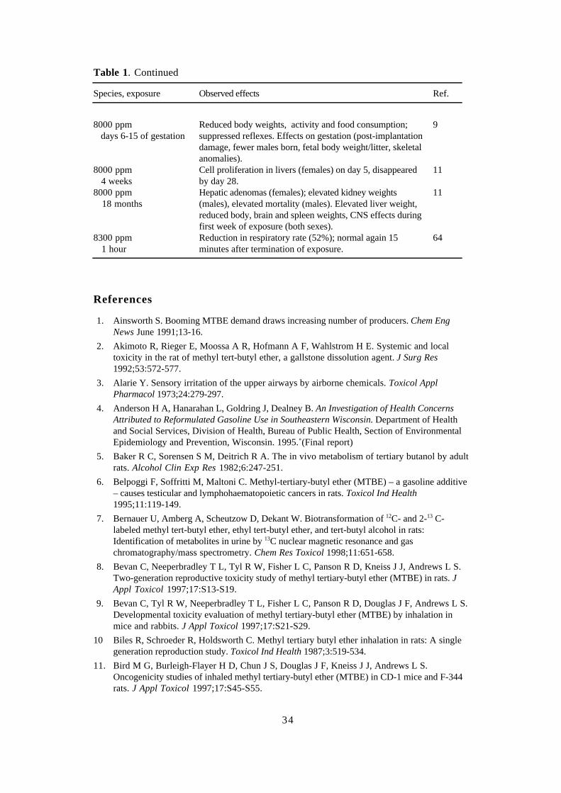
34
Table 1. Continued
Species, exposure Observed effects Ref.
8000 ppm days 6-15 of gestation
Reduced body weights, activity and food consumption;suppressed reflexes. Effects on gestation (post-implantationdamage, fewer males born, fetal body weight/litter, skeletalanomalies).
9
8000 ppm 4 weeks
Cell proliferation in livers (females) on day 5, disappearedby day 28.
11
8000 ppm 18 months
Hepatic adenomas (females); elevated kidney weights(males), elevated mortality (males). Elevated liver weight,reduced body, brain and spleen weights, CNS effects duringfirst week of exposure (both sexes).
11
8300 ppm 1 hour
Reduction in respiratory rate (52%); normal again 15minutes after termination of exposure.
64
References
1. Ainsworth S. Booming MTBE demand draws increasing number of producers. Chem EngNews June 1991;13-16.
2. Akimoto R, Rieger E, Moossa A R, Hofmann A F, Wahlstrom H E. Systemic and localtoxicity in the rat of methyl tert-butyl ether, a gallstone dissolution agent. J Surg Res1992;53:572-577.
3. Alarie Y. Sensory irritation of the upper airways by airborne chemicals. Toxicol ApplPharmacol 1973;24:279-297.
4. Anderson H A, Hanarahan L, Goldring J, Dealney B. An Investigation of Health ConcernsAttributed to Reformulated Gasoline Use in Southeastern Wisconsin. Department of Healthand Social Services, Division of Health, Bureau of Public Health, Section of EnvironmentalEpidemiology and Prevention, Wisconsin. 1995. (Final report)
5. Baker R C, Sorensen S M, Deitrich R A. The in vivo metabolism of tertiary butanol by adultrats. Alcohol Clin Exp Res 1982;6:247-251.
6. Belpoggi F, Soffritti M, Maltoni C. Methyl-tertiary-butyl ether (MTBE) – a gasoline additive– causes testicular and lymphohaematopoietic cancers in rats. Toxicol Ind Health1995;11:119-149.
7. Bernauer U, Amberg A, Scheutzow D, Dekant W. Biotransformation of 12C- and 2-13 C-labeled methyl tert-butyl ether, ethyl tert-butyl ether, and tert-butyl alcohol in rats:Identification of metabolites in urine by 13C nuclear magnetic resonance and gaschromatography/mass spectrometry. Chem Res Toxicol 1998;11:651-658.
8. Bevan C, Neeperbradley T L, Tyl R W, Fisher L C, Panson R D, Kneiss J J, Andrews L S.Two-generation reproductive toxicity study of methyl tertiary-butyl ether (MTBE) in rats. JAppl Toxicol 1997;17:S13-S19.
9. Bevan C, Tyl R W, Neeperbradley T L, Fisher L C, Panson R D, Douglas J F, Andrews L S.Developmental toxicity evaluation of methyl tertiary-butyl ether (MTBE) by inhalation inmice and rabbits. J Appl Toxicol 1997;17:S21-S29.
10 Biles R, Schroeder R, Holdsworth C. Methyl tertiary butyl ether inhalation in rats: A singlegeneration reproduction study. Toxicol Ind Health 1987;3:519-534.
11. Bird M G, Burleigh-Flayer H D, Chun J S, Douglas J F, Kneiss J J, Andrews L S.Oncogenicity studies of inhaled methyl tertiary-butyl ether (MTBE) in CD-1 mice and F-344rats. J Appl Toxicol 1997;17:S45-S55.

35
12. Borghoff S J, Murphy J E, Medinsky M A. Development of physiologically basedpharmacokinetic model for methyl tertiary-butyl ether and tertiary-butanol in male Fischer-344 rats. Fundam Appl Toxicol 1996;30:264-275.
13. Brady J F, Xiao F, Ning S M, Yang C S. Metabolism of methyl tertiary-butyl ether by rathepatic microsomes. Arch Toxicol 1990;64:157-160.
14. Brown S L. Atmospheric and potable water exposures to methyl tert-butyl ether (MTBE).Regul Toxicol Pharmacol 1997;25:256-276.
15. Buckley T J, Prah J D, Ashley D, Zweidinger R A, Wallace L A. Body burden measurementsand models to assess inhalation exposure to methyl tertiary butyl ether (MTBE). J Air WasteManage Assoc 1997;47:739-752.
16. Budavari S, O’Neil M J, Smith A, Heckelman P E, Kinneary J F. In The Merck Index: AnEncyclopedia of Chemicals, Drugs and Biologicals, 12th ed. Whitehouse Station, NJ: Merckand Co. 1996:1032.
17. Cain W S, Leaderer B P, Ginsberg G L et al. Acute exposure to low-level methyl tertiary-butyl ether (MTBE): Human reactions and pharmacokinetic response. Inhal Toxicol1996;8:21-48.
18. Cheminfo. Methyl Tert-Butyl Ether. (Database; CD-ROM). Canadian Centre for OccupationalHealth Safety, Hamilton, Ontario, Canada, 1997:3.
19. Casanova M, Heck H D. Lack of evidence for the involvement of formaldehyde in thehepatocarcinogenicity of methyl tertiary-butyl ether in CD-1 mice. Chem Biol Interact1997;105:131-143.
20. Cederbaum A I, Cohen G. Oxidative demethylation of t-butyl alcohol by rat livermicrosomes. Biochem Biophys Res Commun 1980;97:730-736.
21 Cinelli S, Ciliutti P, Falezza A et al. Absence of mutagenicity of methyl-tertiary-butyl ether.Toxcol Lett 1992;S1:300.
22. Conaway C C, Schroeder R E, Snyder N K. Teratology evaluation of methyl tertiary butylether in rats and mice. J Toxicol Environ Health 1985;16:797-809.
23. Daughtrey W C, Gill M W, Pritts I M, Douglas J F, Kneiss J J, Andrews L S.Neurotoxicological evaluation of methyl tertiary-butyl ether in rats. J Appl Toxicol1997;17:S57-S64.
24. ECETOC. Methyl tert-butyl ether (MTBE). Health risk characterisation. CAS no. 1634-04-4.(EINECS no. 216.653.1). Brussels: European Centre for Ecotoxicology and Toxicology,1997. (Technical report No. 72)
25. Evans T W, Edlund K R. Tertiary alkyl ethers. Preparation and properties. Ind Eng Chem1936;28:1186-1188.
26. Gordian M E, Huelsman M D, Brecht M-L, Fischer D G. Heath effects of methyl tertiarybutyl ether (MTBE) in gasoline in Alaska. Alaska Med 1995;37:101-103, 119.
27. Hakkola M, Honkasalo M L, Pulkkinen P. Neuropsychological symptoms among tankerdrivers exposed to gasoline. Occup Med 1996;46:125-130.
28. HEI. The potential health effects of oxygenates added to gasoline. A review of the currentliterature. HEI Oxygenates Evaluation Committee, Cambridge, Maryland: Health EffectsInstitute, April 1996. (Special report)
29. Hong J-Y, Wang Y-Y, Bondoc F Y, Yang C S, Lee M, Huang W-Q. Rat olfactory mucosadisplays a high activity in metabolizing methyl tert-butyl ether and other gasoline ethers.Fundam Appl Toxicol 1997;40:205-210.
30. Hong J-Y, Yang C S, Lee M et al. Role of cytochrome P450 in the metabolism of methyltert-butyl ether in human livers. Arch Toxicol 1997;71:266-269.
31. Janowitz P, Schumacher K A, Swobodnik W, Kratzer W, Tudyka J, Wechsler J G.Transhepatic topical dissolution of gallbladder stones with MTBE and EDTA. Results, sideeffects, and correlation with CT imaging. Dig Dis Sci 1993;38:2121-2129.
36
32. Kado N Y, Kuzmicky P A, Loarca-Pina G, Mumtaz M M. Genotoxicity testing of methyltertiary-butyl ether (MTBE) in the Salmonella microsuspension assay and mouse bonemarrow micronucleus test. Mutat Res 1998;412:131-138.
33. Kemikalieinspektionen. Metyl-tert-butyleter. En litteratursammanställning. Solna, Sweden:National Swedish Chemicals Inspectorate, 1998;1:1-14.
34. Leuschner U, Hellstern A, Schmidt K, Fischer H. Güldütuna S, Hübner K, Leuschner M.Gallstone dissolution with methyl tert-butyl ether in 120 patients – efficacy and safety. DigDis Sci 1991;36:193-199.
35. Lington A W, Dodd D E, Ridlon S A, Douglas J F, Kneiss J J, Andrews L S. Evaluation ofa 13-week inhalation toxicity study on methyl t-butyl ether (MTBE) in Fischer 344 rats. JAppl Toxicol 1997;17:S37-S44.
36. Lioy P J, Weisel C P, Jo W-K, Pellizzari E, Raymer J H. Microenvironmental and personalmeasurements of methyl-tertiary butyl ether (MTBE) associated with automobile useactivities.J Exp Anal Environ Epidemiol 1994;4:427-441.
37. Little C J, Dale A D, Whatley J A, Wickings J A. Methyl tert.-butyl ether: A newchromatographic eluent. J Chromatogr 1979;169:381-385.
38. Lundberg P, ed. Scientific Basis for Swedish Occupational Standards. IX. Methyl-t-butylether. Arbete och Hälsa 1988;32:35-41.
39. Mackerer C R, Angelosanto F A, Blackburn G R, Schreiner C A. Identification offormaldehyde as the metabolite responsible for the mutagenicity of methyl tertiary-butyl etherin the activated mouse lymphoma assay. Proc Soc Exp Biol Med 1996;212:338-341.
40. Marsh D F, Leake C D. The comparative anesthetic activity of the aliphatic ethers.Anesthesiology 1950;11:455-463.
41. McKee R H, Vergnes J S, Galvin J B, Douglas J F, Kneiss J J, Andrews L S. Assessment ofthe in vivo mutagenic potential of methyl tertiary-butyl ether. J Appl Toxicol 1997;17:S31-S36.
42. Mennear J H. Carcinogenicity studies on MTBE: Critical review and interpretation. RiskAnal 1997;17:673-681.
43. Milas N A. Studies in auto-oxidation reactions. II. The mechanism of the auto-oxidation ofcertain ethers. J Chem Soc 1931;53:221-233.
44. Miller M J, Ferdinandi E S, Klan M, Andrews L S, Douglas J F, Kneiss J J.Pharmacokinetics and disposition of methyl t-butyl ether in Fischer-344 rats. J Appl Toxicol1997;17:S3-S12.
45. Mohr S N, Fielder N, Weisel C, Kelly-McNeil K. Health effects of MTBE among NewJersey garage workers. Inhal Toxicol 1994;6:553-562.
46. Moolenaar R L, Hefflin B J, Ashley D L, Middaugh J P, Etzel R A. Methyl tertiary butylether in human blood after exposure to oxygenated fuel in Fairbanks, Alaska. Arch EnvironHealth 1994;49:402-409.
47. Moser G J, Wolf D C, Sar M, Gaido K W, Janszen D, Goldsworthy T L. Methyl tertiarybutyl ether-induced endocrine alterations in mice are not mediated through the estrogenreceptor. Toxicol Sci 1998;41:77-87.
48. Moser G J, Wong B A, Wolf D C, Fransson-Steen R L, Goldsworthy T L. Methyl tertiarybutyl ether lacks tumor-promoting activity in N-nitrosodiethylamine-initiated B6C3F1 femalemouse liver. Carcinogenesis 1996;17:2753-2761.
49. Moser G J, Wong B A, Wolf D C, Moss O R, Goldsworthy T L. Comparative short-termeffects of methyl tertiary butyl ether and unleaded gasoline vapor in female B6C3F1 mice.Fundam Appl Toxicol 1996;31:173-183.
50. Mount D L, Churchill F C, Bergqvist Y. Determination of mefloquine in blood, filter paper-absorbed blood and urine by 9-fluorenylmethyl chloroformate derivatization followed by liquidchromatography with fluorescence detection. J Chromatogr 1991;564:181-193.

37
51. Nihlén A, Löf A, Johanson G. Liquid/air partition coefficients of methyl and ethyl t-butylethers, t-amyl methyl ether, and t-butyl alcohol. J Expos Anal Environ Epidemiol1995;5:573-582.
52. Nihlén A, Löf A, Johanson G. Experimental exposure to methyl tertiary-butyl ether. I.Toxicokinetics in humans. Toxicol Appl Pharmacol 1998;148:274-280.
53. Nihlén A, Sumner S, Löf A, Johanson G. 13C-labeled methyl t-butyl ether: Toxicokineticsand characterization of urinary metabolites in man. Proceedings from the InternationalCongress of Toxicology – ICT VIII. Paris, 5-9 July 1998. Toxicol Lett 1998;Suppl1/95:103.
54. Nihlén A, Wålinder R, Löf A, Johanson G. Experimental exposure to methyl tertiary-butylether: II. Acute effects in humans. Toxicol Appl Pharmacol 1998;148:281-287.
55. Pekari K, Riihimäki V, Vainiotalo S, Teräväinen E, Aitio A. Experimental exposure tomethyl-tert-butyl ether (MTBE) and methyl-tert-amyl ether (MTAE). Proceedings from theInternational Symposium on Biological Monitoring in Occupational and EnvironmentalHealth, Espoo, Finland, 11-13 September 1996:27-28.
56. Ponchon T, Baroud J, Pujol B, Valette P J, Perrot D. Renal failure during dissolution ofgallstones by methyl-tert-butyl ether [letter]. Lancet 1988;2:276-277.
57. Prah J D, Goldstein G M, Devlin R et al. Sensory, symptomatic, inflammatory, and ocularresponses to and the metabolism of methyl tertiary butyl ether in a controlled humanexposure experiment. Inhal Toxicol 1994;6:521-538.
58. Prescott-Mathews J S, Wolf D C, Wong B A, Borghoff S J. Methyl tert-butyl ether causesα2u-globulin nephropathy and enhanced renal cell proliferation in male Fischer-344 rats.Toxicol Appl Pharmacol 1997;143:301-314.
59. Product register. National Swedish Chemicals Inspectorate, Solna, Sweden: 1998.
60. Riihimäki V, Matikainen E, Akila R et al. Central nervous system effects of the gasolineadditive methyl-tert-butylether (MTBE). Proceedings from the International Symposium onBiological Monitoring in Occupational and Environmental Health, Espoo, Finland, 11-13September 1996:23-24.
61. Robinson M, Bruner R H, Olson G R. Fourteen- and ninety-day oral toxicity studies ofmethyl tertiary-butyl ether in Sprague-Dawley rats. J Am Coll Toxicol 1990;9:525-540.
62. Rosenkranz H S, Klopman G. Predictions of the lack of genotoxicity and carcinogenicity inrodents of two gasoline additives: methyl- and ethyl-t-butyl ethers. In Vitro Toxicol1991;4:49-54.
63. Savolainen H, Pfäffli P, Elovaara E. Biochemical effects of methyl tertiary-butyl ether inextended vapour exposure of rats. Arch Toxicol 1985;57:285-288.
64. Tepper J S, Jackson M C, McGee J K, Costa D L, Graham J A. Estimation of respiratoryirritancy from inhaled MTBE in mice. Inhal Toxicol 1994;6:563-569.
65. Thistle J L, May G R, Bender C E et al. Dissolution of cholesterol gallbladder stones bymethyl tert-butyl ether administered by percutaneous transhepatic catheter. New Engl J Med1989;320:633-639.
66. Turini A, Amato G, Longo V, Gervasi P G. Oxidation of methyl- and ethyl- tertiary-butylethers in rat liver microsomes: Role of the cytochrome P450 isoforms. Arch Toxicol1998;72;207-214.
67. U.S. EPA. Assessment of Potential Health Risks of Gasoline Oxygenated with MethylTertiary Butyl Ether (MTBE). EPA/600/R-93/206, Washington, DC: Office of Research andDevelopment, US Environmental Protection Agency, 1993.
68. White M C, Johnson C A, Ashley D L, Buchta T M, Pelletier D J. Exposure to methyltertiary-butyl ether from oxygenated gasoline in Stamford, Connecticut. Arch Environ Health1995;50:183-189.

38
69. Vojdani A, Mordechai E, Brautbar N. Abnormal apoptosis and cell cycle progression inhumans exposed to MTBE and benzene contaminating water. Human Expir Toxicol1997;16:485-494.
70. Yoshikawa M, Arashidani K, Katoh T, Kawamoto T, Kodama Y. Pulmonary elimination ofmethyl tertiary-butyl ether after intraperitoneal administration in mice. Arch Toxicol1994;68:517-519.
71. Zhang Y P, Macina O T, Rosenkranz H S, Karol M H, Mattison D R, Klopman G.Predictions of the metabolism and toxicological profiles of gasoline oxygenates. InhalToxicol 1997;9:237-254.

39
Consensus Report for Dimethyl Adipate,Dimethyl Glutarate and DimethylSuccinate
December 9, 1998
Chemical and physical data. Uses
dimethyl adipateCAS No.: 627-93-0Synonyms: dimethyl hexanedioate, methyl adipateFormula: C8H14O4
Structure: CH3-O-CO-CH2-CH2-CH2-CH2-CO-O-CH3
Molecular weight: 174.22Boiling point: 231 °CMelting point: 10.3 °CVapor pressure: 0.0016 kPa (20 °C)Conversion factors: 1 ppm = 7.23 mg/m3 (20 °C)
1 mg/m3 = 0.138 ppm (20 °C)
dimethyl glutarateCAS No.: 1119-40-0Synonyms: dimethyl pentanedioate, methyl glutarateFormula: C7H12O4
Structure: CH3-O-CO-CH2-CH2-CH2-CO-O-CH3
Molecular weight: 160.17Boiling point: 214 °CMelting point: - 42.5 °CVapor pressure: 0.0061 kPa (20 °C)Conversion factors: 1 ppm = 6.65 mg/m3 (20 °C)
1 mg/m3 = 0.150 ppm (20 °C)
dimethyl succinateCAS No.: 106-65-0Synonyms: dimethyl butanedioate, methyl succinateFormula: C6H10O4
Structure: CH3-O-CO-CH2-CH2-CO-O-CH3
Molecular weight: 146.14Boiling point: 196.4 °CMelting point: 19 °CVapor pressure: 0.0167 kPa (20 °C)Conversion factors: 1 ppm = 6.06 mg/m3 (20 °C)
1 mg/m3 = 0.165 ppm (20 °C)

40
Dimethyl adipate (DMA), dimethyl glutarate (DMG) and dimethyl succinate (DMS)usually occur together. The mixtures are a clear liquid (1). DMA, DMG and DMSare soluble in substances such as ethanol and ether (13). Solubility in water hasbeen reported to be 29.9 g/l (20 °C) for DMA and 131 g/l (25 °C) for DMS (7).Product information leaflets state that the water solubility of mixtures containing >55% DMG, >15% DMS and > 10% DMA is 5 to 5.5% by weight (20 °C), and thatthe vapor pressure is 0.013 kPa.
Mixtures of DMA, DMG and DMS are used as solvents. The mixtures are usedindustrially in paints and coatings, and also for paint removal and for cleaningpolyurethane foam and unsaturated polyester resin (22).
DMS is also used in the cosmetics and food processing industries. It occurs, forexample, as a flavoring in ice cream, candy, bakery products and drinks (2).
Uptake, biotransformation, excretion
There is little information in the literature on oral uptake of either DMA, DMG orDMS. In one study with rats (16), it was shown that DMS was rapidly absorbed inthe digestive tract. There are no quantitative data on skin uptake for any of the threeesters.
Nearly all of inhaled DMA, DMG and DMS is deposited in the upper respiratorypassages. In one study (19), rats were exposed (one-way air flow) for 40 minutesto 50 –100 mg/m3 DMA, DMG and DMS vapors, either separately or mixed, or to250 – 320 mg/m3 DMG vapor. It was found that deposition in the upper respiratorypassages exceeded 97% in all cases – much the same for all three substances, bothsexes, and all air concentrations tested.
DMA, DMG and DMS are metabolized by hydrolysis and can form monomethylesters, dicarboxylic acids and methanol; this has been demonstrated in vitro withhomogenates, nasal explants and isolated cells (5, 19, 22, 29, 30). In rats, theesters are efficiently hydrolyzed by carboxylesterase, mostly in the airways(especially in olfactory epithelium) and liver (4, 5, 19, 22, 30). There is somedifference between males and females. In a comparative study, the efficiency ofhydrolysis to monomethyl esters was tested with low (“subsaturating”) substrateconcentrations of DMA, DMG and DMS in homogenates of olfactory epitheliumfrom female or male rats. Hydrolysis was equally effective in homogenates fromboth sexes when DMG was used as substrate, whereas DMA was hydrolyzed moreeffectively by the olfactory epithelium homogenate from females and DMS by thatfrom males. A structure-activity relationship was also observed in the hydrolysisreactions: DMA > DMG > DMS (5).
The activity of carboxylesterase in olfactory epithelium from humans (hydrolysisof DBE in 3/6 samples) was reported in an abstract to be 100 to 1000 times lowerthan that of rats (9). (DBE = dibasic esters, i.e. DMA, DMG and DMS.)
Glutaric acid (a metabolite of DMG) is one of the body’s natural components. Itis formed in metabolism of the amino acid lysine (8). Malic acid (a metabolite ofDMS) also occurs naturally in the body’s general metabolism. It is a component, for

41
example, of the energy-producing Krebs cycle, which yields the end productscarbon dioxide and water (8, 14, 29). Increased synthesis of proinsulin andincreased insulin secretion have been shown to follow administration of DMS, andare ascribed primarily to an influx of malic acid to the Krebs cycle in the pancreaticislets (isles of Langerhans) (15, 17, 18, 30).
Toxic effects
Human dataProduct information leaflets from suppliers/manufacturers report that temporaryeffects on vision (foggy vision) may occur after exposure to mixtures of DMG,DMS and DMA. No air concentrations are given, but it is stated that the problemhad occurred at exposure to “high” concentrations of vapor or after direct contactwith the eyes (product mixtures containing > 55% DMG, > 15% DMS and > 10%DMA). The product information leaflets also warn that inhalation of “high”concentrations of the vapors can irritate respiratory passages.
Animal dataThe LD50 for rats given DMA by intraperitoneal injection is reported in one study(21) to be 1.9 g/kg.
Mice were exposed by inhalation to various concentrations of DMA, DMG andDMS, either separately or in mixtures, and their respiratory rates were measured(mixture 1: 63% DMG, 25% DMS, 12% DMA; mixture 2: 57% DMG, 23% DMS,20% DMA). The data were used to calculate the concentration that caused a 50%decrease in respiratory rate (RD50), which is a measure of the irritation potential of asubstance. The RD50 was reported to be 890 mg/m3 for DMG, 910 mg/m3 forDMA, 1600 mg/m3 for DMS, 590 mg/m3 for mixture 1 and 610 mg/m3 for mixture2. DMS appeared to be the least potent of the three esters but was reported to havethe steepest dose-response curve (20).
In an inhalation study (12), male rats were exposed to a vapor-aerosol DBE mixture(90% aerosol) generated from a liquid that was 66% DMG, 17% DMS and 17%DMA. The animals were exposed for 4 hours to an average aerosol concentration of5900 mg/m3 and sacrificed at intervals of 1 to 42 days after the exposure. Damage(degeneration, inflammation, necrosis), which was largely reversible within 6 weeks,was seen in nasal mucosa exposed to air flow. Severe necrosis was noted in olfactoryepithelial cells in the anterior portion of the nasal cavity, for example (these cells didnot recover within 6 weeks), whereas the damage further back was lighter and morelimited. The cells that were most sensitive and affected first were the sustenacular cells(which support the olfactory epithelium).
In another inhalation study, male and female rats were exposed to DBE vaporsgenerated from a liquid mixture of 67% DMG, 17% DMS and 17% DMA, 6 hours/day, 5 days/week for up to 13 weeks. Concentrations were 390, 76 or 20 mg/m3.After seven weeks, animals in the two higher dose groups (both sexes) showeddose-dependent degeneration (minimal to mild) of olfactory epithelium in the nasalcavities. After 13 weeks of exposure, minimal to moderate degenerative changes

42
were observed in the nasal cavities (olfactory epithelium) of females at all doselevels and males at the two higher dose levels. The damage was characterized byinitial cell death and loss of sustenacular and sensory cells. After an exposure-freeperiod of 6 weeks there were indications that the damaged tissue was regenerating.At the highest dose level there were negative effects on weight gain and lower liverweights in the females (reversible changes) (10).
In a reproduction study (1), female rats were exposed for 6 hours/day duringgestation to 990 mg/m3 vapor/aerosol (about 40/60), or to 380 or 150 mg/m3 vaporof a DBE mixture (65% DMG, 18% DMS, 17% DMA). Somewhat reduced abso-lute liver weights were observed in all dose groups (trend, not significant). Animalsin the two higher dose groups had reduced food intake during the first 6 days andreduced growth. In another reproduction study, male and female rats (parentgeneration) were exposed 6 hours/day before, during and after gestation (a total ofabout 22 weeks) to 1000 mg/m3 vapor/aerosol or 400 or 160 mg/m3 vaporgenerated from an identical DBE mixture (65% DMG, 18% DMS, 17% DMA) (11).From week 7 onward, both males and females in the highest dose group gainedweight more slowly. When the animals were killed, however, significantly lowerbody weights were noted only in females in the highest dose group. Some organweights were also significantly different in the high-dose group, especially amongthe females. Liver weights were significantly lower in males (relative liver weightsonly) and females in both the high and medium dose groups. Histopathologicalexamination of nasal tissue revealed squamous cell metaplasias, especially inolfactory epithelia. Their prevalence and severity increased with dose, and femaleswere more sensitive than males.
A dose-dependent cytotoxicity, expressed as increased leakage of acidic phospha-tases, was observed when olfactory and respiratory epithelia from female rats wereincubated with DMA, DMG or DMS (10 – 100 mM). All three substances wereobserved to have significant effects on the olfactory epithelium at concentrations of25 mM and above, whereas effects on respiratory epithelium were not seen below50 mM. The toxicity of DMA, DMG and DMS was shown to be dependent on acarboxylesterase-mediated activation. Liberation of acidic phosphatases wasreduced when the animals were pre-treated with a carboxylesterase inhibitor (22).No significant elevation of acidic phosphatases was observed when nasal explantsfrom rats were incubated with the metabolite methanol (100 mM) (22), but themonomethyl esters and dicarboxylic acids produced by metabolism of DBE werereported in another in vitro study (23) to stimulate liberation of acidic phosphatases(in tests with 25 or 50 mM). A ranking of the monoesters for cytotoxic potencyyielded the result monomethyl adipate > monomethyl glutarate > monomethylsuccinate (23).
In a subsequent study (24), olfactory and respiratory epithelia from female ratswere examined under optical and electron microscopes after incubation with 10, 25or 50 mM DMA. At the lowest dose level mild degenerative changes were noted inthe respiratory epithelium and more pronounced degenerative changes in thesustenacular cells in olfactory epithelium. The two higher dose levels caused severe
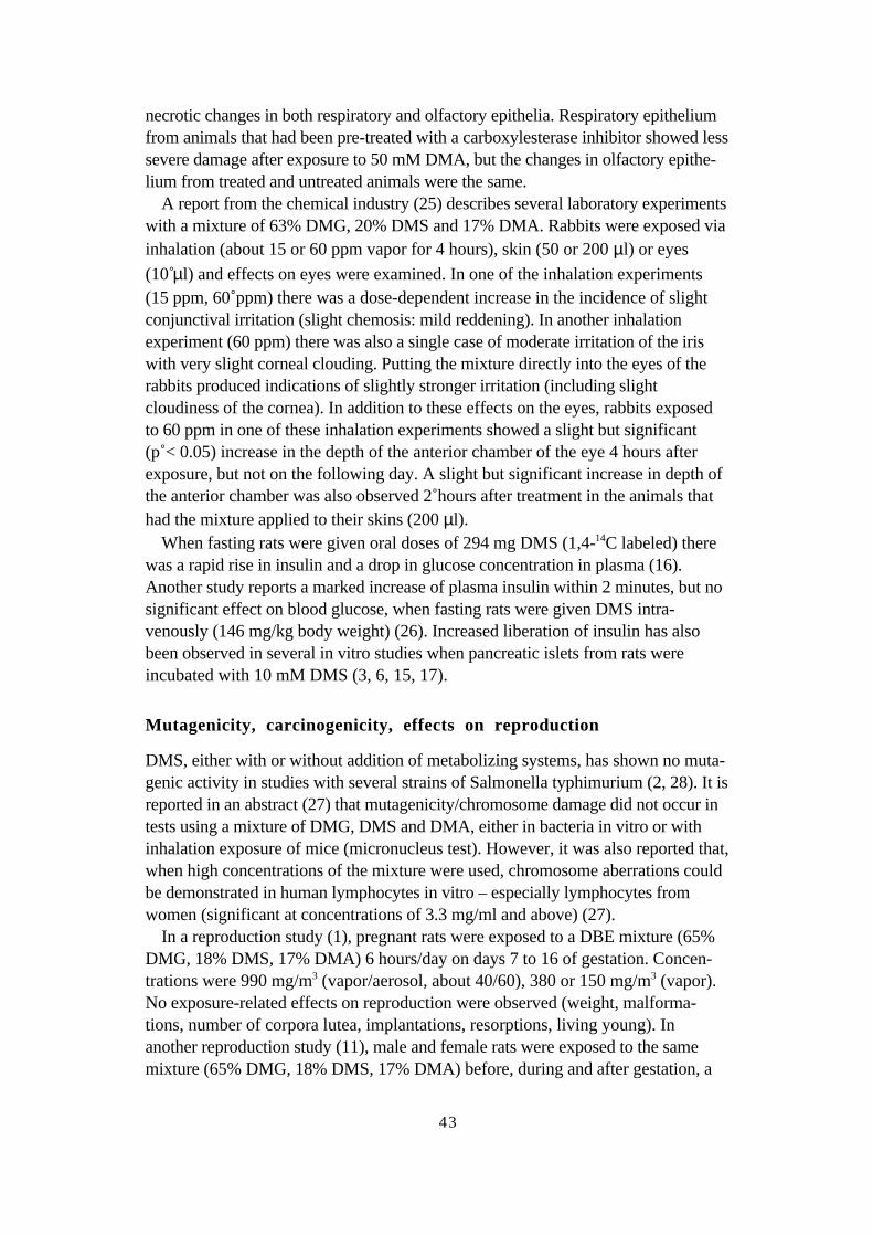
43
necrotic changes in both respiratory and olfactory epithelia. Respiratory epitheliumfrom animals that had been pre-treated with a carboxylesterase inhibitor showed lesssevere damage after exposure to 50 mM DMA, but the changes in olfactory epithe-lium from treated and untreated animals were the same.
A report from the chemical industry (25) describes several laboratory experimentswith a mixture of 63% DMG, 20% DMS and 17% DMA. Rabbits were exposed viainhalation (about 15 or 60 ppm vapor for 4 hours), skin (50 or 200 µl) or eyes
(10 µl) and effects on eyes were examined. In one of the inhalation experiments(15 ppm, 60 ppm) there was a dose-dependent increase in the incidence of slightconjunctival irritation (slight chemosis: mild reddening). In another inhalationexperiment (60 ppm) there was also a single case of moderate irritation of the iriswith very slight corneal clouding. Putting the mixture directly into the eyes of therabbits produced indications of slightly stronger irritation (including slightcloudiness of the cornea). In addition to these effects on the eyes, rabbits exposedto 60 ppm in one of these inhalation experiments showed a slight but significant(p < 0.05) increase in the depth of the anterior chamber of the eye 4 hours afterexposure, but not on the following day. A slight but significant increase in depth ofthe anterior chamber was also observed 2 hours after treatment in the animals thathad the mixture applied to their skins (200 µl).
When fasting rats were given oral doses of 294 mg DMS (1,4-14C labeled) therewas a rapid rise in insulin and a drop in glucose concentration in plasma (16).Another study reports a marked increase of plasma insulin within 2 minutes, but nosignificant effect on blood glucose, when fasting rats were given DMS intra-venously (146 mg/kg body weight) (26). Increased liberation of insulin has alsobeen observed in several in vitro studies when pancreatic islets from rats wereincubated with 10 mM DMS (3, 6, 15, 17).
Mutagenicity, carcinogenicity, effects on reproduction
DMS, either with or without addition of metabolizing systems, has shown no muta-genic activity in studies with several strains of Salmonella typhimurium (2, 28). It isreported in an abstract (27) that mutagenicity/chromosome damage did not occur intests using a mixture of DMG, DMS and DMA, either in bacteria in vitro or withinhalation exposure of mice (micronucleus test). However, it was also reported that,when high concentrations of the mixture were used, chromosome aberrations couldbe demonstrated in human lymphocytes in vitro – especially lymphocytes fromwomen (significant at concentrations of 3.3 mg/ml and above) (27).
In a reproduction study (1), pregnant rats were exposed to a DBE mixture (65%DMG, 18% DMS, 17% DMA) 6 hours/day on days 7 to 16 of gestation. Concen-trations were 990 mg/m3 (vapor/aerosol, about 40/60), 380 or 150 mg/m3 (vapor).No exposure-related effects on reproduction were observed (weight, malforma-tions, number of corpora lutea, implantations, resorptions, living young). Inanother reproduction study (11), male and female rats were exposed to the samemixture (65% DMG, 18% DMS, 17% DMA) before, during and after gestation, a
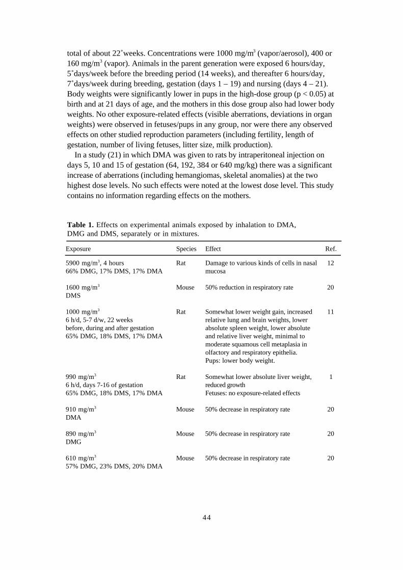
44
total of about 22 weeks. Concentrations were 1000 mg/m3 (vapor/aerosol), 400 or160 mg/m3 (vapor). Animals in the parent generation were exposed 6 hours/day,5 days/week before the breeding period (14 weeks), and thereafter 6 hours/day,7 days/week during breeding, gestation (days 1 – 19) and nursing (days 4 – 21).Body weights were significantly lower in pups in the high-dose group (p < 0.05) atbirth and at 21 days of age, and the mothers in this dose group also had lower bodyweights. No other exposure-related effects (visible aberrations, deviations in organweights) were observed in fetuses/pups in any group, nor were there any observedeffects on other studied reproduction parameters (including fertility, length ofgestation, number of living fetuses, litter size, milk production).
In a study (21) in which DMA was given to rats by intraperitoneal injection ondays 5, 10 and 15 of gestation (64, 192, 384 or 640 mg/kg) there was a significantincrease of aberrations (including hemangiomas, skeletal anomalies) at the twohighest dose levels. No such effects were noted at the lowest dose level. This studycontains no information regarding effects on the mothers.
Table 1. Effects on experimental animals exposed by inhalation to DMA,DMG and DMS, separately or in mixtures.
Exposure Species Effect Ref.
5900 mg/m3, 4 hours66% DMG, 17% DMS, 17% DMA
Rat Damage to various kinds of cells in nasalmucosa
12
1600 mg/m3
DMSMouse 50% reduction in respiratory rate 20
1000 mg/m3
6 h/d, 5-7 d/w, 22 weeksbefore, during and after gestation65% DMG, 18% DMS, 17% DMA
Rat Somewhat lower weight gain, increasedrelative lung and brain weights, lowerabsolute spleen weight, lower absoluteand relative liver weight, minimal tomoderate squamous cell metaplasia inolfactory and respiratory epithelia.Pups: lower body weight.
11
990 mg/m3
6 h/d, days 7-16 of gestation65% DMG, 18% DMS, 17% DMA
Rat Somewhat lower absolute liver weight,reduced growthFetuses: no exposure-related effects
1
910 mg/m3
DMAMouse 50% decrease in respiratory rate 20
890 mg/m3
DMGMouse 50% decrease in respiratory rate 20
610 mg/m3
57% DMG, 23% DMS, 20% DMAMouse 50% decrease in respiratory rate 20

45
Table 1. Continued
Exposure Species Effect Ref.
590 mg/m3
63% DMG, 25% DMS, 12% DMAMouse 50% decrease in respiratory rate 20
400 mg/m3
5-7 d/w, 22 weeksbefore, during and after gestation65% DMG, 18% DMS, 17% DMA
Rat Lower relative and absolute liverweights, minimal to moderate squamouscell metaplasia in olfactory epithelium
11
390 mg/m3
6 h/d, 5 d/w, up to 13 weeks67% DMG, 17% DMS, 17% DMA
Rat Minimal to moderate degeneration ofolfactory epithelium, lower weight gain,lower liver weight
10
380 mg/m3,6 h/d, days 7-16 of gestation65% DMG, 18% DMS, 17% DMA
Rat Somewhat lower absolute liver weight,lower growthPups: no exposure-related effects
1
160 mg/m3
6 h/d, 5-7 d/w, 22 weeksbefore, during and after gestation65% DMG, 18% DMS, 17% DMA
Rat Minimal to mild squamous cellmetaplasia in olfactory epithelium
11
76 mg/m3
6 h/d, 5 d/w, up to 13 weeks67% DMG 17% DMS, 17% DMA
Rat Minimal to mild degeneration ofolfactory epithelium
10
20 mg/m3
6 h/d, 5 d/w, up to 13 weeks67% DMG, 17% DMS, 17% DMA
Rat Minimal degeneration of olfactoryepithelium
10
Dose-effect/dose-response relationships
There are no data from which to derive a dose-effect or dose-response relationshipfor humans.
Effects on experimental animals exposed to DMA, DMG and/or DMS aresummarized in Table 1. Dose-dependent effects on olfactory epithelium have beenobserved in rats exposed to a mixture of DMA, DMG and DMS in airconcentrations of 20 mg/m3 and higher.
Conclusions
There are no data from which to determine a critical effect of occupational exposureto DMA, DMG and DMS. Judging from animal experiments, degeneration ofolfactory epithelium is the critical effect of exposure to mixtures of the threesubstances.
46
References
1. Alvarez L, Driscoll C, Kelly D P, Staples R E, Chromey N C, Kennedy G L. Developmentaltoxicity of dibasic esters by inhalation in the rat. Drug Chem Toxicol 1995;18:295-314.
2. Andersen P H, Jensen N J. Mutagenic investigation of flavourings: Dimethyl succinate, ethylpyruvate and aconitic acid are negative in the Salmonella/mammalian-microsome test. FoodAddit Contam 1984;1:283-288.
3. Bakkali Nadi A, Zhang T M, Malaisse W J. Effects of the methyl esters of pyruvate,succinate and glutamate on the secretory response to meglitinide analogues in rat pancreaticislets. Pharm Res 1996;33:191-194.
4. Bogdanffy M S. Biotransformation enzymes in the rodent nasal mucosa: The value of a histo-chemical approach. Environ Health Perspect 1990;85:177-186.
5. Bogdanffy M S, Kee C R, Hinchman C A, Trela B A. Metabolism of dibasic esters by ratnasal mucosal carboxylesterase. Drug Metab Dispos 1991;19:124-129.
6. Giroix M H, Zhang T M, Leclercq-Meyer V, Sener A, Portha B, Malaisse W J. Restrictedeffect of formycin A and non-glucidic nutrients upon insulin release in islets from rats withhereditary or acquired non-insulin-dependent diabetes. Acta Diabetol 1995;32:198-202.
7. IUCLID. International Uniform Chemical Information Database, European Chemicals Bureau,Environment Institute, Joint Research Centre, European Commission.
8. Karlsson P. Introduction to Modern Biochemistry. 3rd ed. New York: Academic Press,1971:178-179, 216, 218-219.
9. Kee C R, Bogdanffy M S, Keenan C M, Keenan K P, Resau J. Sex and species differences inmetabolism of dibasic esters by nasal carboxylesterase. Toxicologist 1989;9:284.
10. Keenan C M, Kelly D P, Bogdanffy M S. Degeneration and recovery of rat olfactoryepithelium following inhalation of dibasic esters. Fundam Appl Toxicol 1990;15:381-393.
11. Kelly D P, Kennedy G L , Keenan C M. Reproduction study with dibasic esters followinginhalation in the rat. Drug Chem Toxicol 1998;21:253-267.
12. Lee K P, Valentine R, Bogdanffy M S. Nasal lesion development and reversibility in ratsexposed to aerosols of dibasic esters. Toxicol Pathol 1992;20:376-393.
13. Lide D R, Frederikse H P R. CRC Handbook of Chemistry and Physics. New York: CRCPress Inc., 1995-1996:3–93:3–187:3–241.
14. MacDonald M J. Metabolism of the insulin secretagogue methyl succinate by pancreaticislets. Arch Biochem Biophys 1993;300:201-205.
15. MacDonald M J, Fahien L A. Glyceraldehyde phosphate and methyl esters of succinic acid.Diabetes 1988;37:997-999.
16. Malaisse-Lagae F, Bakkali Nadi A, Malaisse W J. Insulinotropic response to enterallyadministered succinic and glutamic acid methyl esters. Arch Int Pharmacodyn 1994;328:235-242.
17. Malaisse W J, Rasschaert J, Villanueva-Penacarrillo M L, Valverde I. Respiratory, ionic, andfunctional effects of succinate esters in pancreatic islets. Am J Physiol 1993;264:E428-433.
18. Malaisse W J, Sener A. Metabolic effects and fate of succinate esters in pancreatic islets. AmJ Physiol 1993;264:E434-440.
19. Morris J B, Clay R J, Trela B A, Bogdanffy M S. Deposition of dibasic esters in the upperrespiratory tract of the male and female Sprague-Dawley rat. Toxicol Appl Pharmacol1991;108:538-546.
20. Nair R S, Dudek B R, Grothe D R, Johannsen F R, Lamb I C, Martens M A, Sherman J H,Stevens M W. Mixture risk assessment: A case study of Monsanto experiences. Food ChemToxicol 1996;34:1139-1145.
21. Singh A R, Lawrence W H, Autian J. Embryonic-fetal toxicity and teratogenic effects ofadipic acid esters in rats. J Pharm Sci 1973;62:1596-1600.

47
22. Trela B A, Bogdanffy M S. Carboxylesterase-dependent cytotoxicity of dibasic esters (DBE) inrat nasal explants. Toxicol Appl Pharmacol 1991;107:285-301.
23. Trela B A, Bogdanffy M S. Cytotoxicity of dibasic esters (DBE) metabolites in rat nasalexplants. Toxicol Appl Pharmacol 1991;110:259-267.
24. Trela B A, Frame S R, Bogdanffy M S. A microscopic and ultrastructural evaluation ofdibasic esters (DBE) toxicity in rat nasal explants. Exper Mol Pathol 1992;56:208-218.
25. Valentine R. Ocular effects of a dibasic ester (DBE) mixture in rabbits. E I du Pont deNemours Co. Haskell Lab, Delaware. NTIS/OTS0535224-1, 1993.
26. Vincent D, Villaneuva-Penacarrillo M L, Malaisse-Lagae F, Leclercq-Meyer V, Valverde I,Malaisse W J. In vivo stimulation of insulin release by succinic acid methyl esters. Arch IntPharmacodyn 1994;327:246-250.
27. Vlachos D A, Arce G T, Rickard L B, Covell D L, Sarrif A M. Evaluation of dibasic esters(DBE), a new class of industrial solvents, in a genotoxicity test battery. Environ MolMutagen 1988;11 suppl 1:109
28. Zeiger E, Anderson B, Haworth S, Lawlor T, Mortelmans K. Salmonella mutagenicity tests:Results from the testing of 311 chemicals. Environ Mol Mutagen 1992;19 suppl 21:2-22,69.
29. Zhang T M, Sener A, Malaisse W J. Metabolic effects and fate of succinic acid methyl estersin rat hepatocytes. Arch Biochem Biophys 1994;314:186-192.
30. Zhang T M, Sener A, Malaisse W J. Hydrolysis of succinic acid dimethyl ester in ratpancreatic islets. Biochem Mol Med 1995;55:131-137.

48
Consensus Report for1,1,1-Trifluoroethane and1,1,1,2,2-Pentafluoroethane
February 24, 1999
Chemical and physical data. Uses
1,1,1-trifluoroethaneCAS No.: 420-46-2Synonyms: methylfluoroform, HFC143a,
FC143a, R143aFormula: CH3CF3
Molecular weight: 84.04Boiling point: - 47.5 °CMelting point: - 111.3 °CVapor pressure: 1267 kPa (25 °C)Conversion factors. 1 ppm = 3.49 mg/m3 (20 °C)
1 mg/m3 = 0.287 ppm (20 °C)
1,1,1,2,2-pentafluoroethaneCAS No.: 354-33-6Synonyms: pentafluoroethane, HFC125,
FC125, HFA125, R125Formula: CHF2CF3
Molecular weight: 120.02Boiling point: - 48.5 °CMelting point: - 103 °CVapor pressure: 1381 kPa (25 °C)Conversion factors: 1 ppm = 4.98 mg/m3 (20 °C)
1 mg/m3 = 0.20 ppm (20 °C)
1,1,1-Trifluoroethane at room temperature is a gas. It is combustible at airconcentrations of about 70,000 ppm or higher (2). It is reported to be soluble inethanol and chloroform (7). 1,1,1,2,2-Pentafluoroethane at room temperature is acolorless, non-combustible gas with low solubility in water (0.97 g/liter) (4, 6).Both substances are used in coolants/refrigerants. 1,1,1,2,2-Pentafluoroethane isalso used in fire extinguishers. In 1977, 77 tons of 1,1,1-trifluoroethane (in 5products) and 104 tons of 1,1,1,2,2-pentafluoroethane (in 16 products) were usedin Sweden (product register, National Chemicals Inspectorate).
49
Uptake, biotransformation, excretion
No studies on the metabolism of 1,1,1-trifluoroethane were found.The metabolism of 1,1,1,2,2-pentafluoroethane has been studied in experiments
with rats. In one study, rats were exposed to up to 50,000 ppm pentafluoroethane6 hours/day, 5 days/week for 4 or 13 weeks, and it is reported that no increase offluoride concentration was found in either plasma or urine (6). In another study (5)rats were exposed to 9700 ppm pentafluoroethane for 6 hours, and it was found thatthe concentration of trifluoroacetic acid in urine was extremely low during the 12hours immediately following the exposure (40 times lower than after exposure to11,000 ppm halothane). Slight trifluoracetylation of protein was demonstrated inliver homogenates from the exposed animals (using immunochemical methods).These data suggest that metabolism of 1,1,1,2,2-pentafluoroethane by rats isextremely limited. To the extent that metabolism does occur, it is effected by theP450 system: the pentafluoroethane is probably transformed via a pentafluoroethylradical to pentafluoroethanol, which may subsequently eliminate hydrogen fluorideand form trifluoroacetyl fluoride. The trifluoroacetyl fluoride may be hydrolyzed,creating trifluoroacetic acid and more hydrogen fluoride. Trifluoroacetyl fluoridecan also eliminate fluoride and bind to protein (1, 3, 5).
The reported oil/gas distribution coefficient in vitro for 1,1,1,2,2-pentafluoro-ethane is 7.3 (9). The reported octanol/water distribution coefficient for 1,1,1,2,2-pentafluoroethane is 1.48 (4).
Toxicity
Human dataThere is no information regarding effects of human health attributed to exposure toeither 1,1,1-trifluoroethane or 1,1,1,2,2-pentafluoroethane.
Animal dataRats exposed for 4 hours to 540,000 or 97,000 ppm 1,1,1-trifluoroethane hadtemporary, dose-dependent weight loss, but there were no deaths. Oxygenconcentration in the exposures was about 20% (2). Dogs were given intravenousinjections of adrenaline and exposed to 1,1,1-trifluoroethane for 10 minutes: atendency to cardiac arrhythmia was noted at an air concentration of 300,000 ppmbut not at 250,000 ppm or lower (2).
Rats exposed for 4 hours to 800,000 ppm 1,1,1,2,2-pentafluoroethane (20%oxygen by volume) developed ataxia and labored breathing, and their reaction tonoise was impaired, but there were no deaths (6). When mice were exposed for sixhours to 600,000 ppm (same oxygen concentration), observed effects included lowactivity, tremor and weight loss (6). When dogs were given intravenous injectionsof adrenaline and exposed to 1,1,1,2,2-pentafluoroethane for 10 minutes, theyshowed a tendency to cardiac arrhythmia at air concentrations of 100,000 ppm orhigher, but none of the dogs showed a positive response to an exposure of75,000 ppm under the same conditions (6). Rats exposed to 50,000 ppm

50
1,1,1,2,2-pentafluoroethane 6 hours/day during gestation had an unsteady gaitduring the exposures. Rabbits exposed to 50,000 ppm 1,1,1,2,2-pentafluoroethane6 hours/day during gestation had somewhat slower weight gain and lower feedintake during the first few days of the exposure (6).
Rats were exposed to 1,1,1-trifluoroethane concentrations of 40,000, 10,000 or2000 ppm 6 hours/day, 5 days/week for 4 weeks or 90 days. Exposures were either“nose only” (4 weeks) or whole-body (4 weeks or 90 days). Clinical signs, bodyweights, organ weights, hematology, biochemical blood analyses, urine analyses,tissue morphology, β-oxidation activity in the liver and effects on the eyes werestudied. No treatment-related effects were seen in the animals after whole-bodyexposure. Males that had been exposed “nose only” for 4 weeks, however, hadrecurring weight loss (no change in food intake) and degenerative changes in testesat all dose levels. The authors regarded this as a result of stress caused mostly byextreme temperature conditions. No significant changes were noted in female ratsafter “nose only” exposures (2).
In another study (6), rats were given whole-body exposures to 50,000, 15,000or 5000 ppm 1,1,1,2,2- pentafluoroethane 6 hours/day, 5 days/week for 4 or 13weeks, and sacrificed at termination of exposure or at intervals for up to 4 weeksafterward. Clinical signs, body weights, organ weights, hematology, biochemicalblood analyses, urine analyses, tissue morphology, β-oxidation activity in the liverand effects on eyes were studied. No treatment-related effects are reported.
Mutagenicity, carcinogenicity, effects on reproduction
One study (8) reports that 1,1,1-trifluoroethane had mutagenic effects on two offour strains of Salmonella typhimurium when tested in vitro, both with and withoutmetabolic activation. Another study (2) reports that mutagenic effects could not beobserved when the substance was tested, either with or without addition ofmetabolizing systems, on a total of six strains of Salmonella typhimurium and twostrains of E coli. In cell transformation tests on mammalian cells in vitro, 1,1,1-trifluoroethane had no observed mutagenic effect (8). There was no significantincrease in chromosome aberrations when human lymphocytes were exposed to thesubstance in vitro (2). Further, there was no significant increase of micronuclei inbone marrow cells of mice that had been exposed to 40,000, 10,000 or 2000 ppm1,1,1-trifluoroethane 6 hours/day for two days (2).
No mutagenic effect was observed when 1,1,1,2,2-pentafluoroethane was testedin vitro, either with or without metabolic activation, on various strains ofSalmonella typhimurium and one strain of E coli (6, 8). In tests on mammalian cellsin vitro there was a statistically significant (p < 0.01) increase of cells with chromo-some aberrations at cytotoxic doses and prolonged exposure times, but not at otherdoses/exposure times (6). The occurrence of chromosome aberrations did notincrease when 1,1,1,2,2,-pentafluoroethane was tested on human lymphocytes invitro (6). Nor was there a significantly higher frequency of micronuclei in the bone
51
marrow cells of mice exposed for 6 hours to 600,000, 120,000 or 24,000 ppm1,1,1,2,2-pentafluoroethane (6).
In one study, a 3% solution of 1,1,1-trifluoroethane in corn oil was given to rats(36 of each sex) by gavage in doses of 300 mg/kg body weight, 5 days/week for52 weeks. The animals were killed on week 125, and routine histopathologicalexaminations of lungs, liver, spleen, kidneys and brain were made, and otherorgans were also examined if they looked abnormal. Males had significantly lowerbody weights from week 28 to week 88. No other exposure-related effects wereobserved: there was no significant increase of cancer incidence in any organ, forexample (8). Shortcomings in the design of the experiment (limited histopatho-logical examinations, brief exposure time, single dose level) make it difficult todraw any definite conclusions from this study.
In a reproduction study (2), rats and rabbits were exposed to 40,000, 10,000or 2,000 ppm 1,1,1-trifluoroethane 6 hours/day on days 6 – 15 (rats) or 6 – 18(rabbits) of gestation. A slight but significant increase of visceral and skeletalanomalies (due to retarded development) was observed in the rats, and a slightincrease of skeletal aberrations in the rabbits, but, considering the unusually lowincidence in the control groups and the lack of a clear dose-response relationship,the effects were judged to be unrelated to the exposures. No significant differenceswere reported for other reproduction parameters (pre- and post-implantation losses,embryo/fetus deaths, fetus weights), and it was concluded that 1,1,1-trifluoroethanewas neither teratogenic nor fetotoxic. No effects on the mothers were observed.
In another reproduction study (6), rats and rabbits were exposed to 50,000,15,000 or 5000 ppm 1,1,1,2,2-pentafluoroethane for 6 hours/day on days 6 – 15(rats) or 6 – 18 (rabbits) of gestation. Pre- and post-implantation losses,embryo/fetus death, deformities/anomalies and fetal weights were registered.Transient effects on the mothers (rat: ataxia; rabbit; initial slow weight gain) wereobserved at 50,000 ppm, but no statistically significant difference in any of thestudied reproduction parameters was found in any dose group.
Dose-effect/dose-response relationships
There are no data from which to derive a dose-effect or dose-response relationshipfor occupational exposure to either of these two substances. Effects on experimentalanimals are summarized in Tables 1 and 2.
Conclusions
There are no data on human exposures that would serve to define a critical effect foroccupational exposure to either 1,1,1-trifluoroethane or 1,1,1,2,2-pentafluoro-ethane. In animal studies, no effects that can be ascribed with certainty to eithersubstance have been reported at exposures to air concentrations below 50,000 ppm.Effects such as tendencies to irregular heartbeat have been reported at higherexperimental exposures.
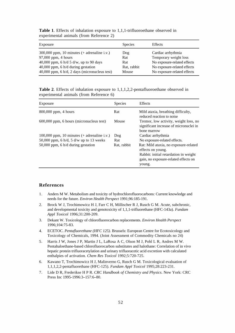
52
Table 1. Effects of inhalation exposure to 1,1,1-trifluoroethane observed inexperimental animals (from Reference 2)
Exposure Species Effects
300,000 ppm, 10 minutes (+ adrenaline i.v.) Dog Cardiac arrhythmia97,000 ppm, 4 hours Rat Temporary weight loss40,000 ppm, 6 h/d 5 d/w, up to 90 days Rat No exposure-related effects40,000 ppm, 6 h/d during gestation Rat, rabbit No exposure-related effects40,000 ppm, 6 h/d, 2 days (micronucleus test) Mouse No exposure-related effects
Table 2. Effects of inhalation exposure to 1,1,1,2,2-pentafluoroethane observed inexperimental animals (from Reference 6)
Exposure Species Effects
800,000 ppm, 4 hours Rat Mild ataxia, breathing difficulty,reduced reaction to noise
600,000 ppm, 6 hours (micronucleus test) Mouse Tremor, low activity, weight loss, nosignificant increase of micronuclei inbone marrow
100,000 ppm, 10 minutes (+ adrenaline i.v.) Dog Cardiac arrhythmia50,000 ppm, 6 h/d, 5 d/w up to 13 weeks Rat No exposure-related effects.50,000 ppm, 6 h/d during gestation Rat, rabbit Rat: Mild ataxia, no exposure-related
effects on young.Rabbit: initial retardation in weightgain, no exposure-related effects onyoung.
References
1. Anders M W. Metabolism and toxicity of hydrochlorofluorocarbons: Current knowledge andneeds for the future. Environ Health Perspect 1991;96:185-191.
2. Brock W J, Trochimowicz H J, Farr C H, Millischer R J, Rusch G M. Acute, subchronic,and developmental toxicity and genotoxicity of 1,1,1-trifluorethane (HFC-143a). FundamAppl Toxicol 1996;31:200-209.
3. Dekant W. Toxicology of chlorofluorocarbon replacements. Environ Health Perspect1996;104:75-83.
4. ECETOC. Pentafluorethane (HFC 125). Brussels: European Centre for Ecotoxicology andToxicology of Chemicals, 1994. (Joint Assessment of Commodity Chemicals no 24)
5. Harris J W, Jones J P, Martin J L, LaRosa A C, Olson M J, Pohl L R, Andres M W.Pentahaloethane-based chlorofluorocarbon substitutes and halothane: Correlation of in vivohepatic protein trifluoracetylation and urinary trifluoracetic acid excretion with calculatedenthalpies of activation. Chem Res Toxicol 1992;5:720-725.
6. Kawano T, Trochimowicz H J, Malinverno G, Rusch G M. Toxicological evaluation of1,1,1,2,2-pentafluorethane (HFC-125). Fundam Appl Toxicol 1995;28:223-231.
7. Lide D R, Frederikse H P R. CRC Handbook of Chemistry and Physics. New York: CRCPress Inc 1995-1996:3–157:6–80.

53
8. Longstaff E, Robinson M, Bradbrook C, Styles J A, Purchase I F H. Genotoxicity andcarcinogenicity of fluorocarbons: assessment by short-term in vitro tests and chronic exposurein rats. Toxicol Appl Pharmocol 1984;72:15-31.
9. Wang Y, Olson M J, Baker M T. Interaction of fluoroethane chlorofluorocarbon (CFC)substitutes with microsomal cytochrome P450. Biochem Pharmacol 1993;46:87-94.

54
Consensus Report for Calcium Oxide andCalcium Hydroxide
February 24, 1999
Chemical and physical data. Occurrence
calcium oxideCAS No.: 1305-78-8Synonyms: unslaked lime, caustic lime,
quicklime, lime*Formula: CaOMolecular weight: 56.08Density: 3.25 – 3.38 g/cm3
Boiling point: 2850 °CMelting point: 2614 °CVapor pressure: extremely lowSolubility: disintegrates on contact with water
calcium hydroxideCAS No.: 1305-62-0Synonyms: slaked lime, hydrated lime, lime*Formula: Ca(OH)2
Molecular weight: 74.10Density: 2.24 g/cm3
Disintegration point: disintegrates, releasing water, at 580 °CVapor pressure: extremely lowSolubility: 1.7 g/liter water–––––––––––––––––––––––––* lime may refer to either calcium oxide or calcium hydroxide.
Calcium oxide is produced by heating limestone (calcium carbonate) to 950 – 1000°C in a process called calcining:
CaCO3 + heat → CaO + CO2
Calcium oxide is a white, amorphous substance that crystallizes with the samestructure as sodium chloride. Most of it is used in chemical processing. Calciumoxide is used as a slag-forming substance and for production of sodium hydroxide,cement, glass, paper pulp, paper and sugar. Other areas of use are purification of

55
drinking water and sewage, ore enrichment and refining, and as an earth stabilizerin laying foundations for buildings. Commercial grade calcium oxide is usuallyabout 90 to 95% calcium oxide. The usual impurities are water, calcium carbonateand iron.
Cement is produced by grinding limestone with clay or sand to a fine powder andheating the powder, which becomes a gravel-like material. During this process thecalcium oxide reacts with the silicon oxide, forming calcium silicate, the mostimportant component of Portland cement. The material is then ground to powderagain, usually with a little gypsum. Addition of water forms calcium silicate hydrateand calcium hydroxide.
Calcium hydroxide is formed when calcium oxide and water are mixed:
CaO + H2O → Ca(OH)2 + heat
This results in the creation of a dry, fine white powder. The process is calledslaking. A surplus of water creates solutions of calcium hydroxide, calledlimewater. Calcium hydroxide is cheap, and is widely used to neutralize acid lakesand farmland. Sugar refining is another area of use.
Mortar is a plastic mixture of calcium hydroxide, sand and water. During thehardening process the calcium hydroxide reacts with the CO2 in the air:
Ca(OH)2 + CO2 → CaCO2 + H2O
The quartz in the sand also reacts with the calcium hydroxide, forming silicate,which gives the mortar additional strength.
Other areas of use are in lubricants and pesticides. Commercially availablecalcium hydroxide is usually 90 to 95% calcium hydroxide. The usual impurities arewater, calcium carbonate and iron. Calcium hydroxide is widely used in dentistryfor root canal treatments, since its high pH (around 12) neutralizes the acidenvironment in the tooth and thus has a bactericidal effect. It also has the ability toinduce re-mineralization of demineralized dentin (5, 6). Calcium hydroxide issometimes chewed in various mixtures that may contain tobacco, areca nut or otheradditives. These mixtures, called pan masala or betel quid, are quite popular in Indiaand the Far East.
The concentration of dust (which consisted mostly of calcium oxide) wasmeasured at a Swedish plant producing paper pulp. The geometric mean for totaldust at the monitoring stations was 1.2 (0.1 – 7.7) mg/m3 before the ventilationsystem was upgraded and 0.1 (0.1 – 0.2) mg/m3 afterward. The two monitoringstations closest to the kiln showed the greatest decrease, and the personal monitorsshowed that the geometric mean for total dust concentration had dropped from 1.2(0.4 - 5.8) mg/m3 to 0.2 (0.1 - 0.6) mg/m3 (25). Personal monitors were also usedto measure the average exposure to calcium oxide at a American sugar refinery.Average exposures were 12.9 mg/m3 around the lime kiln and 4.3 mg/m3 duringhandling after the substance had been powdered (15). An unpublished NIOSH

56
report gives data from an American metal industry where calcium oxide was used asa lubricant in wire drawing: air concentrations ranged from 0.8 to 5.8 mg/m3 forcalcium oxide and from 0.4 to 2.4 mg/m3 for the fraction in respirable form (9).
Uptake, biotransformation, excretion
Calcium oxide and calcium hydroxide react with skin and mucous membranes oncontact by splitting fat and proteins. This facilitates continued penetration into thetissues by the remaining alkalis. Calcium oxide damages mucous membranes anddamp skin by generating heat and dehydrating the tissues as the particles react withthe moisture in the skin, and by the alkalinity of the calcium hydroxide that isformed in this process (1, 2).
Toxic effects
General effectsEffects on humans:It is difficult to separate the effects of the two substances, since calcium oxideimmediately becomes calcium hydroxide on contact with moisture. Calcium oxide,however, is considerably more irritating than calcium hydroxide. Symptomsinvolving the skin, eyes and respiratory passages are the predominant effects, andare described below. When the substances are ingested they cause irritation andsmarting, and may also cause corrosive damage to the lining of the mouth, throatand esophagus. Stomach cramps, vomiting and in severe cases systemic effects canoccur (11, 12). Chronic exposure to calcium hydroxide can result in inflammatoryand ulcerous changes in the mouth as well as damage to the digestive tract (18).
Effects in experimental test systems:The LD50 for calcium hydroxide in water given orally to rats is 7.3 (4.8 – 11.1)g/kg (23). In one study reviewed, male rats were given water containing 50 or350 mg calcium hydroxide/liter. After two months the animals had become restlessand aggressive, and their food intake was lower. After three months they had lostweight and had lower hemoglobin values and lower counts of red and white bloodcells. Examination of the dead animals showed inflammation and damage in thestomach, small intestine, kidneys and liver (18). Two odontological root-treatmentpreparations containing up to 25% calcium hydroxide were applied to rat phrenicnerves in vitro. Nerve impulses were blocked within 30 –100 seconds. Exposuresof 1.5 and 5 minutes yielded proportional degrees of reversibility, but 30 minutes ofexposure resulted in blocking that did not abate during an observation period of afurther 30 minutes (4). Another dental compound containing calcium hydroxide asthe active component was tested on human cells in vitro: it caused degenerativechanges in the cells and changes in various cell components within about a minute(20).
57
EyesEffects on humans:Exposure to calcium oxide dust and to dust or solutions of calcium hydroxide havesimilar effects on humans. These strongly irritating and corrosive substances cancause severe damage, particularly to the cornea, resulting in permanent reduction ofvisual acuity. The severity of the effect is determined by the concentration of thesubstance, its pH and the exposure time. The hydroxyl ion is considered to beprimarily responsible for the damage, which increases steeply between pH 11 and12. Calcium hydroxide penetrates the epithelium of the cornea more slowly thanother alkalis, which may explain why it causes less severe damage.
Slight erosion of the cornea produces a very superficial clouding of Bowman’smembrane, which is just below the epithelium. This damage occurs immediately inhumans and is particularly common when the epithelium has been damaged and theunderlying tissue exposed. More prolonged or intense exposure results in moresevere damage, with clouding and penetration even deeper into the supporting tissue(stroma) of the cornea. This occurs immediately at pH 12 or higher, and removessubstance from the cornea. Penetration of this extent can also damage the deepestlayer (endothelium). With severe corrosive damage the cornea becomes numb forseveral days. When the damage is less severe, the calcium ion plays a critical role inthe clouding of the cornea: a thin layer of calcified material is formed in Bowman’smembrane. With more severe damage there may be plaque formation. This type ofclouding occurs sporadically in people. In the most severe type of damage, thestroma is completely and permanently eroded. Calcium hydroxide seldom penetratesinto the eye far enough to damage the lens and iris, although glaucoma has beenreported after severe damage. In a few of the cases it appeared after a few hours,and in others after six to twelve weeks. Simply rinsing the eye after exposure maynot remove all the calcium hydroxide. Mechanical cleaning may also be necessary(7).
Effects in experimental test systems, effects on animals:When the epithelium was removed from the corneas of rabbits, it was found thatwith up to 10 minutes of exposure and up to pH 11, damage to the stroma fromalkalis was moderate and reversible. The degree of damage became much moresevere when the pH was increased from 11 to 12, and at higher pH there isextensive breakdown of corneal tissue. In some species there is an immediate andsevere clouding of the corneal stroma (7). After rabbits were exposed to a paste ofcalcium hydroxide for 1 minute, there was a gradual reduction of mucopoly-saccharides in the cornea, most pronounced after 24 hours and still not normalizedafter 3 months (18). In another study with rabbits, 0.01, 0.03 or 0.10 ml of a100% calcium hydroxide solution was dropped into one eye and the animals wereobserved for three weeks. The exposures to the two higher doses had to beterminated after 7 and 14 days because of the severity of the irritation. Furthermore,it was apparent that the damage would be permanent. At the lowest concentrationthere was some irritation remaining after 3 weeks (8).
58
SkinEffects on humans:Calcium oxide is strongly irritating and corrosive to skin, and can cause open sores.Sores can also result from exposure to wet cement, which contains calciumhydroxide. Three factors contribute to the development of cement burns: alkalinity(a pH around 12), the mechanical abrasion of the particles in the cement, and theduration of contact with the skin. Two to three hours of exposure can cause severeskin damage. A burning sensation may arise, sometimes not until several hoursafter the contact, and the damage seems to continue even after the area is rinsed.This may be due to the difficulty of getting rid of all the alkali (17, 27). A cementburn often leaves a scar when it heals. In a survey at two cement factories inAustralia, it was found that 5 of 117 employees had an occupation-related, non-allergic dermatitis (26).
Effects on animals:No relevant data were found.
Respiratory passagesEffects on humans:Calcium oxide is strongly irritating and corrosive to respiratory passages.Inflammation of respiratory passages and ulceration and perforation of the nasalseptum, as well as pneumonia, have resulted from inhalation of calcium oxide dust.In an undated publication from the health department in Pennsylvania, it was statedthat exposure caused severe nasal irritation at 25 mg/m3 but not at 9 – 10 mg/m3 (2).In the previously mentioned Swedish study, fifteen Swedish pulp mill workersexposed to calcium oxide were found to have lower nasal clearance than unexposedcontrols matched for age, sex and smoking habits (25). The exposed workersshowed a significant improvement in nasal clearance after the total dust concentra-tions in the workplace had been reduced from 1.2 mg/m3 to 0.1 mg/m3, and thedifference between exposed and unexposed workers disappeared. The improvementwas regarded as due mostly to the reduction in dust exposure, although the possi-bility that it may have been at least partly due to the simultaneous reduction oftemperature in the work environment (42 °C to 28 °C) could not be ruled out. Therewas no observed difference in lung function between the two groups. Air flowthrough the nose, measured as PEF (Peak Expiratory Flow), was somewhat loweramong exposed workers both before and after the plant had been renovated. Nasalinflammation tended to be more common among the exposed workers before thedust concentration was reduced, but there was no statistically significant differencebetween the groups with regard to symptoms or inflammation markers in nasallavage fluid. These results should be interpreted with caution, considering the sizeof the studied group.
An unpublished report from NIOSH states that irritation of nose and throat was awidespread complaint in an industry where measured concentrations of calciumoxide were 0.4 to 5.8 mg/m3 (9). Studies of cement workers have revealed no

59
elevation in risk of lung diseases (13, 16). Atrophy of mucous membranes in noseand throat was a common observation among workers in a Polish cement plant(19).
Exposure to calcium hydroxide can trigger symptoms of acute irritation,coughing, pain and possibly chemical burns of mucous membranes. Massiveexposure can result in pulmonary edema and shock (18). There are no data on therelationship between symptoms and exposure levels of calcium hydroxide.
Effects on animalsNo relevant data were found.
Teratogenicity, mutagenicity, carcinogenicity
Effects on humansA study of the most common form of cancer among the inhabitants of Papua NewGuinea, squamous cell carcinoma in the mouth, showed that the placement ofcalcium hydroxide and the tumor location were in good agreement (the same side ofthe mouth in 106 of 162 cases) among betel nut chewers (24).
Studies of cement plant workers revealed elevated risk of cancer in the stomach(16) and caecum (13). Elevated incidences of cancers in lungs, bronchi, trachea,and bladder have been observed among masons (21). These findings mightpossibly be explained by exposure to quartz and/or chromium.
Effects in experimental test systems, effects on animals:Calcium hydroxide at a concentration of 800 µg/ml showed no cytotoxic effects on
human cells. At 50 - 800 µg/ml there were 20 to 40% more cells after five days of
cultivation (compared with untreated cells), and at 100 µg/ml DNA synthesis was23% higher. No genotoxicity, measured as DNA string breaks, was seen at3 mg/ml, the highest tested concentration (14). Hamsters exposed to 4 mg calciumhydroxide 5 days/week for up to 14 months showed both hyperplasia and papillaryformations in oral mucosa (3). Similar results are reported from other studies, e.g.atypical epithelium in the cheek pouches of hamsters after exposure to calciumhydroxide (10). When calcium hydroxide was applied to mucous membranes in themouths of rats, it caused various degrees of hyperplasia in all of them and thick-ening of the mucous membrane (hyperkeratosis) in most of them. However, nomalignancy was seen during the 12-month follow-up period (22).
Dose-response/dose-effect relationships
It is difficult to determine the smallest dose that has a definite effect on skin andeyes. Exposure of skin or mucous membranes often results in ulceration. Thecorrosive damage and penetration into the skin are considerably worse if the pH isabove 11. Skin exposed to cement for two or three hours can develop severechemical burns. Reduced nasal clearance was found in workers exposed to dust

60
consisting primarily of calcium oxide, where the geometric average for total dustwas 1.2 mg/m3 (25). The available literature supplies no further basis for deter-mining a dose-response relationship for either calcium oxide or calcium hydroxide.
Conclusions
Direct contact with calcium oxide dust and dust or solutions of calcium hydroxidecan cause severe chemical burns, especially of the eyes but also of skin and mucousmembranes. The critical effects of exposure to calcium oxide and calcium hydroxideare reduced nasal clearance and irritation of the respiratory passages. Calcium oxideis the more irritating of the two substances. No conclusions can be drawn regardingthe carcinogenic effects of either substance.
References
1. ACGIH. Calcium hydroxide. Documentation of the Threshold Limit Values and BiologicalExposure Indices, 6th ed. Cincinnati, Ohio: American Conference of Governmental IndustrialHygienists Inc. 1991:199.
2. ACGIH. Calcium oxide. Documentation of the Threshold Limit Values and BiologicalExposure Indices, 6th ed. Cincinnati, Ohio: American Conference of Governmental IndustrialHygienists Inc. 1991:200-201.
3. Agrawal R C, Sarode A V, Bhide S V. Histopathology of hamster cheek and liver followingtopical application of lime. Indian J Med Res 1986;84:542-547.
4. Boiesen J, Brodin P. Neurotoxic effect of two root canal sealers with calcium hydroxide on ratphrenic nerve in vitro. Endod Dent Traumatol 1991;7:242-245.
5. Burke M E, Romano F. Calcium hydroxide uses in dentistry. J Conn State Dent Assoc1989;63:334-336.
6. Foreman P C, Barnes I E. A review of calcium hydroxide. Int Endod J 1990;23:283-297.
7. Grant W M, Schuman J S. Toxicology of the Eye, 4th ed. Springfield, Illinois: CC ThomasPubl, 1993:82-86, 298-304.
8. Griffith J F, Nixon G A, Bruce R D, Reer P J, Bannan E A. Dose-response studies withchemical irritants in the albino rabbit eye as a basis for selecting optimum testing conditionsfor predicting hazard to the human eye. Toxicol Appl Pharmacol 1980;55:501-513.
9. Gunter B J, Schulenberg M K. Health Hazard Evaluation Report: CF & I Steel Corporation.NIOSH, 1981. (HETA 81-115-967).
10. IARC. Tobacco habits other than smoking; betel-quid and areca-nut chewing; and some relatednitrosamines. In Monographs on the Evaluation of Carcinogenic Risks to Humans Vol. 37.Lyon, France: International Agency for Research on Cancer, 1985.
11. IPCS. Calcium hydroxide. International Programme on Chemical Safety (IPCS), WHO,Geneva 1993. (2 pages).
12. IPCS. Calcium oxide. International Programme on Chemical Safety (IPCS), WHO, Geneva1993. (2 pages)
13. Jakobsson K, Horstmann V, Welinder H. Mortality and cancer morbidity among cementworkers. Br J Ind Med 1993;50:264-272.
14. Jeng J H, Kuo M L, Hahn L J, Kuo M Y P: Genotoxic and non-genotoxic effects of betelquid ingredients on oral mucosal fibroblasts in vitro. J Dent Res 1994;73:1043-1049.
15. Mann J H Jr. An industrial hygiene evaluation of beet sugar processing plants. Am Ind HygAssoc J 1990;51:313-318.
61
16. McDowall M E. A mortality study of cement workers. Br J Ind Med 1984;41:179-182.
17. Peters W J. Alkali burns from wet cement. Can Med Assoc J 1984;130:902-904.
18. Pierce J O. Alkaline materials. In Clayton G D, Clayton F E eds. Patty’s Industrial Hygieneand Toxicology, 4th ed, Vol 2a. New York: John Wiley, 1993:762-765.
19. Pilch J. Changes in nasopharyngeal mucosa in people exposed to cement dust. Otolaryng Pol1986;40:9-16. (in Polish, English abstract)
20. Puza V, Novak L. Veränderungen von Zellen und Zellorganellen nach der Einwirkung vonKalziumhydroxid. Dtsch Stomatol 1991;41:203-206.
21. Rafnsson V, Johannesdottir S G. Mortality among masons in Iceland. Br J Ind Med1986;43:522-525.
22. Sirsat S M, Kandarkar S V. Histological changes in the oral mucosa of the Wistar rat treatedwith commercial lime (calcium hydroxide) – an optical and submicroscopic study. Br J Cancer1968;22:303-315.
23. Smyth H F Jr, Carpenter C P, Weil C S, Pozzani U C, Striegel J A, Nycum J S. Range-finding toxicity data. List VII. Am Ind Hyg Assoc J 1969;30:470-476.
24. Thomas S J, MacLennan R. Slaked lime and betel nut cancer in Papua New Guinea. Lancet1992;340:577-578.
25. Torén K, Brisman J, Hagberg S, Karlsson G. Improved nasal clearance among pulp-millworkers after the reduction of lime dust. Scand J Work Environ Health 1996;22:102-107.
26. Varigos G A, Dunt D R. Occupational dermatitis. An epidemiological study in the rubber andcement industries. Contact Dermatitis 1981;7:105-110.
27. Wilson G R, Davidson P M. Full thickness burns from ready-mixed cement. Burns1985;12:139-144.

62
Consensus Report for Cyclohexanone
February 24, 1999
This report is based on two documents previously published here (4, 54) and onresearch published since 1987. It contains some supplementary information fromolder literature.
Chemical and physical data. Uses
CAS No.: 108-94-1Synonyms: cyclohexylketone, ketohexamethylene,
pimelin ketoneFormula: C6H10OStructure:
O
Molecular weight: 98.14Density liquid: 0.95 (20 °C) vapor: 3.4 (air = 1)Boiling point: 155.6 °CMelting point: - 32.1 °CFlash point open cup: 44 °C closed cup: 54 °CVapor pressure: 0.69 kPa (25 °C)Distribution coefficients olive oil/air: 3.83 (30 °C) (1) olive oil/water: 0.79 – 1.23Conversion factors: 1 ppm = 4 mg/m3 (25 °C, 101.3 kPa)
1 mg/m3 = 0.249 ppm (25 °C, 101.3 kPa)Odor threshold: 0.88 ppm (2)
Cyclohexanone is an alicyclic ketone which at room temperature is a colorless, oilyliquid. It has an odor reminiscent of peppermint and acetone. Cyclohexanone isused in the production of polyamide (nylon) and as a solvent for paint, enamel,

63
printing ink, rubber, wax, various kinds of resin (vinyl, cellulose acetate, nitro-cellulose), glue, insecticides and polyvinyl chloride (PVC). PVC is widely used inmedical equipment and supplies, and there is a risk that residual amounts of cyclo-hexanone can escape from the polymer. Small amounts of cyclohexanone havebeen identified in intravenous solutions stored in bags made of PVC plastic (30,57, 62), and leaching from dialysis tubes has also been reported (58). The use ofcyclohexanone as a solvent in the production of audio and video tapes has increasedin recent years. Cyclohexanone has been identified as an emission product in thethermal breakdown of PVC at 170 °C, and it is therefore possible that electro-magnetic welding of PVC produces emissions of cyclohexanone (3). Cyclo-hexanone is also used as a monomer in the production of cyclohexanone resin.
Uptake, biotransformation, excretion
UptakeIt has been demonstrated in animal experiments that cyclohexanone is taken up bythe lungs, digestive tract and skin. The blood/air distribution coefficient is 2150,implying high uptake via respiratory passages (26). This was confirmed in aninhalation study with human subjects, which reports a relative uptake during rest of57 – 59% (40). Skin uptake of liquid cyclohexanone by persons (n = 3) who heldone hand immersed up to the wrist in pure solvent for 30 minutes was estimated tobe 1 – 2% of the dose absorbed by inhalation during 8 hours of exposure to acyclohexanone concentration of 200 mg/m3 (50 ppm) (40).
Biotransformation and excretion
In inhalation studies in which eight human subjects were exposed to 100 –400 mg/m3 cyclohexanone for 8 hours, 1,2- and 1,4-cyclohexanediol in urineaccounted for 56 – 62% of the amount of uptake, and only 1% was excreted ascyclohexanol (40). Similar results were obtained when three volunteers wereexposed to 415 mg/m3 for 8 hours. Cyclohexanol and 1,2-cyclohexanediol wereexcreted primarily as glucuronides, whereas the 1,4-diol was excreted only inunconjugated form. The study also indicated that cyclohexanediol in urine occursalmost entirely as trans isomer (12). Repeated exposure to cyclohexanone vapor(200 mg/m3, 8 hours/day, 5 days) resulted in a gradually increasing excretion ratefor cyclohexanediol, at least for the first few days (40).
The urine of newborn babies that had been given intravenous nutrient solutionscontaminated with cyclohexanone contained mostly unconjugated trans-1,2-cyclo-hexanediol, with small amounts of the 1,3- and 1,4-isomers. The cis form of 1,2-cyclohexanediol was also detected in a few samples (38).
In contrast to the studies described above, large amounts of glucuronidizedcyclohexanol were seen in a case of acute poisoning. The half time for cyclo-hexanol in the patient’s plasma was 4.75 hours (50). Cyclohexanol has also beenidentified in the urine of workers occupationally exposed to cyclohexanone (43,44).
64
It has been proposed that the primary metabolic pathway in man is initialreduction to cyclohexanol by alcohol dehydrogenase, followed by cytochromeP-450 mediated hydroxylation to cyclohexanediol, which is excreted in urine (38).Available data do not indicate whether the glucuronide of the 1,2-diol is formedexclusively by glucuronidation of the diol or if oxidation of glucuronidizedcyclohexanol occurs as well.
In laboratory animals, cyclohexanone is reduced to cyclohexanol, which conju-gates with glucuronic acid and is excreted in urine. Rabbits excreted 66% of an oraldose as glucuronides, mostly of cyclohexanol (11). In beagles, the total excretionof cyclohexanol accounted for 74 – 100% of the given dose, and most of it (60% ofthe dose) was glucuronide. Less than 1% of the dose was excreted in urine asunconjugated cyclohexanone and cyclohexanol. Repeated exposure to cyclo-hexanone had no effect on the fraction of the dose that was transformed to cyclo-hexanol (34). When Wistar and Gunn rats were given an intravenous infusion of50 mg/kg, conjugates of cyclohexanol accounted for 15 – 20% (Wistar) and 19 –29% (Gunn) of the dose. After infusions of 100 mg/kg the conjugates accountedfor 17 – 25% and 24 – 34% respectively (17). After oral administration of cyclo-hexanone to rabbits (1.9 mmol/kg) and rats (2.5 mmol/kg), trace amounts of cis-2-hydroxycyclohexyl mercapturic acid were found in their urine (28).
When beagles were given bolus doses of cyclohexanone by intravenous infusion(284 mg/kg/day, 18 days), cyclohexanone in plasma followed a two-compartmentmodel with half times of 6.6 minutes and 81 minutes (31).
Since alcohol dehydrogenase catalyzes both the reduction of cyclohexanone tocyclohexanol and the oxidation of ethanol to acetaldehyde, interaction betweenethanol and cyclohexanone can be expected. A man who drank 720 ml sake (10%ethanol w/v), mixed with about 40 g cyclohexanone and some acetone and methylethyl ketone, showed unusually rapid elimination of the ethanol and low levels ofcyclohexanone in plasma (50). Rabbits that were given cyclohexanone and ethanol(4.8 mmol/kg) either separately or together showed accelerated metabolism of bothsubstances when they were given simultaneously (51). This is probably becausemetabolism of one of the substances generates the right oxidation state of thecofactor NAD+ / NADH for metabolism of the other substance.
Cyclohexanone induces the cytochrome P-450 system and has been shown topotentiate the hepatotoxicity of other chemicals (5).
Toxic effects
Human dataAbout ten people, both men and women, were exposed to cyclohexanone vapor for3 to 5 minutes and asked to assess the degree of eye, nose and throat irritation andsay whether they believed they could work in that atmosphere for 8 hours. Theresults were reported as the concentrations that a majority of the subjects consideredirritating/acceptable. Irritation of eyes, nose and throat was reported after exposureto 75 ppm. Exposure to 50 ppm “was not tolerable,” mostly because of the

65
resulting throat irritation. The highest concentration judged acceptable for theproposed 8-hour exposure was 25 ppm (42).
Allergic contact eczema has been reported after exposure to paint containingcyclohexanone resin, but cyclohexanone alone showed no sensitizing ability whentested on guinea pigs (6). A later case report, however, describes allergic contacteczema caused by cyclohexanone (53).
Two cases of poisoning have been reported. The first was a man who drank720 ml sake (10% alcohol) mixed with 100 ml of a liquid cement for PVCcontaining 39% cyclohexanone, 18% acetone and 28% methyl ethyl ketone. Theman lost consciousness and was found to have metabolic acidosis and low bloodoxygen. During the following days the man’s blood-sugar level rose – probablycaused by the acetone – and later he also had elevated serum levels of transaminases(liver enzymes). The kinetics of the three solvents and the time the man wasunconscious indicate that cyclohexanol was the probable reason for the loss ofconsciousness. Whether the man recovered or not is not mentioned (50).
A 15-year-old boy who drank cyclohexanone became mentally affected and wentinto shock, with metabolic acidosis, chemical hepatitis, renal insufficiency andmuscular degeneration (pain, elevated serum levels of creatine phosphokinase,myoglobulinuria) (63).
Workers (n = 75) in a furniture factory who coated wood with cyclohexanonereported symptoms such as mood swings, irritability, forgetfulness, insomnia andheadaches more often than controls (n = 85). Irritation of eyes, upper respiratorypassages and skin were also more common among the exposed workers thanamong controls, as were aching joints, muscles and bones. The statistical analysisof the symptom data (if one was made) is not given. Significant effects on both thecentral and peripheral nervous system were found. Reduced conductivity wasobserved in all three studied nerves (medianus, ulnaris and peroneus). Latency timeand amplitude were also affected. As to CNS function, there were delayed reactiontimes to both visual and auditory stimuli. The average exposure time was 14 ± 3 (±SD) years (39). According to the authors, exposure measurements were made at thebeginning, middle and end of twelve consecutive 8-hour shifts and in differentplaces where cyclohexanone was used: the measured air concentrations rangedfrom 162 to 368 mg/m3 (40 – 92 ppm). There may have been exposure to othersubstances. The control group was matched for socioeconomic factors, workloadand shift (39).
Workers (n = 23) who had been exposed for 4 years to cyclohexanone (150 –630 mg/m3 or 37 – 158 ppm; unclear what the interval refers to), methyl ethylketone and minor amounts of toluene and acetone, showed declines in alertness andverbal memory on tests of cognitive performance (37).
A case report describes a 58-year-old man with lifelong exposure to solvents,mostly cyclohexanone, white spirit and isopropanol. For nearly 30 years the manhad had repeated seizures resembling epilepsy, with reduced consciousness andoccasionally a total loss of consciousness. There were never any convulsions. Theman’s exposure to solvents stopped, and the seizures stopped within a year. Four

66
years later the man was again exposed to high solvent levels (not further described)for three days and had a seizure resembling those he had had earlier (27).
Anosmia (loss of the sense of smell), irritation of mucous membranes, loss ofappetite, headaches, dizziness, concentration difficulty and alcohol intolerance werereported in a man who had been exposed for 20 years to dichloromethane,tetrahydrofuran, methyl ethyl ketone, cyclohexanone and small amounts ofdimethylformamide. The inability to discern odors lingered for 15 months afterexposure was terminated (41).
Animal dataThe acute toxicity of cyclohexanone is low or moderate. Reported LD50 values arein the range 0.9 – 2.2 g/kg (20, 29, 61), depending on species and method ofadministration. When rats were exposed to 4000 ppm for 4 hours, all of them died(n = 6) (56). In a later study with rats, 4000 ppm was reported to be the level atwhich mortality was first observed (“approximate lethal concentration”) (29).Simultaneous administration of oxime (not specified) increased the toxicity ofcyclohexanone, measured as LD50, for female rats (23). Oxime occurs togetherwith cyclohexanone in the production of caprolactam, which is transformed topolyamide fiber by heating.
Female guinea pigs (n = 10) exposed to 4000 ppm for 6 hours showed effects onrespiration and declining rectal temperature, and after about 1.5 hours of exposuredeclining pulse rate also (59).
When cyclohexanone was given to dogs in intravenous infusions (284 mg/kg/day,18 – 21 days) it affected respiration, caused “vocalization,” stimulated urineproduction, and caused salivation, watering of the eyes and ataxia (loss of muscularcoordination). The effects intensified with increased concentration and infusion rate.Despite the indications of CNS effects, no damage to the brain could be detectedhistologically. The dogs given the dose as a 6% solution, 75 ml/minute, had to bekilled after 18 days. Examination of these animals revealed metabolic acidosis,elevated absolute and relative liver weights with depletion of glycogen in the liver,plasma cell infiltrate around hepatic veins, hemosiderin deposits (accumulation ofiron outside the blood) in the liver, spleen and lymph nodes, as well as mildextramedulary blood formation and bone marrow hyperplasia. An increase in thenumber of white blood cells, reduction in the number of erythrocytes and reducedhemoglobin contents were also observed, and in some cases elevated concentrationsof hemoglobin in plasma. These findings, taken together with the enlarged andthickened epithelial cells and the protein droplets in the kidneys, indicate hemolysis.There were no pathological findings in the liver, kidneys or spleen in the groups ofdogs given the same daily dose in a more diluted solution (0.75%) (31).
High doses of cyclohexanone (1000 – 1600 mg/kg) given orally to rabbitscaused pronounced damage to lungs, heart muscle, liver, spleen and kidneys.Repeated skin application of high doses of cyclohexanone caused a pronounceddrop in rectal temperature, convulsions and narcosis (61).

67
Slight narcosis, effects on respiration, ataxia and increased salivation wereobserved in rabbits exposed to 3000 ppm cyclohexanone vapor 6 hours/day,5 days/week for 3 weeks. Eye irritation was observed at 300 ppm, but not at190 ppm (10 weeks). After exposure to 190 ppm, low-grade degenerative changeswere observed in liver and kidneys. There was no observed effect on hemoglobinlevels or on number of red or white blood cells at any dose level (60).
Mice exposed to 19,000 mg/m3 cyclohexanone vapor (4730 ppm) for up to 2hours developed edema and local hemorrhages in the lungs, respiratory effects andCNS depression. Average survival time was about 100 minutes. The mice thatsurvived the exposure were sacrificed 7 days later: histological examinationrevealed hyperplasia in the white pulp of the spleen. The same study reports aconcentration-dependent weakening of contraction ability in isolated perfused rabbitheart after exposure to 1.93 – 19.3 mM (perfusate, 30 minutes). Cyclohexanone inoil instilled in one eye (5 – 40%) or applied to the skin under occlusion (12.4 –99%) resulted in concentration-dependent irritation (rabbits) (20).
Groups of 10 newborn rats were given repeated intravenous injections ofcyclohexanone (1, 10, or 25 mg/kg/day, 18 days): there were no observed histo-pathological changes in brain, heart, lungs, liver, spleen, kidneys, eyes, stomach,caecum or duodenum that could be related to the cyclohexanone. Nor were thereany observed effects on clinical chemical or hematological parameters (16).
Male rats (Wistar and Gunn) were given cyclohexanone (0, 50 or100 mg/kg/day) by repeated intravenous infusion for 28 days: no effects werenoted on weight gain, hematological or clinical chemical parameters, other thansignificantly lower levels of serum calcium. No macroscopic (heart, lungs, liver,spleen and kidneys) or histopathological (spleen, skeletal muscles, kidneys, lungs,liver, stomach, duodenum, pancreas, bladder, brain and eyes) changes werereported. Nor were there any observed effects on the lens of the eye (17).
In another study, however, lens clouding was reported in 3 of 12 guinea pigsafter repeated application of cyclohexanone directly to the skin. No such effectswere noted in the controls (49). This result, however, could not be confirmed in alater study with rabbits and guinea pigs (18). The latter study reports changes in thelens after intravenous (0.5 or 5 mg/kg) and percutaneous (0.5 ml) administration (3times a week for 3 weeks) of cyclohexanone in all guinea pig groups including thecontrols. There were no changes in exposed rabbits. The authors conclude that thechanges observed in the guinea pigs are natural to the species and make guinea pigsunsuitable experimental animals in this context.
No damage to the peripheral nervous system could be demonstrated in experi-mental animals given cyclohexanone intraperitoneally in doses of 200 mg/kg, twicea day, 5 days/week for 13 weeks (46).
The duration of an electrically stimulated muscle contraction in male rats after4 hours of exposure to cyclohexanone vapor was compared with an unexposedcontrol group, and the air concentration that shortened the duration by 30% wasdetermined to be 440 ppm. Female mice were exposed for 2 hours and the time to

68
maximum contraction after electrical stimulation was measured. Exposure to490 ppm reduced the speed to contraction by 30%, i.e. latency time increased (13).
The mechanism behind cyclohexanone’s neurotoxicity was investigated bystudying its ability to induce or prevent convulsions. Intraperitoneal administrationof cyclohexanone did not cause convulsions in female mice – it had an anti-convulsive action. Cyclohexanone impeded both chemically and electrically inducedconvulsions. The doses that had effect in 50 % of animals (ED50). were 238 mg/kg(chemical induction) and 397 mg/kg (electrical induction). Cyclohexanone alsoacted as a competitive inhibitor for a ligand specific for picrotoxin receptors, whichsuggests that cyclohexanone has its effect via this mechanism (24).
Adult male rats exposed to 8 ppm cyclohexanone constantly for 10 weeks (45)and young rats exposed to 2 ppm constantly for 3 weeks (48) developed morpho-logical changes in cells in their olfactory bulbs. Exposure to completely odorless aircaused similar changes in adult animals, though not so pronounced. The resultshould be regarded as an adaptation to a restricted olfactory environment.
Cyclohexanone (0.01 M) reduced the uptake of 3H-thymidine by human lympho-cytes in vitro (47) and was cytotoxic to mouse fibroblasts in vitro (20). Aconcentration of 0.02 M in the culture medium reduced cell growth by 50%.
Mice exposed for 4 hours to cyclohexanone vapor (184, 255, 282, 334 or577 ppm) showed changed behavior in swimming tests immediately following theexposures. The effect was dose-dependent and reversible (9). This performancemeasure does not differentiate between effects on the central nervous system andthose on other organs.
Teratogenicity mutagenicity, carcinogenicity
TeratogenicityCyclohexanone given intraperitoneally to female mice (50 mg/kg/day, 28 days) hadno observable effect on their fertility (21).
Sprague-Dawley rats were exposed to 100, 250 or 500 ppm cyclohexanonevapor 7 hours/day on days 5 to 20 of gestation: birth weight, sex distribution,resorptions and fetal deaths were not significantly different from controls. Nosignificant increase in the frequency of aberrations was noted, although there was aweak increase in the proportion of rudimentary ribs per litter in the groups exposedto 250 and 500 ppm. In the absence of conventional indications of embryotoxicity,the small number of deformities noted in the cyclohexanone group was judged to beunrelated to the exposure (52).
Cyclohexanone given orally to pregnant mice in doses of 800 mg/kg/day on days8 –12 of gestation had no effect on litter size or weight of pups on days 1 and 3 (7),nor on their performance in a maze on days 21, 58 and 200 (15). Cyclohexanonegiven orally to pregnant mice in doses of 2200 mg/kg/day on days 8 –12 ofgestation caused significantly lower birth weights in a screening test with pregnantmice (n = 28). The treatment was lethal for 6 of the 28 mice (55).

69
Development of fertile chicken eggs exposed to an unspecified concentration ofcyclohexanone vapor was retarded, and the neuromotor ability of the chicks wasworse than that of controls (19).
When female mice were given 1% cyclohexanone in food during gestation andlactation, their pups were smaller and mortality was higher (14).
Mutagenicity, carcinogenicityCyclohexanone was not mutagenic in tests with Salmonella typhimurium (22) or inthe L5178Y tk+ / tk- mouse lymphoma cell forward mutation assay, either with orwithout addition of metabolic systems (36). In another study, however, cyclo-hexanone was positive in Ames tests with Salmonella typhimurium. The number offorward mutations in Bacillus subtilis was also elevated (35). This study has beencriticized on several points: the frequency of spontaneous mutations was elevated,the number of controls small and the procedures done only once (10).
Cyclohexanone (0.1 – 10 mM) has induced chromosome aberrations in humanleukocytes in vitro (8, 32).
In a 2-year study with rats and mice, elevated incidence of some cancer formswas reported in exposed animals. Since the expected dose-response relationship didnot appear, the evidence of carcinogenicity was judged to be minimal and the effectweak to nonexistent (33).
In 1989 the IARC stated in its assessment that cyclohexanone was “not classi-fiable as to its carcinogenicity to humans” (Group 3). Evidence of carcinogenicityto experimental animals was judged to be inadequate. There were no human data(25). No subsequent studies were found in the literature.
Dose-response/dose-effect relationships
Dose-response and dose-effect relationships observed in experimental animalsexposed to cyclohexanone (inhalation and other means of administration) aresummarized in Tables 1 and 2.
Occupationally exposed persons experience effects on the nervous system andeye, nose and throat irritation at air concentrations of 40 to 92 ppm. A majority ofsubjects exposed to cyclohexanone vapor for 3 to 5 minutes reported throatirritation at 50 ppm, and at 75 ppm irritation of eyes and nose as well. The highestconcentration the majority of them judged tolerable for a hypothetical 8-hourworking day was 25 ppm.
Rats showed no effects at 100 mg/kg. In a study of rabbits, marginal liver andkidney effects were observed at 190 ppm (760 mg/m3) and eye irritation at300 ppm (1200 mg/m3).
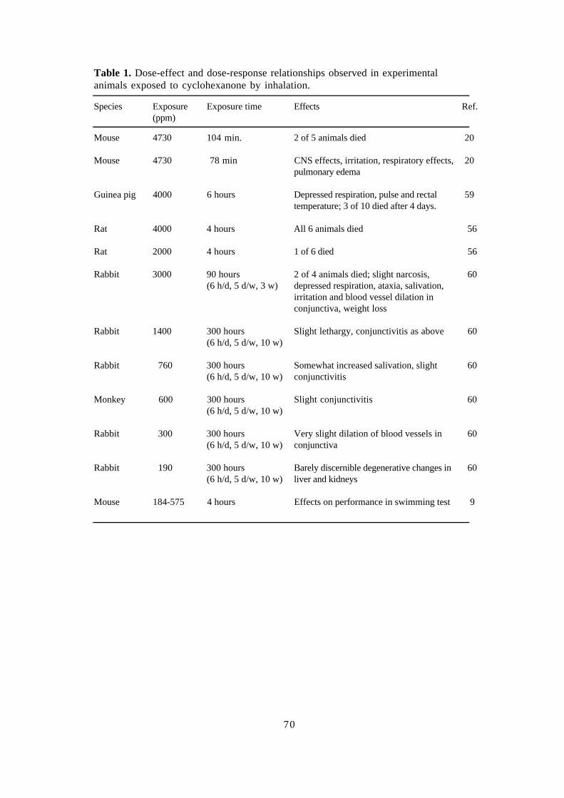
70
Table 1. Dose-effect and dose-response relationships observed in experimentalanimals exposed to cyclohexanone by inhalation.
Species Exposure(ppm)
Exposure time Effects Ref.
Mouse 4730 104 min. 2 of 5 animals died 20
Mouse 4730 78 min CNS effects, irritation, respiratory effects,pulmonary edema
20
Guinea pig 4000 6 hours Depressed respiration, pulse and rectaltemperature; 3 of 10 died after 4 days.
59
Rat 4000 4 hours All 6 animals died 56
Rat 2000 4 hours 1 of 6 died 56
Rabbit 3000 90 hours(6 h/d, 5 d/w, 3 w)
2 of 4 animals died; slight narcosis,depressed respiration, ataxia, salivation,irritation and blood vessel dilation inconjunctiva, weight loss
60
Rabbit 1400 300 hours(6 h/d, 5 d/w, 10 w)
Slight lethargy, conjunctivitis as above 60
Rabbit 760 300 hours(6 h/d, 5 d/w, 10 w)
Somewhat increased salivation, slightconjunctivitis
60
Monkey 600 300 hours(6 h/d, 5 d/w, 10 w)
Slight conjunctivitis 60
Rabbit 300 300 hours(6 h/d, 5 d/w, 10 w)
Very slight dilation of blood vessels inconjunctiva
60
Rabbit 190 300 hours(6 h/d, 5 d/w, 10 w)
Barely discernible degenerative changes inliver and kidneys
60
Mouse 184-575 4 hours Effects on performance in swimming test 9

71
Table 2. Dose-effect and dose-response relationships observed in experimentalanimals exposed to cyclohexanone by means other than inhalation.
Species Dose(mg/kg/d)
Administrationmethod
Effects Ref.
Rabbit 1600-1900 oral Narcosis 61
Mouse 2200 oral 6/28 pregnant mice died. Pups had lowbirth weights
55
Mouse 800 oral No effect on locomotor activity of pups 15
Mouse 800 oral, days 8-12of gestation
No effect on litter size or weight of pups 7
Rat 200 i.p.5 d/w, 13 weeks
No effect on neural conductivity inperipheral nerves
46
Dog 284 i.v. 18-21 days6% solution75 ml/minute
CNS effects that increased with concentra-tion and rate of administration; respiratoryeffects, elevated liver and adrenal weight,metabolic acidosis
31
Dog 284 i.v. 18-21 days0.75% solution5 ml/minute
Lethargy, dilated pupils 31
Rat 0, 50, 100 i.v.28 days
No histopathological changes in brain,no effects on clinical/chemical parameters
17
Rat 1, 10, 25 i.v., 18 days No effects on hematology, chemistry,histopathology or organ weights
16
Rabbit 12.4-99% epidermal Concentration-dependent skin irritation,from mild to very severe
20
Rabbit 5-40% instillation in eyes Concentration-dependent irritation, mildto severe
20
Rabbit 2.5% instillation in eyes No eye irritation 20
Conclusions
The critical effect of occupational exposure to cyclohexanone is judged to be itseffect on the nervous system. Occupationally exposed persons (40 – 92 ppm)report symptoms including CNS effects and irritation. Throat irritation wasobserved in volunteers exposed to 50 ppm for a few minutes.

72
References
1. Alarie Y, Schaper M, Nielsen G D, Abraham M H. Structure-activity relationships ofvolatile organic chemicals as sensory irritants. Arch Toxicol 1998;72:125-140.
2. Amoore J E, Hautala E. Odor as an aid to chemical safety: Odor thresholds compared withthreshold limit values and volatilities for 214 industrial chemicals in air and water dilution.J Appl Toxicol 1983;3:272-289.
3. Andersson B. Thermal degradation of weldable poly(vinyl chloride) samples at lowtemperature. J Chrom 1988;445:353-361.
4. Anonymous. Scientific Basis for Swedish Occupational Standards 3. Cyclohexanone. Arbeteoch Hälsa 1982;24:75-79.
5. Brondeau M T, Ban M, Bonnet P, Guenier J P, de Ceaurriz J. Acetone compared to otherketones in modifying the hepatotoxicity of inhaled 1,2-dichlorobenzene in rats and mice.Toxicol Lett 1989;49:69-78.
6. Bruze M, Boman A, Bergqvist-Karlsson A, Björkner B, Wahlberg J E, Voog E. Contactallergy to a cyclohexanone resin in humans and guinea pigs. Contact Derm 1988;18:46-49.
7. Chernoff N, Kavlock R J. A teratology test system which utilizes postnatal growth andviability in the mouse. Environ Sci Res 1983;27:417-427.
8. Collin J P. Effet cytogénétique du cyclamate de soude, de la cyclohéxanone et ducyclohéxanol. Diabéte 1971;19:215-221.
9. de Ceaurriz J, Desiles J P, Bonnet P, Marignac B, Muller J, Guenier J P. Concentration-dependent behavioral changes in mice following short-term inhalation exposure to variousindustrial solvents. Toxicol Appl Pharmacol 1983;67:383-389.
10 DFG (Deutsche Forschungsgemeinschaft). Toxikologisch-arbeitsmedizinische Begründungenvon MAK-Werten. Cyclohexanon. Weinheim: VCH-Verlagsgesellschaft 1994:14 pp.
11. Elliot T H, Parke D V, Williams R T. Studies in detoxication. 79. The metabolism ofcyclo[14C]hexane and its derivatives. Biochem J 1959;72:193-200.
12. Flek J, Sedivec V. Identifikace a stanovení metabolitu cyklohexanonu v lidské moci.(Identification and determination of metabolites of cyclohexanone in human urine). PracovLék 1989;41:259-263. (English abstract)
13. Frantík E, Hornychová M, Horváth M. Relative acute neurotoxicity of solvents: Isoeffectiveair concentrations of 48 compounds evaluated in rats and mice. Environ Res 1994;66:173-185.
14. Gondry E. Recherches sur la toxicité de la cyclohexylamine, de la cyclohexanone et ducyclohexanol, métabolites du cyclamate. Eur J Toxicol Environ Hyg 1972;4:227-238.
15. Gray L E, Kavlok R J, Ostby J, Ferrell J, Rogers J, Gray K. An evaluation of figure-eightmaze activity and general behavioral development following prenatal exposure to fortychemicals: Effects of cytosine arabinoside, dinocap, nitrofen, and vitamin A. Neurotoxicol1986;7:449-462.
16. Greener Y, Gillies B, Wienckowski D, Schmitt D, Woods E, Youkilis E. Assessment of thesafety of chemicals administered intravenously in the neonatal rat. Teratol 1987;35:187-194.
17. Greener Y, Martis L, Indacochea-Redmond N. Assessment of the toxicity of cyclohexanoneadministered intravenously to Wistar and Gunn rats. J Toxicol Environ Health 1982;10:385-396.
18. Greener Y, Youkilis E. Assessment of the cataractogenic potential of cyclohexanone inguinea pigs and rabbits. Fundam Appl Toxicol 1984;4:1055-1066.
19. Griggs J H, Weller E M, Palmisano P A, Niedermeier W. The effect of noxious vapors onembryonic chick development. Ala J Med Sci 1971;8:342-345.
20. Gupta P K, Lawrence W H, Turner J E, Autian J. Toxicological aspects of cyclohexanone.Toxicol Appl Pharmacol 1979;49:525-533.
73
21. Hall I H, Carlson G L, Abernethy G S, Piantadosi C. Cycloalkanones. 4. Antifertilityactivity. J Med Chem 1974;17:1253-1257.
22. Haworth S, Lawlor T, Mortelmans K, Speck W, Zeiger E. Salmonella mutagenicity testresults for 250 chemicals. Environ Mutagen 1983;5 Suppl 1:3-38.
23. Henkel W, Rublack H. Kombinierte Wirkung von Cyklohexanon und Oxim – Bewertung derKombinationswirkung. Z Ges Hyg 1976;22:234-235.
24. Holland K D, Naritoku D K, McKeon A C, Ferrendelli J A, Covey D F. Convulsant andanticonvulsant cyclopentanones and cyclohexanones. Mol Pharmacol 1990;37:98-103.
25. IARC. Cyclohexanone. Monographs on the Evaluation of Carcinogenic Risks to Humans,Vol 47. Lyon: International Agency for Research on Cancer 1989;47:157-169.
26. Imbriani M, Ghittori S, Pezzagno G, Capodaglio E. Urine/air partition coefficients for someindustrially important substances. G Ital Med Lav 1985;7:133-140.
27. Jacobsen M, Baelum J, Bonde J P. Temporal epileptic seizures and occupational exposure tosolvents. Occup Environ Med 1994;51:429-430.
28. James S P, Waring R H. The metabolism of alicyclic ketones in the rabbit and rat.Xenobiotica 1971;1:573-580.
29. Kennedy G, Graepel J. Acute toxicity in the rat following either oral or inhalation exposure.Toxicol Lett 1991;56:317-326.
30. Khalfi F, Dine T, Luyckx M et al. Determination of cyclohexamone after derivatization with2,4-dinitrophenyl hydrazine in intravenous solutions stored in PVC bags by highperformance liquid chromatography. Biomed Chromatogr 1998;12:69-72.
31. Koeferl M, Miller T, Fisher J, Martis L, Garvin P, Dorner J. Influence of concentration andrate of intravenous administration on the toxicity of cyclohexanone in beagle dogs. ToxicolAppl Pharmacol 1981;59:215-229.
32. Lederer J, Collin J P, Pottier-Arnould A-M, Gondry E. L’action cytogénétique et tératogènedu cyclamate et de ses métabolites. Thérapeutique 1971;47:357-363.
33. Lijinsky W, Kovatch R. Chronic toxicity study of cyclohexanone in rats and mice. JNCI1986;77:941-949.
34. Martis L, Tolhurst T, Koeferl M, Miller T, Darby T. Disposition kinetics of cyclohexanonein beagle dogs. Toxicol Appl Pharmacol 1980;55:545-553.
35. Massoud A A, Ali A M M, Shafik H M. Mutagenic-carcinogenic effects of cyclohexanone inBacillus subtilis and Salmonella typhimurium. Egypt J Microbiol 1983;18:213-224.
36. McGregor D, Brown A, Cattanach P et al. Responses of the L5178Y tk+/tk- mouselymphoma cell forward mutation assay: III. 72 coded chemicals. Environ Mol Mutagen1988;12:85-154.
37. Milanovic L, Spilich G, Vucinic G, Knezevic S, Ribaric B, Mubrin Z. Effects ofoccupational exposure to organic solvents upon cognitive performance. Neurotoxicol Teratol1990;12:657-660.
38. Mills G. Walker V. Urinary excretion of cyclohexanediol, a metabolite of the solvent cyclo-hexanone, by infants in a special care unit. Clin Chem 1990;36:870-874.
39. Mitran E, Callender T, Orha B, Dragnea P, Botezatu G. Neurotoxicity associated withoccupational exposure to acetone, methyl ethyl ketone and cyclohexanone. Environ Res1997;73:181-188.
40. Mráz J, Gálová E, Nohová H. Uptake, metabolism and elimination of cyclohexanone inhumans. Int Arch Occup Environ Health 1994;66:203-208.
41. Muttray A, Konietzko J. Störungen des Riechvermögens durch und für Arbeitsstoffe.Arbeitsmed Sozialmed Umweltmed 1994;29:409-413.
42. Nelson K W, Ege J F, Ross M, Woodman L E, Silverman L. Sensory response to certainindustrial solvent vapors. J Ind Hyg Toxicol 1943;25:282-285.

74
43. Ong C, Chia S, Phoon W, Tan K, Kok P. Monitoring of exposure to cyclohexanonethrough the analysis of breath and urine. Scand J Work Environ Health 1991;17:430-435.
44. Ong C N, Sia G L, Chia S E. Determination of cyclohexanol in urine and its use in environ-mental monitoring of cyclohexanone exposure. J Anal Toxicol 1991;15:13-16.
45. Panhuber H, Mackay-Sim A, Laing D G. Prolonged odor exposure causes severe cellshrinkage in the adult rat olfactory bulb. Dev Brain Res 1987;31:307-311.
46. Perbellini L, De Grandis D, Semenzato F, Bongiovanni L G. Studio sperimentale sullaneurotossicitá del cicloesanolo e del cicloesanone. Med Lavoro 1981;2:102-106.
47. Perocco P, Bolognesi S, Alberghini W. Toxic activity of seventeen industrial solvents andhalogenated compounds on human lymphocytes cultured in vitro. Toxicol Lett 1983;16:69-75.
48. Rehn B, Pahuber H, Laing D G, Breipohl W. Spine density on olfactory granule celldendrites is reduced in rats reared in a restricted olfactory environment. Dev Brain Res1988;40:143-147.
49. Rengstorff R H, Petrali J P, Sim V M. Cataracts induced in guinea pigs by acetone,cyclohexanone, and dimethyl sulfoxide. Am J Optom Arch Am Acad Optom 1972;49:308-319.
50. Sakata M, Kikuchi J, Haga M. Disposition of acetone, methyl ethyl ketone andcyclohexanone in acute poisoning. Clin Toxicol 1989;27:67-77.
51. Sakata M, Take J, Watanabe T, Sakata K, Wada K, Haga M. Metabolic interaction of ethanoland cyclohexanone in rabbits. J Toxicol Environ Health 1993;38:33-42.
52. Samimi B, Harris S, de Peyster A. Fetal effects of inhalation exposure to cyclohexanonevapor in pregnant rats. Toxicol Ind Health 1989;5:1035-1043.
53. Sanmartín O, de la Cuadra J. Occupational contact dermatitis from cyclohexanone as a PVCadhesive. Contact Derm 1992;27:189-190.
54. Savolainen H. Nordic Expert Group for Documentation of Occupational Exposure Limits.63. Cyklohexanon och cyklopentanon. Arbete och Hälsa 1985;42:1-33. (English abstract)
55. Seidenberg J M, Anderson D G, Becker R A. Validation of an in vivo developmental toxicityscreen in the mouse. Teratogen Carcinogen Mutagen 1986;6:361-374.
56. Smyth H F, Carpenter C P, Weil C S, Pozzani U C, Striegel J A, Nycum J S. Range-finding toxicity data: List VII. Am Ind Hyg Ass J 1969;30:470-476.
57. Snell R. Capillary GC analysis of compounds leached into parenteral solutions packaged inplastic bags. J Chrom Sci 1989;27:524-528.
58. Snell R. Gas chromatographic determination of cyclohexanone leached from hemodialysistubing. J AOAC Int 1993;76:1127-1132.
59. Specht H, Miller J W, Valaer P J, Sayers R R. Acute response of guinea pigs to theinhalation of ketone vapors. National Institute of Public Health Bulletin No 176. USGovernment Printing Office, Washington, DC 1940:1-66.
60. Treon J F, Crutchfield W E, Kitzmiller K V. The physiological response of animals tocyclohexane, methylcyclohexane, and certain derivatives of these compounds. II. Inhalation. JInd Hyg Toxicol 1943;25:323-347.
61. Treon J F, Crutchfield W E, Kitzmiller K V. The physiological response of rabbits to cyclo-hexane, methylcyclohexane, and certain derivatives of these compounds. I. Oraladministration and cutaneous application. J Ind Hyg Toxicol 1943;25:199-214.
62. Ulsaker G A, Korsnes R M. Determination of cyclohexanone in intravenous solutions storedin PVC bags by gas chromatography. Analyst 1977;102:882-883.
63. Zuckerman G B, Lam S C, Santos S M. Rhabdomyolysis following oral ingestion of thehydrocarbon cyclohexanone in an adolescent. J Environ Pathol Toxicol Oncol 1998;17:11-15.

75
Consensus Report for Some LactateEsters
June 2, 1999
This report is based primarily on a criteria document compiled jointly by the NordicExpert Group and the Dutch Expert Committee (9). That document, in turn, isbased on two recently published survey articles (5, 6). The Criteria Grouppublished an earlier consensus report for lactate esters in 1995 (8). The presentreport takes up the following lactate esters: methyl lactate, ethyl lactate, isopropyllactate, isobutyl lactate, n-butyl lactate, 2-ethylhexyl lactate, myristyl lactate andcetyl lactate. Some information on a few of the other lactate esters is given in thecriteria document (9).
Lactates occur in two enantiomorphous (mirror image) forms, D (dextro) and L(levo). The forms often occur together: the DL (dextrolevo) form. 2-Ethylhexyllactate also has diastereoisomeric forms.
Chemical and physical data. Uses
methyl lactateCAS No.: 547-64-8 (DL form)
27871-49-4 (L form)Formula: C4H8O3
CH3CH(OH)COOCH3
Molecular weight: 104.1Melting point: - 66 °CBoiling point: 144 °CFlash point: 57 °CDensity: 1.092 g/ml (20 °C)Vapor pressure: 0.34 kPa (20 °C)
23 kPa (100 °C)Saturation concentration: 3302 ppm (20 °C)Distribution coefficient: log Po/w = - 0.53Conversion factors: 1 ppm = 4.3 mg/m3 (20 °C, 101.3 kPa)
1 mg/m3 = 0.23 ppm (20 °C, 101.3 kPa)
Methyl lactate is a colorless, transparent liquid. It mixes with water at roomtemperature, and is also soluble in alcohol and ether. It is used as a solvent forcellulose acetate.

76
ethyl lactateCAS No.: 97-64-3 (DL form)
687-47-8 (L form)Formula: C5H10O3
CH3CH(OH)COOCH2CH3
Molecular weight: 118.1Melting point: - 25 °CBoiling point: 153 °CFlash point: 61 °CDensity: 1.033 g/ml (20 °C)Vapor pressure: 0.22 kPa (20 °C)
17 kPa (100 °C)Distribution coefficient: log Po/w = 0.06Conversion factors: 1 ppm = 4.9 mg/m3 (20 °C, 101.3 kPa)
1 mg/m3 = 0.20 ppm (20 °C, 101.3 kPa)
Ethyl lactate at room temperature is a colorless liquid with a mild, characteristicodor. The reported odor threshold is 0.89 mg/m3, and the odor becomes irritating at65 mg/m3 (5). Ethyl lactate mixes with water, alcohols, ketones, esters, hydro-carbons and ethers. It occurs naturally, most notably in several different kinds offruit. It is used as a solvent for nitrocellulose, cellulose acetate and many celluloseethers. It is an ingredient in enamels, paints, polishes and various cosmeticproducts. It is used as a replacement for trichloroethylene in de-greasing andcleaning (9). An air concentration of 0.6 ppm was measured at a Swedish companythat used ethyl lactate for de-greasing metal. There were peaks of 10 ppm aroundsome operations, and an 8-hour average was calculated to be 4.2 ppm (4).
isopropyl lactateCAS No.: 617-51-6 (DL form)
63697-00-7 (L form)Formula: C6H12O3
CH3CH(OH)COOCH(CH3)2
Molecular weight: 132.2Boiling point: 157 °CFlash point: 60 °CDensity: 0.991 g/ml (20 °C)Vapor pressure: 0.17 kPa (20 °C)
15 kPa (100 °C)Distribution coefficient: log Po/w = 0.39Conversion factors: 1 ppm = 5.5 mg/m3 (20 °C, 101.3 kPa)
1 mg/m3 = 0.18 ppm (20 °C, 101.3 kPa)
Isopropyl lactate is soluble in water, alcohol, ether and benzene.

77
isobutyl lactateCAS No.: 585-24-0 (DL form)
702-84-0 (L form)Formula: C7H14O3
CH3CH(OH)COOCH2CH(CH3)2
Molecular weight: 146.2Boiling point: 182 °CFlash point: 76 °CDensity: 0.979 g/ml (20 °C)Vapor pressure: 0.05 kPa (20 °C)Distribution coefficient: log Po/w = 1.10Conversion factors: 1 ppm = 6.1 mg/m3 (20 °C, 101.3 kPa)
1 mg/m3 = 0.165 ppm (20 °C, 101.3 kPa)
Isobutyl lactate is soluble in water: 5.1 g/100 ml at 20 °C.
n-butyl lactateCAS No.: 138-22-7 (DL form)
34451-19-9 (L form)Formula: C7H14O3
CH3CH(OH)COO(CH2)3CH3
Molecular weight: 146.2Melting point: - 43 °CBoiling point: 187 °CFlash point: 79 °CDensity: 0.984 g/ml (20 °C)Vapor pressure: 0.03 kPa (20 °C)
4.7 kPa (100 °C)Distribution coefficient: log Po/w = 1.10Conversion factors: 1 ppm = 6.1 mg/m3 (20 °C, 101.3 kPa)
1 mg/m3 = 0.165 ppm (20 °C, 101.3 kPa)
At room temperature, n-butyl lactate is a watery liquid with a mild odor. It mixeswith water (4.5 g/100 ml), alcohol, ether and many solvents. In acids and alkalis itis hydrolyzed to lactic acid and butyl alcohol. The odor threshold is reported in onesource (5) to be 0.095 mg/m3, and in another (2) to be 7 ppm (42.6 mg/m3). Thereported irritation threshold for the odor is 9 mg/m3. Butyl lactate has been used as asolvent for synthetic polymers, enamels, paints etc. It occurs in low concentrations(< 0.03%) in cosmetic products (9).
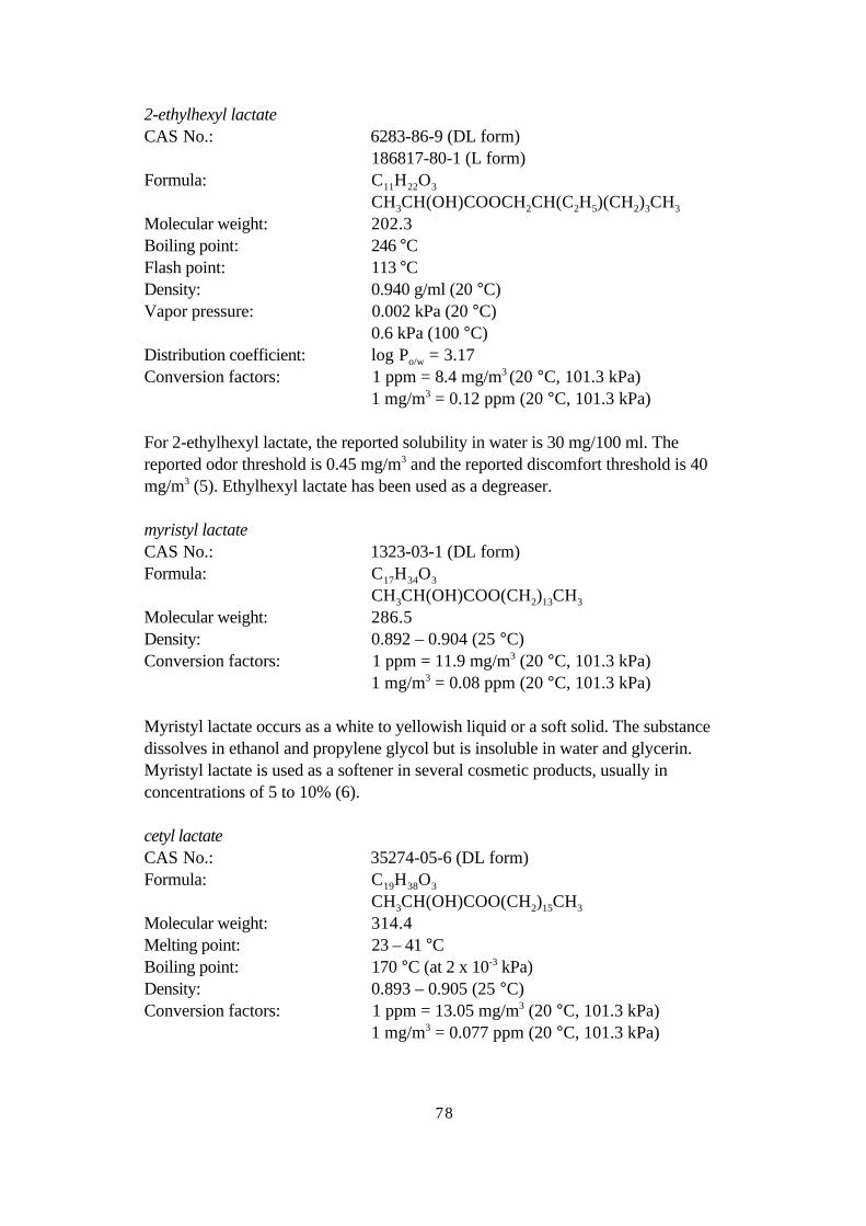
78
2-ethylhexyl lactateCAS No.: 6283-86-9 (DL form)
186817-80-1 (L form)Formula: C11H22O3
CH3CH(OH)COOCH2CH(C2H5)(CH2)3CH3
Molecular weight: 202.3Boiling point: 246 °CFlash point: 113 °CDensity: 0.940 g/ml (20 °C)Vapor pressure: 0.002 kPa (20 °C)
0.6 kPa (100 °C)Distribution coefficient: log Po/w = 3.17Conversion factors: 1 ppm = 8.4 mg/m3 (20 °C, 101.3 kPa)
1 mg/m3 = 0.12 ppm (20 °C, 101.3 kPa)
For 2-ethylhexyl lactate, the reported solubility in water is 30 mg/100 ml. Thereported odor threshold is 0.45 mg/m3 and the reported discomfort threshold is 40mg/m3 (5). Ethylhexyl lactate has been used as a degreaser.
myristyl lactateCAS No.: 1323-03-1 (DL form)Formula: C17H34O3
CH3CH(OH)COO(CH2)13CH3
Molecular weight: 286.5Density: 0.892 – 0.904 (25 °C)Conversion factors: 1 ppm = 11.9 mg/m3 (20 °C, 101.3 kPa)
1 mg/m3 = 0.08 ppm (20 °C, 101.3 kPa)
Myristyl lactate occurs as a white to yellowish liquid or a soft solid. The substancedissolves in ethanol and propylene glycol but is insoluble in water and glycerin.Myristyl lactate is used as a softener in several cosmetic products, usually inconcentrations of 5 to 10% (6).
cetyl lactateCAS No.: 35274-05-6 (DL form)Formula: C19H38O3
CH3CH(OH)COO(CH2)15CH3
Molecular weight: 314.4Melting point: 23 – 41 °CBoiling point: 170 °C (at 2 x 10⁻³ kPa)Density: 0.893 – 0.905 (25 °C)Conversion factors: 1 ppm = 13.05 mg/m3 (20 °C, 101.3 kPa)
1 mg/m3 = 0.077 ppm (20 °C, 101.3 kPa)
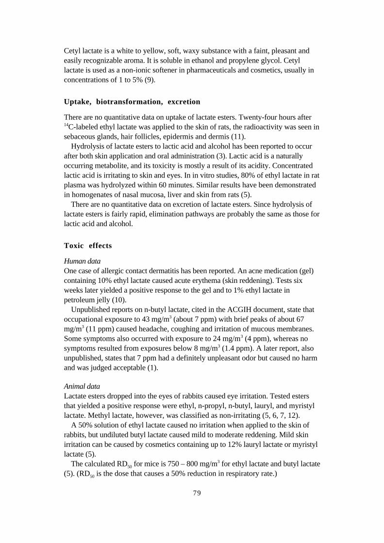
79
Cetyl lactate is a white to yellow, soft, waxy substance with a faint, pleasant andeasily recognizable aroma. It is soluble in ethanol and propylene glycol. Cetyllactate is used as a non-ionic softener in pharmaceuticals and cosmetics, usually inconcentrations of 1 to 5% (9).
Uptake, biotransformation, excretion
There are no quantitative data on uptake of lactate esters. Twenty-four hours after14C-labeled ethyl lactate was applied to the skin of rats, the radioactivity was seen insebaceous glands, hair follicles, epidermis and dermis (11).
Hydrolysis of lactate esters to lactic acid and alcohol has been reported to occurafter both skin application and oral administration (3). Lactic acid is a naturallyoccurring metabolite, and its toxicity is mostly a result of its acidity. Concentratedlactic acid is irritating to skin and eyes. In in vitro studies, 80% of ethyl lactate in ratplasma was hydrolyzed within 60 minutes. Similar results have been demonstratedin homogenates of nasal mucosa, liver and skin from rats (5).
There are no quantitative data on excretion of lactate esters. Since hydrolysis oflactate esters is fairly rapid, elimination pathways are probably the same as those forlactic acid and alcohol.
Toxic effects
Human dataOne case of allergic contact dermatitis has been reported. An acne medication (gel)containing 10% ethyl lactate caused acute erythema (skin reddening). Tests sixweeks later yielded a positive response to the gel and to 1% ethyl lactate inpetroleum jelly (10).
Unpublished reports on n-butyl lactate, cited in the ACGIH document, state thatoccupational exposure to 43 mg/m3 (about 7 ppm) with brief peaks of about 67mg/m3 (11 ppm) caused headache, coughing and irritation of mucous membranes.Some symptoms also occurred with exposure to 24 mg/m3 (4 ppm), whereas nosymptoms resulted from exposures below 8 mg/m3 (1.4 ppm). A later report, alsounpublished, states that 7 ppm had a definitely unpleasant odor but caused no harmand was judged acceptable (1).
Animal dataLactate esters dropped into the eyes of rabbits caused eye irritation. Tested estersthat yielded a positive response were ethyl, n-propyl, n-butyl, lauryl, and myristyllactate. Methyl lactate, however, was classified as non-irritating (5, 6, 7, 12).
A 50% solution of ethyl lactate caused no irritation when applied to the skin ofrabbits, but undiluted butyl lactate caused mild to moderate reddening. Mild skinirritation can be caused by cosmetics containing up to 12% lauryl lactate or myristyllactate (5).
The calculated RD50 for mice is 750 – 800 mg/m3 for ethyl lactate and butyl lactate(5). (RD50 is the dose that causes a 50% reduction in respiratory rate.)

80
Cosmetics containing small amounts of lauryl lactate or cetyl lactate were testedon the skin of guinea pigs (the Magnusson-Kligman test). Both substances werejudged to be non-sensitizing (6).
In inhalation studies, groups of rats (both sexes) were exposed 6 hours/day,5 days/week for 28 days. Exposure levels for ethyl lactate were 0, 25, 75, 150,200, 600 or 2500 mg/m3. In the two highest dose groups there were degenerativechanges in olfactory epithelia with hyperplasia in goblet cells. Similar effects wereobserved at high doses when the animals were exposed to isobutyl lactate or n-butyllactate. The NOAEL for these three lactates is 200 mg/m3. Further information ispresented in Table 1 (9).
At the same laboratory, rats exposed by inhalation to 2-ethylhexyl lactate showedthe same effects even at 75 mg/m3, the lowest tested dose. The effects were thesame whether the ester was in gas or aerosol phase. See also Table 1 (9).
Groups of rats (both sexes) were given myristyl lactate in oral doses of 0, 0.5,2.5 or 5.0 mg/kg body weight, 5 days/week for 13 weeks. Elevated relative liverweights and enlargement and thickening of the walls of the stomach and duodenumwere seen in rats in the two higher dose groups. Histological examination revealeddiffuse hyperplasia in the mucous membranes of the duodenum. These changeswere not observed at the lowest dose (6).
Mutagenicity, carcinogenicity, teratogenicity
Ethyl lactate has been tested on several different strains of Salmonella typhimurium,both with and without metabolizing systems. No mutagenic activity was observed.No mutagenic activity was observed when 2-ethylhexyl lactate was tested onSalmonella and E. coli (5).
No carcinogenicity studies were found on any of these lactate esters.Ethyl lactate (doses of 0, 517, 1551 or 3619 mg/kg body weight/day) was
applied to the skin of rats on days 6 – 15 of gestation. There was slight reddening atthe site of application, but no clinical effects were seen in the mothers and therewere no observed effects on the young (5).
Pregnant rats were exposed to an aerosol of 2-ethylhexyl lactate (0, 200, or 600mg/m3) 6 hours/day on days 6 to 15 of gestation. Animals in the high-dose grouphad lower food intake, but showed no toxic effects. Retarded ossification was notedin pups in both dose groups, but was attributed to stress rather than the toxicity ofthe exposure (5).
Dose-response/dose effect relationships
For most of the lactate esters, there are no data from which to derive a dose-effect ordose-response relationship. Data on human exposures are particularly sparse.
Data from inhalation exposure studies with rats are presented in Table 1.Myristyl lactate given orally to rats increased liver weights at a daily dose of 2.5
mg/kg body weight or higher. The NOAEL in this study was 0.5 mg/kg bodyweight (6).
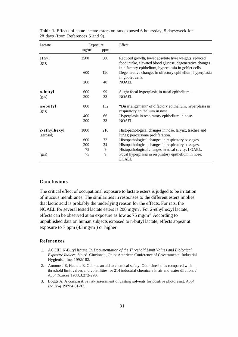
81
Table 1. Effects of some lactate esters on rats exposed 6 hours/day, 5 days/week for28 days (from References 5 and 9).
Lactate Exposure Effect mg/m3 ppm
ethyl(gas)
2500 500 Reduced growth, lower absolute liver weights, reducedfood intake, elevated blood glucose, degenerative changesin olfactory epithelium, hyperplasia in goblet cells.
600 120 Degenerative changes in olfactory epithelium, hyperplasiain goblet cells.
200 40 NOAEL
n-butyl 600 99 Slight focal hyperplasia in nasal epithelium.(gas) 200 33 NOAEL
i sobutyl(gas)
800 132 “Disarrangement” of olfactory epithelium, hyperplasia inrespiratory epithelium in nose.
400 66 Hyperplasia in respiratory epithelium in nose.200 33 NOAEL
2-ethylhexyl(aerosol)
1800 216 Histopathological changes in nose, larynx, trachea andlungs; peroxisome proliferation.
600 72 Histopathological changes in respiratory passages.200 24 Histopathological changes in respiratory passages.75 9 Histopathological changes in nasal cavity; LOAEL.
(gas) 75 9 Focal hyperplasia in respiratory epithelium in nose;LOAEL
Conclusions
The critical effect of occupational exposure to lactate esters is judged to be irritationof mucous membranes. The similarities in responses to the different esters impliesthat lactic acid is probably the underlying reason for the effects. For rats, theNOAEL for several tested lactate esters is 200 mg/m3. For 2-ethylhexyl lactate,effects can be observed at an exposure as low as 75 mg/m3. According tounpublished data on human subjects exposed to n-butyl lactate, effects appear atexposure to 7 ppm (43 mg/m3) or higher.
References
1. ACGIH. N-Butyl lactate. In Documentation of the Threshold Limit Values and BiologicalExposure Indices, 6th ed. Cincinnati, Ohio: American Conference of Governmental IndustrialHygienists Inc. 1992:182.
2. Amoore J E, Hautala E. Odor as an aid to chemical safety: Odor thresholds compared withthreshold limit values and volatilities for 214 industrial chemicals in air and water dilution. JAppl Toxicol 1983;3:272-290.
3. Boggs A. A comparative risk assessment of casting solvents for positive photoresist. ApplInd Hyg 1989;4:81-87.

82
4. Carlsson H, Andersson Sköld Y, Janhäll S, Solyom P, Ancker K. Rengöring med laktater.Miljöteknisk utvärdering. IVL Rapport B 1160. Stockholm: The Swedish Institute for Waterand Air Pollution Research: 1995. (In Swedish)
5. Clary J J, Feron V J, van Velthuijsen J A. Safety assessment of lactate esters. Regul ToxicolPharmacol 1998;27:88-97.
6. Cosmetic Ingredient Review Panel. Final report on the safety assessment of glycolic acid,ammonium, calcium, potassium, and sodium glycolates, methyl, ethyl, propyl, and butylglycolates, and lactic acid, ammonium, calcium, potassium, sodium, and TEA-lactates,methyl, ethyl, isopropyl, and buytl lactates, and lauryl, myristyl, and cetyl lactates. Int JToxicology 1998;17 suppl 1:1-241.
7. Latven A R, Molitor H. Comparison of the toxic, hypnotic and irritating properties of eightorganic solvents. J Pharm Exp Ther 1939;65:89-94.
8. Lundberg P, ed. Scientific Basis for Swedish Occupational Standards. 16. Lactates. Arbete ochHälsa 1995;19:68-73.
9. Lundberg P. DECOS and SCG basis for an occupational standard. Lactate esters. Arbete ochHälsa 1999;9:1-21.
10. Marot L, Grosshans E. Allergic contact dermatitis to ethyl lactate. Contact Dermatitis1987;17:45-46.
11. Prottey C, George D, Leech R W et al. The mode of action of ethyl lactate as a treatment foracne. Br J Dermatol 1984;110:475-485.
12. Sanderson D M. A note on glycerol formal as a solvent in toxicity testing. J PharmPharmacol 1959;11:150-155.
83
Consensus Report for Ethylene GlycolMethylether and Ethylene GlycolMethylether Acetate
June 2, 1999
This report is based on a criteria document published in Arbete och Hälsa (22).
Chemical and physical characteristics. Uses
Ethylene glycol methylether (EGME)CAS No.: 109-86-4Synonyms: glycol methylether, 2-methoxyethanol,
methyl glycolFormula: CH3–O–CH2–CH2–OHMolecular weight: 76.09Density: 0.96 (20 °C)Boiling point: 124 °CMelting point: - 85.1 °CVapor pressure: 1.3 kPa (9.7 mm Hg) (20 °C)Evaporation rate: 0.5 (butyl acetate = 1)Saturation concentration: 12,800 ppm (25 °C)Relative density: 2.6 (air = 1)Conversion factors: 1 ppm = 3.11 mg/m3 (20 °C)
1 mg/m3 = 0.322 ppm (20 °C)
Ethylene glycol methylether acetate (EGMEA)CAS No.: 110-49-6Synonyms: ethylene glycol monomethylether acetate
2-methoxyethyl acetate, methyl glycol acetateFormula: CH3–O–CH2–CH2–O–CO–CH3
Molecular weight: 118.13Density: 1.005 (20 °C)Boiling point: 145 °CMelting point: - 65 °CFlash point: 55.6 °C (open cup)Vapor pressure: 0.27 – 0.50 kPa (2.0 – 3.7 mm Hg) (20 °C)Evaporation rate: 0.3 (butyl acetate = 1)Saturation concentration: 3100 – 6000 ppm (25 °C)Relative density: 4.07 (air = 1)Conversion factors: 1 ppm = 4.90 mg/m3 (20 °C)
1 mg/m3 = 0.200 ppm (20 °C)
84
EGME and EGMEA at room temperature are flammable, volatile, clear liquids witha weak, sweetish odor and bitter taste. Both substances dissolve readily andcompletely in water as well as polar and non-polar solvents.
EGME is produced by a reaction between methanol and ethylene oxide. EGMEAis produced from EGME by conventional estrification. Known impurities in EGMEare reported to be < 0.1% methanol, < 0.1% diethylene glycol methylether and< 0.02% ethylene glycol (22).
EGME and EGMEA do not occur in nature. World-wide reported uses for thetwo glycol ethers are in paints and enamels; printer’s ink; plastic packaging forfoodstuffs; pigments for silk-screen printing; photographic and photolithographicprocesses (including the production of offset plates); CDs, circuit boards andintegrated circuits; cleaners for household and industrial use; and antifreeze inhydraulic fluids and airplane fuel. In 1993 there were 23 Swedish chemicalproducts containing EGME, and total annual use was about 260 tons of the puresubstance. EGMEA was not listed in the product register. EGME was usedprimarily as a solvent, but it was also an ingredient in paints and enamels. In 1997there were 27 Swedish products containing EGME, and annual use was 19 tons,most of which was used as photoresist in the telecommunications industry.EGMEA was a listed ingredient in 3 products, with an annual use of less than0.1 ton. In 1994 EGME and EGMEA were classified by the EU as toxic toreproduction and their use in consumer products was prohibited (22).
Reported average exposure levels are in the range <0.1 to 23 mg/m3 for EGME,and from <0.1 to 143 mg/m3 for EGMEA. Exposure has been reported from semi-conductor and circuit board manufacture, printing, painting (especially automobileand ship painting), furniture finishing, paint production and automobile repair (22).No data were found on exposures in Swedish workplaces.
Uptake, biotransformation, excretion
As suggested by the chemical structure and the ready solubility of EGME andEGMEA, both substances are efficiently absorbed via all paths of uptake andrapidly distributed throughout the body. Uptake via respiratory passages has beenmeasured at 76% of the amount inhaled (22).
Uptake of EGME by frozen and thawed human epidermis in vitro was2.8 mg/cm2/hour (12). An average absorption rate of 2.9 mg/cm2/hour, with largeinter-individual variations, was measured for liquid EGME in an in vivo study withvolunteers. Exposure of hands and lower arms to EGME in liquid form was calcu-lated to yield an absorption rate 100 times that of exposure to 5 ppm in the air. Theauthors also calculated that, with whole-body exposure to EGME vapor, 55% oftotal uptake occurs via the skin (24).
EGME is distributed fairly evenly between blood and other tissues, with theexception of low solubility in adipose tissue. Methoxyacetic acid (MAA) also hasrelatively even distribution in body tissues (22).

85
EGMEA is efficiently hydrolyzed to EGME by the carboxylesterases in nasalmucosa, liver, kidneys, lungs and blood. The most important metabolic pathwayfor EGME is oxidation via methoxyacetaldehyde (MALD) to MAA. This meta-bolism can be inhibited by ethanol, and the importance of alcohol dehydrogenase isillustrated by the fact that metabolism of EGME is almost completely suppressed inrats that have been pre-treated with pyrazol. When men were exposed to 5 ppmEGME for 4 hours (resting), an estimated 86% of the inhaled amount of EGMEwas excreted in urine as MAA. The reported half time for MAA in human urine is77 hours. The half time for MAA in serum and plasma has been reported to beabout 6 hours for mice and 20 hours for monkeys (22).
In addition to MAA, methoxyethyl glucuronide, methoxyethyl sulfate, ethyleneglycol, glycolic acid, glycine, methoxyacetyl glucuronide, methoxyacetyl glycine,methoxycitrate and methoxybutenic acid were identified in nucleomagneticresonance spectrometry (NMR) analysis of urine samples from mice and rats givendoses of 13C -labeled EGME. Simultaneous administration of acetate, anendogenously formed substance and a precursor in the Krebs cycle, increased theproportion of EGME-related metabolites and reduced the proportion of MAA-relatedmetabolites. These results show that ether cleavage can occur, and also that EGMEafter oxidation can form methoxyacetyl-coenzyme A. It has been suggested that this“false substrate” in the Krebs cycle may be related EGME’s toxic effects onreproduction (22).
Mechanism studies
EGME had no effect when incubated with human erythrocytes, whereas 0.5 mM ofMAA increased their osmotic fragility. When human erythrocyte membranes(ghosts) were incubated with MAA or EGME, membrane-bound acetyl-cholinesterase (IC50 = 5.5 mM) and ATPase (IC50 = 1.4 mM) were inhibited byMAA but not by EGME (26).
Simultaneous administration of a number of other substances (formate, acetate,glycine, glucose, serine, sarcosine) involved in the formation of pyridine and purine– which in turn are needed for synthesis of DNA and RNA – reduces or completelyeliminates the malformed sperm and disruption of spermatogenesis caused byEGME in experimental animals (22).
Addition of 10 µM MAA, but not 1 µM, reduced the proliferative capacity of fetalmouse liver cells in vitro, observed as reduced incorporation of tritium-labeledthymidine. No effect on survival of the cells was observed, however (20).
Toxic effects
Animal dataEGME and EGMEA have moderate acute toxicity. The reported LD50 values forEGME range from 0.9 to 3.4 g/kg body weight, depending on species and methodof administration. The reported LC50 for inhalation is 4600 mg/m3 (1480 ppm).
86
Four hours of exposure to 1000 ppm resulted in atrophied sperm in male rats, and625 ppm produced damaged spermatids within 24 hours. The reported LD50 valuesfor EGMEA range from 1.3 to 5.6 g/kg. The reported LD50 for MAA with oraladministration (in water) is 1 to 1.5 g/kg (22).
Short-term exposures via gavage, skin application, feed and inhalation havesimilar effects in several species, including reduced thymus, spleen and testesweights, lower counts of white and red blood cells and platelets, lower hematocrit,hemoglobin levels and bone marrow cellularity, higher numbers of immaturegranulocytes and disturbance of spermatogenesis. Spermatogenesis is disrupted at aparticular phase, the late pachytene, and the effect shows up later in lower spermcounts or aspermia. Toxicity is about the same regardless of the method of exposure– gavage, drinking water, skin application or inhalation (22).
After tests with rabbits, EGME and EGMEA were classed according to EECcriteria as non-irritating to skin, and EGME as non-irritating to eyes (22).
Human dataOlder studies report that repeated occupational exposure to products containingEGME can cause headaches, weakness, dizziness, ataxia, toxic encephalopathy anddampened reflexes (22). Further case reports are summarized in Table 1.
In a cross-sectional study of 65 workers who produced and packaged EGME,measured concentrations in workplace air were 4 to 20 ppm and personal monitorsindicated 5.4 to 8.5 ppm (time-weighted averages). Trends (not significant) tolower leukocyte counts and lower Hb were seen in the 40 exposed workers whenthey were compared with the 25 unexposed workers. Closer study of a sub-groupof 6 exposed and 9 unexposed workers showed tendencies to reduced leukocytecounts, lower hemoglobin, reduced testicle size, lower sperm counts, elevatedlevels of luteinizing hormone (LH) and lower levels of testosterone and follicle-stimulating hormone (FSH) in serum, none of which was statistically significant(7).
Of 73 painters at a shipyard, 10% had anemia and 5% had granulocytopenia,compared with 0% in an unexposed control group. No other hematologic differ-ences between the groups were observed. Measured exposure levels were 0 – 5.6(mean 0.8, median 0.4) ppm for EGME and 0 – 21.5 (mean 2.6) ppm for ethyleneglycol ethylether (EGEE). A review of patient journals revealed that the conditionshad arisen during employment as painters. The authors listed about 60 substancesthat painters at a shipyard might be exposed to, and of these lead, benzene andglycol ethers were identified as potentially harmful to blood-forming organs. Allblood-lead levels were below 40 µg/dl and most of them were below 20 µg/dl. Airmonitoring and product reviews indicated negligible exposure to benzene, and theauthors concluded that the observed hematologic effects could not be explained byexposure to lead or benzene (33).
No reports on skin irritation, eye irritation or sensitization were found in theliterature.

87
Mutagenicity
With the exception of the studies reviewed below, EGME and its metabolite MAAhave been negative in all genotoxicity studies, including Ames tests, in all testedSalmonella strains, both with and without addition of metabolizing systems (for asurvey see Reference 25).
EGME caused mutations in the gpt gene in a cell line from Chinese hamsters, butno mutations in the hprt gene in another cell line. The metabolite MALD was weaklymutagenic in Salmonella (TA97a) and gave rise to an increased number of muta-tions, sister chromatid exchanges and chromosome aberrations in Chinese hamstercells in vitro, in the concentration interval 5 – 40 mM. Chromosome damage fromMALD was also seen in human lymphocytes after 1 hour at 40 mM and after 24hours at 2.5 mM. No chromosome damage was observed in mice that had beengiven up to 1000 mg/kg MALD or up to 2500 mg/kg EGME by gavage (22).
EGME and its metabolites were tested for genotoxicity and epigenetic effects indifferent test systems. Increased numbers of micronuclei and mitotic irregularitieswere seen in vitro at 65 mM EGME, 0.12 mM MALD and 3.2 mM MAA. WithMALD, an elevated frequency of mutations was seen at 1 – 10 mM, of sisterchromatid exchanges and chromosome aberrations at 0.1 – 1 mM, and ofmorphological transformations at 0.1 – 0.3 mM. The authors regard the results asweakly positive for EGME and MAA and clearly positive for MALD (13).
EGMEA was tested with a number of Salmonella strains, both with and withoutmetabolic activation, in two different laboratories. One judged it to be weakly muta-genic, and the other judged it to be possibly mutagenic (36).
Carcinogenicity
There are no reports of animal studies on the carcinogenicity of EGME or EGMEA.In McGregor’s assessment, aside from a few positive results in Ames tests (seeMutagenicity), there is no experimental evidence that either of the substances iscarcinogenic (25).
A review of 198 cases of acute myelotic leukemia (AML) was made in a Frenchcase-control study. Blind estimates of exposure to different types of glycol ethersand potential exposure levels were made by an expert panel. No relationshipbetween AML and glycol ethers was seen (21).
Reproduction toxicity
Animal dataA large number of animal studies have provided a clear and unambiguous picture ofthe toxic effects on reproduction in both sexes. Males given low doses have reducedtestes weights, histological changes in testes and low sperm counts; higher dosescause testicular atrophy and aspermia. The effects are temporary. Females havelower fertility and higher numbers of dead and resorbed fetuses. Young have lowerpost-natal survival rates and higher frequencies of skeletal anomalies, malformed

88
extremities and severe deformities. Effects on fetuses appear at doses too low tohave visible effects on the mothers. At higher doses there is 100% fetal mortality.The degree of fetal damage is extremely sensitive to the time of exposure. Theseeffects have been demonstrated for all administration methods and in severaldifferent species (22). A few of the more recent studies are summarized below.
Rabbits were given EGME in drinking water, 12.5 – 50 mg/kg/day, 5 days/weekfor 12 weeks: there were dose-dependent declines in several parameters of spermquality. The effects were significant at 37.5 and 50 mg/kg, and the most markedeffect was reduced number of sperm per ejaculate. Histologically, spermatogenesis(number of round spermatids per Sertoli’s cell) was somewhat reduced at 25 andseverely disrupted at 37.5 mg/kg. At 50 mg/kg spermatogenesis ceased almostcompletely in 5 of 7 rabbits. No effects were seen on the libido or fertility of therabbits that still had functional sperm production, and no other pathological orhistopathological effects were observed. The authors concluded that spermato-genesis in rabbits is about 10 times more sensitive to EGME than that in rats ormice (4, 16).
EGME given to female rats in doses of 300 mg/kg/day completely eliminated theestrus cycle: inhibition of ovulation, luteal body hypertrophy, permanently elevatedprogesterone levels and permanently low levels of estradiol, follicle-stimulatinghormone (FSH), luteinizing hormone (LH) and prolactin. Addition of MAA toluteal cells in vitro resulted in elevated progesterone levels in the cultivation mediumat all levels; 1 mM was the lowest tested concentration (9). MAA was also tested inan in vitro system with luteinized granulosa cells from humans. Incubation with0 – 5 mM of MAA for 6 to 48 hours yielded a duration- and concentration-dependent increase of progesterone. The effect was significant at 1 mM, but thetendency was also apparent at 0.1 and 0.5 mM. The authors associate theseobservations with the effects on menstrual cycle and ovarian function in humans(1).
Pregnant monkeys (Macaca fascicularis) were given EGME by gavage in dosesof 12, 24, or 36 mg/kg/day on days 20 to 45 of gestation (during organogenesis).Effects on the mothers (moderate to extreme loss of appetite, weight loss) wereobserved at all dose levels. Most of the animals in the two higher dose groups weretherefore given nourishment and/or electrolytes by gavage. Caesarean sections wereperformed after 100 days: all 8 fetuses in the high-dose group were dead orresorbed, as were 3 of 11 in the middle group and 4 of 14 in the low-dose group.This can be compared with 0 of 6 in the untreated control group and 0 of 3 in acontrol group treated with ethanol (0.47 mmol/kg/day). The authors noted that thedead fetuses looked different from those seen in spontaneous, drug-induced orethanol-induced fetal deaths, and drew the conclusion that the effect was notsecondary to the maternal toxicity but a direct effect of the EGME. One of the deadfetuses in the highest dose group had no digits on the forelimbs. Deformities of thistype had not previously been seen in monkeys, but had been observed earlier inmice and rabbits given EGME. Other deformities were also observed, but the
89
possibility that these were secondary to the death of the fetus could not be ruled out.No deformities were seen in the living fetuses (31).
Human dataIn examination of sperm quality in painters at a shipyard, it was found that they hadoligospermia (10/79 compared to 0/40) and azoospermia (4/79 compared to 0/40)more often than controls, as well as a tendency to a lower number of sperm perejaculate. Average exposures of the painters were 0.8 ppm for EGME and 2.6 ppmfor EGEE. Urine analyses for ethoxyacetic acid indicated considerable skinexposure (34).
A women whose job during two pregnancies was to rinse laboratory glassware inEGMEA had in both cases boys with genital defects (hypospadia, micropenis, bifidtype of scrotum). The authors could find no other factors in the work environment,the home environment or heredity that could explain them (5).
There is a report on 44 patients in Matamoros, Mexico, who had a syndrome withcharacteristic facial distortions and mental retardation. All of them were born in1971 – 1977 and were children of mothers who during their pregnancies hadworked at a factory making condensers. There are no quantitative data on exposure,but during work the women had dipped their hands into a solution consistingmainly of EGME and ethylene glycol. There was no ventilation, and workers usedno protective gloves or face masks. Indications of acute poisoning, with fatigue,dizziness, nausea and vomiting, had occurred during work. A closer examination of28 of the cases revealed that all of them also had musculoskeletal defects and thatabout half of them had eye and ear defects as well. There was no familial relation-ship between the cases and birth defects of this nature had not occurred previouslyin any of the affected families (30).
An elevated frequency of spontaneous abortions (compared with unexposedcontrols) was noted in women in the semiconductor industry, and was associatedparticularly with diffusion/dipping and photolithography. (For a brief description ofthe production of integrated circuits, see e.g. Britannica Online (6)). Exposure toglycol ethers, xylene, toluene and hexamethyl disilazane was reported to occurduring photolithographic work, and to arsine, phosphine and diborane duringdiffusion work. No exposure measurements were made (27). This report served toinitiate several further epidemiological studies in the semiconductor industry.
In a cohort study of 6088 women employed in 14 semiconductor factories, 904pregnancies and 113 miscarriages were examined. After control for age, smokinghabits, ethnic background, education, income, date of pregnancy and stress levels,there was a tendency for women in production work to have a higher proportion ofmiscarriages than other employees. A significantly higher frequency was seenamong women who worked with masking. In this group, the highest risk ofmiscarriage was associated with etching (3). In a sub-study, the outcomes of 891pregnancies were sorted according to exposure during the first trimester. Womenworking with photolithography, who were exposed to ethylene glycol ethers

90
(EGME, EGEE and their acetates), fluorides and other substances, had a signifi-cantly higher risk of miscarriage (32).
In a prospective study made at the same companies, 403 women were followedfor 6 months by analysis of chorionic gonadotropin in urine. After control forpossibility of conceiving, use of contraceptives and age, there was significantlylower fertility in the women working in dipping and the same tendency in thoseexposed to glycol ethers (14). In addition, female production workers had a signifi-cantly higher risk of spontaneous abortions than those who held other types of jobs.All three pregnancies among the women who were exposed to ethylene glycolethers terminated in miscarriages (14). The same group of women also kept diarieson their menstruations. Prolonged menstrual cycles were seen in women whoworked with dipping, and shortened cycles and a greater number of irregularmenstruations were seen in the photolithography group (17).
In exposure assessments made in the workplaces at the same time, it is stated that15 – 20% of the factories used photochemicals (negative photoresist), usuallycontaining 3% EGME. All personal monitors registered EGME levels below10 ppb, average exposure to ethylene glycol ethylether acetate (EGEEA) was22 ppb, and exposure to 1-methoxypropyl acetate was 8 ppb (18). Exposures toglycol ethers were strongly correlated to exposures to xylene and n-butyl acetate(19).
In a study of 454 pregnancies among 1368 women employed in the semi-conductor industry, risk of spontaneous abortion tended to be higher for those whoworked in chip production, and those with chemical exposure outside of chipproduction, than for unexposed subjects. The proportion of stillbirths also tended tobe higher in the two exposed groups. The authors report that chip productioninvolves exposure to glycol ethers and a number of other solvents, which theylisted, but exposures were not measured (29).
Another study in the semiconductor industry covered both female employees(561 pregnancies) and the wives of male employees (589 pregnancies). For thefemale employees, those with the highest likelihood of exposure to ethylene glycolethers had significantly reduced fertility and elevated risk of spontaneous abortion.No increased risk of miscarriage was found among the wives of employed men, butthere was a tendency to lower fertility. No personal monitoring measurements weretaken, and only general information on exposures is given. A few measurementsyielded glycol ether levels below 0.2 ppm in the highest exposure group. The glycolethers named in the study are diethylene glycol dimethylether (DEGDME) andethylene glycol ethylether acetate (EGEEA); EGME was not mentioned. Simul-taneous exposure to glycol ethers and hexamethyl disilazane occurred. No increasein frequency of the studied effects was noted with exposure to n-butyl acetate, N-methyl-2-pyrrolidone or xylene, unless there was simultaneous exposure to glycolether (8).
None of the epidemiological studies made in the semiconductor industry containsdetailed information on exposure levels. About 400 air samples were analyzed in aseparate study, and average levels of 0.1 ppm EGME and 0.01 ppm EGMEA were
91
found (28). The studies are also alike in that the authors claim there was exposure tono factors, other than glycol ethers, known to have toxic effects on reproduction.
Immunotoxicity
Animal dataAll clinical, morphological and histological indications of leukemia disappeared in malerats that had been given subcutaneous injections of human leukemia cells when theywere given drinking water containing 2.5 mg/ml EGME. Addition of 0.25 mg/ml,equivalent to a daily dose of 15 mg/kg, halved the leukemia response. EGEE alsoretarded the leukemia response, but was ten times less potent than EGME. Seven othertested glycols and glycol ethers had no effect. In vitro tests with the same cell lineshowed a concentration-dependent reduction in number of cells in the dose interval1 – 100 µM EGME. The metabolite MAA was about half as effective, which theauthors regard as an indication that the mitosis-inhibiting effect of EGME is not due toa cytotoxic mechanism alone (11).
Mice given EGME by gavage in doses of 500 or 100 mg/kg/day for 5 to 10 daysdeveloped atrophy and decline in mature thymocytes in the thymal cortex, but themedulla was unaffected (23).
Female rats were exposed to EGME in drinking water, 2000 or 6000 mg/liter(equivalent to 161 or 486 mg/kg/day), for 21 days: the treatment resulted in a dose-dependent reduction of thymus weight, increased activity of killer cells, reducedantibody production and a lower number of cells in the spleen. At 6000 mg/l therewas also reduced production of gamma interferon. Male rats exposed to 1600 or4800 ppm in drinking water (200 or 531 mg/kg/day) showed all these effects aswell as reduced testes weights at both dose levels. Thymus atrophy and reducedinterleukin-2 production were also seen at the higher dose (15).
Single oral doses of 125 or 500 mg/kg caused a 3 or 8 times higher apoptosisindex (programmed cell death) in the thymus, compared with unexposed rats. Therewas a parallel increase in the liver’s capacity to metabolize MALD to MAA. Pre-treatment with phenobarbital suppressed this effect almost completely (2).
Immune response was studied in rats and mice that had been given EGME,EGMEA, MALD or MAA in oral doses of 50 to 400 mg/kg/day for 10 days. In therats, the four substances yielded similar immunosuppression, expressed as reducedthymus and spleen weights and reduced antibody plaque-forming cell (PFC)response. The effects were significant at the lowest dose level, and equimolar dosesof the four substances produced equivalent immunosuppression. Pre-treatment with4-methylpyrazole caused these effects to disappear, which indicates that metabolicactivation is necessary. This immunosuppression was observed in all of the ratstrains but in none of the mouse strains. Nor did MAA in subcutaneous doses of upto 1920 mg/kg/day produce immunosuppression in the mice, which indicates thatthe difference between the species can not be explained by differences inbioavailability or metabolic rate (22).

92
Atrophy, dose-dependent reduction in cellularity, and changes in thymocytepatterns indicating disturbances in thymocyte maturation were observed in thymusglands from the young of mice given EGME in doses of 100 – 200 mg/kg/day ondays 10 to 17 of gestation (20).
There are a number of other animal studies that support these findings on theimmunotoxic effects of EGME (22).
Human dataEffects on several kinds of leukocytes were observed in 9 floorlayers when theywere compared with an unexposed, matched control group. The changes comprisedreduced numbers of eosinophils and segmented neutrophils and increased numbersof rod neutrophils and lymphocytes. Among the lymphocytes there were lowernumbers of T cells and helper cells, but higher numbers of NK and B cells.According to the authors, this lymphocyte pattern resembles the one seen inimmune-deficiency diseases. Tendencies to lower hemoglobin values and lowernumbers of erythrocytes were also observed. The floorlayers were exposed to anumber of solvents, including EGME (mean 6.1, maximum 150 mg/m3), EGEE,EGBE, butanol, isobutanol, toluene, xylene, methyl ethyl ketone and methylisobutyl ketone. Solvent levels in blood indicated that EGME was the predominantexposure (10).
Dose-effect/dose-response relationships
The addition of 10 µM MAA reduced mitosis in fetal liver cells in vitro (20).According to a toxicokinetic model, this level is equivalent to 8 hours of inhalationexposure to 1 ppm EGME (35).
Increased fetal death was observed in monkeys given EGME in oral doses of12 mg/kg/day during gestation (31). At 25 mg/kg/day there were effects on testesand sperm in rabbits (4, 16), and in rats prolonged gestation, smaller litters anddeformed pups. Doses of 50 mg/kg/day affect the thymus, suppress the immuneresponse, are toxic and teratogenic to fetuses of rodents and completely eliminatespermatogenesis in rabbits. At doses around 100 mg/kg/day these effects areconsiderably stronger, and bone marrow depression, disturbances in hematopoiesisand reduced fertility are also seen. Inhalation exposure to 50 ppm has fetotoxiceffects on rodents, producing skeletal aberrations and deformities. It is noteworthythat most of these effects are seen in all species and with all methods of administra-tion, although the necessary exposure level varies. These variations can probably bepartly explained by differences in study design. It is therefore difficult to give asingle critical effect. Immunological effects of inhalation exposure have not beenstudied with modern methods.
Relationships between occupational exposure and effects are given in Table 1.Because the exposure situations are not fully known – particularly with regard toskin exposure – it is difficult to draw quantitative conclusions on a dose-effect ordose-response relationship from the existing data on human exposures.

93
Table 1. Effects on human health associated with occupational exposure to EGME.
Level ppm
Exposuresituation
Number ofpersons
Observed effects Ref.
mean 0.8median 0.4
Shipyard painters; highskin exposure,including EGEE (mean2.6 ppm)
73 men 10% had anemia and 5% hadgranulocytopenia(0% in controls).Low sperm counts.
33, 34
mean 2peak 48
Floorlayers; alsoexposed to EGEE andother solvents
9 men Higher numbers of rod neutrophils,lymphocytes, NK and B cells.Lower numbers of eosinophils,segmented neutrophils, T cells andhelper cells. Tendencies to lower Hbvalues and erythrocyte counts.
10
5 – 9 Production andpackaging of EGME
65 men Tendencies to lower white bloodcell counts, Hb values, testicle size,sperm counts and testosterone andFSH levels in serum. Tendency toelevated serum level of leuteinizinghormone (studied in different sub-groups).
7
about 8 Manual cleaning, skinexposure
2 men Bone-marrow depression, pancyto-penia
22
18 – 58 Production andcleaning of microfilm
1 man Apathy, fatigue, increased need forsleep, low counts of red and whiteblood cells and platelets, low Hband hematocrit.
22
60 – 4000(reconstruc-tion)
Printshop cleaning;large surfaces, skinexposure
6 men Symptoms of poisoning, CNSeffects. Hypocellular bone marrow(examined in only one of the men)
22
Effects on blood composition, immune system, testes and spermatogenesis havebeen seen with occupational exposure to EGME at air levels of 0.4 to 10 ppm,combined with unknown, but probably quite high, skin exposure. Theseobservations are in good agreement with the results of animal experiments, andshould be attributed to EGME even though other agents can not be unequivocallyruled out.
Two case reports (5, 30) contain descriptions of 44 and 2 children with birthdefects born to mothers who were massively exposed to EGME during theirpregnancies, and where there seems to be no other explanation for the deformities.
Effects on the menstrual cycle, reduced fertility and higher frequencies of sponta-neous abortions have been seen among women production workers in the semicon-ductor industry. These observations are also in accordance with the effects ofEGME observed in laboratory animals, and ethylene glycol ethers are presented by
94
the authors as the only plausible agents that could be identified. The importance ofEGME relative to that of other glycol ethers is unclear. To the extent that exposureto EGME occurs, the air levels in this context should be below 1 ppm.
Conclusions
Judging from both animal data and experience of occupational exposures, thecritical effects of ethylene glycol methylether (EGME) are its toxic effects onreproduction and blood formation.
EGME and its acetate ester EGMEA are efficiently absorbed via both inhalationand skin exposure. Skin penetration can account for a large portion of the totaluptake if the skin is exposed to liquids or vapors containing EGME or EGMEA.EGMEA is rapidly transformed to EGME in the body, and in animal experimentsthe two substances are equally toxic. The health hazards of EGMEA shouldtherefore be regarded as equivalent to those of EGME.
Effects on blood composition, testes and spermatogenesis have been observed inmen occupationally exposed to EGME at air levels of 0.4 to 10 ppm, probablycombined with considerable skin exposure. Severe birth defects and disturbances inhematopoiesis have been associated with occupational exposure to EGME andEGMEA at unknown, but probably high levels. Daily oral doses of 12 mg/kg bodyweight caused fetal death in monkeys, and 25 mg/kg/day reduced spermatogenesisin rabbits.
Several studies report elevated incidences of miscarriage, menstrual irregularitiesand low fertility among women in the semiconductor industry. The importance ofEGME in relation to other agents is not clear.
References
1. Almekinder J L, Lennard D E, Walmer D K, Davis B J. Toxicity of methoxyacetic acid incultured human luteal cells. Fundam Appl Toxicol 1997;38:191-194.
2. Balasubramanian H, Kaphalia L, Campbell G A, Moslen M T. Induction of apoptosis in therat thymus by 2-methoxyethanol is decreased by phenobarbital pretreatment. Occup Hyg1995;2:275-281.
3. Beaumont J J, Swan S H, Hammond S K et al. Historical cohort investigation ofspontaneous abortion in the semiconductor health study: Epidemiologic methods and analysesof risk in fabrication overall and in fabrication work groups. Am J Ind Med 1995;28:735-750.
4. Berndtson W E, Foote R H. Disruption of spermatogenesis in rabbits consuming ethyleneglycol monomethyl ether. Reprod Toxicol 1997;11:29-36.
5. Bolt H M, Golka K. Maternal exposure to ethylene glycol monomethyl ether acetate andhypospadia in offspring: A case report. Br J Ind Med 1990;47:352-353.
6. Britannica Online. Electronics: Principal devices and components, Integrated circuits,Manufacturing technology. 1999: http:www.eb.com.
7. Cook R R, Bodner K M, Kolesar R C et al. A cross-sectional study of ethylene glycolmonomethyl ether process employees. Arch Environ Health 1982;37:346-351.
8. Correa A, Gray R H, Cohen R et al. Ethylene glycol ethers and risks of spontaneous abortionand subfertility. Am J Epidemiol 1996;143:707-717.

95
9. Davis B J, Almekinder J L, Flagler N, Travlos G, Wilson R, Maronpot R R. Ovarian lutealcell toxicity of ethylene glycol monomethyl ether and methoxy acetic acid in vivo and invitro. Toxicol Appl Pharmacol 1997;142:328-337.
10. Denkhaus W, Steldern D, Botzenhardt U, Konietzko H. Lymphocyte subpopulations insolvent-exposed workers. Int Arch Occup Environ Health 1986;57:109-115.
11. Dieter M P, Jameson C W, Maronpot R R, Langenbach R, Braun A G. Thechemotherapeutic potential of glycol alkyl ethers: Structure-activity studies of ninecompounds in a Fischer-rat leukemia transplant model. Cancer Chemother Pharmacol1990;26:173-180.
12. Dugard P H, Walker M, Mawdsley S J, Scott R C. Absorption of some glycol ethers throughhuman skin in vitro. Environ Health Perspect 1984;57:193-197.
13. Elias Z, Danière M C, Marande A M, Poirot O, Terzetti F, Schneider O. Genotoxic and/orepigenetic effects of some glycol ethers: Results of different short-term tests. Occup Hyg1996;2:187-212.
14. Eskenazi B, Gold E B, Samuels S J et al. Prospective monitoring of early fetal loss andclinical spontaneous abortion among female semiconductor workers. Am J Ind Med1995;28:833-846.
15. Exon J H, Mather G G, Bussiere J L, Olson D P, Talcott P A. Effects of subchronicexposure of rats to 2-methoxyethanol or 2-butoxyethanol: Thymic atrophy andimmunotoxicity. Fundam Appl Toxicol 1991;16:830-840.
16. Foote R H, Farrell P B, Schlafer D H et al. Ethylene glycol monomethyl ether effects onhealth and reproduction in male rabbits. Reprod Toxicol 1995;9:527-539.
17. Gold E B, Eskenazi B, Hammond S K et al. Prospectively assessed menstrual cyclecharacteristics in female water-fabrication and nonfabrication semiconductor employees. Am JInd Med 1995;28:799-815.
18. Hammond S K, Hines C J, Hallock M F, Woskie S R, Kenyon E M, Schenker M B.Exposures to glycol ethers in the semiconductor industry. Occup Hyg 1996;2:355-366.
19. Hines C J, Selvin S, Samuels S J et al. Hierarchical cluster analysis for exposure assessmentof workers in the semiconductor health study. Am J Ind Med 1996;28:713-722.
20. Holladay S D, Comment C E, Kwon J, Luster M I. Fetal hematopoietic alterations aftermaternal exposure to ethylene glycol monomethyl ether: Prolymphoid cell targeting. ToxicolAppl Pharmacol 1994;129:53-60.
21. Hours M, Dananche B, Caillat-Vallet E et al. Glycol ethers and myeloid acute leukemia: Amulticenter case control study. Occup Hyg 1996;2:405-410.
22. Johanson G. SCG basis for an occupational health standard – Ethylene glycol monomethylether and ethylene glycol monomethyl ether acetate. Arbete och Hälsa 1999;13:1-43.
23. Kayama F, Yamashita U, Kawamoto T, Kodama Y. Selective depletion of immaturethymocytes by oral administration of ethylene glycol monomethyl ether. Int JImmunopharmacol 1991;13:531-540.
24. Kezic S, Mahieu K, Monster A C, de Wolff F A. Dermal absorption of vaporous and liquid2-methoxyethanol and 2-ethoxyethanol in volunteers. Occup Environ Med 1997;54:38-43.
25. McGregor D. A review of some properties of ethylene glycol ethers relevant to theircarcinogenic evaluation. Occup Hyg 1996;2:213-235.
26. Mori K, Kaido M, Fujishiro K, Inoue N. Testicular toxicity and alterations of glutathionemetabolism resulting from chronic inhalation of ethylene oxide in rats. Toxicol ApplPharmacol 1989;101:299-309.
27. Pastides H, Calabrese E J, Hosmer D W, Harris D R. Spontaneous abortion and generalillness symptoms among semiconductor manufacturers. J Occup Med 1988;30:543-551.
28. Paustenbach D J. Assessment of the developmental risks resulting from occupationalexposure to select glycol ethers within the semiconductor industry. J Toxicol Environ Health1988;23:29-75.

96
29. Pinney S M, Lemasters G K. Spontaneous abortions and stillbirths in semiconductoremployees. Occup Hyg 1996;2:387-401.
30. Saavedra D, Arteaga M, Tena M. Industrial contamination with glycol ethers resulting interatogenic damage. Ann N Y Acad Sci 1997;837:126-137.
31. Scott W J, Fradkin R, Wittfoht W, Nau H. Teratologic potential of 2-methoxyethanol andtransplacental distribution of its metabolite, 2-methoxyacetic acid, in non-human primates.Teratology 1989;39:363-373.
32. Swan S H, Beaumont J J, Hammond S K et al. Historical cohort study of spontaneousabortion among fabrication workers in the semiconductor health study: Agent-level study. AmJ Ind Med 1995;28:751-769.
33. Welch L S, Cullen M R. Effect of exposure to ethylene glycol ethers on shipyard painters.III. Hematologic effects. Am J Ind Med 1988;14:527-536.
34. Welch L S, Schrader S M, Turner T W, Cullen M R. Effects of exposure to ethylene glycolethers on shipyard painters. II. Male reproduction. Am J Ind Med 1988;14:509-526.
35. Welsch F, Blumenthal G M, Conolly R B. Physiologically based pharmacokinetic modelsapplicable to organogenesis: Extrapolation between species and potential use in prenataltoxicity risk assessments. Toxicol Lett 1995;82-83:539-547.
36. Zeiger E, Anderson B, Haworth S, Lawlor T, Mortelmans K. Salmonella mutagenicity tests.V. Results from testing of 311 chemicals. Environ Mol Mutagen 1992;19:2-141.

97
Consensus Report for Thiourea
June 2, 1999
This report is an update of the Consensus Report published in 1988 (3).
Chemical and physical data
CAS No.: 62-56-6Synonym: thiocarbamideFormula: H2N-CS-NH2
Molecular weight: 76.12Melting point: 176 – 178 °CBoiling point: disintegrates below boiling pointDensity: 1.405 g/cm3 (20 °C)Solubility in water: 136 g/liter (20 °C) in ethanol: 37 g/liter (20 °C)Volatility: does not evaporate from an aqueous solution (10)
Thiourea at room temperature is a white, crystalline powder. The compound occursin two tautomeric forms, i.e. the molecule has three different reactive groups:amino, imino and sulfhydryl.
C S
H2N
H2N
C SH
H2N
HN
Occurrence and use
Thiourea is used as a catalyst in the production of fumaric acid, as an intermediate inthe production of chemicals such as the dioxide of thiourea (formamidine sulfinicacid, FASA), thiouracil, thiobarbiturates and thiazol dyes, in metal refining, and asan antioxidant in photosensitive paper (diazo paper). Some metal polishes containthiourea. Thiourea was once used medicinally as a treatment for hyperthyroidism(10).
Thiourea has been found to occur naturally in laburnum (14).World production of thiourea in 1994 was about 10,000 tons, and Germany,
Japan and China were the major producers (10).
98
Air monitoring during the years 1988-1991 at German workplaces where thioureawas used showed levels ranging from below the detection limit (not specified) to0.32 mg/m3, with an average of 0.085 mg/m3 (10).
Uptake, distribution, excretion
Thiourea is taken up rapidly and completely from the digestive tracts of bothanimals and man (84). A peak concentration in blood was measured in humansubjects 30 minutes after oral intake of 200 g thiourea, and at that point thioureacould also be found in urine.
Studies of skin uptake (rabbits) showed that 4% was absorbed if the thiourea(2.0 g/kg) was dissolved in water, whereas only 0.1% was absorbed if it wasapplied to the skin in solid form (10). Thiourea dissolved in acetone was applied tothe lower arms of human subjects (4 µg/cm2; total exposed surface was 13 cm2 )and they were instructed not to wash for 24 hours. Uptake was measured bydetermination of excretion in urine within the following 5 days: less than 1% wasabsorbed (20). In an in vivo study on hairless rats the flow of thiourea through thestratum corneum was determined to be 3.5 nmol/cm2/hour (66). A Russian studywith rats reports systemic effects on the thyroid after single exposures to 500 mgthiourea/kg body weight, applied to the back in the form of a 3% aqueous solution.The amount of uptake was not determined (45).
Thiourea is rapidly spread throughout the body. A whole-body autoradiographystudy with mice (14C-thiourea, i.v.) showed that radioactivity began to accumulatein the thyroid after only 5 minutes, and remained higher in this tissue than in anyother organ during the entire 4-day observation period. Elevated concentrationswere also seen in the walls of the larger blood vessels, the adrenal cortex andmammary tissue, as well as liver, lungs and kidneys. Thiourea passes the placentalbarrier and also accumulates in the thyroid of the fetus (74). Red blood cells (25)and lungs (33, 34) contain proteins with high affinity for thiourea. Thiourea alsoseems to accumulate in melanomas in mice during the process of melanin synthesis(52). In an older study with rats, about 2% of an injected (i.p.) dose of 35S-labeledthiourea was deposited in the thyroid mostly in the form of sulfate (56%) and boundto proteins (13%) (36).
In an experiment in which rats were given thiourea (100 mg/kg) by intraperitonealinjection, the half time in plasma was calculated to be 3.3 hours (25).
Thiourea is excreted primarily via the kidneys: when rats were given 35S-labeledthiourea (1 mg, i.p.), 98% of the radioactivity was excreted in urine within 48hours. Most of it was unchanged, but 6% was in the form of inorganic sulfate andanother 6% in the form of ether sulfate (68). When rats were injected with 14C-labeled thiourea (0.6 mg/kg, i.p.), 80 to 90% of the radioactivity was recovered inurine within 24 hours (35). When 12 healthy volunteers were given 500 mgthiourea intravenously, about a third of the dose was recovered in urine within 24hours. Only a few percent were excreted during the following 24-hour period, andnone thereafter (84).

99
Thiourea has been found in the urine of workers exposed to carbon disulfide(58).
Biotransformation
As mentioned previously, most thiourea is excreted unchanged, but some metabolictransformation may occur (see Figure 1). The microsomal flavin-containing mono-oxygenase (FMO) catalyzes an NADPH and O2-dependent S-oxidation of thioureato formamidine sulfinic acid (FASA) with the corresponding sulfenic acid as anintermediate product (60). This means that a more toxic product is formed (86, 88).Thiourea can also undergo autooxidiation to FASA (88). The oxidation of thioureacan also increase the formation of oxidized glutathione (GSSG) in the cells, sincereduced glutathione (GSH) can reduce the sulfenic acid back to thiourea (46).Cyanamide and urea are other possible metabolites of thiourea (86). FMO occurs asmultiple iso–enzymes in mammals. Five different isoenzymes of FMO have beenidentified in humans: FMO3 and FMO5 are expressed in the liver, and FMO1 inkidneys. It is not clear which isoenzymes oxidize thiourea. FMO1 from rats (37)and mice (38) has been shown in vitro to use thiourea as substrate. Inhibitionexperiments have shown that thiourea is probably the substrate for FMO3 from bothmice and humans (18).
Thiourea functions as an antioxidant and has the ability to capture hydroxyl andsuperoxide radicals as well as hydrogen peroxide (42) and peroxynitrite (83).Experiments with yeast suggest that thiourea can also generate oxygen radicals (9).
Figure 1. Metabolism of thiourea
Toxic effects
Human dataOf 525 patients given thiourea in initial doses of up to 2 – 3 g/day as a treatment forhyperthyroidism (antithyroid), 9% developed side effects in the form of fever,digestive upsets etc. (78). A maintenance dose in treatment of hyperthyroidism was25 – 70 mg/day, whereas 10 – 15 mg/day had no effect (cited in Reference 15).
C SH
H2N
HN
C SO2
H2N
HN
C SO
H2N
HN
thiourea formamidine sulfenic acid formamidine sulfinic acid
FMO
NADPH, O2
- -FMO
NADPH, O2
GSH
100
A pronounced drop in thrombocyte and granulocyte levels was seen in a womanwho was treated with a total of 83 g thiourea over a period of 5 weeks. The toxiceffect on bone marrow was reversible (54).
Indications of reduced thyroid function were observed in a Russian study ofworkers employed in thiourea manufacture. The study covered 45 exposed workersand 20 unexposed controls. Reported air concentrations of thiourea were in therange 0.6 to 12 mg/m3. In the middle of the production hall the air concentrationwas 3.9 ± 1.0 mg/m3, and concentrations around loading and cleaning were higher(9.0 ± 0.9 mg/m3). The workers had been exposed for 9.5 ± 1.1 years, 73% hadbeen exposed for at least 5 years, and 54.5% of them were over 40 years of age.The concentrations of thyroid hormones T4 and T3 were significantly lower in theexposed workers than in the controls (T4: 78.0 ± 5.2 vs 109.4 ± 2.0 nmol /l,p < 0.05; T3: 1.2 ± 0.1 vs 3.8 ± 0.1 nmol/l, p < 0.001). Thyroid hyperplasia wasobserved in 17 of the 45 exposed workers. Concentrations of T4 and T3 in this sub-group were 80.6 ± 1.8 and 0.9 ± 0.1 nmol/l respectively (76).
An elevated prevalence of hypothyroidism was observed, especially among men,in a study of workers in an English textile factory where thiourea (as well asresorcinol, which can also inhibit thyroid function) was used. The study wasinitiated because four cases of clinical hypothyroidism, three of which were men intheir 40s, had been diagnosed among the factory workers within a period of 6years. In a follow-up survey of 189 men and 48 women (44% of the employees:115 production workers and 122 from offices, management and laboratories)12 new cases of hypothyroidism of varying degrees of severity were discovered(classification according to Evered). Most of these cases were in the groupconsidered to be unexposed. The air concentration of thiourea in the production hallwas measured in the vicinity of the exhaust fans, and was reported to be about5 µg/m3, i.e. extremely low. The resorcinol concentration was also low: 20 µg/m3.The authors suggest the possibility of exposure to chemicals outside the productionfacilities (63). The proportion of participants was low, the exposure situation wasunclear, and no dose-response relationship was seen.
There are number of case reports of contact allergies from contact with photocopypaper containing thiourea (diazo paper), sometimes in combination with light(photoallergy) (16, 24, 41, 55). One case of contact and photoallergy after exposureto thiourea in silver polish has also been described (17). The number of reportedcases of contact allergy to thiourea is low, considering how much of it is used (29,39).
Exposure to thiourea in silver polish was recently suggested as a possible causeof a case of hepatitis (11).
Animal dataThe LD50 for thiourea ranges from 125 to 10,000 mg/kg with oral exposure(mouse, rat, rabbit), and from 4 to 1340 mg/kg with intraperitoneal administrationto various strains of rats (15). This indicates a wide variation in sensitivity between

101
different species and strains. The LC50 for rats (4 hours of inhalation) is above170 mg/m3 (10, 14).
Thiourea does not seem to be particularly irritating to eyes or skin (14).Studies of lung damage have sometimes used chemicals including a thiourea
group. Rats given 1.25 mg/kg thiourea by intraperitoneal injection (the LD50 forthiourea in this experiment was 3.55 mg/kg) developed pulmonary edema,probably because of effects on the endothelium that increased its permeability (26).Several studies of the mechanism behind this effect have shown that young rats areless sensitive, and that tolerance develops if the animals are first treated with a lowerdose of thiourea (35). Toxicity to the lungs is correlated to the amount of thioureathat forms covalent bonds to proteins in the alveolar walls (35, 69) and to thehistamine content of plasma (27, 28).
Repeated exposure to thiourea inhibits thyroid function in laboratory animals.Secondary effects are enlargement of the pituitary and atrophy of ovaries, uterusand prostate. Toxic effects on blood-forming organs have also been reported. Miceseem to be less sensitive than rats (15).
Young female rats (21 – 30 days old) were exposed for 10 days to variousamounts of thiourea in drinking water (2 to 4 animals per dose). Inhibition ofthyroid activity was registered in the form of induced compensatory thyroidhyperplasia. Animals exposed to 0.1% thiourea in drinking water (equivalent to131 mg/kg body weight/day) developed greatly enlarged thyroids (hyperplasia).Exposure to 1% in drinking water (1.17 g/kg/day) had the same effect on thethyroid. A weak effect was observed at 21 mg/kg, and 12 mg/kg had no observedeffect (5).
In a study with Sprague-Dawley rats, thiourea was given in drinking water(0.02 – 2.5 ppm) for 13 weeks. No clinical or histopathological effects wereobserved (reviewed in Reference 10).
Long-term exposure (3 to 63 weeks) of 11-month old female mice (0.25 –0.375% thiourea in feed) caused changes in the adrenals, pituitary, ovaries, uterusand blood vessels, in addition to the effects on the thyroid (13).
Genotoxicity, mutagenicity
The results of a large number of experiments in which thiourea was tested forgenotoxic/mutagenic activity have recently been summarized in a German report (4).The report concludes that thiourea is either not genotoxic or is only weakly so.
There are no data from bacterial test systems that clearly indicate genotoxicity (53,85). Thiourea was not genotoxic in tests with Aspergillus nidulans (12). A weakmutagenic effect was observed in V79 cells (87) and in a host-mediated test system(72). Thiourea, without metabolic activation, was positive in a DNA repair test withE. coli (31) and in an in vitro micronucleus test (22). A cell transformation test withhamster embryo cells was considered negative, since a positive effect was seen onlyat the lowest dose (59). The results of various tests for DNA damage and repair inrat hepatocytes have been contradictory (1, 19, 47, 73, 87). Weak positive or

102
positive results (inter- and intrachromosomal recombinations), sometimes at toxicconcentrations, have been observed in yeasts (23, 67). Formation of free radicalsseems to be involved in the recombination effect in yeast (9). Some in vivo studiesof genotoxicity with Drosophila have had positive results (8, 81) while others havebeen negative (6, 7, 64). Thiourea was not clastogenic in an in vivo micronucleustest with rats (study reviewed in Reference 4). Results of mouse lymphoma tests (invivo) have been inconsistent (48, 51, 82).
It is not clear just how thiourea exercises its genotoxicity, but metabolism to amore reactive substance is probably necessary. Formamidine sulfinic acid, or FASA(see Biotransformation) is a conceivable candidate. FASA has been shown to begenotoxic in several in vitro tests (88). Chemical oxidation of 14C-labeled thioureawith H2O2 in the presence of calf thymus DNA caused the formation of FASA,cyanamide and urea, as well as covalent bonding of the radioactivity to DNA (88).
Carcinogenicity
Thiourea has been tested for carcinogenic activity in several older studies. Theresults of these studies have been well summarized by the IARC (36) and theGerman working group for MAK values (15). The IARC has placed thiourea inGroup 2B: “possibly carcinogenic to humans.” The EU has made a similarassessment, and placed thiourea in Category 3 (“substances that may be carcino-genic to humans”) in accordance with Directive 67/548/EEC. There are no reportedstudies in which thiourea was tested according to present praxis for cancer tests onanimals (See Table 1).
Oral administration of thiourea to rats has caused tumors in the thyroid (61), liver(21), ear canal (Zymbal’s gland), and eyelid (Meibom’s gland) (65). These differenttumor locations have usually been observed in different studies. In a 2-year studywith rats in which thiourea was mixed in feed (80 ppm), no increase of tumorfrequency was observed (62).
Thiourea’s ability to cause tumors in the thyroid is attributed to hormonal distur-bances. Rats are regarded as a sensitive species in this respect. Thiourea inhibits theenzyme thyroid peroxidase, causing a drop of thyroid hormones T3 and T4 inserum. This in turn stimulates the hypothalamus and pituitary to produce morethyroid-stimulating hormone (TSH). TSH stimulates thyroid growth, andchronically elevated levels of TSH in serum can result in thyroid hyperplasia, whichmay eventually develop into tumors (2, 30, 32).
A medium-term study, in which rats were exposed to 0.25% thiourea in drinkingwater (which would be about 200 mg/kg/day) for up to 26 weeks, showed thatthiourea had a promotive effect on the formation of thyroid tumors in animalspreviously initiated with a nitrosamine. The hormonal changes were greater in theinitiated animals than in those receiving thiourea alone (40). Elevated TSH levelsseem to be most important during the earliest stages of tumor development (57, 71).
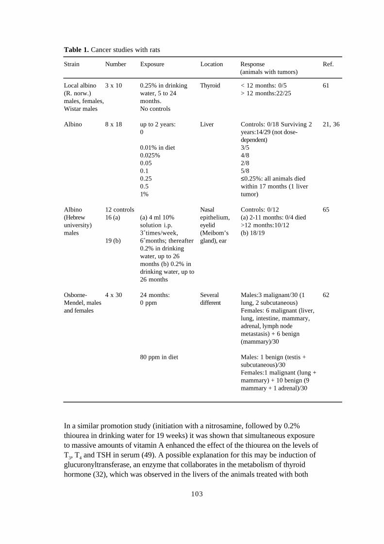
103
Table 1. Cancer studies with rats
Strain Number Exposure Location Response(animals with tumors)
Ref.
Local albino(R. norw.)males, females,Wistar males
3 x 10 0.25% in drinkingwater, 5 to 24months.No controls
Thyroid < 12 months: 0/5> 12 months:22/25
61
Albino 8 x 18 up to 2 years:0
0.01% in diet0.025%0.050.10.250.51%
Liver Controls: 0/18 Surviving 2years:14/29 (not dose-dependent)3/54/82/85/8≤0.25%: all animals diedwithin 17 months (1 livertumor)
21, 36
Albino(Hebrewuniversity)males
12 controls16 (a)
19 (b)
(a) 4 ml 10%solution i.p.3 times/week,6 months; thereafter0.2% in drinkingwater, up to 26months (b) 0.2% indrinking water, up to26 months
Nasalepithelium,eyelid(Meibom’sgland), ear
Controls: 0/12(a) 2-11 months: 0/4 died>12 months:10/12(b) 18/19
65
Osborne-Mendel, malesand females
4 x 30 24 months:0 ppm
80 ppm in diet
Severaldifferent
Males:3 malignant/30 (1lung, 2 subcutaneous)Females: 6 malignant (liver,lung, intestine, mammary,adrenal, lymph nodemetastasis) + 6 benign(mammary)/30
Males: 1 benign (testis +subcutaneous)/30Females:1 malignant (lung +mammary) + 10 benign (9mammary + 1 adrenal)/30
62
In a similar promotion study (initiation with a nitrosamine, followed by 0.2%thiourea in drinking water for 19 weeks) it was shown that simultaneous exposureto massive amounts of vitamin A enhanced the effect of the thiourea on the levels ofT3, T4 and TSH in serum (49). A possible explanation for this may be induction ofglucuronyltransferase, an enzyme that collaborates in the metabolism of thyroidhormone (32), which was observed in the livers of the animals treated with both

104
thiourea and vitamin A. Thiourea (0.2%) in drinking water, either with or withoutvitamin A, also increased the level of CYP2E1 in the liver (75).
Thiourea showed no initiating or promoting ability in a liver foci test with rats.Instead, treatment with 0.05 – 0.2% thiourea in drinking water for 51 to 70 daysreduced both the number and the volume of foci (which lacked ATPase) in diethyl-nitrosamine-initiated male and female Sprague-Dawley rats (56). In another studywith rats (F-344 males, initiation with another nitrosamine) thiourea (0.1% indrinking water for 19 weeks) increased the number of GSTP-positive foci. Thistreatment also resulted in nodules and neoplasias in thyroids. Thiourea andphenobarbital were observed to have a slight synergistic effect on liver foci (70).
Thyroid tumors have not been observed in mice after exposure to thiourea. In onestudy with mice in which thiourea was given orally (5 g/kg in diet), benign bonetumors were observed in the skull. According to the authors, the bone tumors wereprobably a side-effect of a direct toxic effect on the pituitary (50). In an older studywith C3H mice it was found that high doses of thiourea inhibited the occurrence ofspontaneous mammary tumors in surviving animals – probably because ofthiourea’s toxic effect on ovaries in this strain of mice and the consequent reductionin estrogen production (79).
No significant elevation in risk of bladder cancer was found in an epidemiologicalcase-control study (relative risk 1.2; 95% confidence interval 0.4 – 3.3) in whichexposure to thiourea was estimated from job or business sector (80).
Teratogenicity
High doses of thiourea given orally to pregnant rats (1 or 2 g/kg, day 12) and mice(1 g/kg, day 10) were fetotoxic, causing an increased number of resorbedembryos. No weight reductions and no deformities were observed in survivingfetuses (77).
When pregnant rats were exposed to thiourea (0.2% in drinking water) during thethird or the second and third week of gestation, there were effects on the thyroids ofthe fetuses (day 20), the newborn pups and the mothers. The treatment had noeffect on the body weights of the mothers. Exposure during the entire period ofgestation was altogether too toxic for the fetuses. Increased thyroid weights andreduced iodine content were also observed in the young when the females wereexposed to the same dose of thiourea during the nursing period. The effects on thethyroid were reversible (44). In another study by the same research group, femalerats were given 0.2% thiourea in drinking water during the first 14 days ofgestation. The exposure caused deformities in the bones and nervous systems of theyoung. General hemorrhaging was also observed (43).
Dose-effect/dose-response relationships
It was found in one study that occupational exposure to thiourea inhibited thyroidfunction, measured as lower levels of the thyroid hormones T3 and T4. Measured airconcentrations in the factory were reported to be between 0.6 and 12 mg/m3.

105
Thyroid hyperplasia was observed in 17 of 45 workers (76). If it is assumed thatthe workers weighed 70 kg and inhaled 1 m3 per hour for 8 hours/day, and thatuptake was complete, this air concentration is equivalent to a dose of 0.07 to 1.4 mgthiourea/kg body weight/day. Data from therapeutic use of thiourea indicate clearlythat a daily oral dose of 10 to 15 mg/day (about 0.1 - 0.2 mg/kg/day) has no effecton the thyroid, and that doses of 25 to 70 mg/day (about 0.4 - 1.0 mg/kg/day) dohave an effect (15).
The Dutch expert group has reported a NOEL of 4 mg/kg (80 ppm in diet,Osborne-Mendel rats) for the tumorigenic effect on rats with oral exposure (14, 62).An LD50 of about the same size, 3.55 mg/kg, has been reported for Sprague-Dawley rats after intraperitoneal injection of thiourea (26).
For rats, The NOEL for thiourea in drinking water (13-week study) wasdetermined to be > 2.5 ppm (10), equivalent to a dose of about > 0.25 mg/kg/day.
Conclusions
The critical effect of thiourea is inhibition of thyroid function, and this has beenreported with occupational exposure. Thiourea causes tumors in experimentalanimals. In addition to thyroid tumors, which are probably caused by hormonaldisturbances to which the test animals are particularly sensitive, tumors in otherlocations – notably liver, ear canal and eyelid – have been observed. Documentationof this carcinogenic activity is weak, however. The results of tests for genotoxicityare inconsistent. Skin contact with thiourea can cause contact allergy andphotoallergy. Thiourea can cross the placental barrier.
References
1. Althaus FR, Lawrence SD, Sattler GL, Longfellow DG, Pitot HC. Chemical quantificationof unscheduled DNA synthesis in cultured hepatocytes as an assay for the rapid screening ofpotential chemical carcinogens. Cancer Research 1982;42:3010-3015.
2. Andrae U, Greim H. Initiation and promotion in thyroid carcinogenesis. In: Dekant W,Neumann H, eds. Tissue Specific Toxicity: Biochemical Mechanisms. London: AcademicPress, 1992:71-93.
3. Anonymous. Scientific Basis for Swedish Occupational Standards 9. Lundberg P, ed.Thiourea. Arbete och Hälsa 1988;32:67-74.
4. Anonymous. Thioharnstoff. Berufsgenossenschaft der chemischen Industrie. Heidelberg,Germany 1995, No. 251. (Issued 06/95).
5. Astwood E. The chemical nature of compounds which inhibit the function of the thyroidgland. J Pharmacol Exp Ther 1943;78:79-89.
6. Batiste-Alentorn M, Xamena N, Creus A, Marcos R. Further studies with the somatic white-ivory system of Drosophila melanogaster. Genotoxicity testing of ten carcinogens. EnvironMol Mutagen 1994;24:143-147.
7. Batiste-Alentorn M, Xamena N, Creus A, Marcos R. Genotoxic evaluation of ten carcinogensin the Drosophila melanogaster wing spot test. Experientia 1995;51:73-76.
8. Batiste-Alentorn M, Xamena N, Creus A, Marcos R. Genotoxicity studies with the unstablezeste-white (UZ) system of Drosophila melanogaster. Results with ten carcinogeniccompounds. Environ Mol Mutagen 1991;18:120-125.

106
9. Brennan RJ, Schiestl RH. Free radicals generated in yeast by the Salmonella test-negativecarcinogens benzene, urethane, thiourea and auramine O. Mutat Res 1998;403:65-73.
10. BUA (Berategremium für Umweltrelevante Altstoffe). Thioharnstoff. BUA-Stoffbericht 179(Oktober 1995). Stuttgart: Wissenschaftliche Verlagsgesellschaft 1996.
11. Buffet C, Garnier R, Efthymiou M, Levillain J, Galliot M, Dussaix E. Hépatite au décoursd’une exposition à la thiourée? La Presse Médicale 1997;26:464-465.
12. Crebelli R, Bellincampi D, Conti G, Conti L, Morpurgo G, Carere A. A comparative studyon selected chemical carcinogens for chromosome malsegregation, mitotic crossing-over andforward mutation induction in Aspergillus nidulans. Mutat Res 1986;172:139-149.
13. Dalton A, Morris H, Dubnik C. Morphologic changes in the organs of female C3H miceafter long-term ingestion of thiourea and thiouracil. J Natl Cancer Inst 1948;9:201-223.
14. DECOS. Health-based recommended occupational exposure limits for thiourea. Dutch ExpertCommittee for Occupational Standards, Directorate General of Labour, the Netherlands, 1990(RA11/90).
15. DFG (Deutsche Forschungsgemeinschaft). Toxikologisch-arbeitsmedizinische Begründungenvon MAK-Werten. Thioharnstoff. Weinheim: VCH-Verlagsgesellschaft, 1988:15 pages.
16. Dooms-Goossens A, Chrispeels M, De Veylder H, Roelandts R, Willems L, Degreef H.Contact and photocontact sensitivity problems associated with thiourea and its derivatives:A review of the literature and case reports. Br J Dermatol 1987;116:573-579.
17. Dooms-Goossens A, Debusschère K, Morren M, Roelandts R, Coopman S. Silver polish:Another source of contact dermatitis reactions to thiourea. Contact Dermatitis 1988;19:133-135.
18. Falls J, Cherrington N, Clements KM, Philpot RM, Levi PE, Rose RL, Hodgson E.Molecular cloning, sequencing, and expession in Escherichia coli of mouse flavin-containingmonooxygenase 3 (FMO3): Comparison with the human isoform. Arch Biochem Biophys1997;347:9-18.
19. Fautz R, Forster R, Hechenberger CMA, Hertner T, von der Hude W, Kaufmann G, Madle H,Madle S, Miltenburger HG, Müller L, Pool-Zobel BL, Puri E, Schmezer P, Seeberg AH,Strobel R, Suter W, Baumeister M. Report of a comparative study of DNA damage and repairassays in primary rat hepatocytes with five coded chemicals. Mutat Res 1991;260:281-294.
20. Feldmann R, Maibach H. Absorption of some organic compounds through the skin in man.J Invest Dermatol 1970;54:399-404.
21. Fitzhugh O, Nelson A. Liver tumors in rats fed thiourea or thioacetamide. Science1948;108:626-628.
22. Fritzenschaf H, Kohlpoth M, Rusche B, Schiffmann D. Testing of known carcinogens andnoncarcinogens in the Syrian hamster embyo (SHE) micronucleus test in vitro; correlationswith in vivo micronucleus formation and cell transformation. Mutat Res 1993;319:47-53.
23. Galli A, Schiestl R. Salmonella test positive and negative carcinogens show different effectson intrachromosomal recombination in G2 cell cycle arrested cells. Carcinogenesis1995;16:659-663.
24. Geier J, Fuchs T. Contact allergy due to 4-N,N-dimethylaminobenzene diazonium chlorideand thiourea in diazo copy paper. Contact Dermatitis 1993;28:304-305.
25. Giri S, Combs A. Thiourea binding by rat erythrocyte, resistant to trichloroacetic aciddenaturation of protein. Chem Biol Inteactions 1972;5:97-105.
26. Giri S, Hollinger M, Cross C, Dungworth D. Effects of thiourea on pulmonary edema,pleural and peritoneal effusions and toxicity in rats pretreated with Actinomycin D.Toxicology 1974;2:211-222.
27. Giri S, Hollinger M, Rice S. Effects of thiourea on pulmonary vascular permeability and onlung and plasma histamine levels in rats. Toxicol Lett 1991;57:283-290.
28. Giri SN, Hollinger MA, Rice SA. Effects of thiourea tolerance on plasma histamine, andlung vascular permeability. Arch Toxicol 1991;65:603-605.

107
29. Greim H. Thioharnstoff. In Greim H, ed. Gesundheitsschädliche Arbeitsstoffe. Toxikologisch-arbeitsmedizinische Begründungen von MAK-Werten. Weinheim: VCH, 1997;24:1-5.
30. Hard GC. Recent developments in the investigation of thyroid regulation and thyroidcarcinogenesis. Environ Health Perspect 1998;106:427-436.
31. Hellmer L, Bocsfoldi G. An evaluation of the E. coli K-12 uvrB/recA DNA repair host-mediated assay. I. In vitro sensitivity of the bacteria to 61 compounds. Mutat Res1992;272:145-160.
32. Hill R, Crisp T, Hurley P, Rosenthal S, Singh D. Risk assessment of thyroid follicular celltumors. Environ Health Perspect 1998;106:447-457.
33. Hollinger M, Giri S. Interaction of thiourea with rat lung protein. Toxicology 1990;60:245-251.
34. Hollinger M, Giri S. Non-enzymatic covalent binding of radioactivity from [14C]thiourea torat lung protein. Toxicol Lett 1990;52:1-5.
35. Hollinger M, Giri S, Budd E. A pharmacodynamic study of [14C]thiourea toxicity in mature,immature, tolerant and nontolerant rats. Toxicol Appl Pharmacol 1976;37:545-556.
36. IARC. Some anti-thyroid and related substances, nitrofurans and industrial chemicals.Thiourea. IARC Monographs on the Evaluation of Carcinogenic Risks to Humans1974;7:95-109.
37. Itoh K, Kimura T, Yokoi T, Itoh S, Kamataki T. Rat liver flavin-containing monooxygenase(FMO): cDNA cloning and expression in yeast. Biochim Biophys Acta 1993;1173:165-171.
38. Itoh K, Nakamura K, Kimura T, Itoh S, Kamataki T. Molecular cloning of mouse liverflavin containing monooxygenase (FMO1) cDNA and characterization of the expressionproduct: Metabolism of the neurotoxin, 1,2,3,4-tetrahydroisoquinoline (TIQ). J Toxicol Sci1997;22:45-56.
39. Kanerva L, Estlander T, Jolanki R. Occupational allergic contact dermatitis caused by thioureacompounds. Contact Dermatitis 1994;31:242-248.
40. Kanno J, Matsuoka C, Furuta K, Onodera H, Miyajima H, Maekawa A, Hayashi Y. Tumorpromoting effect of goitrogens on the rat thyroid. Toxicol Pathol 1990;18:239-246.
41. Kellett J, Beck M, Auckland G. Contact sensitivity to thiourea in photocopy paper. ContactDermatitis 1984;11:124.
42. Kelner M, Bagnell R, Welch K. Thioureas react with superoxide radicals to yield a sulfhydrylcompound. J Biol Chem 1990;265:1306-1311.
43. Kern M, Tatar-Kiss Z, Kertai P, Foldes I. Teratogenic effect of 2’-thiourea in the rat. ActaMorphol Acad Sci Hung 1980;28:259-267.
44. Kertai P, Remenar I. Effect of 2-thiourea administered to pregnant rats on the thyroid and theprotein bound iodine content of the offspring. Acta Med Acad Sci Hung 1975;32:271-277.
45. Kosova L. On the toxic effects of thiourea and its dioxide after absorption through the skin.Hygiene and Sanitation. Washington, D.C.: Environmental Protection Agency and NationalScience Foundation, 1971;38-42. (translated from Russian)
46. Krieter PA, Ziegler DM, Hill KE, Burk RF. Increased biliary GSSG efflux from rat liversperfused with thiocarbamide substrates for the flavin-containing monooxygenase. MolPharmacol 1984;26:122-127.
47. Lonati-Galligani M, Lohman PH, Berends F. The validity of the autoradiographic method fordetecting DNA repair synthesis in rat hepatocytes in primary culture. Mutat Res1983;113:145-160.
48. Mitchell AD, Rudd CJ, Caspary WJ. Evaluation of the L5178Y mouse lymphoma cellmutagenesis assay: Intralaboratory results for sixty-three coded chemicals tested at SRIInternational. Environ Mol Mutagen 1988;12 Suppl 13:37-101.
49. Mitsumori K, Onodera H, Takahashi M, Shimo T, Yasuhara K, Takegawa K, Takahashi M,Hayashi Y. Promoting effect of large amounts of vitamin A on cell proliferation of thyroid

108
proliferative lesions induced by simultaneous treatment with thiourea. Cancer Lett1996;103:19-31.
50. Muranyi-Kovacs I, Rudali G, Arnaud D. Effect of thiourea on intracranial bone tumorformation in AkR mice. Hormone Res 1979;10:79-87.
51. Myhr BC, Caspary WJ. Evaluation of the L5178Y mouse lymphoma cell mutagenesis assay:Intralaboratory results for sixty-three coded chemicals tested at Litton Bionetics, Inc. EnvironMol Mutagen 1988;12 Suppl 13:103-194.
52. Mårs U, Larsson B. Thiourea as a melanoma targeting agent. Melanoma Res 1996;6:113-120.
53. Nakamura SI, Oda Y, Shimada T, Oki I, Sugimoto K. SOS-inducing activity of chemicalcarcinogens and mutagens in Salmonellla typhimurium TA1535/pSK1002: Examination with151 chemicals. [erratum published in Mutat Res 1988;207:213]. Mutat Res 1987;192:239-246.
54. Newcombe P, Deane E. Thiourea causing granulopenia and thrombopenia. Lancet1944;246:179.
55. Nurse D. Sensitivity to thiourea in plan printing paper. Contact Dermatitis 1980;6:153-154.
56. Oesterle D, Deml E. Lack of initiating and promoting activity of thiourea in rat liver focibioassay. Cancer Lett 1988;41:245-249.
57. Onodera H, Mitsumori K, Takahashi M, Shimo T, Yasuhara K, Kituara K, Takahashi M,Hayashi Y. Thyroid proliferative lesions induced by anti-thyroid drugs in rats are not alwaysaccompanied by sustained increases in serum TSH. J Toxicol Sci 1994;19:227-234.
58. Pergal M, Vukojevic N, Djuric D. Carbon disulfide metabolites excreted in the urine ofexposed workers. II. Isolation and identification of thiocarbamide. Arch Environ Health1972;25:42-44.
59. Pienta RJ, Poiley JA, Lebherz WB 3rd. Morphological transformation of early passage goldenSyrian hamster embryo cells derived from cryopreserved primary cultures as a reliable in vitrobioassay for identifying diverse carcinogens. Int J Cancer 1977;19:642-655.
60. Poulsen LL, Hyslop RM, Ziegler DM. S-Oxygenation of N-substituted thioureas catalyzedby the pig liver microsomal FAD-containing monooxygenase. Arch Biochem Biophys1979;198:78-88.
61. Purves H, Griesbach W. Studies on experimental goitre. VIII. Thyroid tumors in rats treatedwith thiourea. Br J Exp Pathol 1947;28:46-53.
62. Radomski J, Deichmann W, MacDonald W, Glass E. Synergism among oral carcinogens. I.Results of the simultaneous feeding of four tumorigens to rats. Toxicol Appl Pharmacol1965;7:652-656.
63. Roberts F, Wright A, O’Hagan S. Hypothyroidism in textile workers. J Soc Occup Med1990;40:153-156.
64. Rodriguez-Arnaiz R. Genotoxic activation of hydrazine, two dialkylhydrazines, thiourea andethylene thiourea in the somatic w/w+ assay of Drosophila melanogaster. Mutat Res1997;395:229-242.
65. Rosin A, Ungar H. Malignant tumors in the eyelids and the auricular region of thiourea-treated rats. Cancer Res 1957;17:302-305.
66. Rougier A, Rallis M, Krien P, Lotte C. In vivo percutaneous absorption: A key role forstratum corneum/vehicle partitioning. Arch Dermatol Res 1990;282:498-505.
67. Schiestl R, Gietz R, Mehta R, Hastings P. Carcinogens induce intrachromosomalrecombination in yeast. Carcinogenesis 1989;10:1445-1455.
68. Schulman J Jr, Keating R. Studies on the metabolism of thiourea. I. Distribution andexcretion in the rat of thiourea labeled with radioactive sulfur. J Biol Chem 1950;183:215-221.
69. Scott A, Powell G, Upshall D, Curtis C. Pulmonary toxicity of thioureas in the rat. EnvironHealth Perspect 1990;85:43-50.

109
70. Shimo T, Mitsumori K, Onodera H, Yasuhara K, Takahashi M, Takahashi M, Ueno Y,Hayashi Y. Synergistic effects of phenobarbital and thiourea on proliferative lesions in the ratliver. Cancer Lett 1994,81:45-52.
71. Shimo T, Mitsumori K, Onodera H, Yasuhara K, Kitaura K, Takahashi M, Kanno J, HayashiY. Time course observation of thyroid proliferative lesions and serum TSH levels in ratstreated with thiourea after DHPN initiation. Cancer Lett 1994;85:141-149.
72. Simmon VF, Rosenkranz HS, Zeiger E, Poirier LA. Mutagenic activity of chemicalcarcinogens and related compounds in the intraperitoneal host-mediated assay. J Natl CancerInst 1979;62:911-918.
73. Sina JF, Bean CL, Dysart GR, Taylor VI, Bradley MO. Evaluation of the alkaline elution/rathepatocyte assay as a predictor of carcinogenic/mutagenic potential. Mutat Res 1983;113:357-391.
74. Slanina P, Ullberg S, Hammarstrom L. Distribution and placental transfer of 14C-thiourea and14C-thiouracil in mice studied by whole-body autoradiography. Acta Pharmacol Toxicol1973;32:358-368.
75. Takegawa K, Mitsumori K, Onodera H, Mutai M, Kitaura K, Takahashi M, Uneyama C,Yasuhara K, Takahashi M, Yanai T, Masegi T, Hayashi Y. UDP-GT involvement in theenhancement of cell proliferation in thyroid follicular cell proliferative lesions in rats treatedwith thiourea and vitamin A. Arch Toxicol 1997;71:661-667.
76. Talakin Y, Kolomoiskaya M, Melekhin V, Grishina R, Chernykh L, Kondratenko L.Functional status of the thyroid gland of workers employed in thiourea manufacture. Gig TrProf Zabol 1985;9:50-51. (in Russian)
77. Teramoto S, Kaneda M, Aoyama H, Shirasu Y. Correlation between the molecular structureof N-alkylureas and N-alkylthioureas and their teratogenic properties. Teratology1981;23:335-342.
78. Vanderlaan W, Storrie V. A survey of the factors controlling thyroid function, with especialreference to newer views on antithyroid substances. Pharmacol Rev 1955;7:301-334.
79. Vazquez-Lopez E. The effects of thiourea on the development of spontaneous tumors on mice.Br J Cancer 1949;3:401-414.
80. Vineis P, Magnani C. Occupation and bladder cancer in males: A case-control study. Int JCancer 1985;35:599-606.
81. Vogel EW, Nivard MJ. Performance of 181 chemicals in a Drosophila assay predominantlymonitoring interchromosomal mitotic recombination. Mutagenesis 1993;8:57-81.
82. Wangenheim J, Bolcsfoldi G. Mouse lymphoma L5178Y thymidine kinase locus assay of 50compounds. Mutagenesis 1988;3:193-205.
83. Whiteman M, Halliwell B. Thiourea and dimethylthiourea inhibit peroxynitrite-dependentdamage: Nonspecificity as hydroxyl radical scavengers. Free Radical Biol & Med1997;22:1309-1312.
84. Williams R, Kay G. Absorption, distribution and excretion of thiourea. Am J Physiol1945;143:715-722.
85. Zeiger E, Anderson B, Haworth S, Lawlor T, Mortelmans K. Salmonella mutagenicity tests:IV. Results from the testing of 300 chemicals. Environ Mol Mutagen 1988;11 Suppl 12:1-157.
86. Ziegler DM. Intermediate metabolites of thiocarbamides, thioureylenes and thioamides:Mechanism of formation and reactivity. Biochem Soc Transact 1978;6:94-96.
87. Ziegler-Skylakakis K, Rossberger S, Andrae U. Thiourea induces DNA repair synthesis inprimary rat hepatocyte cultures and gene mutations in V79 Chinese hamster cells. ArchToxicol 1985;58:5-9.
88. Ziegler-Skylakakis K, Nill S, Pan JF, Andrae U. S-Oxygenation of thiourea results in theformation of genotoxic products. Environ Mol Mutagen 1998;31:362-373.
110
Summary
Montelius J (ed). Scientific Basis for Swedish Occupational Standards. XX.Arbete och Hälsa 1999:26, pp 1-116.
Critical evaluation of those scientific data which are relevant as a background fordiscussion of Swedish occupational exposure limits. This volume consists of theconsensus reports given by the Criteria Group at the Swedish National Institute forWorking Life between July, 1998 and June, 1999.
Key Words: Calcium oxide, Calcium hydroxide, Cyanamide, Cyclohexanone,Dimethyl adipate, Dimethyl glutarate, Dimethyl succinate, Ethyleneglycol monomethyl ether, Ethylene glycol monomethyl ether acetate,Glutaraldehyde, Lactate esters, Methyl tertiary-butyl ether,Occupational Exposure Limit (OEL), Pentafluoroethane, Phosphorustrichloride, Phosphorus pentachloride, Phosphoryl chloride, ScientificBasis, Thiourea, Trifluoroethane.
Sammanfattning
Montelius J (ed). Vetenskapligt underlag för hygieniska gränsvärden. XX. Arbeteoch Hälsa 1999:26, s 1-116.
Sammanställningar baserade på kritisk genomgång och värdering av devetenskapliga fakta, vilka är relevanta som underlag för fastställande av hygienisktgränsvärde. Volymen omfattar de underlag som avgivits från Kriteriegruppen förhygieniska gränsvärden under perioden juli 1998 - juni 1999.
Nyckelord: Cyanamid, Cyklohexanon, Dimetyladipat, Dimetylglutarat,Dimetylsuccinat, Etylenglykolmetyleter, Etylenglykolmetyleteracetat,Fosfortriklorider, Fosforpentaklorid, Fosforylklorid, Glutaraldehyd,Hygieniskt gränsvärde, Kalciumoxid, Kalciumhydroxid, Laktatestrar,Metyl-tert-butyleter, Pentafluoretan, Tiourinämne, Trifluoretan,Vetenskapligt underlag.
En svensk version av dessa vetenskapliga underlag finns publicerad i Arbete ochHälsa 1999:25.

111
APPENDIX
Consensus reports in this and previous volumes
Substance Consensus Volume in Arbetedate och Hälsa (No.)
Acetaldehyde February 17, 1987 1987:39 (VIII)Acetamide December 11, 1991 1992:47 (XIII)Acetic acid June15, 1988 1988:32 (IX)Acetone October 20, 1987 1988:32 (IX)Acetonitrile September 12, 1989 1991:8 (XI)Acrylamide April 17, 1991 1992:6 (XII)Acrylates December 9, 1984 1985:32 (VI)Acrylonitrile April 28, 1987 1987:39 (VIII)Aliphatic amines August 25, 1982 1983:36 (IV)Aliphatic hydrocarbons, C10-C15 June 1, 1983 1983:36 (IV)Aliphatic monoketons September 5, 1990 1992:6 (XII)Allyl alcohol September 9, 1986 1987:39 (VIII)Allylamine August 25, 1982 1983:36 (IV)Allyl chloride June 6, 1989 1989:32 (X)Aluminum April 21, 1982 1982:24 (III)
revised September 14, 1994 1995:19 (XVI)p-Aminoazobenzene February 29, 1980 1981:21 (I)Ammonia April 28, 1987 1987:39 (VIII)Amylacetate March 23, 1983 1983:36 (IV)Aniline October 26, 1988 1989:32 (X)Anthraquinone November 26,1987 1988:32 (IX)Arsenic, inorganic December 9, 1980 1982:9 (II)
revised February 15, 1984 1984:44 (V)Arsine October 20, 1987 1988:32 (IX)Asbestos October 21, 1981 1982:24 (III)
Barium June 16, 1987 1987:39 (VIII)revised January 26, 1994 1994:30 (XV)
Benzene March 4, 1981 1982:9 (II)revised February 24, 1988 1988:32 (IX)
Benzoyl peroxide February 13, 1985 1985:32 (VI)Beryllium April 25, 1984 1984:44 (V)Borax October 6, 1982 1983:36 (IV)Boric acid October 6, 1982 1983:36 (IV)Boron Nitride January 27 1993 1993:37 (XIV)Butadiene October 23, 1985 1986:35 (VII)1-Butanol June 17, 1981 1982:24 (III)Butanols June 6, 1984 1984:44 (V)Butyl acetate June 6, 1984 1984:44 (V)Butyl acetates February 11, 1998 1998:25 (XIX)Butylamine August 25, 1982 1983:36 (IV)Butyl glycol October 6, 1982 1983:36 (IV)
Cadmium January 18, 1980 1981:21 (I)revised February 15, 1984 1984:44 (V)revised May 13, 1992 1992:47 (XIII)
Calcium hydroxide February 24, 1999 1999:26 (XX)Calcium nitride January 27, 1993 1993:37 (XIV)Calcium oxide February 24, 1999 1999:26 (XX)
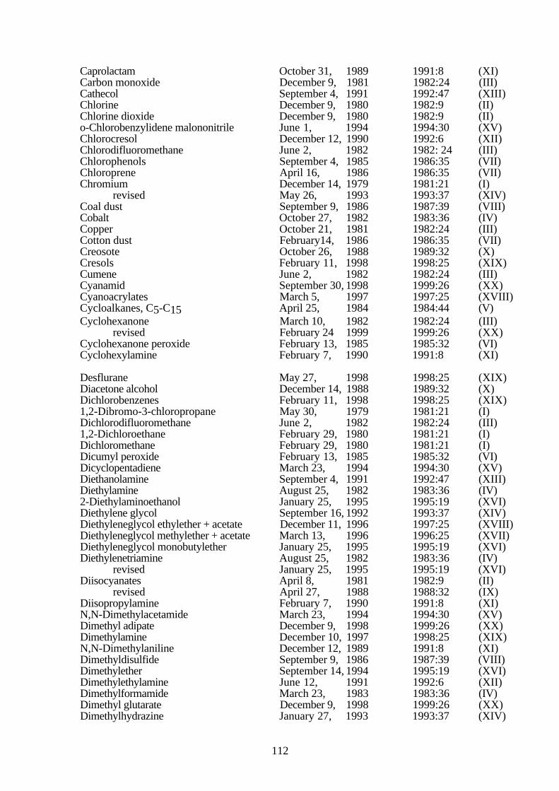
112
Caprolactam October 31, 1989 1991:8 (XI)Carbon monoxide December 9, 1981 1982:24 (III)Cathecol September 4, 1991 1992:47 (XIII)Chlorine December 9, 1980 1982:9 (II)Chlorine dioxide December 9, 1980 1982:9 (II)o-Chlorobenzylidene malononitrile June 1, 1994 1994:30 (XV)Chlorocresol December 12, 1990 1992:6 (XII)Chlorodifluoromethane June 2, 1982 1982: 24 (III)Chlorophenols September 4, 1985 1986:35 (VII)Chloroprene April 16, 1986 1986:35 (VII)Chromium December 14,1979 1981:21 (I)
revised May 26, 1993 1993:37 (XIV)Coal dust September 9, 1986 1987:39 (VIII)Cobalt October 27, 1982 1983:36 (IV)Copper October 21, 1981 1982:24 (III)Cotton dust February14, 1986 1986:35 (VII)Creosote October 26, 1988 1989:32 (X)Cresols February 11, 1998 1998:25 (XIX)Cumene June 2, 1982 1982:24 (III)Cyanamid September 30, 1998 1999:26 (XX)Cyanoacrylates March 5, 1997 1997:25 (XVIII)Cycloalkanes, C5-C15 April 25, 1984 1984:44 (V)Cyclohexanone March 10, 1982 1982:24 (III)
revised February 24 1999 1999:26 (XX)Cyclohexanone peroxide February 13, 1985 1985:32 (VI)Cyclohexylamine February 7, 1990 1991:8 (XI)
Desflurane May 27, 1998 1998:25 (XIX)Diacetone alcohol December 14, 1988 1989:32 (X)Dichlorobenzenes February 11, 1998 1998:25 (XIX)1,2-Dibromo-3-chloropropane May 30, 1979 1981:21 (I)Dichlorodifluoromethane June 2, 1982 1982:24 (III)1,2-Dichloroethane February 29, 1980 1981:21 (I)Dichloromethane February 29, 1980 1981:21 (I)Dicumyl peroxide February 13, 1985 1985:32 (VI)Dicyclopentadiene March 23, 1994 1994:30 (XV)Diethanolamine September 4, 1991 1992:47 (XIII)Diethylamine August 25, 1982 1983:36 (IV)2-Diethylaminoethanol January 25, 1995 1995:19 (XVI)Diethylene glycol September 16, 1992 1993:37 (XIV)Diethyleneglycol ethylether + acetate December 11,1996 1997:25 (XVIII)Diethyleneglycol methylether + acetate March 13, 1996 1996:25 (XVII)Diethyleneglycol monobutylether January 25, 1995 1995:19 (XVI)Diethylenetriamine August 25, 1982 1983:36 (IV)
revised January 25, 1995 1995:19 (XVI)Diisocyanates April 8, 1981 1982:9 (II)
revised April 27, 1988 1988:32 (IX)Diisopropylamine February 7, 1990 1991:8 (XI)N,N-Dimethylacetamide March 23, 1994 1994:30 (XV)Dimethyl adipate December 9, 1998 1999:26 (XX)Dimethylamine December 10, 1997 1998:25 (XIX)N,N-Dimethylaniline December 12,1989 1991:8 (XI)Dimethyldisulfide September 9, 1986 1987:39 (VIII)Dimethylether September 14, 1994 1995:19 (XVI)Dimethylethylamine June 12, 1991 1992:6 (XII)Dimethylformamide March 23, 1983 1983:36 (IV)Dimethyl glutarate December 9,1998 1999:26 (XX)Dimethylhydrazine January 27, 1993 1993:37 (XIV)
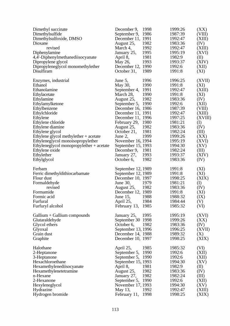
113
Dimethyl succinate December 9,1998 1999:26 (XX)Dimethylsulfide September 9, 1986 1987:39 (VIII)Dimethylsulfoxide, DMSO December 11,1991 1992:47 (XIII)Dioxane August 25, 1982 1983:36 (IV)
revised March 4, 1992 1992:47 (XIII)Diphenylamine January 25, 1995 1995:19 (XVI)4,4'-Diphenylmethanediisocyanate April 8, 1981 1982:9 (II)Dipropylene glycol May 26, 1993 1993:37 (XIV)Dipropyleneglycol monomethylether December 12,1990 1992:6 (XII)Disulfiram October 31, 1989 1991:8 (XI)
Enzymes, industrial June 5, 1996 1996:25 (XVII)Ethanol May 30, 1990 1991:8 (XI)Ethanolamine September 4, 1991 1992:47 (XIII)Ethylacetate March 28, 1990 1991:8 (XI)Ethylamine August 25, 1982 1983:36 (IV)Ethylamylketone September 5, 1990 1992:6 (XII)Ethylbenzene December 16,1986 1987:39 (VIII)Ethylchloride December 11,1991 1992:47 (XIII)Ethylene December 11,1996 1997:25 (XVIII)Ethylene chloride February 29, 1980 1981:21 (I)Ethylene diamine August 25, 1982 1983:36 (IV)Ethylene glycol October 21, 1981 1982:24 (III)Ethylene glycol methylether + acetate June 2, 1999 1999:26 (XX)Ethyleneglycol monoisopropylether November 16, 1994 1995:19 (XVI)Ethyleneglycol monopropylether + acetateSeptember 15, 1993 1994:30 (XV)Ethylene oxide December 9,1981 1982:24 (III)Ethylether January 27, 1993 1993:37 (XIV)Ethylglycol October 6, 1982 1983:36 (IV)
Ferbam September 12,1989 1991:8 (XI)Ferric dimethyldithiocarbamate September 12, 1989 1991:8 (XI)Flour dust December 10, 1997 1998:25 (XIX)Formaldehyde June 30, 1979 1981:21 (I)
revised August 25, 1982 1983:36 (IV)Formamide December 12,1989 1991:8 (XI)Formic acid June 15, 1988 1988:32 (IX)Furfural April 25, 1984 1984:44 (V)Furfuryl alcohol February 13, 1985 1985:32 (VI)
Gallium + Gallium compounds January 25, 1995 1995:19 (XVI)Glutaraldehyde September 301998 1999:26 (XX)Glycol ethers October 6, 1982 1983:36 (IV)Glyoxal September 13, 1996 1996:25 (XVII)Grain dust December 14, 1988 1989:32 (X)Graphite December 10,1997 1998:25 (XIX)
Halothane April 25, 1985 1985:32 (VI)2-Heptanone September 5, 1990 1992:6 (XII)3-Heptanone September 5, 1990 1992:6 (XII)Hexachloroethane September 15, 1993 1994:30 (XV)Hexamethylenediisocyanate April 8, 1981 1982:9 (II)Hexamethylenetetramine August 25, 1982 1983:36 (IV)n-Hexane January 27, 1982 1982:24 (III)2-Hexanone September 5, 1990 1992:6 (XII)Hexyleneglycol November 17, 1993 1994:30 (XV)Hydrazine May 13, 1992 1992:47 (XIII)Hydrogen bromide February 11, 1998 1998:25 (XIX)

114
Hydrogen fluoride April 25, 1984 1984:44 (V)Hydrogen peroxide April 4, 1989 1989:32 (X)Hydrogen sulfide May 4, 1983 1983:36 (IV)Hydroquinone October 21, 1989 1991:8 (XI)
Indium March 23, 1994 1994:30 (XV)Industrial enzymes June 5, 1996 1996:25 (XVII)Isophorone February 20, 1991 1992:6 (XII)Isopropanol December 9,1981 1982:24 (III)Isopropylamine February 7, 1990 1991:8 (XI)Isopropylbenzene June 2, 1982 1982:24 (III)
Lactates March 29, 1995 1995:19 (XVI)Lactate esters June 2, 1999 1999:26 (XX)Lead, inorganic February 29, 1980 1981:21 (I)
revised September 5, 1990 1992:6 (XII)Lithium boron nitride January 27, 1993 1993:37 (XIV)Lithium nitride January 27, 1993 1993:37 (XIV)
Maleic anhydride September 12, 1989 1991:8 (XI)Manganese February 15, 1983 1983:36 (IV)
revised April 17, 1991 1992:6 (XII)revised June 4, 1997 1997:25 (XVIII)
Man made mineral fibers March 4, 1981 1982:9 (II)revised December 1,1987 1988:32 (IX)
Mercury, inorganic April 25, 1984 1984:44 (V)Mesityl oxide May 4, 1983 1983:36 (IV)Metal stearates, some September 15,1993 1994:30 (XV)Methacrylates September 12, 1984 1985:32 (VI)Methanol April 25, 1985 1985:32 (VI)Methyl acetate March 28 1990 1991:8 (XI)Methylamine August 25, 1982 1983:36 (IV)Methylamyl alcohol March 17, 1993 1993:37 (XIV)Methyl bromide April 27, 1988 1988:32 (IX)Methyl chloride March 4, 1992 1992:47 (XIII)Methyl chloroform March 4, 1981 1982:9 (II)Methylene chloride February 29, 1980 1981:21 (I)4,4'-Methylene dianiline June 16, 1987 1987:39 (VIII)Methyl ethyl ketone February 13, 1985 1985:32 (VI)Methyl ethyl ketone peroxide February 13, 1985 1985:32 (VI)Methyl formare December 12,1989 1991:8 (XI)Methyl glycol October 6, 1982 1983:36 (IV)Methyl iodide June 30, 1979 1981:21 (I)Methylisoamylamine September 5, 1990 1992:6 (XII)Methyl mercaptane September 9, 1986 1987:39 (VIII)Methyl methacrylate March 17, 1993 1993:37 (XIV)Methyl pyrrolidone June 16, 1987 1987:39 (VIII)Methyl-t-butyl ether November 26, 1987 1988:32 (IX)
revised September 30, 1998 1999:26 (XX)Mixed solvents, neurotoxicity April 25, 1985 1985:32 (VI)Molybdenum October 27, 1982 1983:36 (IV)Monochloroacetic acid February 20, 1991 1992:6 (XII)Monochlorobenzene September 16,1993 1993:37 (XIV)Monomethylhydrazine March 4, 1992 1992:47 (XIII)Mononitrotoluene February 20, 1991 1992:6 (XII)Monoterpenes February 17, 1987 1987:39 (VIII)Morpholine December 8,1982 1983:36 (IV)
revised June 5, 1996 1996:25 (XVII)

115
Naphthalene May 27, 1998 1998:25 (XIX)Natural crystallinic fibers (except asbestos)June 12, 1991 1992:6 (XII)Nickel April 21, 1982 1982:24 (III)Nitroethane April 4, 1989 1989:32 (X)Nitrogen oxides December 11, 1985 1986:35 (VII)Nitroglycerin February 13, 1985 1985:32 (VI)Nitroglycol February 13, 1985 1985:32 (VI)Nitromethane January 6, 1989 1989:32 (X)Nitropropane October 28, 1986 1987:39 (VIII)2-Nitropropane March 29, 1995 1995:19 (XVI)Nitroso compounds December 12, 1990 1992:6 (XII)Nitrosomorpholine December 8,1982 1983:36 (IV)Nitrotoluene February 20, 1991 1992:6 (XII)Nitrous oxide December 9,1981 1982:24 (III)
Oil mist April 8, 1981 1982:9 (II)Organic acid anhydrides, some September 12, 1989 1991:8 (XI)Oxalic acid February 24, 1988 1988:32 (IX)Ozone April 28, 1987 1987:39 (VIII)
Paper dust February 7, 1990 1991:8 (XI)Pentaerythritol November 16, 1994 1995:19 (XVI)1,1,1,2,2-Pentafluoroethane February 24, 1999 1999:26 (XX)Peroxides, organic February 13, 1985 1985:32 (VI)Phenol February 13, 1985 1985:32 (VI)Phosphorous chlorides September 30, 1998 1999:26 (XX)Phosphorous oxides February 11, 1998 1998:25 (XIX)Phthalates December 8,1982 1983:36 (IV)Phthalic anhydride September 12,1989 1991:8 (XI)Piperazine September 12, 1984 1985:32 (VI)Plastic dusts December 16, 1986 1987:39 (VIII)Platinum June 4, 1997 1997:25 (XVIII)Polyaromatic hydrocarbons February 15, 1984 1984:44 (V)Polyisocyanates April 27, 1988 1988:32 (IX)Potassium aluminium fluoride June 4, 1997 1997:25 (XVIII)2-Propanol December 9,1981 1982:24 (III)Propene September 13, 1996 1996:25 (XVII)Propionic acid November 26, 1987 1988:32 (IX)Propylacetate September 14, 1994 1995:19 (XVI)Propylene glycol June 6, 1984 1984:44 (V)Propylene glycol-1,2-dinitrate May 4, 1983 1983:36 (IV)Propylene glycol monomethylether October 28,1986 1987:39 (VIII)Propylene oxide June 11, 1986 1986:35 (VII)Pyridine May 13, 1992 1992:47 (XIII)
Quartz March 13, 1996 1996:25 (XVII)
Resorcinol September 4, 1991 1992:47 (XIII)
Selenium December 11,1985 1986:35 (VII)revised February 22, 1993 1993:37 (XIV)
Sevoflurane May 27, 1998 1998:25 (XIX)Silica March 13, 1996 1996:25 (XVII)Silver October 28, 1986 1987:39 (VIII)Stearates, metallic, some September 15, 1993 1994:30 (XV)Stearates, non-metallic, some November 17, 1993 1994:30 (XV)Strontium January 26, 1994 1994:30 (XV)

116
Styrene February 29, 1980 1981:21 (I)revised October 31, 1989 1991:8 (XI)
Sulfur dioxide April 25, 1985 1985:32 (VI)Sulfur fluorides March 28, 1990 1991:8 (XI)Synthetic inorganic fibers March 4, 1981 1982:9 (II)
revised December 1,1987 1988:32 (IX)Synthetic organic and inorganic fibers May 30, 1990 1991:8 (XI)
Talc dust June 12, 1991 1992:6 (XII)Terpenes, mono- February 17, 1987 1987:39 (VIII)Tetrabromoethane May 30, 1990 1991:8 (XI)Tetrachloroethane June 4, 1997 1997:25 (XVIII)Tetrachloroethylene February 29, 1980 1981:21 (I)1,1,1,2-Tetrafluoroethane March 29, 1995 1995:19 (XVI)Tetrahydrofuran October 31, 1989 1991:8 (XI)Tetranitromethane April 4, 1989 !989:32 (X)Thioglycolic acid June 1, 1994 1994:30 (XV)Thiourea December 1,1987 1988:32 (IX)
revised June 2, 1999 1999:26 (XX)Thiram October 31, 1989 1991:8 (XI)Thiurams, some October 31, 1989 1991:8 (XI)Titanium dioxide February 21, 1989 1989:32 (X)Toluene February 29, 1980 1981:21 (I)Toluene-2,4-diisocyanate April 8, 1981 1982:9 (II)Toluene-2,6-diisocyanate April 8, 1981 1982:9 (II)1,1,1-Trifluoroethane February 24, 1999 1999:26 (XX)Trichlorobenzene September 16, 1993 1993:37 (XIV)1,1,1-Trichloroethane March 4, 1981 1982:9 (II)Trichloroethylene December 14,1979 1981:21 (I)Trichlorofluoromethane June 2, 1982 1982:24 (III)1,1,2-Trichloro-1,2,2-trifluoroethane June 2, 1982 1982:24 (III)Triethanolamine August 25, 1982 1983:36 (IV)Triethylamine December 5,1984 1985:32 (VI)Trimellitic anhydride September 12, 1989 1991:8 (XI)Trimethylolpropane November 16, 1994 1995:19 (XVI)Trinitrotoluene April 17, 1991 1992:6 (XII)
Vanadium March 15, 1983 1983:36 (IV)Vinyl acetate June 6, 1989 1989:32 (X)Vinyl toluene December 12,1990 1992:6 (XII)
White spirit December 16,1986 1987:39 (VIII)Wood dust June 17, 1981 1982:9 (II)
Xylene February 29, 1980 1981:21 (I)
Zinc April 21, 1982 1982:24 (III)Zinc dimethyl dithiocarbamate September 12, 1989 1991:8 (XI)Ziram September 12, 1989 1991:8 (XI)
Sent for publication December 1999