scientific basis for swedish occupational standards xxii · nr 2001:20 scientific basis for swedish...
TRANSCRIPT
arbete och hälsa | vetenskaplig skriftserie
isbn 91-7045-624-0 issn 0346-7821 http://www.niwl.se/
nr 2001:20
Scientific Basis for SwedishOccupational Standards XXII
Ed. Johan MonteliusCriteria Group for Occupational Standards
National Institute for Working LifeS-112 79 Stockholm, Sweden
Translation:Frances Van Sant
National Institute for Working Life
ARBETE OCH HÄLSAEditor-in-chief: Staffan MarklundCo-editors: Mikael Bergenheim, Anders Kjellberg,Birgitta Meding, Bo Melin, Gunnar Rosén and EwaWigaeus Tornqvist
© National Institut for Working Life & authors 2001
National Institute for Working LifeS-112 79 StockholmSweden
ISBN 91–7045–624–0ISSN 0346–7821http://www.niwl.se/Printed at CM Gruppen, Bromma
Arbete och Hälsa
Arbete och Hälsa (Work and Health) is ascientific report series published by theNational Institute for Working Life. Theseries presents research by the Institute’sown researchers as well as by others, bothwithin and outside of Sweden. The seriespublishes scientific original works, disser-tations, criteria documents and literaturesurveys.
Arbete och Hälsa has a broad target-group and welcomes articles in differentareas. The language is most often English,but also Swedish manuscripts arewelcome.
Summaries in Swedish and English as wellas the complete original text are availableat www.niwl.se/ as from 1997.
Preface
The Criteria Group of the Swedish National Institute for Working Life (NIWL) has thetask of gathering and evaluating data which can be used as a scientific basis for theproposal of occupational exposure limits given by the Swedish Work EnvironmentAuthority (SWEA). In most cases a scientific basis is written on request from the SWEA.The Criteria Group shall not propose a numerical occupational exposure limit value but,as far as possible, give a dose-response/dose-effect relationship and the critical effect ofoccupational exposure.
In searching of the literature several data bases are used, such as RTECS, Toxline,Medline, Cancerlit, Nioshtic and Riskline. Also information in existing criteria documentsis used, e.g. documents from WHO, EU, US NIOSH, the Dutch Expert Committee forOccupational Standards (DECOS) and the Nordic Expert Group. In some cases criteriadocuments are produced within the Criteria Group, often in collaboration with DECOS orUS NIOSH.
Evaluations are made of all relevant published original papers found in the searches. Insome cases information from handbooks and reports from e.g. US NIOSH and US EPAis used. A draft consensus report is written by the secretariat or by a scientist appointedby the secretariat. The author of the draft is indicated under Contents. A qualifiedevaluation is made of the information in the references. In some cases the information canbe omitted if some criteria are not fulfilled. In some cases such information is included inthe report but with a comment why the data are not included in the evaluation. Afterdiscussion in the Criteria Group the drafts are approved and accepted as a consensusreport from the group. They are sent to the SWEA.
This is the 22nd volume which is published and it contains consensus reports approvedby the Criteria Group during the period July 2000 to June 2001. These and previouslypublished consensus reports are listed in the Appendix (p 90). Technical editing forprinting was made by Karin Sundström.
Johan Högberg Johan MonteliusChairman Secretary
The Criteria Group has the following membership (as of June, 2001)
Maria Albin Dept Environ Occup Medicine,University Hospital, Lund
Olav Axelson Dept Environ Occup Medicine,University Hospital, Linköping
Sture Bengtsson Swedish Industrial Workers Union
Sven Bergström Swedish Trade Union Confederation
Anders Boman Dept Environ Occup Dermatology,Karolinska Hospital, Stockholm
Christer Edling Dept Environ Occup Medicine,University Hospital, Uppsala
Sten Flodström National Chemicals Inspectorate
Lars Erik Folkesson Swedish Metal Workers' Union
Johan Högberg chairman Toxicology and Risk assessment,Natl Inst for Working Life
Anders Iregren Toxicology and Risk assessment,Natl Inst for Working Life
Gunnar Johanson v. chairman Toxicology and Risk assessment,Natl Inst for Working Life
Bengt Järvholm Dept Environ Occup Medicine,University Hospital, Umeå
Kjell Larsson Respiratory health and Climate,Natl Inst for Working Life
Carola Lidén Dept Environ Occup Dermatology,Karolinska Hospital, Stockholm
Johan Montelius secretary Toxicology and Risk assessment,Natl Inst for Working Life
Bengt Olof Persson observer Swedish Work Environment Authority
Bengt Sjögren Toxicology and Risk assessment,Natl Inst for Working Life
Harri Vainio Dept Environmental Medicine,Karolinska Institutet
Kerstin Wahlberg observer Swedish Work Environment Authority
Olof Vesterberg Respiratory health and Climate,Natl Inst for Working Life
Contents
Consensus report for:Ethylenethiourea1 1Toluene-2,4-diamine and Toluene-2,6-diamine2 25α-Methylstyrene3 37Hydrogen Cyanide, Sodium Cyanide and Potassium Cyanide4 43Toluene Diisocyanate (TDI), Diphenylmethane Diisocyanate (MDI), 60
Hexamethylene Diisocyanate (HDI)5
Summary 89
Sammanfattning (in Swedish) 89
Appendix: Consensus reports in this and previous volumes 90
1 Drafted by Agneta Rannug, Margareta Warholm, Institute of Environmental Medicine, Karolinska
Institutet/National Institute for Working Life.2 Drafted by Ulla Stenius, Institute of Environmental Medicine, Karolinska Institutet/National Institute for
Working Life.3 Drafted by Niklas Finnberg, Institute of Environmental Medicine, Karolinska Institutet/National Institute for
Working Life.4 Drafted by Birgitta Lindell, Toxicology and Risk assessment, National Institute for Working Life.5 Drafted by Kjell Larsson, Programme for Respiratory Health and Climate, National Institute for Working Life;
Jan-Olof Levin, Programme for chemical exposure assessment, National Institute for Working Life (the section“Measuring air concentrations of TDI, MDI and HDI”);Margareta Littorin, Staffan Skerfving, Department of Occupational and Environmental Medicine, UniversityHospital, Lund (the section ”Biological measures of exposure”).
1
Consensus Report for Ethylenethiourea
September 27, 2000
This Consensus Report is based largely on a criteria document from the DutchExpert Committee for Occupational Standards (DECOS) (15), and takes intoaccount research published through 1999. The last literature search was made inMay, 2000.
Chemical and physical data
CAS No.: 96-45-7Synonyms: ETU
imidazoline-2-thiol2-imidazolidinethione2-mercaptoimidazoline
Formula: C3H6N2S
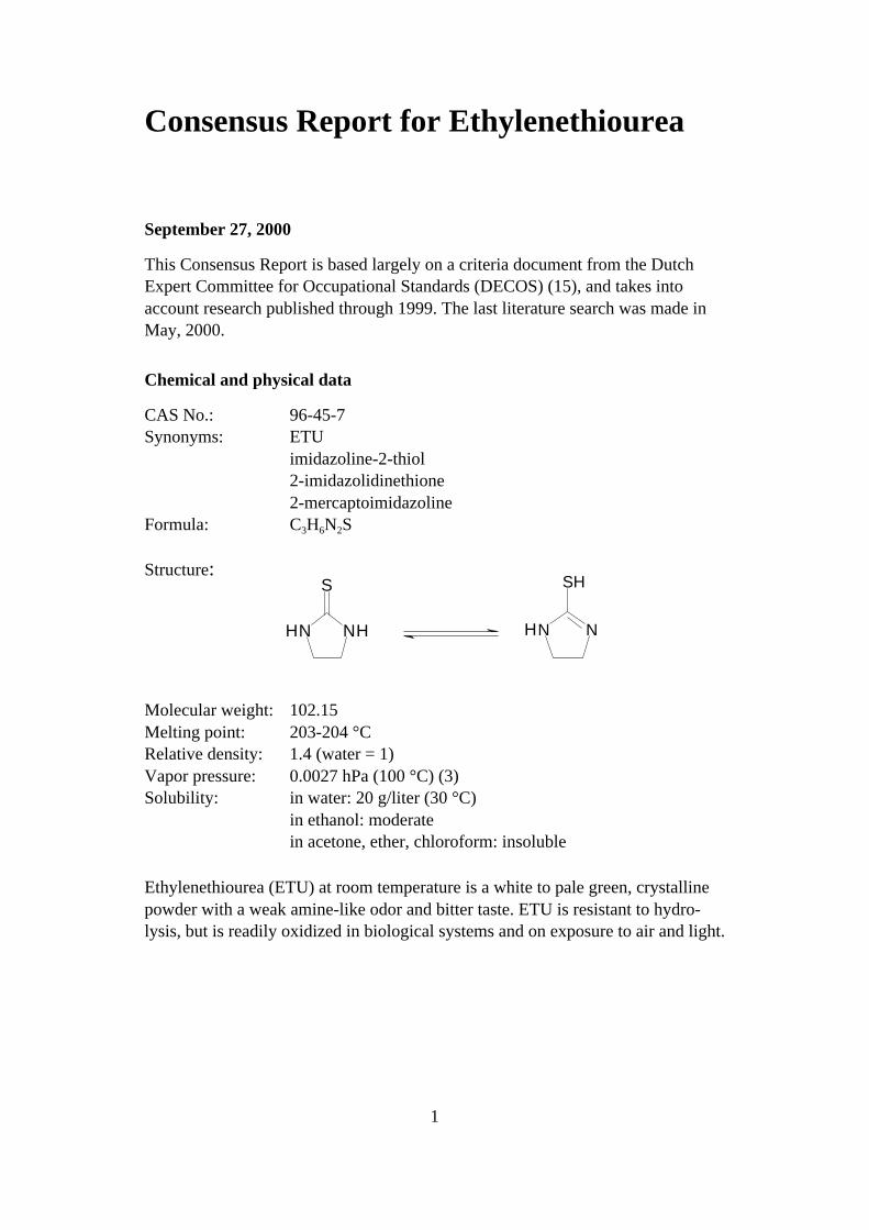
Structure:
Molecular weight: 102.15Melting point: 203-204 °CRelative density: 1.4 (water = 1)Vapor pressure: 0.0027 hPa (100 °C) (3)Solubility: in water: 20 g/liter (30 °C)
in ethanol: moderatein acetone, ether, chloroform: insoluble
Ethylenethiourea (ETU) at room temperature is a white to pale green, crystallinepowder with a weak amine-like odor and bitter taste. ETU is resistant to hydro-lysis, but is readily oxidized in biological systems and on exposure to air and light.
NN
S
NN
SH
H H H
2
Occurrence, use
Occupational exposure to ETU may occur in the rubber industry, where it is usedfor vulcanization of polyacrylate rubber and as an accelerator in the manufactureof neoprene rubber. ETU has also been used in production of antioxidants andsynthetic resins. Exposure to ETU may also occur in forestry and agriculture,where metallic salts of ethylenebisdithiocarbamate (e.g. maneb, mancozeb, zineb)are used as fungicides. These products usually contain ETU as an impurity. ETUis formed in biological systems by the breakdown of ethylenebisdithiocarbamate.
ETU can be synthesized by a reaction between ethylene diamine and carbondisulfide, followed by addition of hydrochloric acid to close the ring.
ETU does not occur naturally in the environment. Non-occupational exposurein Poland was estimated by measuring the concentration of ETU in various foods,and intake was found to be between 0.01 and 1 µg/kg body weight/day (33). In thepopulations of four Italian towns, measured excretion of ETU in urine was in therange <0.1 to 8.3 µg/g creatinine (5). In a wine district where ethylenebisdi-thiocarbamate was used as a fungicide, excretion of ETU in urine was higher:up to 61.4 µg/g creatinine. The highest values were found in smokers and winedrinkers (5). In a laboratory study with five volunteers in which the amount ofETU in diet was determined by analysis, it was found that most of the ETU inurine originated from intake of wine (4). ETU has been found in tobacco smoke (8to 27 ng/cigarette in 4 of 12 tested brands) (7). FAO/WHO have proposed 4 µg/kgbody weight/day as an acceptable intake of ETU (19). The EU threshold limitvalue for ETU in foods is 50 µg/kg (cited in Reference 17).
In a Finnish study of groups of agricultural and forestry workers who usedmancozeb or maneb fungicides (containing ethylenebisdithiocarbamate), airconcentrations of ETU around spraying ranged from 0.14 to 0.6 µg/m3 (averagevalues within the groups). Air levels were higher around weighing (highest averagevalue 1.81 µg/m3). The highest concentration of ETU measured in urine was23 µg/liter (49). Another Finnish study of 29 potato farmers (probably including atleast some of the participants in the previously mentioned study) showed airconcentrations of 0.004 to 3.3 µg/m3 in the breathing zone and 0.006 to 0.8 µg/m3
in the tractor cab. In the 24 hours immediately after the exposure, excretion of ETUin urine was in the range 0.09–2.5 µg/mmol creatinine (0.8-22.1 µg/g creatinine)(52). In 1980, air concentrations of 120 to 160 µg/m3 were measured in an Englishrubber factory where ETU was used in a process that generated dust (84). In anETU production plant in England, air levels shown on personal monitors were 10to 240 µg/m3, with a single reading of 330 µg/m3 (84).
Uptake, distribution, excretion
Data from animal experiments show that ETU is rapidly absorbed from thedigestive tract. IPCS/WHO report that ETU was identified in the blood of rats asearly as 5 minutes after oral administration of 14C-ETU (100 mg/kg body weight)
3
(33). A study with guinea pigs (reviewed in Reference 3) showed that uptake ofETU through intact skin was relatively slow: 14% of 2-14C-ETU (15 mg/ml, 1 mlapplied to an area of 4 x 4 cm) was absorbed within 24 hours. If the skin wasdamaged, uptake within 24 hours was 42%. The only laboratory data indicatingrespiratory uptake of ETU are in unpublished studies on rats (3). There are noquantitative data on ETU uptake by humans, although the substance has beenfound in urine of occupationally exposed persons (49, 52). It was found that ETUin the urine of workers producing fungicides was correlated to the amounts ofmancozeb and ETU on their hands (6). It was concluded that most of the ETUexposure in this work environment was due to skin uptake (6).
Regardless of the path of absorption, ETU accumulates in the thyroid (15).Single doses of ETU (20 mg/kg body weight) were given to rats and guinea pigsby gavage, and 96 hours later there was a much higher accumulation of ETU inthyroid than in liver, kidney, heart and muscle tissue, where concentrations wereabout the same (64). When 2-14C-ETU was given to pregnant rats, the radio-activity was evenly distributed in all examined tissues except the thyroid, whereaccumulation was particularly marked after 24 hours (>30 times). After 2 and 6hours, the concentration in the thyroid was two to three times higher than in othertissues. The concentration of ETU was somewhat lower in fetal tissue than in themothers. This study also shows that ETU can cross the placental barrier (38).Rhesus monkeys (2 females) were given single oral doses of ETU (40 mg/kg bodyweight) and no accumulation in thyroid was noted 48 hours later (1).
Groups of 6 rats of each sex were given 0, 2, 20, 200, 1000 or 2000 µg 14C-ETU/day for 7 days. Doses were equivalent to 0, 0.1, 1, 10, 50 or 100 ppm in feed.The amount of 14C in thyroid increased with increasing dose, but only up to 50ppm. No further increase was noted at 100 ppm. Seventeen days after the last doseof ETU, the 14C level in thyroids had declined by 80 to 94%. This shows that ETUand/or its metabolites do not accumulate permanently in the thyroid (57).
Most ETU is eliminated in urine. In an experiment in which two female rhesusmonkeys were exposed to 14C-ETU (40 mg/kg body weight, gavage) 47% and64% of the radioactivity was recovered in urine within 48 hours. Feces containedless than 1.5% (1). In a similar experiment with rats and guinea pigs (20 mg/kgbody weight) 65% (rats) and 47% (guinea pigs) of radioactivity was recovered inurine within 48 hours (61% and 45% within 24 hours) (64). In a 28-day study withrats it was found that the relative amounts of ETU in urine increased with in-creasing dose, possibly indicating that metabolism of ETU became saturated.At daily doses of 10.6, 17.6 and 23.4 mg/kg body weight, excretion in urine wason average 25%, 36% and 49% of the dose respectively (50).
For ETU and its metabolites, IPCS/WHO report a half time of 28 hours inmonkeys (9.3 mg 2-14C-ETU, per os), 9-10 hours in rats (240 mg/kg body weight,per os) and 5 hours in mice (240 mg/kg body weight, per os) (33, 71). The halftime for 14C-ETU (4 mg/kg body weight, i.v.) in the blood of 2 female cats was3.5 hours (34). In humans, the half time for elimination of ETU via kidneys is
4
estimated to be between 32 and 100 hours (49, 52). It is possible that the long halftime is due to slow uptake through the skin (52).
Biotransformation
Rats and cats were given oral doses of 14C-ETU (4 mg/kg b.w.); 24-hour urinesamples from the rats contained mostly unchanged ETU, ethyleneurea, 4-imidazoline-2-one and imidazoline, and those from the cats contained mostly S-methyl ETU, unchanged ETU and ethyleneurea (34). Biotransformation was moreextensive in the cats than in the rats (34). Very small amounts of 1-methyl thio-urea were found in plasma of rats after oral administration of ETU (48). It wasshown in a study with mice that biotransformation of ETU involves oxidation ofthe sulfur atom, with 2-imidazoline-2-yl-sulfenate as the primary product (78).There are no data on metabolic pathways in humans.
In mice, ETU is metabolized primarily by the microsomal flavin-containingmonooxygenase system (FMO) (30). The FMO-dependent binding of ETUmetabolites to proteins in the liver may contribute to the chronic liver toxicity thathas been observed in mice (15, 30). Mice metabolize ETU more rapidly than ratsdo, which may explain why ETU shows acute toxicity but not teratogenicity inmice (see below). Oral administration of ETU (50 to 1000 mg/kg body weight)induced cytochrome P-450 (aniline hydroxylase: CYP2E1) activity in mice (61),but reduced the activity in rats (54, 61).
NitrosationN-nitroso-ethylenethiourea, a nitrosamide containing sulfur, may be formed fromETU in the presence of nitrite in acid environments. Nitrosamides spontaneouslybreak down to carbonium ions at physiological pH, and are mutagenic withoutmetabolic activation (47).
Sodium nitrite, which is used to preserve meat, is the primary dietary source ofnitrites. In Europe, the daily intake of sodium nitrite is about 4 mg per person.Nitrates may also play some role, since they can be reduced to nitrites in themouth. Intake of nitrate is mostly from vegetables, and on average amounts toabout 100 mg per person per day. It can be assumed that about 6% of this (6 mg)is transformed to nitrite, increasing the daily nitrite intake to about 10 mg perperson (81). The formation of N-nitroso-ETU is probably much less likely withinhalation or skin uptake of ETU than with oral exposure.
Biological monitoring
As mentioned previously, urine samples can be used for biological monitoring thatreflects the past 24 hours of exposure to ETU. Analysis of ETU bound to hemo-globin has been proposed as a method for estimating longer exposures (up to 4months). Of 15 workers occupationally exposed to mancozeb, 40% hadidentifiable Hb adducts of ETU (0.5-1.42 pmol/mg Hb) (69). It has beendemonstrated in studies with rats that ETU, after metabolic activation –
5
presumably to a reactive sulfenic acid (see under Biotransformation) – formscovalent bonds to cysteine in hemoglobin in the form of a mixed disulfide. Sinceglutathione has the same ability to bind the reactive metabolite of ETU, only avery small proportion is bound to Hb. It appears that, at comparable exposures,more Hb adducts are formed in humans than in rats (69).
Toxic effects
Human dataIn an English study from 1984, thyroid function was examined in eight productionworkers from a plant that produced ETU and five workers (mixers) from a factorywhere ETU was used in rubber production (84). Air concentrations of ETU rangedfrom 10 to 330 µg/m3 in the production plant and from 120 to 160 µg/m3 in therubber factory. Thyroid function was measured as levels of T4 (thyroxine), TSH(thyroid-stimulating hormone) and TBG (thyroxine-binding globulin) in serum. Itwas found that T4 levels were lower in the mixers (geometric mean 80.5 nmol/l)than in the process workers (geometric mean 96.4 nmol/l) and an unexposedcontrol group (geometric mean 105.7 nmol/l), but the individual values werewithin the range of normal reference values for T4 (50 to 150 nmol/l) (53). TSHand TBG levels were normal in all the subjects except one mixer, who had anelevated TSH level (84).
The question of whether ETU is teratogenic was addressed in a study of 699women of childbearing age who had come into contact with ETU at a rubberfactory in Birmingham, England. Of these, 255 women were traced who hadborne a total of 420 children. Only 59 of the women had worked at the factoryduring early pregnancy, and none of these had borne children with birth defects.In the entire group of 420 children there were 11 with birth defects; no more thanpredicted. Three of these children had been born before their mothers began workat the factory, and the other eight had been born at least a year after their mothershad quit working there (83).
There is a study on the incidence of thyroid cancer among 1,929 workers whohad worked in several rubber factories and in a factory for production of ETU inEngland. No cases of thyroid cancer in this group had been reported to theregional cancer register between 1957 and 1971. The expected incidence ofthyroid cancer was 2.6 per 100,000 (0.6 for men and 2.0 for women), whichwould be less than one case (0.05) in a group of this size (83).
An ecological study, not reviewed by referees (von Meyer WC, Philadelphia,PA: Rohm & Haas Company, 1977) is cited by Houeto et al. (29). In this studythere was a trend (not statistically significant) to elevated incidences of liver andthyroid cancer in several parts of the United States where use of dithiocarbamatepesticides had increased.
A study of 49 Mexican workers who sprayed tomatoes with ethylenebisdithio-carbamate fungicides without using protective clothing or masks revealed elevatedTSH levels (2.13 ± 0.15 mIU/liter; 1.61 ± 0.l9 mIU/liter in 24 unexposed
6
controls). Levels of T4 were unaffected, however, and no symptoms of changes inthyroid function were observed, although no clinical examination was made.Exposure to ETU was estimated by measuring the concentration of ETU inmorning urine the day after taking the blood samples used for the other analyses.The average level among the exposed subjects was 58 ± 26 ppb. All the controlsand 34% of the exposed subjects had urine levels below the detection limit of 10ppb. A cytogenetic examination revealed that the exposed workers hadsignificantly elevated levels of sister chromatid exchanges and chromosomeaberrations in the form of total translocations, but it is impossible to determinewhether this damage was due to ETU or to other substances in the fungicides (85).Elevated frequencies of chromosome aberrations and sister chromatid exchangesare also reported in an earlier study of 44 workers exposed to mancozeb (36).
Patch tests with ETU (2% in vaseline) were given to 200 patients at a Polishdermatology clinic: there was one positive response (0.5%) (74). There is areported case of allergic contact dermatitis in a 53-year-old woman who hadworked in production of rubber goods for 13 years. She had a positive reaction toa patch test with ETU (1-0.01% in water). Results for 20 controls were negative(9). A positive reaction to ETU has also been reported in a dentist with contacteczema on the fingertips (37).
Among 11 cases of contact allergy after use of a rubber heat retainer, 6 of 7tested patients had positive reactions to patch tests with ETU (1%), and all 7 ofthem had positive reactions to diphenylthiourea. This chemical could be identifiedin the heat retainers, and was probably the cause of the contact allergy. The role ofETU is less clear, since this substance could not be identified in the heat retainers(60).
Animal data: Short-term effects (up to 4 month
The acute toxicity of ETU is low. The reported LD50 for ETU given orally to ratsis between 545 and 1830 mg/kg body weight. For oral doses to mice and hamsters,the LD50 is more than 3000 mg/kg body weight (58). Cats seem to be moresensitive (45). A lethal dose for skin exposure of pregnant rats (ETU dissolved inDMSO) has been reported to be about 2250 mg/kg body weight (86).
SkinEthylenethiourea apparently causes little skin irritation. The threshold value for aneffect of ETU on the skin of guinea pigs was >10% in water (59). ETU was testedfor allergenic potential in the guinea pig maximization test, and ranked as weak(59).
ThyroidRepeated exposure to ETU inhibits thyroid function in laboratory animals (15).Rats (Wistar males) were given ETU in drinking water (0 to 300 mg/l, ad libitum)for 28 days. The treatment resulted in a dose-dependent (11-23 mg/kg bodyweight/day) inhibition in secretion of T3 and T4 and a tenfold increase of TSH. No
7
changes in thyroid were detected under an optical microscope, but electronmicroscopy showed some changes in thyroid follicular cells (51).
In a 13-week study, F344/N rats (10 of each sex per group) were given feedcontaining 60, 125, 250, 500 or 750 ppm ETU (66). Histopathological changeswere seen in thyroid and pituitary of both males and females. Diffuse hyperplasiain thyroid follicular cells was observed in both sexes at all dose levels. TheNOAEL was therefore reported to be below 60 ppm in feed (≈ 3.0 mg/kg bodyweight/day for males and 4.3 mg/kg body weight/day for females) (66).
In a 90-day study, Sprague-Dawley rats (both sexes, 12 per group) wereexposed to 75 or 100 ppm ETU in feed. At 100 ppm the serum level of T4 wasreduced and the T3/T4 quotient and TSH levels were elevated in the males, whilethere was a smaller effect on the females. At 75 ppm the T4 levels were reduced inboth sexes, but since neither T3, TSH nor thyroid weights were affected, theanimals were regarded as having normal thyroid function (67).
In another 90-day study with rats, it was found that 125 mg ETU/kg feed(125 ppm) reduced levels of T3 and T4 and markedly raised levels of TSH, andalso enlarged thyroids, whereas 25 ppm yielded lower levels of T4 and thyroidhyperplasia on day 60 – which, however, was not seen on either day 30 or day 90.The authors gave a NOAEL of 25 ppm (≈ 1.8-2.2 mg/kg body weight/day) for 90days of exposure to ETU in feed (22). The Dutch criteria group made a differentassessment, and gave a NOAEL of 5 ppm (≈ 0.4 mg ETU/kg body weight/day)(15).
Groups of 10 Osborne-Mendel rats (males) were given feed containing 0, 50,100, 500 or 750 ppm ETU for up to 120 days (25). Relative thyroid weights wereelevated at all examination times (30, 60, 90 and 120 days) in the animals re-ceiving at least 100 ppm ETU in feed, but only at the last examination in the ratsreceiving 50 ppm. Thyroid weights were slightly but significantly elevated at thetwo lowest doses (at most 133% of controls), but thyroid weights in animalsexposed to 500 or 750 ppm were about 5 times those of controls. An effect onthyroid function, measured as reduced uptake of 131I, was observed only in the twohighest dose groups after 4 hours, and also in the 100-ppm dose group after 24hours. No histological changes were observed in the thyroids of rats given 50 ppmETU in feed (25). In the assessments of the IARC (31) and DECOS (15), 50 ppmETU in feed (according to DECOS, about 3.7 mg/kg body weight/day) should beregarded as the NOAEL in this study.
Young Wistar rats (males, 80-90 g) were exposed to ETU in feed for 5 days. Aslight but significant elevation of TSH and reduction in levels of free T4 wereobserved at 5 ppb, but not 500 ppb (63). The authors suggest that the reverseddose-response relationship might be due to tolerance development or more rapiddetoxification.
An unpublished report (reviewed in Reference 3) describes an inhalation study(nose-only exposure) with Wistar rats, in which groups of 5 males and 5 femaleswere exposed to 0, 10, 40, or 200 mg/m3 ETU 6 hours/day, 5 days/week for 4weeks. The particle size suggests that the ETU penetrated deep into the lungs. The
8
animals in the two highest dose groups had lower body weights and lower feedintake. The number of reticulocytes in the highest dose group (200 mg/m3) washalf that of controls. Effects on thyroid – lower T4, histological changes – weredose-dependent, and observed at 40 mg/m3 and above. Hyperplasias in the anteriorpituitary and in mandibular glands were also observed. The NOEL was reported tobe between 10 and 40 mg/m3.
A series of biochemical experiments made to elucidate the mechanism behindETU’s effect on thyroid showed that ETU inhibits thyroid peroxidase. Theinhibition occurred only in the presence of iodide, and involved simultaneousoxidation of ETU to imidazoline and bisulfite. Inhibition of thyroid peroxidaseceased when all the ETU had been oxidized. ETU did not form covalent bonds tothyroid peroxidase. Since the inhibition was reversible, occasional exposure tosmall amounts of ETU should not have much effect on thyroid function (16).In summary, several short-term studies of thyroid effects have shown that theNOAEL for rats is in the range 0.4 to 4 mg/kg body weight/day. For mice, whichare less sensitive than rats, the NOAEL for thyroid effects is 50 mg/kg bodyweight/day (15).
LiverEffects on liver (increased liver weight, triglycerides in liver, fatty degeneration)were observed in rats 24 hours after doses of 920 mg ETU/kg body weight(gavage) (90). DECOS (15) reports a study of male rats that received ETU indrinking water for up to 8 months. Liver morphology was not affected by 50 mg/l(15 mg/kg body weight/day) whereas 500 mg/l had effects which includedincreasing the amount of smooth endoplasmic reticulum.
Nervous systemEffects on the peripheral nervous system were observed in rats given 600 ppmETU in feed for 4 weeks (90). Toxic effects on the central nervous system wereobserved in 4 of 7 pregnant cats given 10 mg ETU/kg body weight/day for 20days (45). The results of a study in which ETU was given to rats in drinking water(0 to 300 mg/l) led the authors to state that the target organ for ETU’s neurotoxiceffect was cholinergic peripheral nerves rather than the CNS (77).
KidneysIn a 28-day study, rats were given drinking water containing 0, 100, 200 or300 mg/l ETU (≈ 0, 11, 18, or 23 mg ETU/kg body weight/day) and effects onkidneys were examined (50). Weight gain in the two highest dose groups waslower than in controls, possibly because of slight dehydration, since these animalsdrank less than normal. No significant changes in urine composition were found(Na, K, protein, glucose, uric acid, specific gravity, vasopressin). Examinationunder optical microscope revealed no histological changes in the kidneys, butelectron microscopy revealed changes in the proximal tubuli of animals exposedto 300 mg/l. In another study with rats, in which ETU was given by gavage insingle doses of 50 to 500 mg/kg body weight, dose-dependent indications of

9
kidney damage (including proteinuria) were observed at doses of 100 mg/kg orhigher (55).
Animal data: long-term effects
Mice and ratsThe NTP made a long-term exposure study in which B6C3F1 mice and F-344/Nrats were given ETU in feed (66). The study combined perinatal exposure with aconventional NTP protocol for studies of chronic toxicity. Long-term exposure ofthe mice caused non-neoplastic damage to thyroid, liver and pituitary (11, 66)Vacuolization of cytoplasm in thyroid follicular cells was observed in both maleand female mice exposed to 330 ppm ETU (≈ 66 mg/kg b.w./day) for 2 years(LOAEL). Levels of T4 were significantly lower in both sexes, and TSH levelswere slightly elevated (11).
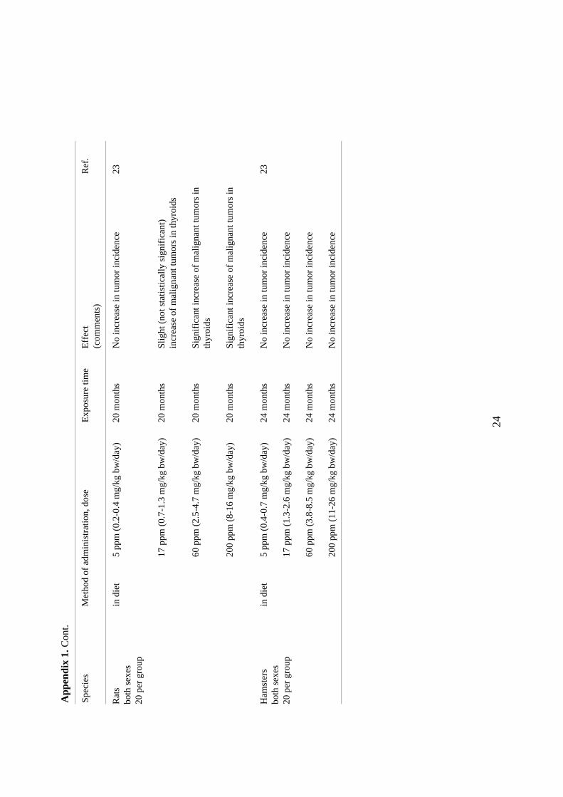
The ETU-exposed rats showed thyroid damage but no non-neoplastic damage toliver or pituitary (11, 66). Thyroid hyperplasia was observed in both male andfemale rats exposed to 83 ppm for 9 months, and was accompanied by significantreductions of T3 and T4 and elevated TSH. A lower concentration (25 ppm) alsohad effects on T3 , T4 and TSH in the animals that had been exposed perinatally to9 ppm. At the close of the two-year study no histopathological effects wereobserved in the rats exposed to levels below 83 ppm, but 60 to 90% of thoseexposed to 83 ppm and 250 ppm had hyperplasias in thyroid follicular cells (11).In a French study, groups of 20 male and 20 female rats were exposed to 0, 5, 17,60 or 200 ppm ETU in feed for two years (23). Reduced food intake and effectson body weight were reported at 17 ppm and higher. Significantly elevated serumcholesterol levels were found in all dose groups and both sexes. The elevationswere constant over time (3 to 24 months) and dose-dependent: 5 ppm ETU raisedthe cholesterol level by about 30%, and 200 ppm by about 80%. Slightly elevatedserum levels of the hepatic enzymes alkaline phosphatase (ALP) and alanineaminotransferase (ALT) were also observed, but they were temporary and notclearly related to the dose of ETU. The intake of ETU at 5 ppm in feed wascalculated to be about 0.37 mg/kg b.w./day at one month of age, and 0.22 to 0.26mg/kg b.w./day at 3 months and older.
HamstersIn conjunction with the study described above, 20 hamsters of each sex per groupwere exposed to ETU in feed for 20 months. Dose levels were 0, 5, 17, 60 or200 ppm (23). Reduced food intake and lower body weights were observed at60 ppm and higher. As with the rats, cholesterol levels in serum in both sexes andat all dose levels and all times were significantly above those of controls. At theend of the 20-month exposure, ALP and ALT levels were also significantlyelevated (about 40% at most) in both sexes at all dose levels. Glucose-6-phosphatedehydrogenase in the liver was significantly lower (as much as 60%) in both sexesat all dose levels.
10
DogsBeagles of both sexes have also been experimentally exposed to ETU. Exposureshave been for 4, 13, or 52 weeks. These studies have not been published, but havebeen assessed by the FAO/WHO expert panel (20). In the 4-week study, the dogs(2 of each sex per group) were exposed to concentrations of 0, 200, 980 and4900 ppm in feed. Reduced weight gain, lower T4 and T3 levels and thyroidenlargement were observed at 980 ppm. In the 13-week study, the dogs (4 of eachsex per group) were exposed to 0, 10, 150 and 2000 ppm in feed. No effects werenoted at 10 ppm (NOAEL), which according to the WHO expert group is equi-valent to 0.39 mg/kg b.w./day. At 150 and 2000 ppm there were statisticallysignificant reductions of hemoglobin, hematocrit and red blood cells, and astatistically significant elevation of cholesterol level. Effects on the thyroid wereseen only at 2000 ppm. In the 52-week study, the dogs (4 of each sex per group)were exposed to 0, 5, 50 or 500 ppm ETU in feed. No effects were observed at5 ppm (NOAEL). The 50 ppm exposure resulted in reduced weight gain, thyroidhypertrophy with colloid accumulation, slight increase in thyroid weight and anaccumulation of pigment in the liver (20).
MonkeysRhesus monkeys caught in the wild (5 of each sex per group) were exposed toETU in diet for about 6 months in two studies which have not been published butare mentioned by the FAO/WHO expert panel (20). The studies report increaseduptake of 125I at a concentration of 50 ppm, and elevated thyroid and spleenweights in males at 150 ppm and above and in females at 50 ppm and above.These studies were judged to be unreliable, however, since the monkeys werenot entirely healthy (20).
Genotoxicity, mutagenicity
The results of various short-term tests published prior to 1993 have beensummarized and evaluated by Dearfield (14). The overall impression from thelarge number of bacterial tests made with ETU is that the substance has weak butdose-dependent mutagenic activity which is not apparent at concentrations below1000 µg per plate (20 ml medium), and that the mutations are base-substitutions.High concentrations have caused aneuploidy (incomplete chromosome separation)in yeast cells, mutations in Tradescantia plants, and gene mutations and chromo-some aberrations in mammalian cells. In vivo tests with mammals have usuallybeen negative (14).
Subsequently published studies report aneuploidy in yeast at a concentrationof about 500 µg/l, and inhibited mitosis and elevated numbers of chromosomeaberrations in onions at concentrations of 2.5 and 25 µg/ml (21). Increasednumbers of somatic mutations were observed in two insecticide-resistant strainsof Drosophila when the larvae were raised on food containing 50 or 100 mgETU/liter (70). The Comet assay was used to identify and quantify the DNAdamage (alkaline labile sites) in mice that had been treated with ETU (76). ETU
11
was tested along with 7 other substances that cause hepatic cancers in experi-mental animals but have not been shown to cause micronuclei in the bone marrowcells of mice. The mice were killed 3 hours and 24 hours after receiving a singleintraperitoneal dose of 2000 mg/kg body weight. The ETU caused DNA damagein cells from liver, lung, spleen, kidney and bone marrow.
Mutagenicity in bacteria is greatly increased if ETU is combined with nitrite,and N-nitroso ETU is strongly mutagenic in bacterial tests (79, 80, 82). A re-markable sensitivity to ETU plus sodium nitrite was observed in two studies usingthe host-mediated assay method (8, 82). In mice given an oral dose of 50 mgNaNO2/kg body weight, there was a significant, dose-dependent increase in thenumber of mutations in Salmonella typhimurium G46 when the mice weresimultaneously given ETU in doses of 1 to 25 mg/kg body weight (82).
The interaction between ETU and sodium nitrite has also been studied withregard to induction of dominant-lethal mutations in mice (88). Daily doses of ETU(150 mg/kg b.w.) and NaNO2 (50 mg/kg b.w.) were given orally for five conse-cutive days to male mice, which were mated with groups of untreated females forthe following six weeks. The females mated six weeks after the treatment had agreatly reduced proportion of pregnancies as well as lower numbers of implantsand living embryos. The delayed effects were regarded as an indication that ETUin the presence of NaNO2 forms N-nitroso ETU and damages the stem cells(spermatogonia). An increase in the number of genetic aberrations was also seenin stem cells of mice after treatment with 100 mg N-nitroso ETU/kg b.w. NeitherETU nor N-nitroso ETU has been tested on mammals with methods that canreveal hereditary (non-lethal) changes (e.g. specific locus test, mouse spot test).
In brief, ETU is regarded as weakly genotoxic because of the dose-dependentincreases of gene mutations observed in bacteria and the results of a few tests withyeast cells, plants, fruit flies, mammalian cells and laboratory mammals (in vivo),which have shown genotoxic effects at high exposure levels. Most in vivo testswith mammals have been negative, however. N-nitroso ETU, on the other hand, isa powerful genotoxin both in vitro and in vivo. Endogenous formation of N-nitroso ETU, which occurs mostly in acidic environments, must be consideredwhen assessing both the genotoxicity of ETU and its potential carcinogenic effect.The probability of N-nitroso ETU formation with ETU exposure must be muchlower with inhalation or skin uptake than it is with oral exposure.
Earlier assessments of genotoxicityIn 1987, the IARC summarized data from genotoxicity tests in the form of agenotoxicity profile (32). Positive results were reported only from tests withprokaryotes and lower eukaryotes. ETU was classified as non-genotoxic in anassessment of pesticides made for a FAO/WHO program (20). The NTP reportedthat ETU had been thoroughly tested for genotoxicity using numerous testmethods both in vivo and in vitro, and with few exceptions results had beennegative (66). DECOS, which set a health-based exposure limit for ETU (15),reported that ETU per se is non-mutagenic. A 1995 review article described ETU
12
as non-genotoxic in mammalian systems and proposed that ETU causes livertumors in mice by a non-genotoxic mechanism (18). Dearfield (at the EPA) madethe overall assessment that ETU could not be regarded as lacking genotoxicactivity (14). The genotoxic effect was judged to be weak, but it was pointed outthat nitrosation creates a mutagenic product that may be more potent.
Carcinogenicity
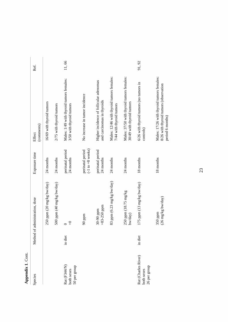
The results of cancer tests are summarized in Appendix 1. Most of these studieswere made with rats. Both short-term and long-term toxicity studies have shownthat species differ in both sensitivity to ETU and the organs affected.
MiceElevated incidences of hepatic adenomas and carcinomas have been reported inmice at a dose of 66 mg/kg/day (330 ppm ETU in feed) (11, 66). Male and femalemice were exposed perinatally from minus one up to eight weeks of age (F0)and/or as adults (F1) to between 0 and 1000 ppm ETU in feed. The mice exposedto 330 ppm perinatally only showed no tumors after two years. Those exposed to330 ppm as adults only had tumors in liver, pituitary or thyroid. For adult animalsexposed to 330 ppm, the incidences of thyroid and pituitary tumors were margi-nally higher in animals that had also been exposed perinatally. Perinatal exposureto 300 ppm, however, had no effect on tumor incidence in mice exposed to thehighest dose (1000 ppm) when they were compared to the group not exposedperinatally.
Yoshida et al. (93) made a study in which ETU was given to mice (Crj:CD1) incombination with sodium nitrite. The mice were given ETU and sodium nitrite (inwater) by gavage once a week for ten weeks, in the following combinations (ETU+ NaNO2): 0 + 0; 100 + 0; 0 + 70; 25 + 17.5; 50 + 35 and 100 + 70 mg/kg bodyweight. The animals were observed for 18 months beginning with the first treat-ment. It was found that ETU combined with sodium nitrite caused an earlierappearance of tumors and/or a dose-dependent increase of tumors in lymphatictissue, lung, stomach, Harder’s gland and uterus. The tumor locations were thusnot the same as those observed after administration of ETU alone (see 11, 66).No carcinogenic effect was observed in mice given ETU or sodium nitrite alone.A dose-dependent increase of pulmonary adenomas and adenocarcinomas wasobserved in both females and males, and the number of females with pulmonaryadenomas or adenocarcinomas was significantly elevated in the group given (ETU+ NaNO2 ) 25 + 17.5 mg/kg b.w./week. These results suggest that ETU istransformed in vivo to N-nitroso ETU and that N-nitroso ETU has a strongercarcinogenic effect on mice than ETU alone. This has been confirmed in a studyof tumor induction in mice (ICR females), in which oral administration of N-nitroso ETU in doses of 0.66 to 2.64 mg (26.4 to 105.6 mg/kg b.w.) once a weekfor ten weeks increased the incidence of pulmonary tumors and lymphocyticneoplasms (62).
13
RatsFor rats, the thyroid has been found to be the organ most sensitive to both short-term and long-term exposures. Dose-dependent increases of thyroid tumors havebeen observed in a number of different studies with rats (11, 23, 24, 26, 91, 92).Graham et al. reported increased appearance of thyroid tumors at 250 ppm ormore in feed (24, 26), but not at 125 ppm. Calculating from information given bythe authors, exposure to 125 ppm in feed is equivalent to about 10 mg/kg bodyweight/day (NOAEL). Gak et al. (23) reported no thyroid tumors after 20 monthsof exposure to 17 ppm in feed (according to the authors, equivalent to 1.27 mg/kgb.w./day or less).
In the NTP study, male and female rats were exposed perinatally from minus oneup to eight weeks of age (F0) and as adults (F1) to 0-250 ppm ETU in feed. Therewas a clear increase of hyperplasia in thyroid follicular cells after 2 years ofexposure to 83 or 250 ppm. Thyroid follicular cell adenomas or carcinomas wereseen in about 20% of animals exposed to 83 ppm and in about 60% of thoseexposed to 250 ppm. Male rats were more sensitive than females to the carcino-genic effects of ETU. In addition to the dose-dependent increases of thyroid tumors,there was also a small but significant increase of tumors in Zymbal’s glands (bothsexes at F0 90 ppm, F1 250 ppm) and mononuclear cell leukemia (both sexes at F0
90 ppm, F1 250 ppm; males at F0 90 ppm, F1 83 ppm) (11). The LOEL was 83 ppm,which according to DECOS (15) is equivalent to 6.23 mg/kg b.w./day.
The ability of thioureas (e.g. ETU and thiourea) to cause thyroid tumors isattributed to hormonal disturbances. Rats are regarded as a sensitive species in thisrespect. Thiourea inhibits the enzyme thyroid peroxidase, which causes serumlevels of thyroid hormones T3 and T4 to decline. This in turn stimulates the hypo-thalamus and pituitary, and more thyroid-stimulating hormone (TSH) is produced.TSH stimulates thyroid growth, and chronically elevated levels of TSH in serumcan cause thyroid hyperplasia which may eventually develop into tumors (2, 27,28).
When female rats were given simultaneous doses of ETU (80 mg/kg bodyweight) and NaNO2 (56 mg/kg body weight) once per week from 11 to 51 weeksof age, 13% of animals developed adenocarcinomas in uterine endometrium (65).No such tumors were observed in controls.
Teratogenicity
ETU is strongly teratogenic to rats (10, 12, 41, 56, 72, 75). It can also haveembryotoxic effects on mice (10, 39), rabbits (41), cats (45), hamsters (10, 46),and guinea pigs (10). ETU causes elevated mortality and a low incidence ofmalformations in some of these species, but only at high dose levels (12, 40). Inan aquatic in vitro assay for embryotoxic effects on water fleas (Daphnia magna),a significant increase in the incidence of malformations was seen at an ETUconcentration of 20 mg/liter (68).
14
The lowest single oral dose that yields developmental anomalies in rats(LOAEL) is 40 mg/kg body weight. For repeated doses, the lowest exposure is10 to 20 mg/kg/day on days 6 to 15 of gestation (41, 72). Maternal toxicity wasobserved at 80 mg/kg/day, and somewhat retarded ossification was observed in athird of fetuses after repeated administration of 5 mg/kg (41). Brain damage is themost common teratogenic effect in rats. ETU causes craniocele, meningo-encephalocele, hydrocephalus, obliteration of the neural canal and enlarged brainventricles. Skeletal damage is also common, and includes club foot, short andcrooked tails, and rib anomalies. ETU shows different types of teratogenic effects,determined by the stage of gestation at which the mother was exposed. It wasshown in one study (72) that effects on the eyes appeared only after treatment ondays 10 and 11, tail defects after treatment on days 11– 14, and cleft palate aftertreatment on days 12-16. Damage to toes on forepaws appeared at earlierexposures than damage to toes on hindpaws. In a study of teratogenic effects ofETU in thyroidectomized females and controls given false thyroidectomies, it wasconcluded that ETU-dependent changes in thyroid function or thyroxine levels inthe mothers was probably not the explanation for the teratogenic effects of ETU(56).
Rat embryos examined after in vitro exposure to ETU show damage, primarilyto the tail and head, at concentrations of 10 mg/liter or higher (13, 35, 42, 89).Both early (days 10-13) and late (day 19) prenatal exposure damages nervoustissue (13, 40). Neural cells have been identified as particularly sensitive to thetoxic effects of ETU, both by examination of cells and tissues from exposedembryos and by exposure of cultured embryonic cells (13, 40, 89).
For mice, the lowest single dose with embryotoxic effect (LOAEL) is1600 mg/kg (39); for repeated exposures, the lowest dose level is more than200 mg/kg (10). Rats are thus twenty to forty times more sensitive than mice,although the teratogenic effects of ETU are of the same types in both species (13).The difference in metabolic capacity between rats and mice leads to higher bloodlevels of ETU in rats than in mice (see Biotransformation) (73). The effect ofmaternal metabolism was examined by adding S9 from Aroclor-1254 induced rator mouse liver together with a NADPH-generating system to embryos exposed toETU in vitro (13). S9 from mouse completely neutralized the teratogenic effect ofETU on both rat and mouse embryos. The differences in metabolism may explainsome of the differences in sensitivity between rats and mice, but rat embryos andcultured brain cells from rats are also more sensitive to the toxic effects of ETUthan similar tissues from mice with exposure in vitro (13, 89).
Effects of nitrosationIn the presence of NaNO2 ETU is nitrosated to a substance that is teratogenic tomice (87): 400 mg/kg ETU given together with 200 mg/kg NaNO2 is embryotoxicand teratogenic, causing primarily skeletal anomalies when administered on day 6,8, or 10, but not when administered on day 12 of gestation. The NaNO2 has effectonly when given within about an hour of the ETU exposure (87). The reported

15
damage includes deformed tails and ribs, omphalocele, cleft palate, highfrequency of deformed vertebra, fused lung lobes, missing kidneys (kidneyagenesis), small or missing eyes and swollen brain ventricles (87).
N-nitroso ETU causes hydrocephalus in rat embryos (44). It has been observed,however, that NaNO2 almost completely neutralizes the teratogenic effects ofETU in rats treated on day 13 or 15 of gestation (43).
Dose-effect/dose-response relationships
It was found in one study that occupational exposure to ETU at air concentrationsin the range 120–160 mg/m3 inhibits thyroid function, measured as somewhatlower levels of the thyroid hormone T4 (thyroxine). Levels of T4 were lower inexposed workers (geometric mean 80.5 nmol/l) than in unexposed controls(geometric mean 105.7 nmol/l), but the individual values were all within normalreference limits for T4 (84). Assuming an air intake of 10 m3/day and a bodyweight of 70 kg, exposure to an air concentration of 120 µg/m3 would result inuptake of about 17 µg/kg/day. Skin uptake is likely, and may be high.
Elevated TSH levels were observed in Mexican farm workers exposed to fungi-cides containing ETU. ETU levels in urine were on average 58 ppb (µg/liter) (85).Assuming a urine volume of 2 liters, that half of absorbed ETU is excreted in urine,and that uptake is complete, this is equivalent to a single dose of 3 to 4 µg/kg.
Considering the widespread use of ETU, there are few reported cases of contactallergy.
Relevant information from animal experiments with oral exposure is summarizedin Table 1. In the only (unpublished) inhalation study with rats, effects on thyroidwere noted at exposure to 40 mg/m3 ETU 6 hours/day (3). If it is assumed that airintake is 0.2 m3/day and body weight is 0.33 kg, this exposure is equivalent to6 mg/kg/day. Exposure to 10 mg/m3 had no effect.
Conclusions
Data from occupational exposures indicate that the critical effect of occupationalexposure to ETU is its effect on the thyroid. This effect has also been observed inexperimental animals. ETU is carcinogenic to experimental animals. Simultaneousexposure to ETU and nitrite in food yields tumors at lower ETU levels and inother organs. ETU is considered to be slightly genotoxic, whereas N-nitroso ETUis strongly genotoxic. ETU is teratogenic to experimental animals. The teratogeniceffect of ETU in different species seems to be inversely related to the biotrans-formation rate. There are neither qualitative nor quantitative data on biotrans-formation in humans. A few cases of contact allergy have been reported after skincontact with ETU, but the allergenic potential of ETU is probably low. Animalstudies suggest that skin uptake may be high.

16
Table 1. Effects observed in experimental animals given ETU in feed.
Dose(mg/kg/day)
Concentration infeed (ppm)
Duration ofexposure
Level value for effect, species,effect
Ref.
0.2 - 0.4 5 24 months LOEL, ratelevated serum cholesterol
23
0.4 - 0.7 5 20 months LOEL, hamsterelevated serum cholesterol
23
ca. 2 25 60 days LOEL, rateffects on thyroid
22
ca. 2 25 90 days NOEL, rateffects on thyroid
22
ca. 2 50 52 weeks LOEL, dogeffects on thyroid
20
3.7 50 120 days NOAEL, rateffects on thyroid
25
3 - 4.3 60 13 weeks LOAEL, ratthyroid hyperplasia
66
5 until day 15 ofgestation
LOAEL, ratretarded ossification
41
6.2 83 24 months LOAEL, ratthyroid cancer, effects on thyroid
11
10 until day 15 ofgestation
LOAEL, ratteratogenic effects
41
10 20 days LOAEL, catCNS toxicity
45
23 28 days LOEL, ratstructural changes in renal tubules
50
66 330 24 months lowest tested dose, mouseliver cancer, effects on thyroid
11
References
1. Allen JR, Van Miller JP, Seymour JL. Absorption, tissue distribution and excretion of 14Cethylenethiourea by the rhesus monkey and rat. Res Commun Chem Pathol Pharmacol1978;20:109-115.
2. Andrae U, Greim H. Initiation and promotion in thyroid carcinogenesis. In: Dekant W,Neumann H, eds. Tissue-specific Toxicity: Biochemical Mechanisms. London: AcademicPress, 1992:71-93.
3. Anonymous. Ethylenthioharnstoff. Berufsgenossenschaft der chemischen Industrie.Heidelberg, Germany, 1995. No. 1, June 1995.
4. Aprea C, Betta A, Catenacci G, Colli A, Lotti A, Minoia C, Olivieri P, Passini V, Pavan I,Roggi C, Ruggeri R, Sciarra G, Turci R, Vannini P, Vitalone V. Urinary excretion ofethylenethiourea in five volunteers on a controlled diet (multicentric study). Sci Total Environ1997;203:167-179.
5. Aprea C, Betta A, Catenacci G, Lotti A, Minoia C, Passini W, Pavan I, Saverio Robustellidella Cuna F, Roggi C, Ruggeri R, Soave C, Sciarra G, Vannini P, Vitalone V. Referencevalues of urinary ethylenethiourea in four regions of Italy (multicentric study). Sci TotalEnviron 1996;192:83-93.
17
6. Aprea C, Sciarra G, Sartorelli P, Mancini R, Di Luca V. Environmental and biologicalmonitoring of exposure to mancozeb, ethylenethiourea, and dimethoate during industrialformulation. J Toxicol Environ Health 1998;53:263-281.
7. Autio K. Determination of ethylenethiourea (ETU) as a volatile N,N´-dimethyl derivative byGLC-MS and GLC-NPSD. Applications for determining ETU residues in berries andcigarette smoke condensate. Finn Chem Lett 1983;4:10-14.
8. Autio K, von Wright A, Pyysalo H. The effect of oxidation of the sulfur atom on themutagenicity of ethylenethiourea. Mutat Res 1982;106:27-31.
9. Bruze M, Fregert S. Allergic contact dermatitis from ethylene thiourea. Contact Dermatitis1983;9:208-212.
10. Chernoff N, Kavlock RJ, Rogers EH, Carver BD, Murray S. Perinatal toxicity of maneb,ethylene thiourea, and ethylenebisisothiocyanate sulfide in rodents. J Toxicol Environ Health1979;5:821-834.
11. Chhabra RS, Eustis S, Haseman JK, Kurtz PJ, Carlton BD. Comparative carcinogenicity ofethylene thiourea with or without perinatal exposure in rats and mice. Fundam Appl Toxicol1992;18:405-417.
12. Daston GP. Advances in understanding mechanisms of toxicity and implications for riskassessment. Reprod Toxicol 1997;11:389-396.
13. Daston GP, Yonker JE, Powers JF, Heitmeyer SA. Difference in teratogenic potency ofethylenethiourea in rats and mice: relative contribution of embryonic and maternal factors.Teratology 1989;40:555-566.
14. Dearfield KL. Ethylene thiourea (ETU). A review of the genetic toxicity studies. Mutat Res1994;317:111-132.
15. DECOS. Health-based Recommended Occupational Exposure Limits for Ethylene Thiourea.Dutch Expert Committee for Occupational Standards. Directorate General of Labour, TheNetherlands, 1999;03:1-64.
16. Doerge DR, Takazawa RS. Mechanism of thyroid peroxidase inhibition by ethylenethiourea.Chem Res Toxicol 1990;3:98-101.
17. Dubey JK, Heberer T, Stan HJ. Determination of ethylenethiourea in food commodities by atwo-step derivatization method and gas chromatography with electron-capture and nitrogen-phosphorus detection. J Chromatogr A 1997;765:31-38.
18. Elia M, Arce G, Hurt SS, O’Neill PJ, Scribner HE. The genetic toxicology ofethylenethiourea: a case study concerning the evaluation of a chemical’s genotoxic potential.Mutat Res 1995;341:141-149.
19. FAO/WHO. Ethylenethiourea (ETU). In: Pesticide residues in food - 1993. Report sponsoredjointly by FAO and WHO. FAO Plant Production and Protection paper 122. 1993:52-56.
20. FAO/WHO.Ethylenethiourea. In: Pesticide residues in food - 1993. Toxicology evaluations.WHO 1994:167-213.
21. Franekic J, Bratulic N, Pavlica M, Papes D. Genotoxicity of dithiocarbamates and theirmetabolites. Mutat Res 1994;325:65-74.
22. Freudenthal RI, Kerchner G, Persing R, Baron RL. Dietary subacute toxicity of ethylenethiourea in the laboratory rat. J Environ Pathol Toxicol 1978;1:147-161.
23. Gac JC, Graillot C, Truhaut R. Difference in the sensitivity of the hamster and the rat to theeffects of long-term administration of ethylenethiourea. Eur J Toxicol Environ Hyg1976;9:303-312. (in French, English abstract)
24. Graham SL, Davis KJ, Hansen WH, Graham CH. Effects of prolonged ethylene thioureaingestion on the thyroid of the rat. Food Cosmet Toxicol 1975;13:493-499.
25. Graham SL, Hansen WH. Effects of short-term administration of ethylenethiourea uponthyroid function of the rat. Bull Environ Contam Toxicol 1972;7:19-25.
26. Graham SL, Hansen WH, Davis KJ, Perry CH. Effects of one-year administration ofethylenethiourea upon the thyroid of the rat. J Agric Food Chem 1973;21:324-329.
18
27. Hard GC. Recent developments in the investigation of thyroid regulation and thyroidcarcinogenesis. Environ Health Perspect 1998;106:427-436.
28. Hill R, Crisp T, Hurley P, Rosenthal S, Singh D. Risk assessment of thyroid follicular celltumors. Environ Health Perspect 1998;106:447-457.
29. Houeto P, Bindoula G, Hoffman JR. Ethylenebisdithiocarbamates and ethylenethiourea:possible human helalth hazards. Environ Health Perspect 1995;103:568-573.
30. Hui QY, Armstrong C, Laver G, Iverson F. Monooxygenase-mediated metabolism andbinding of ethylene thiourea to mouse liver microsomal protein. Toxicol Lett 1988;41:231-237.
31. IARC. Some anti-thyroid and related substances, nitrofurans and industrial chemicals.Ethylenethiourea. IARC Monographs on the Evaluation of Carcinogenic Risk of Chemicals toMan. 1974;7:45-52.
32. IARC. Overall evaluations of carcinogenicity: An updating of IARC monographs Volumes 1to 42. IARC Monographs on the Evaluation of Carcinogenic Risks to Humans. 1987;suppl7:207-208.
33. IPCS. Dithiocarbamate pesticides, ethylenethiourea, and propylenethiourea: A generalintroduction. Environmental Health Criteria 78. Geneva: International Programme onChemical Safety, World Health Organization, 1988:1-140.
34. Iverson F, Khera KS, Hierlihy SL. In vivo and in vitro metabolism of ethylenethiourea in therat and the cat. Toxicol Appl Pharmacol 1980;52:16-21.
35. Iwase T, Yamamoto M, Shirai M, Akahori F, Masaoka T, Takizawa T, Arishima K, EguchiY. Effect of ethylene thiourea on cultured rat embryos in the presence of hepatic microsomalfraction. J Vet Med Sci 1997;59:59-61.
36. Jablonicka A, Polakova H, Karelova J, Vargova M. Analysis of chromosome aberrations andsister-chromatid exchanges in peripheral blood lymphocytes of workers with occupationalexposure to the mancozeb-containing fungicide Novozir Mn80. Mutat Res 1989;224:143-146.
37. Kanerva L, Estlander T, Jolanki R. Occupational allergic contact dermatitis caused bythiourea compounds. Contact Dermatitis 1994;31:242-248.
38. KatoY, Odanaka Y, Teramoto S, Matano O. Metabolic fate of ethylenethiourea in pregnantrats. Bull Environ Contam Toxicol 1976;16:546-555.
39. Khera KS. Ethylenethiourea-induced hindpaw deformities in mice and effects of metabolicmodifiers on their occurrence. J Toxicol Environ Health 1984;13:747-756.
40. Khera KS. Ethylenethiourea: a review of teratogenicity and distribution studies and anassessment of reproduction risk. Crit Rev Toxicol 1987;18:129-139.
41. Khera KS. Ethylenethiourea: teratogenicity study in rats and rabbits. Teratology 1973;7:243-252.
42. Khera KS. Neuronal degeneration caused by ethylenethiourea in neuronal monocell layers invitro and in fetal rat brain in vivo. Teratology 1987;36:87-93.
43. Khera KS. Reduction of teratogenic effects of ethylenethiourea in rats by interaction withsodium nitrite in vivo. Food Chem Toxicol 1982;20:273-278.
44. Khera KS, Iverson F. Hydrocephalus induced by N-nitrosoethylenethiourea in the progeny ofrats treated during gestation. Teratology 1980;21:367-370.
45. Khera KS, Iverson F. Toxicity of ethylenethiourea in pregnant cats. Teratology 1978;18:311-313.
46. Khera KS, Whalen C, Iverson F. Effects of pretreatment with SKF-525A, N-Methyl-2-thioimidazole, sodium phenobarbital, or 3-methylcholanthrene on ethylenethiourea-inducedteratogenicity in hamsters. J Toxicol Environ Health 1983;11:287-300.
47. Klaassen CD, ed. Casarett and Doull’s Toxicology: The Basic Science of Poisons. New York:McGraw-Hill, 1996.
19
48. Kobayashi H, Kaneda M, Teramoto S. Identification of 1-methylthiourea as the metabolite ofethylenethiourea in rats by high-performance liquid chromatography. Toxicol Lett1982;12:109-113.
49. Kurttio P, Savolainen K. Ethylenethiourea in air and in urine as an indicator of exposure toethylenebisdithiocarbamate fungicides. Scand J Work Environ Health 1990;16:203-207.
50. Kurttio P, Savolainen K, Naukkarinen A, Kosma VM, Tuomisto L, Penttila I, Jolkkonen J.Urinary excretion of ethylenethiourea and kidney morphology in rats after continuous oralexposure to nabam or ethylenethiourea. Arch Toxicol 1991;65:381-385.
51. Kurttio P, Savolainen K, Tuominen R, Kosma VM, Naukkarinen A, Mannisto P, Collan Y.Ethylenethiourea and nabam induced alterations of function and morphology of thyroid glandin rats. Arch Toxicol 1986;Suppl. 9:339-344.
52. Kurttio P, Vartiainen T, Savolainen K. Environmental and biological monitoring of exposureto ethylenebisdithiocarbamate fungicides and ethylenethiourea. Br J Ind Med 1990;47:203-206.
53. Laurell C-B, Lundh B, Nosslin B. Klinisk kemi i praktisk medicin (fourth edition). Lund:Studentlitteratur, 1980.
54. Lewerenz HJ, Plass R. Contrasting effects of ethylenethiourea on hepatic monooxygenases inrats and mice. Arch Toxicol 1984;56:92-95.
55. Lewerenz HJ, Plass R. Effect of ethylenethiourea on kidney function in the rat. Z GesamteHyg 1988;34:304-307. (in German, English abstract)
56. Lu MH, Staples RE. Teratogenicity of ethylenethiourea and thyroid function in the rat.Teratology 1978;17:171-178.
57. Lyman WR, Lacoste RJ. New developments in the chemistry and fate ofethylenebisdithiocarbamate fungicides. In: Proceedings of the 3rd International IUPACCongress on Pesticide Chemistry, Helsinki, 3-9 July, 1974. Stuttgart: George ThiemePublishers, 1974:67-74.
58. MAK, DFG (Deutsche Forschungsgemeinschaft). Toxikologisch-arbeitsmedizinischeBegründungen von MAK-Werten. Ethylenthioharnstoff. Weinheim: VCH-Verlagsgesellschaft, 1995 (Lieferung 21).
59. Matsushita T, Arimatsu Y, Nomura S. Experimental study on contact dermatitis caused bydithiocarbamates maneb, mancozeb, zineb, and their related compounds. Int Arch OccupEnviron Health 1976;37:169-178.
60. Meding B, Baum H, Bruze M, Roupe G, Trulsson L. Allergic contact dermatitis fromdiphenylthiourea in Vulkan heat retainers. Contact Dermatitis 1990;22:8-12.
61. Meneguz A, Michalek H. Induction of hepatic microsomal mixed function oxidase system byethylenethiourea in mice. Arch Toxicol 1986;Suppl. 9:346-350.
62. Moriya M, Mitsumori K, Kato K, Miyazawa T, Shirasu Y. Carcinogenicity of N-nitroso-ethylenethiourea in female mice. Cancer Lett 1979;7:339-342.
63. Nebbia C, Fink-Gremmels J. Acute effects of low doses of zineb and ethylenethiourea onthyroid function in the male rat. Bull Environ Contam Toxicol 1996;56:847-852.
64. Newsome WH. The excretion of ethylenethiourea by rat and guinea pig. Bull EnvironContam Toxicol 1974;11:174-176.
65. Nishiyama K, Ando-Lu J, Nishimura S, Takahashi M, Yoshida M, Sasahara K, Miyajima K,Maekawa A. Initiating and promoting effects of concurrent oral administration ofethylenethiourea and sodium nitrite on uterine endometrial adenocarcinoma development inDonryu rats. In Vivo 1998;12:363-368.
66. NTP. Technical report on the toxicology and carcinogenesis studies of ethylene thiourea inF344/N rats and B6C3F1 mice (feed studies). Research Triangle Park, NC: NatiounalToxicology Program, 1992 (Report No. 388).
67. O’Neil WM, Marshall WD. Goitrogenic effects of ethylenethiourea on rat thyroid. PesticBiochem Physiol 1984;21:92-101.
20
68. Ohta T, Tokishita S, Shiga Y, Hanazato T, Yamagata H. An assay system for detectingenvironmental toxicants with cultured cladoceran eggs in vitro: malformations induced byethylenethiourea. Environ Res 1998:77:43-48.
69. Pastorelli R, Allevi R, Romagnano S, Meli G, Fanelli R, Airoldi L. Gas chromatography-mass spectrometry determination of ethylenethiourea hemoglobin adducts: a possibleindicator of exposure to ethylene bis dithiocarbamate pesticides. Arch Toxicol 1995;69:306-311.
70. Rodriguez-Arnaiz R. Genotoxic activation of hydrazine, two dialkylhydrazines, thiourea andethylene thiourea in the somatic w/w + assay of Drosophila melanogaster. Mutat Res1997;395:229-242.
71. Rose D, Pearson CM, Zuker M, Roberts JR. Ethylenethiourea: Criteria for the Assessment ofits Effects on Man. National Research Council Canada, Associate Committee on ScientificCriteria for Environmental Quality, 1980 (NRCC No. 18469).
72. Ruddick JA, Khera KS. Pattern of anomalies following single oral doses of ethylenethioureato pregnant rats. Teratology 1975;12:277-281.
73. Ruddick JA, Newsome WH, Iverson F. A comparison of the distribution, metabolism andexcretion of ethylenethiourea in the pregnant mouse and rat. Teratology 1977;16:159-162.
74. Rudzki E.Ostaszewski K, Grzywa Z, Kozlowska A. Sensitivity to some rubber additives.Contact Dermatitis 1976;2:24-27.
75. Saillenfait AM, Sabate JP, Langonne I, de Ceaurriz J. Difference in the developmentaltoxicity of ethylenethiourea and three N,N´-substituted thiourea derivatives in rats. FundamAppl Toxicol 1991;17:399-408.
76. Sasaki YF, Izumiyama F, Nishidate E, Matsusaka N, Tsuda S. Detection of rodent livercarcinogen genotoxicity by the alkaline single-cell gel electrophoresis (Comet) assay inmultiple mouse organs (liver, lung, spleen, kidney, and bone marrow). Mutat Res1997;391:201-214.
77. Savolainen K, Hervonen H, Komulainen H, Kurttio P. Peripheral and central nervous systemeffects of nabam and ethylenethiourea in rats. Arch Toxicol 1986;Suppl. 9:345.
78. Savolainen K, Pyysalo H. Identification of the main metabolite of ethylenethiourea in mice. JAgric Food Chem 1979;27:1177-1181.
79. Seiler JP. In vivo mutagenic interaction of nitrite and ethylenethiourea. Experientia1975;31:214-215.
80. Seiler JP. Nitrosation in vitro and in vivo by sodium nitrite, and mutagenicity of nitrogenouspesticides. Mutat Res 1977;48:225-236.
81. Shephard SE, Schlatter C, Lutz WK. Assessment of the risk of formation of carcinogenic N-nitroso compounds from dietary precursors in the stomach. Food Chem Toxicol 1987;25:91-108.
82. Shirasu Y, Moriya M, Kato K, Lienard F, Tezuka H, Teramoto S, Kada T. Mutagenicityscreening on pesticides and modification products: a basis of carcinogenicity evaluation. ColdSpring Harbor Conference Cell Proliferation 1977;4:267-285.
83. Smith D. Ethylene thiourea – a study of possible teratogenicity and thyroid carcinogenicity. JSoc Occup Med 1976;26:92-94.
84. Smith DM. Ethylene thiourea: thyroid function in two groups of exposed workers. Br J IndMed 1984;41:362-366.
85. Steenland K, Cedillo L, Tucker J, Hines C, Sorensen K, Deddens J, Cruz V. Thyroidhormones and cytogenetic outcomes in backpack sprayers using ethylenebis(dithiocarbamate)(EBDC) fungicides in Mexico. Environ Health Perspect 1997;105:1126-1130.
86. Stula EF, Krauss WC. Embryotoxicity in rats and rabbits from cutaneous application ofamide-type solvents and substituted ureas. Toxicol Appl Pharmacol 1977;41:35-55.
87. Teramoto S, Saito R, Shirasu Y. Teratogenic effects of combined administration ofethylenethiourea and nitrite in mice. Teratology 1980;21:71-78.
21
88. Teramoto S, Shingu A, Shirasu Y. Induction of dominant-lethal mutations afteradministration of ethylenethiourea in combination with nitrite of the n-nitroso-ethylenethiourea in mice. Mutat Res 1978;56:335-340.
89. Tsuchiya T, Nakamura A, Iio T, Takahashi A. Species differences between rats and mice inthe teratogenic action of ethylenethiourea: in vivo/in vitro tests and teratogenic activity ofsera using an embryonic cell differentiation system. Toxicol Appl Pharmacol 1991;109:1-6.
90. Ugazio G, Brossa O, Grignolo F. Hepato- and neuro-toxicity by ethylenethiourea. ResCommun Chem Pathol Pharmacol 1985;48:401-414.
91. Ulland BM, Weisburger JH, Weisburger EK, Rice JM, Cypher R. Thyroid cancer in rats fromethylene thiourea intake. J Natl Cancer Inst 1972;49:583-584.
92. Weisburger EK, Ulland BM, Nam J, Gart JJ, Weisburger JH. Carcinogenicity tests of certainenvironmental and industrial chemicals. J Natl Cancer Inst 1981;67:75-88.
93. Yoshida A, Harada T, Maita K. Tumor induction by concurrent oral administration ofethylenethiourea and sodium nitrite in mice. Toxicol Pathol 1993;21:303-310.

22
App
endi
x 1.
Res
ults
fro
m c
arci
noge
nici
ty te
sts
with
exp
erim
enta
l ani
mal
s ex
pose
d to
eth
ylen
ethi
oure
a (E
TU
).
Spec
ies
Met
hod
of a
dmin
istr
atio
n, d
ose
Exp
osur
e tim
eE
ffec
t(c
omm
ents
)R
ef.
Mou
se (
B6C
3F1)
both
sex
es50
per
gro
up
in d
iet
330
ppm
peri
nata
l per
iod
(-1
thro
ugh
+8
wee
ks)
No
incr
ease
in tu
mor
inci
denc
e11
, 66
33 p
pm+
100
ppm
peri
nata
l per
iod
24 m
onth
sN
o in
crea
se in
tum
or in
cide
nce
330
ppm
+10
00 p
pmpe
rina
tal p
erio
d24
mon
ths
Ele
vate
d nu
mbe
rs o
f ad
enom
as a
ndca
rcin
omas
in li
ver,
thyr
oid
and
pitu
itary
330
ppm
(66
mg/
kgbw
/day
)24
mon
ths
Mal
es: 3
2/50
with
live
r tu
mor
s fe
mal
es:
44/5
0 w
ith li
ver
tum
ors
1000
ppm
(20
0 m
g/kg
bw/d
ay24
mon
ths
Ele
vate
d nu
mbe
rs o
f ad
enom
as a
ndca
rcin
omas
in li
ver,
thyr
oid
and
pitu
itary
Mou
se (
Crj
;CD
-1)
both
sex
es60
per
gro
up
gava
ge10
0 m
g/kg
bw
once
a w
eek
10 w
eeks
obse
rved
18
mon
ths
No
incr
ease
in tu
mor
inci
denc
e(2
5 m
g/kg
/wee
k E
TU
+ 1
7.5
mg/
kg/w
eek
NaN
O2
incr
ease
d th
e in
cide
nce
ofpu
lmon
ary
tum
ors
in f
emal
es)
93
Rat
(C
harl
es R
iver
)bo
th s
exes
69-7
3 pe
r gr
oup
in d
iet
5 pp
m (
0.4
mg/
kgbw
/day
)24
mon
ths
2/75
with
thyr
oid
tum
ors
(2/7
2 co
ntro
lsw
ith th
yroi
d tu
mor
s)24
, 26
25 p
pm (
2 m
g/kg
bw/d
ay)
24 m
onth
s1/
73 w
ith th
yroi
d tu
mor
125
ppm
(10
mg/
kgbw
/day
)24
mon
ths
2/73
with
thyr
oid
tum
or (
met
asta
ses
obse
rved
in lu
ngs)
23
App
endi
x 1.
Con
t.
Spec
ies
Met
hod
of a
dmin
istr
atio
n, d
ose
Exp
osur
e tim
eE
ffec
t(c
omm
ents
)R
ef.
250
ppm
(20
mg/
kg b
w/d
ay)
24 m
onth
s16
/69
with
thyr
oid
tum
ors
500
ppm
(40
mg/
kg b
w/d
ay)
24 m
onth
s2/
75 w
ith th
yroi
d tu
mor
s
Rat
(F3
44/N
)bo
th s
exes
50 p
er g
roup
in d
iet
0 +0
peri
nata
l per
iod
24 m
onth
sM
ales
: 1/4
9 w
ith th
yroi
d tu
mor
s fe
mal
es:
3/50
with
thyr
oid
tum
ors
11, 6
6
90 p
pmpe
rina
tal p
erio
d(-
1 to
+8
wee
ks)
No
incr
ease
in tu
mor
inci
denc
e
30-9
0 pp
m+
83-2
50 p
pmpe
rina
tal p
erio
d24
mon
ths
Hig
her
inci
denc
e of
fol
licul
ar a
deno
mas
and
carc
inom
as in
thyr
oids
83 p
pm (
6.23
mg/
kg b
w/d
ay)
24 m
onth
sM
ales
: 12/
46 w
ith th
yroi
d tu
mor
s fe
mal
es:
7/44
with
thyr
oid
tum
ors
250
ppm
(18
.75
mg/
kgbw
/day
)24
mon
ths
Mal
es: 3
7/50
with
thyr
oid
tum
ors
fem
ales
:30
/49
with
thyr
oid
tum
ors
Rat
(C
harl
es R
iver
)bo
th s
exes
26 p
er g
roup
in d
iet
175
ppm
(13
mg/
kg b
w/d
ay)
18 m
onth
s6/
26 w
ith th
yroi
d tu
mor
s (n
o tu
mor
s in
cont
rols
)91
, 92
350
ppm
(26
mg/
kg b
w/d
ay)
18 m
onth
sM
ales
: 17/
26 w
ith th
yroi
d tu
mor
s fe
mal
es:
8/26
with
thyr
oid
tum
ors
(obs
erva
tion
peri
od 6
mon
ths)
24
App
endi
x 1.
Con
t.
Spec
ies
Met
hod
of a
dmin
istr
atio
n, d
ose
Exp
osur
e tim
eE
ffec
t(c
omm
ents
)R
ef.
Rat
sbo
th s
exes
20 p
er g
roup
in d
iet
5 pp
m (
0.2-
0.4
mg/
kg b
w/d
ay)
20 m
onth
sN
o in
crea
se in
tum
or in
cide
nce
23
17 p
pm (
0.7-
1.3
mg/
kg b
w/d
ay)
20 m
onth
sSl
ight
(no
t sta
tistic
ally
sig
nifi
cant
)in
crea
se o
f m
alig
nant
tum
ors
in th
yroi
ds
60 p
pm (
2.5-
4.7
mg/
kg b
w/d
ay)
20 m
onth
sSi
gnif
ican
t inc
reas
e of
mal
igna
nt tu
mor
s in
thyr
oids
200
ppm
(8-
16 m
g/kg
bw
/day
)20
mon
ths
Sign
ific
ant i
ncre
ase
of m
alig
nant
tum
ors
inth
yroi
ds
Ham
ster
sbo
th s
exes
in d
iet
5 pp
m (
0.4-
0.7
mg/
kg b
w/d
ay)
24 m
onth
sN
o in
crea
se in
tum
or in
cide
nce
23
20 p
er g
roup
17 p
pm (
1.3-
2.6
mg/
kg b
w/d
ay)
24 m
onth
sN
o in
crea
se in
tum
or in
cide
nce
60 p
pm (
3.8-
8.5
mg/
kg b
w/d
ay)
24 m
onth
sN
o in
crea
se in
tum
or in
cide
nce
200
ppm
(11
-26
mg/
kg b
w/d
ay)
24 m
onth
sN
o in
crea
se in
tum
or in
cide
nce
25
Consensus Report for Toluene-2,4-diamine and Toluene-2,6-diamine
November 1, 2000
Physical and chemical data. Uses
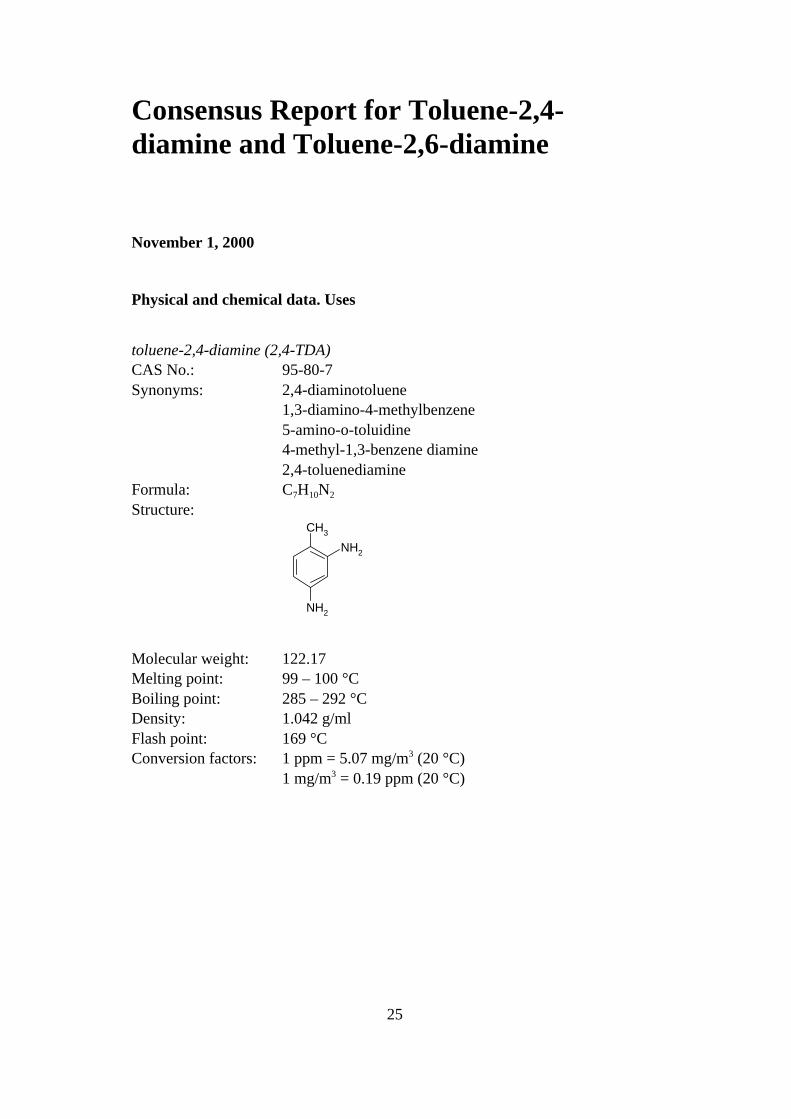
toluene-2,4-diamine (2,4-TDA)CAS No.: 95-80-7Synonyms: 2,4-diaminotoluene
1,3-diamino-4-methylbenzene5-amino-o-toluidine4-methyl-1,3-benzene diamine2,4-toluenediamine
Formula: C7H10N2
Structure:
Molecular weight: 122.17Melting point: 99 – 100 °CBoiling point: 285 – 292 °CDensity: 1.042 g/mlFlash point: 169 °CConversion factors: 1 ppm = 5.07 mg/m3 (20 °C)
1 mg/m3 = 0.19 ppm (20 °C)
CH3
NH2
NH2
26
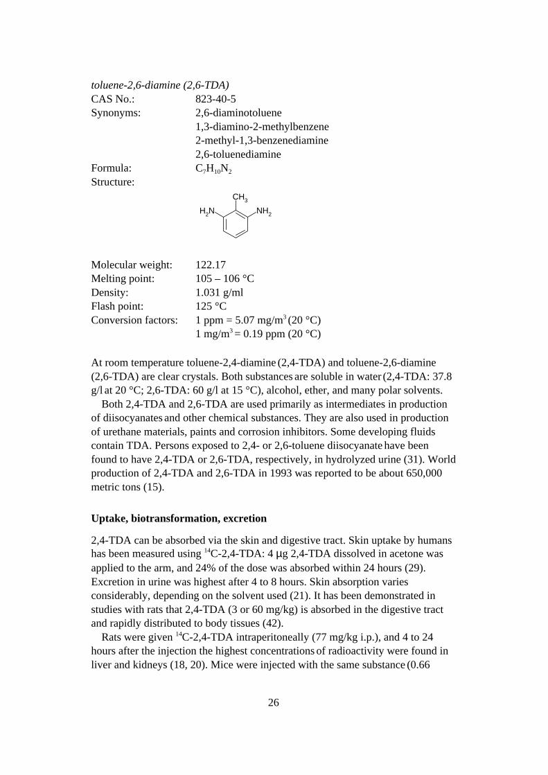
toluene-2,6-diamine (2,6-TDA)CAS No.: 823-40-5Synonyms: 2,6-diaminotoluene
1,3-diamino-2-methylbenzene2-methyl-1,3-benzenediamine2,6-toluenediamine
Formula: C7H10N2
Structure:
Molecular weight: 122.17Melting point: 105 – 106 °CDensity: 1.031 g/mlFlash point: 125 °CConversion factors: 1 ppm = 5.07 mg/m3 (20 °C)
1 mg/m3 = 0.19 ppm (20 °C)
At room temperature toluene-2,4-diamine (2,4-TDA) and toluene-2,6-diamine(2,6-TDA) are clear crystals. Both substances are soluble in water (2,4-TDA: 37.8g/l at 20 °C; 2,6-TDA: 60 g/l at 15 °C), alcohol, ether, and many polar solvents.
Both 2,4-TDA and 2,6-TDA are used primarily as intermediates in productionof diisocyanates and other chemical substances. They are also used in productionof urethane materials, paints and corrosion inhibitors. Some developing fluidscontain TDA. Persons exposed to 2,4- or 2,6-toluene diisocyanate have beenfound to have 2,4-TDA or 2,6-TDA, respectively, in hydrolyzed urine (31). Worldproduction of 2,4-TDA and 2,6-TDA in 1993 was reported to be about 650,000metric tons (15).
Uptake, biotransformation, excretion
2,4-TDA can be absorbed via the skin and digestive tract. Skin uptake by humanshas been measured using 14C-2,4-TDA: 4 µg 2,4-TDA dissolved in acetone wasapplied to the arm, and 24% of the dose was absorbed within 24 hours (29).Excretion in urine was highest after 4 to 8 hours. Skin absorption variesconsiderably, depending on the solvent used (21). It has been demonstrated instudies with rats that 2,4-TDA (3 or 60 mg/kg) is absorbed in the digestive tractand rapidly distributed to body tissues (42).
Rats were given 14C-2,4-TDA intraperitoneally (77 mg/kg i.p.), and 4 to 24hours after the injection the highest concentrations of radioactivity were found inliver and kidneys (18, 20). Mice were injected with the same substance (0.66
CH3
NH2NH2
27
mg/kg i.p.), and 30 minutes later the highest tissue concentrations of radioactivitywere in kidney, testis, epididymis and lung; after 1 hour the concentration washighest in liver (12% of the dose) (43).
In general, it appears that in most species only a small portion (0.1 to 3%) of2,4-TDA is excreted unchanged, and the rest is metabolized (42, 44). The firststep is hydroxylation, followed by N-acetylation. Both mono- and diacetylderivatives have been identified in urine (3, 42, 44). The primary metabolite inurine is 5-hydroxy-2,4-TDA, but mice and rats differ in the occurrence andrelative quantities of the various metabolites and conjugates (43). In vitro studieshave shown that N-acetylation occurs mostly in the liver (17). The reactivemetabolites are formed by P-450-dependent N-hydroxylation and sulfation (2,12, 13, 20). It has been demonstrated in several studies that 2,4-TDA can formadducts with DNA and hemoglobin (4, 11, 25, 27).
2,4-TDA is excreted rapidly, most of it in urine. In rodents, elimination isbiphasic: rapid elimination for 7 hours is followed by a slower phase (18, 43).In a study with rats it was determined that in the slower phase the half time forelimination in urine was 4.6 hours after intravenous administration (3 mg/kg)and 8 hours after oral administration (3 mg/kg) (42). With intraperitonealadministration (77 mg/kg, rats), 69% of the dose was recovered in urine and feceswithin 24 hours (18). Chronic exposure has been found to increase elimination inurine and decrease excretion in feces (43). 2,6-TDA can form hemoglobin adducts(34, 47, 49).
2,6-TDA is rapidly absorbed in the digestive tract. It undergoes hydroxylationand N-acetylation, and the primary metabolites found in urine are 2,6-diacetyl-aminotoluene and 6-acetylamino-2-amino-3-hydroxytoluene (8). Rats given 10-milligram oral doses of 14C-2,6-TDA eliminated 85% of the radioactivity in urinewithin 24 hours. No unmetabolized 2,6-TDA was found in the urine. After 6 days,1% of the radioactivity could still be detected in tissues (7).
Both 2,4-TDA and 2,6-TDA have been identified in hydrolyzed urine ofpersons exposed to toluene diisocyanates (TDI), and there are some studies ofexcretion of 2,4-TDA and 2,6-TDA by persons exposed to TDI (31). In one studyof TDI-exposed workers, the half time in urine was determined to be 18 days for2,4-TDA and 19 days for 2,6-TDA, and the half time in plasma 7.8 days for 2,4-TDA and 9.6 days for 2,6-TDA (28). In another study, in which volunteers wereexposed to TDI for 4 hours, the half time in plasma for 2,4-TDA and 2,6-TDAwas 2 to 5 hours during the initial elimination phase and more than 6 days in theslower phase (5).
Toxic effects
Human dataThere is no published information regarding effects on human health resultingfrom exposure to 2,4-TDA or 2,6-TDA.
28
Animal data
2,4-TDAOne published study reports that 2,4-TDA causes very little or no skin irritation(14).
The reported LD50 values for 2,4-TDA range from 73 to 500 mg/kg bodyweight, depending on species and method of administration. The reported LC50
values for inhalation exposure are 120 – 150 mg/m3 for mice and 916 mg/m3 forrats (15). High doses of 2,4-TDA cause methemoglobin formation in severalspecies (37, 45). Cats were found to be the most sensitive species: a dose of 10mg/kg (i.p.) resulted in elevated numbers of Heinz inclusion bodies (37), regardedas an indication of oxidative stress.
Mice were given 2,4-TDA in oral doses of 25, 50 or 100 mg/kg/day for 14days. The treatment resulted in changes in hepatic enzymes in serum, and thehighest dose resulted in higher liver weights. Immunotoxic effects – highernumbers of B cells in spleens and lower spleen weights – were observed at allexposures. The immunotoxic effects were attributed to disruptions indifferentiation and maturation of leukocytes (6, 46).
In a 7-week study, rats and mice were given 2,4-TDA in diet. The rats givenfeed containing 1000 ppm 2,4-TDA (≈75 mg/kg body weight/day) had lowerbody weights, elevated hematopoiesis and liver changes. The same diet given tomice resulted in reduced body weights but no histopathological changes. TheNOEL for the 7-week exposure was reported to be 250 ppm for rats and 200 ppmfor mice (32). In a two-year NTP cancer study, groups of rats and mice weregiven 2,4-TDA in diet for 103 weeks. Initial exposure levels for the rats were 125and 250 ppm in feed, but because of low weight gain these amounts were reducedto 50 and 100 ppm after 40 weeks. Elevated mortality was seen in the high-dosegroup from 80 weeks onward. The histological findings included liver damageand kidney changes (32). Exposure levels for the mice were 100 and 200 ppm(≈15 and 30 mg/kg body weight/day), and liver hyperplasias and lower weightgain were observed in both exposure groups (32).
2,6-TDAThe lethal single dose with oral administration was 3000 mg/kg body weigbt formale rats and 1000 mg/kg b.w. for female rats (33). Rats exposed to 2,6-TDA indiet (> 1000 ppm) for 14 days lost weight. There was elevated mortality amongmice given a diet containing 3000 ppm 2,6-TDA for 14 days. No effect was seenon body weights of either rats or mice after 14 days on a diet containing 300 ppm(33).
In a 13-week study, rats and mice were given 2,6-TDA in diet. Lower weightgain was noted in male rats at 100 ppm (the lowest dose) and in female rats at1000 ppm. This effect was also noted in mice, at 300 ppm for males and at 1000ppm for females: 100 ppm had no effect on weight gain in the mice (33). The ratsdeveloped thyroid hyperplasias at exposures of 3000 ppm or higher. Renal hyper-pigmentation and a papilloma in the forestomach were observed in the mice at

29
1000 ppm (33). In a cancer study, rats were given 250 or 500 ppm and mice 50 or100 ppm 2,6-TDA in diet for 103 weeks. Lower weight gain was registered forthe male rats in the high-dose group, the female rats in both dose groups, and thefemale mice (33).
Genotoxicity
2,4- and 2,6-TDA have been found to be equally mutagenic in several in vitrotests, including Ames’ tests. For a more detailed review of in vitro tests, referenceis made to the GDCH and IPCS documents (15, 23).
2,4-TDAIn vivo genotoxicity studies of 2,4-TDA have yielded positive results, especiallywith high doses. A dose-dependent increase of single-strand breaks was observedin the livers of rats after intraperitoneal administration of 2,4-TDA (37 – 500mg/kg) (5, 22), although oral administration (50 or 150 mg/kg) had negativeresults (24). An increase of unscheduled DNA synthesis was noted in rat hepato-cytes after oral administration of 2,4-TDA (150 mg/kg) (16, 30). Intraperitonealadministration of 2,4-TDA (9 or 18 mg/kg) increased sister chromatid exchangein the bone marrow of mice (35). There have been some studies on the ability of2,4-TDA to produce chromosome damage (15). Elevated frequencies of micro-nuclei or chromosome aberrations have been seen in laboratory rodents only afterhigh doses. 2,4-TDA induced mutations in Big Blue™ transgenic mice (19, 38).
2,4-TDA given to rats induces dose-dependent increases in DNA adducts,especially in liver and mammary glands. Three different types of DNA adductswere identified, and 60% of the adducts remained two weeks later (25). Thelowest dose that induced DNA adducts in liver was 0.5 mg/kg (single dose, i.p.)(4, 25). In another study, it was shown that an oral dose of 50 mg/kg yieldedgreater numbers of adducts and more persistent adducts when administered over10 days than when given all at once (27). The authors regard this as an indicationthat activation of 2,4-TDA was saturated. It has been demonstrated that thereactive metabolites are formed via P-450-dependent N-hydroxylation andsulfation (2, 12, 13, 20), and the mutagenic intermediate of 2,4-TDA is believed tobe 4-acetoxyamino-2-aminotoluene (9). In a subsequent study with rats, adductswere identified 6 months after 6 weeks of exposure to 40 or 180 ppm 2,4-TDA infeed (11). In rats, 2,4-TDA has been shown to form covalent bonds to DNA, RNAand microsomal protein, and to the microsomal fraction of liver (12, 15).
2,6-TDAFor 2,6-TDA, most in vivo mutagenicity studies have been negative. Positiveresults are associated with extremely high doses (1).
2,6-TDA has been shown to give rise to hemoglobin adducts (34, 47, 49),although no DNA adducts could be detected in rat liver after intraperitonealadministration (150 mg/kg) (26). Both 2,4-TDA and 2,6-TDA have been shownto form hemoglobin adducts in rats (single doses of 0.5 to 250 mg/kg, i.p.), but
30
only 2,4-TDA induced DNA binding. When compared with 2,4-TDA, 2,6-TDAshowed about one third as much binding to hemoglobin and plasma protein, andone third the tissue concentrations in liver and other organs (34).
Carcinogenicity
There are several older studies in which toluene diamines were tested for carcino-genicity. These results have been summarized by the IARC (22), and the IARChas classified 2,4-TDA as carcinogenic (Group 2B). In a later NTP cancer study(32), groups of 50 rats were given 125 or 250 ppm 2,4-TDA dihydrochloride indiet for 40 weeks. Levels were then reduced to 50 and 100 ppm due to toxicity,and animals in the low-dose group were exposed for a further 63 weeks. Due tothe high mortality in the high-dose group, the animals were killed after 39 and 44weeks. Dose-dependent increases were observed in the numbers of animals(males and females) with hepatocellular carcinomas. An increase in the number ofanimals with non-neoplastic changes was also noted. There were also elevatednumbers of females with carcinomas or adenomas in the mammary glands andmales with subcutaneous fibromas. In the same study, groups of mice wereexposed to 100 or 200 ppm 2,4-TDA in diet for 103 weeks. A dose-dependentincrease in hepatocellular carcinomas was seen in the females, and there was alsoan elevated frequency of lymphoma among females in the low-dose group. Nosignificant increase of tumors was observed in the male mice (32).
2-6-TDA was tested in the same way (33). The dose levels tested were 200 and500 ppm for rats and 50 and 100 ppm for mice. No significant increases in tumorswere observed.
2,4-TDA (25 mg/kg/day, p.o., 30 days) increased the growth of pre-neoplasticfoci in rat livers, whereas 2,6-TDA (50 mg/kg p.o., 30 days) was negative (39).
The difference in carcinogenic effect between 2,4-TDA and 2,6-TDA has beendiscussed in the literature. No differences in absorption or metabolism have beenfound that might explain it (8). The two substances have been found to be equallymutagenic in several different in vitro tests. Results of in vivo mutagenicity testsshowed some differences, however, and 2,6-TDA was usually negative. Exposureto 2,4-TDA in doses about the same as those used in the NTP study has also beenshown to increase cell proliferation in rat liver (10).
Teratogenicity
There have been several epidemiological studies addressing the question ofwhether mixed exposure to toluene diamine and dinitrotoluene has teratogeniceffects. Teratogenic effects were found, but because of the mixed exposure thesestudies are of no value in assessing the teratogenicity of 2,4-TDA (15).
Effects on spermatogenesis have been documented in several animal studieswith 2,4-TDA. They are ascribed to damage to Sertoli’s cells. In one study, ratswere given 2,4-TDA (15 mg/kg/day) in diet for 10 weeks. The treatment induceddegenerative changes in Sertoli’s cells and reduced spermatogenesis (48). In
31
another study with rats, the same exposure resulted in lowered testosterone levelsand reduced spermatogenesis. The NOEL in this study was 5 mg/kg/day given indiet for 10 weeks (40). The NOEL was the same for the fertility index of male ratsin another study (41). Mice were exposed to 2,4-TDA in a dominant-lethal test(40 mg/kg i.p. or p.o for 2 days), and no effect on fertility was observed (36). Adose of 150 mg/kg/day given to pregnant mice by gavage on days 7 to 14 ofgestation had teratogenic effects, but the dose was too toxic to the mothers toallow the teratogenicity to be assessed (15). 2,6-TDA has not been tested forteratogenicity.
Dose-response / dose-effect relationships
There are no data on which to base a dose-response or dose-effect relationship foroccupational exposure to 2,4-TDA or 2,6-TDA. The relationships between dose,response and effect observed in laboratory animals are summarized in Table 1(2,4-TDA) and Table 2 (2,6-TDA).
Conclusions
There are no studies of the effects of TDA on human health. In animalexperiments, the critical effect of 2,4-TDA is cancer. 2,4-TDA is a potentcarcinogen for experimental animals, with the liver as the primary target organ.Dose-dependent increases of hepatocellular carcinomas have been observed in ratsand female mice. 2-4-TDA also affects the reproductive systems of male rats.Both 2,4-TDA and 2,6-TDA are genotoxic.
There is no information that can be used to establish a critical effect foroccupational exposure to 2,6-TDA. 2,6-TDA has shown no carcinogenic effect inanimal experiments.
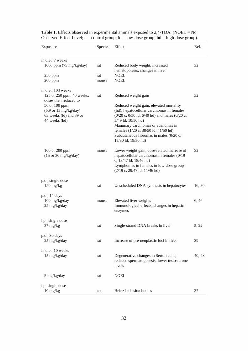
32
Table 1. Effects observed in experimental animals exposed to 2,4-TDA. (NOEL = NoObserved Effect Level; c = control group; ld = low-dose group; hd = high-dose group).
Exposure Species Effect Ref.
in diet, 7 weeks 1000 ppm (75 mg/kg/day) rat Reduced body weight, increased
hematopoiesis, changes in liver32
250 ppm rat NOEL 200 ppm mouse NOEL
in diet, 103 weeks 125 or 250 ppm. 40 weeks; doses then reduced to 50 or 100 ppm, (5.9 or 13 mg/kg/day) 63 weeks (ld) and 39 or 44 weeks (hd)
rat Reduced weight gain
Reduced weight gain, elevated mortality(hd); hepatocellular carcinomas in females(0/20 c; 0/50 ld; 6/49 hd) and males (0/20 c;5/49 ld; 10/50 hd)Mammary carcinomas or adenomas infemales (1/20 c; 38/50 ld; 41/50 hd)Subcutaneous fibromas in males (0/20 c;15/30 ld; 19/50 hd)
32
100 or 200 ppm (15 or 30 mg/kg/day)
mouse Lower weight gain, dose-related increase ofhepatocellular carcinomas in females (0/19c; 13/47 ld; 18/46 hd)Lymphomas in females in low-dose group(2/19 c; 29/47 ld; 11/46 hd)
32
p.o., single dose 150 mg/kg rat Unscheduled DNA synthesis in hepatocytes 16, 30
p.o., 14 days 100 mg/kg/day 25 mg/kg/day
mouse Elevated liver weightsImmunological effects, changes in hepaticenzymes
6, 46
i.p., single dose 37 mg/kg rat Single-strand DNA breaks in liver 5, 22
p.o., 30 days 25 mg/kg/day rat Increase of pre-neoplastic foci in liver 39
in diet, 10 weeks 15 mg/kg/day rat Degenerative changes in Sertoli cells;
reduced spermatogenesis; lower testosteronelevels
40, 48
5 mg/kg/day rat NOEL
i.p. single dose 10 mg/kg cat Heinz inclusion bodies 37
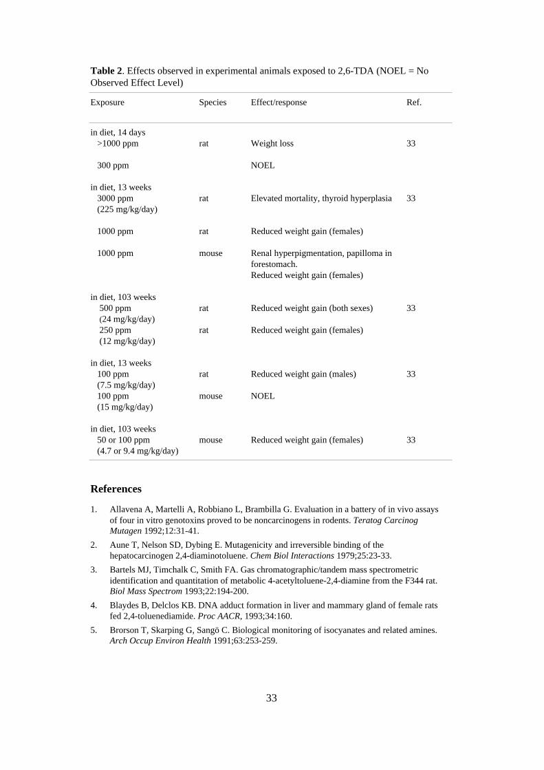
33
Table 2. Effects observed in experimental animals exposed to 2,6-TDA (NOEL = NoObserved Effect Level)
Exposure Species Effect/response Ref.
in diet, 14 days >1000 ppm
300 ppm
rat Weight loss
NOEL
33
in diet, 13 weeks 3000 ppm (225 mg/kg/day)
rat Elevated mortality, thyroid hyperplasia 33
1000 ppm rat Reduced weight gain (females)
1000 ppm mouse Renal hyperpigmentation, papilloma inforestomach.Reduced weight gain (females)
in diet, 103 weeks 500 ppm (24 mg/kg/day) 250 ppm (12 mg/kg/day)
rat
rat
Reduced weight gain (both sexes)
Reduced weight gain (females)
33
in diet, 13 weeks 100 ppm (7.5 mg/kg/day) 100 ppm (15 mg/kg/day)
rat
mouse
Reduced weight gain (males)
NOEL
33
in diet, 103 weeks 50 or 100 ppm (4.7 or 9.4 mg/kg/day)
mouse Reduced weight gain (females) 33
References
1. Allavena A, Martelli A, Robbiano L, Brambilla G. Evaluation in a battery of in vivo assaysof four in vitro genotoxins proved to be noncarcinogens in rodents. Teratog CarcinogMutagen 1992;12:31-41.
2. Aune T, Nelson SD, Dybing E. Mutagenicity and irreversible binding of thehepatocarcinogen 2,4-diaminotoluene. Chem Biol Interactions 1979;25:23-33.
3. Bartels MJ, Timchalk C, Smith FA. Gas chromatographic/tandem mass spectrometricidentification and quantitation of metabolic 4-acetyltoluene-2,4-diamine from the F344 rat.Biol Mass Spectrom 1993;22:194-200.
4. Blaydes B, Delclos KB. DNA adduct formation in liver and mammary gland of female ratsfed 2,4-toluenediamide. Proc AACR, 1993;34:160.
5. Brorson T, Skarping G, Sangö C. Biological monitoring of isocyanates and related amines.Arch Occup Environ Health 1991;63:253-259.
34
6. Burns LA, Bradley SG, White KL, McCay JA, Fuchs BA, Stern M, Brown RD, MusgroveDL, Holsapple MP, Luster MI, Munson AE. Immunotoxicity of 2,4-diaminotoluene infemale B6C3F1 mice. Drug Chem Toxicol 1994;17:401-426.
7. Cunningham ML, Burka LT, Matthews HB. Identification and mutagenicity of the urinarymetabolites of the mutagenic noncarcinogen, 2,6-diaminotoluene. Liquid Chrom1989;12:1407-1416.
8. Cunningham ML, Burka LT, Matthews HB. Metabolism, disposition and mutagenicity of2,6-diaminotoluene, a mutagenic noncarcinogen. Drug Metabol Dispos 1989;17:612-617.
9. Cunningham ML, Matthews HB. Evidence for an acetoxyarylamine as the ultimatemutagenic reactive intermediate of the carcinogenic aromatic amine 2,4-diaminotoluene.Mutat Res 1990;242:101-110.
10. Cunningham ML, Matthews HB. Cell proliferation as a determining factor for thecarcinogenicity of chemicals: studies with mutagenic carcinogens and mutagenicnoncarcinogens. Toxicol Lett 1995;82:9-14.
11. Delclos KB, Blaydes B, Heflich RH, Smith BA. Assessment of DNA adducts and thefrequency of 6-thioguanine resistant T-lymphocytes in F344 rats fed 2,4-toluenediamine orimplanted with toluenediisocyanate-containing polyester polyurethane foam. Mutat Res1996;367:209-218.
12. Dybing E, Aune T, Nelson SD. Covalent binding of 2,4-diaminoanisole and 2,4-diaminotoluene in vivo. Arch Toxicol 1978;1:213-217.
13. Furlong BB, Weaver RP, Goldstein JA. Covalent binding to DNA and mutagenicity of 2,4-diaminotoluene metabolites produced by isolated hepatocytes and 9000g supernatant fromFischer 344 rats. Carcinogenesis 1987;8:247-251.
14. Gad SC, Walsh RD, Dunn BJ. Correlation of ocular and dermal irritancy of industrialchemicals. J Toxicol – Cut Ocular Toxicol 1986;5:195-213.
15. GDCH. Advisory committee on existing chemicals of environmental relevance. 2,4-Toluenediamine and 2,6-toluenediamine. S. Hirzel Verlag, Stuttgart, (BUA) Report 192,1995.
16. George E, Westmoreland C. Evaluation of the in vivo genotoxicity of the structuralanalogues 2,6-diaminotoluene and 2,4-diaminotoluene using the rat micronucleus test and ratliver UDS assay. Carcinogenesis 1991;12:2233-2237.
17. Glinsukon T, Benjamin T, Grantham PH, Weisburger EK, Roller PP. Enzymic N-acetylationof 2,4-toluenediamide by liver cytosols from various species. Xenobiotica 1975;5:475-483.
18. Grantham PH, Mohan L, Benjamin T, Roller PP, Miller JR, Weisburger EK. Comparison ofthe metabolism of 2,4-toluenediamine in rats and mice. J Environ Pathol Toxicol1979;3:149-166.
19. Hayward JJ, Shane BS, Tindall KR, Cunningham ML. Differential in vivo mutagenicity ofthe carcinogen/non-carcinogen pair 2,4- and 2,6-diaminotoluene. Carcinogenesis1995;16:2429-2433.
20. Hiasa Y. m-Toluenediamine carcinogenesis in rat liver. J Nara Med Ass 1970;21:1-19.
21. Hruby R. The absorption of p-toluenediamide by the skin of rats and dogs. Food CosmetToxicol 1977;15:595-599.
22. IARC. Some aromatic amines and related nitro compounds - hair dyes, colouring agents andmiscellaneous industrial chemicals. IARC Monographs on the Evaluation of CarcinogenicRisks of Chemicals to Man, Vol 16. Lyon: International Agency for Research on Cancer1978;16:83-95.
23. IPCS. Environmental Health Criteria 74. Diaminotoluenes. Geneva, InternationalProgramme on Chemical Safety. World Health Organization 1987.
24. Kitchin KT, Brown JL. Dose-response relationship for rat liver DNA damage caused by 49rodent carcinogens. Toxicology 1994;88:31-49.
35
25. La DK, Froines JR. 32P-postlabelling analysis of DNA adducts from Fischer-344 ratsadministered 2,4-diaminotoluene. Chem Biol Interact 1992;83:121-134.
26. La DK, Froines JR. Comparison of DNA binding between the carcinogen 2,6-dinitrotolueneand its noncarcinogenic analog 2,6-diaminotoluene. Mutat Res 1993;301:79-85.
27. La DK, Froines JR. Formation and removal of DNA adducts in Fischer-344 rats exposed to2,4-diaminotoluene. Arch Toxicol 1994;69:8-13.
28. Lind P, Dalene M, Tinnerberg H, Skarping G. Biomarkers in hydrolysed urine, plasma anderythrocytes among workers exposed to thermal degradation products from toluenediisocyanate foam. Analyst 1997;122:51-56.
29. Marzulli FN, Anjo DM, Maibach HI. In vivo skin penetration studies of 2,4-toluenediamine,2,4-diaminoanisole, 2-nitro-p-phenylenediamine, p-dioxane and n-nitro-diethanolamine incosmetics. Food Cosmet Toxicol 1981;19:743-747.
30. Mirsalis JC, Tyson CK, Butterworth BE. Detection of genotoxic carcinogens in the in vivo-invitro hepatocyte DNA repair assay. Environ Mutagen 1982;4:553-562.
31. Montelius J (ed). Scientific Basis for Swedish Occupational Standards. XXII. Arbete ochHälsa 2001;20:60-88. National Institute for Working Life, Solna, Sweden.
32. National Cancer Institute. Bioassay of 2,4-diaminotoluene for possible carcinogenicity. U.S.Department of Health, Education and Welfare, Public Health Service, National Institutes ofHealth, NIH Publication No. 78-1718; Technical Report No. 162, 1979.
33. National Cancer Institute. Bioassay of 2,6-toluenediamide dihydrochloride for possiblecarcinogenicity. U.S. Department of Health, Education and Welfare, Public Health Service,National Institutes of Health, NIH Publication No. 80-1756; Technical Report No. 200, 1980.
34. Neumann HG, Birner G, Kowallik P, Schütze D, Zwirner-Baier I. Hemoglobin adducts in N-substituted aryl compounds in exposure control and risk assessment. Environ Health Persp1993;99:65-69.
35. Parodi S, Taningher M, Russo P, Pala M, Tamaro M, Monti-Bragadin C. DNA-damagingactivity in vivo and bacterial mutagenicity of sixteen aromatic amines and azoderivatives, asrelated quantitatively to their carcinogenicity. Carcinogenesis 1981;2:1317-1326,
36. Soares ER, Lock LF. Lack of an indication of mutagenic effects of dinitrotoluenes anddiaminotoluenes in mice. Environ Mutagen 1980;2:111-124.
37. Stahl KE, Jung F. Über Blutgiftwirkungen des Phenylen-diamins und des Toluen-diamins.Arch Exper Path Pharmacol 1953;220:503-518.
38. Suter W, Ahiabor R, Blanco B, Locher F, Mantovani F, Robinson M, Sreenan G, Staedtler F,Swingler T, Vignutelli A, Perentes A. Evaluation of the in vivo genotoxic potential of threecarcinogenic aromatic amines using Big Blue™ transgenic mouse mutation assay. EnvironMol Mutagen 1996;28:352-362.
39. Taningher M, Peluso M, Parodi S, Ledda-Columbano G, Columbano A. Genotoxic and non-genotoxic activities of 2,4- and 2,6-diaminotoluene, as evaluated in Fischer-344 rat liver.Toxicology 1995;99:1-10.
40. Thysen B, Bloch E, Varma SK. Reproductive toxicity of 2,4-toluenediamine in the rat. 2.Spermatogenic and hormonal effects. J Toxicol Environ Health 1985;16:763-769.
41. Thysen B, Varma SK, Bloch E. Reproductive toxicity of 2,4-toluenediamine in the rat. 1.Effects on male fertility. J Toxicol Environ Health 1985;16:753-761.
42. Timchalk C, Smith FA, Bartels MJ. Route-dependent metabolism of [14C]toluene 2,4-diisocyanate and [14C]toluene 2,4-diamine in Fischer 344 rats. Toxicol Appl Pharmacol1994;124:181-190.
43. Unger PD, Salerno AJ, Ness WG, Friedman MA. Tissue distribution and excretion of2,4[14C]-toluenediamide in the mouse. J Toxicol Environ Health 1980;6:107-114.
44. Waring RH, Pheasant AE. Some phenolic metabolites of 2,4-diaminotoluene in the rabbit, ratand guinea-pig. Xenobiotica 1976;6:257-262.
36
45. Weisbrod D, Stephan U. Untersuchungen zur toxischen, menthämoglobinbildenden underythrozytenschädigenden Wirkung von Diaminotoluen nach einmalinger Applikation. Z GesHyg 1983;29:395-397.
46. White KL, McCay JA, Musgrove DL, Brown RD, Stern ML, Holsapple MP, Munson AE.Immunotoxicity of 2,4-diaminotoluene in female B6C3F1 mice. Toxicologist 1989;9:200.(Abstract)
47. Wilson PM, La DK, Froines JR. Hemoglobin and DNA adduct formation in Fischer-344 ratsexposed to 2,4- and 2,6-toluene diamine. Arch Toxicol 1996;70:591-598.
48. Varma SK, Bloch E, Gondos B, Rossi V, Gunsalus GL, Thysen B. Reproductive toxicity of2,4-toluenediamine in the rat. 3. Effects on androgen-binding protein levels, selectedseminiferous tubule characteristics, and spermatogenesis. J Toxicol Environ Health1988;25:435-451.
49. Zwirner-Baier I, Kordowich FJ, Neumann HG. Hydrolyzable hemoglobin adducts ofpolyfunctional monocyclic N-substituted arenes as dosimeters of exposure and markers ofmetabolism. Environ Health Perspect 1994;102:43-45.
37
Consensus Report for α-Methylstyrene
November 1, 2000
Physical and chemical data. Uses
CAS No.: 98-83-9Synonyms: 2-phenylpropene
isopropyl benzene1-methyl-1-phenylethylene2-phenylpropyleneα-methylstyrol1-phenyl-1-methylethylene1-methylethenylbenzene
Formula: C9H10
Structure:
Molecular weight: 118.19Boiling point: 165 °CMelting point: - 23.2 °CVapor pressure: 0.307 kPa (20 °C)Flash point: 54 °CDensity: 0.91 g/cm3
Conversion factors: 1 ppm = 4.82 mg/m3
1 mg/m3 = 0.21 ppm
At room temperature α-methylstyrene is a clear liquid with a characteristic odor(odor threshold: 0.1 mg/m3). It is soluble in benzene, diethylether, chloroform,acetone and carbon tetrachloride (in acetone and carbon tetrachloride in anyproportions). It has an affinity for oils and fats (log Po/w = 3.34). Some t-butylcatechol may be added to α-methylstyrene to prevent peroxide formation duringstorage. Contact with heat or catalysts may cause α-methylstyrene to polymerize.A common use for α-methylstyrene is as a solvent and polymer in plasticproduction. It occurs in waxes, paints and other surface coatings.
38
Uptake, biotransformation, excretion
Both pure α-methylstyrene and α-methylstyrene emulsified in water can beabsorbed through human skin. Undiluted α-methylstyrene (0.1 ml) spread on ameasured glass surface was pressed against the skin of 8 volunteers for 8 to 10minutes. Both the glass and the skin were then washed with alcohol, and the α-methylstyrene in the alcohol used for washing was determined with spectro-photometry (244 nm). The absorption rate for α-methylstyrene in water emulsionwas determined by having 8 volunteers place their hands in 1-liter beakers ofemulsion. After 1 hour of exposure the amount absorbed was determined byspectrophotometry. For undiluted α-methylstyrene, the absorption rate was19.5 mg/cm2/hour. For α-methylstyrene in water the absorption rate varied withconcentration and temperature, and was 50% higher at 36 °C than at 24 °C (2).
Metabolism of α-methylstyrene is like that of styrene, its structural analogue. Itis activated by P-450 monooxygenase to α-methylstyrene oxide (7-methylstyrene-7,8-oxide), and this metabolite is then transformed to α-methylstyrene glycol andvarious glutathione conjugates. Oxidation of α-methylstyrene glycol yields α-hydroxy-α-phenylpropionic acid (9), which has been identified in the urine ofpersons exposed to α-methylstyrene by inhalation and animals exposed byinhalation and intraperitoneal injection (1, 4, 5).
Toxic effects
Human dataSubjects (number not given) in an odor and irritation study were briefly exposed(duration of exposure not given) to α-methylstyrene vapors in a test chamber. Atair concentrations of 2892 mg/m3 (600 ppm) or higher they reported a very strongodor and experienced severe irritation of eyes and nose. A concentration of482 mg/m3 (100 ppm) had a strong odor but did not cause excessive discomfort.At an air concentration of 241 mg/m3 (50 ppm) the subjects reported apronounced odor but no irritation (15).
A change in the light sensitivity of the eye was observed in three odor-sensitivepersons after 15 to 20 minutes of exposure to a concentration of 0.1 mg/m3
(0.02 ppm) α-methylstyrene (odor threshold 0.1 mg/m3). These exposure condi-tions also produced changes in the α-rhythm on their EEGs. Neither effect wasobserved at 0.08 mg/m3 or 0.04 mg/m3 (10). These effects are not considered to betoxicologically relevant.
There is a study describing a correlation between clinical symptoms ofpoisoning and deterioration of renal circulation in 69 occupationally exposedsubjects employed in the production of divinyl-α-methylstyrene rubber. Thesymptoms appeared after seven to eight years of employment. No further detailsof this study were available (8). A case report describes workers handling divinyl-α-methylstyrene rubber who developed ‘neurocirculatory dystonia’ as a result ofoccupational exposure to 1,3-butadiene and α-methylstyrene. They also had some
39
deterioration in liver function, but the cause of this is unclear. Further informationwas not available (3).
Animal dataEffects of α-methylstyrene on skin were examined in a study with rabbits:undiluted α-methylstyrene was painted on their ears and shaved rumps (10-20strokes spaced over 2 to 4 weeks). The treatment resulted in moderate to markederythema as well as some necrotic effect in the form of exfoliation. In anotherstudy, undiluted α-methylstyrene (two drops) was applied to the eyes of rabbitsand the eyes were examined after 3 minutes, 1 hour, 24 hours, 2 days and 7 days.The substance caused slight inflammation of the conjunctiva, but no visibledamage to the cornea (15).
The LD50 for rats given α-methylstyrene per os was 4.9 g/kg. Liver damage wasfound when the animals were dissected (15).
Mice were exposed to α-methylstyrene in air (2900, 3860 or 4800 mg/m3)6 hours/day for 12 days. Mortality for the females was 6% (1/18) at 2900 mg/m3,56% (10/18) at 3860 mg/m3, and 21% (5/24) at 4800 mg/m3. The cause of deathcould not be determined. No male mice died during the exposures. During thefirst hour of exposure the mice in all exposed groups were hyperactive and did notreact to noise. By the end of the 6-hour exposure the animals had calmed downand appeared drowsy. The mice adapted to this ‘tranquilizing’ effect of α-methyl-styrene after a week of exposure (5 days/week). Higher liver weights and lowerspleen weights were found in both males and females in all exposed groups. Liverweights were significantly higher in the males after a single 6-hour exposure to3860 mg/m3, and in the females after 5 days of exposure to 2900 mg/m3. After 5days at 3860 mg/m3 reduced spleen weights were seen in both sexes. Only themale mice had significantly lower body weights after 5 days of exposure. Histo-pathological examinations revealed no exposure-related tissue changes. After 1and 5 days of exposure there was a reduction of glutathione in the liver in all threeexposed groups (2900, 3860 and 4800 mg/m3) (9). In a short-term test, male micethat inhaled α-methylstyrene had a higher threshold for seizures induced bypentetrazole. A concentration of 3581 mg/m3 (743 ppm) α-methylstyrene caused a50% seizure threshold increase (STI50) for pentetrazole (6).
In an inhalation study, exposure to 2900 or 4800 mg/m3 α-methylstyrene 6hours/day for 12 days resulted in higher liver weights in F-344 rats. Hyalin dropformation could be observed in the kidneys of F-344 males after nine days ofexposure to 1205 mg/m3 but could not be observed in F-344 females or male NBRrats (which lack α2u-globulin production) under the same exposure conditions.The effect is considered to be related to α2u-globulin, a protein that occurs inmuch greater concentrations in male rats than in female rats. There was no hyalindrop formation in the F-344 males exposed to 603 mg/m3 (9).
Histological examination of male white rats exposed to 5 mg/m3 α-methyl-styrene 24 hours/day for 3 months revealed interstitial pneumonia, bronchialinflammation (desquamative bronchitis), abnormal growth (hyperplasia) of
40
bronchial lymph nodes, tissue changes in the walls of blood vessels, degenerativechanges in kidneys and heart muscles, disturbances of glycogen-related functionsin the liver (not explained) and neural cell dystrophy in the cortex, cerebellum andspinal cord. After 2 weeks of exposure to 5 mg/m3 the rats had lower copropor-phyrin levels in urine (not further elucidated) and a higher number of leukocytesthat showed abnormal coloration (green to yellow/yellow-orange) when dyed withacridine orange (not explained). Only minor changes were observed in lungs, liverand acridine-orange dyed leukocytes of rats exposed to 0.5 mg/m3 for 3 months,and no effects were observed in rats exposed to 0.05 mg/m3 for 3 months (10).Data in this study are inadequately presented and ‘end points’ are unclear.
In an inhalation study (15), Wistar rats (10 to 25 per group, both sexes) wereexposed to α-methylstyrene concentrations of 970 mg/m3 (for 139 7-hoursessions), 2900 mg/m3 (for 149 x 7 hours), 3860 mg/m3 (28 x 7 hours) or14,490 mg/m3 (for 3 or 4 x 7 hours) (= 200, 600, 800 or 3000 ppm). The exposureto 14,490 mg/m3 resulted in high mortality (not quantified). The group exposed to3860 mg/m3 lost weight, and kidney and liver weights were affected (no quantita-tive data given). Effects on kidney and liver weights were also observed at2900 mg/m3. No effects were observed at 970 mg/m3.
Guinea pigs were exposed according to the same protocol. Those exposed to3860 mg/m3 lost weight, and liver and kidney weights were affected. Liver weightwas affected at 2900 mg/m3. No effects were seen at 970 mg/m3. Rabbits wereexposed to 970 mg/m3 (139 x 7 hours) or 2900 mg/m3 (149 x 7 hours). The lowerdose group showed no effect. In the higher dose group the exposure resulted inhigher mortality and weight loss. Rhesus monkeys (1 or 2) of both sexes wereexposed along with the rabbits, and neither exposure level resulted in observablehistopathological changes or effects on organ weight (15).
Mutagenicity, carcinogenicity, teratogenicity
Ames’ tests with α-methylstyrene were negative, both with and without metabolicactivation (16), although the intermediate metabolite α-methylstyrene oxide hasyielded positive results (14). Sister-chromatid analysis of human lymphocytes invitro revealed a significant increase in the frequency of sister chromatidexchanges (11). Unlike styrene, which is activated by hemoglobin, α-methyl-styrene’s ability to induce sister chromatid exchanges is not dependent onerythrocytes (11, 12, 13).
Methylstyrene (unspecified isomer) was tested for teratogenicity in a study withrats: 250 mg/kg body weight (in corn oil) was injected intraperitoneally. Thetreatment had no observed effect on the mothers, but there were changes in thesex ratio of the embryos (fewer females) and a significantly higher frequency ofresorptions (7).
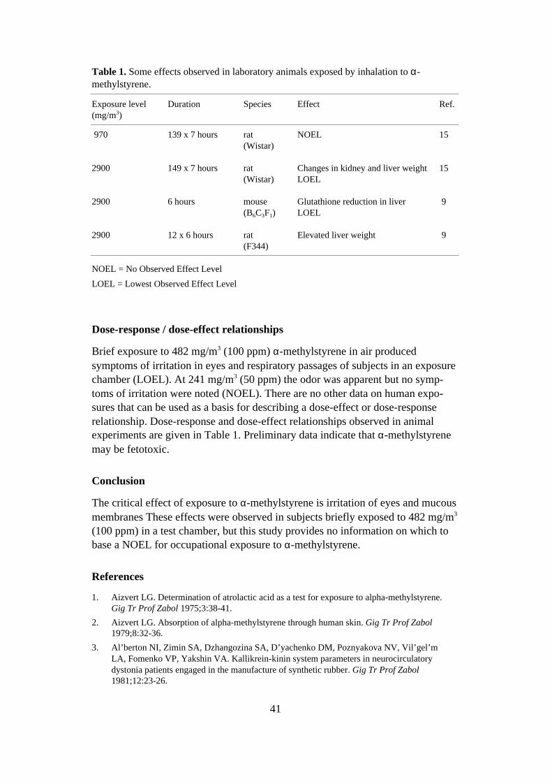
41
Table 1. Some effects observed in laboratory animals exposed by inhalation to α-methylstyrene.
Exposure level(mg/m3)
Duration Species Effect Ref.
970 139 x 7 hours rat(Wistar)
NOEL 15
2900 149 x 7 hours rat(Wistar)
Changes in kidney and liver weightLOEL
15
2900 6 hours mouse(B6C3F1)
Glutathione reduction in liverLOEL
9
2900 12 x 6 hours rat(F344)
Elevated liver weight 9
NOEL = No Observed Effect Level
LOEL = Lowest Observed Effect Level
Dose-response / dose-effect relationships
Brief exposure to 482 mg/m3 (100 ppm) α-methylstyrene in air producedsymptoms of irritation in eyes and respiratory passages of subjects in an exposurechamber (LOEL). At 241 mg/m3 (50 ppm) the odor was apparent but no symp-toms of irritation were noted (NOEL). There are no other data on human expo-sures that can be used as a basis for describing a dose-effect or dose-responserelationship. Dose-response and dose-effect relationships observed in animalexperiments are given in Table 1. Preliminary data indicate that α-methylstyrenemay be fetotoxic.
Conclusion
The critical effect of exposure to α-methylstyrene is irritation of eyes and mucousmembranes These effects were observed in subjects briefly exposed to 482 mg/m3
(100 ppm) in a test chamber, but this study provides no information on which tobase a NOEL for occupational exposure to α-methylstyrene.
References
1. Aizvert LG. Determination of atrolactic acid as a test for exposure to alpha-methylstyrene.Gig Tr Prof Zabol 1975;3:38-41.
2. Aizvert LG. Absorption of alpha-methylstyrene through human skin. Gig Tr Prof Zabol1979;8:32-36.
3. Al’berton NI, Zimin SA, Dzhangozina SA, D’yachenko DM, Poznyakova NV, Vil’gel’mLA, Fomenko VP, Yakshin VA. Kallikrein-kinin system parameters in neurocirculatorydystonia patients engaged in the manufacture of synthetic rubber. Gig Tr Prof Zabol1981;12:23-26.
42
4. Bardodej Z, Bardodejova E. Atrolactic acid as a metabolite of alpha-methylstyrene. CeskHygiena 1966;11:302.
5. Bardodej Z, Bardodejova E. Biotransformation of ethyl benzene, styrene, and alpha-methylstyrene in man. Am Ind Hyg Assoc J 1970;31:206-209.
6. Ceaurriz J, Bonnet P, Certin C, Muller J, Guenier JP. Chemicals as central nervous systemdepressants – Possibilities of an animal model. Cahiers de notes documentaires - Securite ethygiene du travail, 3rd quarter 1981;104:351-355.
7. Hardin BD, Bond GP, Sikov MR, Andrew FD, Beliles RP, Niemeier RW. Testing of selectedworkplace chemicals for teratogenic potential. Scand J Work Environ Health 1981;7 Suppl4:66-75.
8. Konstantinovskaja AS. The renal function in workers employed in the production of divinyl-alpha-methylstyrene rubber. Gig Tr Prof Zabol 1970;14:10-12.
9. Morgan DL. Mahler JF, Kirkpatrick DT, Price HC, O’Connor RW, Wilson RE, MoormanMP. Characterization of inhaled α-methylstyrene vapor toxicity for B6C3F1 mice and F344rats. Toxicol Sci 1999;47:187-194.
10. Minaev AA. Determination of the maximum permissible concentration of alpha methylstyrene vapor in the atmosphere. Hygiene and Sanitation 1966;31:157-161.
11. Norppa H, Sorsa M, Pfäffli P, Vainio H. Styrene and styrene oxide induce SCEs and aremetabolised in human lymphocyte cultures. Carcinogenesis 1980;1:357-361.
12. Norppa H, Vainio H. Induction of sister-chromatid exchanges by styrene analogues incultured human lymphocytes. Mutat Res 1983;116:379-387.
13. Norppa H, Tursi F. Erythrocyte-mediated metabolic activation detected by SCE. Basic LifeSci 1984;29B:547-559.
14. Rosman LB, Beylin VG, Gaddamidi V, Hooberman BH, Sinsheimer JE. Mutagenicity ofpara-substituted alpha-methylstyrene oxide derivatives with Salmonella. Mutat Res1986;171:63-70.
15. Wolf MA, Rowe VK, McCollister DD, Hollingsworth RL, Oyen F. Toxicological studies ofcertain alkylated benzenes and benzene. Arch Ind Health 1956;14:387-398.
16. Zeiger E, Anderson B, Haworth S, Lawlor T, Mortelmans K. Salmonella mutagenicity tests:V. Results from the testing of 311 chemicals. Environ Mol Mutagen 1992;19, Suppl 21:2-141.
43
Consensus Report for Hydrogen Cyanide,Sodium Cyanide and Potassium Cyanide
February 7, 2001
Chemical and physical data. Uses
hydrogen cyanide
CAS No.: 74-90-8Synonyms: hydrocyanic acid, prussic acid, formonitrileFormula: HCNMolecular weight: 27.04Boiling point: 25.70°CMelting point: -13.24°CVapor pressure: 83.8 kPa (20°C)Saturation concentration: 827,246 ppmConversion factors: 1 mg/m3 = 0.89 ppm (20°C)
1 ppm = 1.12 mg/m3 (20°C)
sodium cyanide
CAS No.: 143-33-9Synonyms: cyanide of sodiumFormula: NaCNMolecular weight: 49.01Boiling point: 1496°CMelting point: 563.7°CVapor pressure: -
potassium cyanide
CAS No.: 151-50-8Synonym: cyanide of potassium, cyanide of potashFormula: KCNMolecular weight: 65.12Boiling point: -Melting point: 634.5°CVapor pressure: -
44
HCN occurs as a colorless gas or as a clear, extremely volatile and flammableliquid. It has an odor of bitter almonds. It is soluble in ethanol and similarsolvents and mixes with water, forming a weak acid (4, 44). One source reports anodor threshold of 0.2-5 ppm for HCN (21), but there are many people who can notperceive the odor at all (39, 40). NaCN and KCN occur as clear or whitish solids(4). They are readily soluble in water (about 50 g/100 ml cold water), producingstrongly alkaline solutions (59, 68).
Occupational exposure to cyanide can occur during industrial production anduse of cyanide compounds, and may also result from exposure to industrialchemicals that are metabolized to cyanide, such as simple aliphatic nitriles (4).Cyanides also occur naturally in many foods (e.g. cassava/manioc, passionfruit,bamboo shoots, bean sprouts and almonds, and the pits of apricots, peaches,cherries and plums) in the form of cyanogenic glycosides, which can be brokendown to HCN in the intestine (4, 34, 46). Cyanide may occur in foods as pesticideresidue (4, 34). Cyanide exposure may also have several other sources, includingtobacco smoke, smoke from fires, vehicle exhausts and some medicines (4, 34).
Cyanides are used primarily in the steel, mining, and chemical industries and inelectroplating (4). NaCN is used, for example, to extract gold and silver from oreand in the production of adiponitrile (for nylon), pigments, chelating agents andpesticides (34, 40). NaCN and KCN are used together to treat steel. Largeamounts of cyanide salts such as NaCN and KCN are used in electroplating –KCN particularly with silver plating (4, 34). Many metal polishes contain NaCNor KCN. HCN is used in the production of adiponitrile (for nylon), methylmeth-acrylate, NaCN, chelating agents, and pharmaceuticals. HCN may also be used tofumigate grain storage facilities to kill rodents and insects (4).
Uptake, biotransformation, excretion
HCN, NaCN and KCN are rapidly absorbed in the intestine (4, 6, 73). HCN is alsotaken up very rapidly by the lungs (4, 6). Monkeys were exposed by inhalation to100-150 ppm HCN, and a constant blood level was reached within about 10minutes (67). In an older study, volunteers were exposed to 0.5-18 ppm (0.5-20 mg/m3) HCN by breathing through the mouth. Samples were taken from thestart of exposure for up to about 3 minutes, and lung retention was calculated tobe about 40-80% (about 60% with ‘normal’ breathing). The results were notconcentration-dependent (52). When volunteers were exposed to 1-10 ppm (1.1-11 mg/m3) HCN by breathing through the nose, retention was reported to be 13-23% regardless of concentration (52). Uptake of NaCN and KCN via the lungs isprobably also quite high, but no studies or estimates were found.
Skin uptake varies. In a study with rabbits, application of NaCN dust to dry,intact skin was reported to result in absorption of amounts too small to produceindications of systemic toxicity. Damp NaCN (paste), however, and HCN, NaCNor KCN in solution, were rapidly absorbed through intact skin in amountssufficient to cause deaths (6). In vitro studies (20) have revealed that the
45
absorption rate of NaCN in aqueous solution is strongly dependent on pH in theinterval 9-12. The steady-state absorption rates of CN (pH 11.2-11.4) withapplication of a 1%, 10%, or 40% solution of NaCN were reported to be 2.3, 58and 62 µg/cm2/hour, respectively. These studies also showed that HCN is rapidlyabsorbed through the skin. The reported permeability constant for the cyanide ionwas 3.5 x 104 cm/hour, and that for HCN 100 x 104 cm/hour (in aqueous solution).This means that, at steady-state, HCN is absorbed 30 times faster than CN (20).
After uptake, cyanide is reversibly bound to the methemoglobin in red bloodcells and efficiently distributed by the blood throughout the body (4, 14, 73). Thetotal binding capacity of methemoglobin at normal physiological levels has beenreported to be about 8 mg HCN (73). The cyanide that is not bound to methemo-globin is metabolized in various tissues, notably the liver, kidneys and nasalepithelium (6, 12, 14, 31, 55). Several biotransformation pathways have beenidentified (Figure 1). The main one involves enzymatic transsulfurization tothiocyanate (6, 46, 88). The detoxification rate in humans after intravenousadministration of HCN is reported in one work to be 0.017 mg CN/kg bodyweight/minute (McNamara 1976, cited in 6; 24, 53). In a pharmacological paper,however, it is reported that the maximum detoxification rate for cyanide inhumans is only 0.6 to 0.9 µg/kg body weight/ minute, and that the earlierassumption that the human body can transform 0.017 mg CN/kg bodyweight/minute is erroneous (74). This author also reports that the detoxificationrate is considerably lower in humans than in laboratory rodents or dogs (73).Access to a sulfur substrate (especially thiosulfate) usually determines the speedand efficiency of the transformation of cyanide to thiocyanate (6, 12, 31, 46, 74).It has been reported that metabolism of cyanide is affected by nutrientimbalances, especially protein deficiency (13, 74, 82).
Most absorbed cyanide is excreted in urine, primarily as thiocyanate, but smallamounts are also eliminated in urine and exhaled air as HCN, carbon dioxide andother biotransformation products (6, 56, 60, 90). The average half time forexcretion of thiocyanate has been reported to be 2.7 days in healthy subjects and9 days in subjects with renal insufficiency (73). The portion excreted in humanurine as thiocyanate has been found to be many times larger than the portiondetected in urine as cyanide, with both ‘normal’ exposure (via food, tobaccosmoke etc.) and occupational exposure (15, 57). Smokers excrete much morethiocyanate in urine than non-smokers do (15).
A correlation between air concentration of cyanide and excretion of thiocyanatein urine was determined in a study: the amount of thiocyanate in urine (24-hoursamples) = 0.65 x air concentration of cyanide (ppm) (22). “Air concentration”refers to the average concentration of cyanide in the air for the last three days ofthe work week, and the correlation applies to the average concentration ofthiocyanate in urine during the same period.
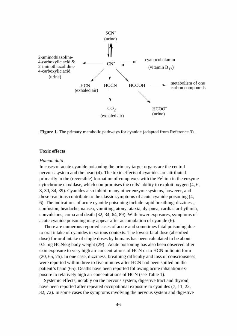
46
Toxic effects
Human dataIn cases of acute cyanide poisoning the primary target organs are the centralnervous system and the heart (4). The toxic effects of cyanides are attributedprimarily to the (reversible) formation of complexes with the Fe3 ion in the enzymecytochrome c oxidase, which compromises the cells’ ability to exploit oxygen (4, 6,8, 30, 34, 39). Cyanides also inhibit many other enzyme systems, however, andthese reactions contribute to the classic symptoms of acute cyanide poisoning (4,6). The indications of acute cyanide poisoning include rapid breathing, dizziness,confusion, headache, nausea, vomiting, atony, ataxia, dyspnea, cardiac arrhythmia,convulsions, coma and death (32, 34, 64, 89). With lower exposures, symptoms ofacute cyanide poisoning may appear after accumulation of cyanide (6).
There are numerous reported cases of acute and sometimes fatal poisoning dueto oral intake of cyanides in various contexts. The lowest fatal dose (absorbeddose) for oral intake of single doses by humans has been calculated to be about0.5 mg HCN/kg body weight (29) . Acute poisoning has also been observed afterskin exposure to very high air concentrations of HCN or to HCN in liquid form(20, 65, 75). In one case, dizziness, breathing difficulty and loss of consciousnesswere reported within three to five minutes after HCN had been spilled on thepatient’s hand (65). Deaths have been reported following acute inhalation ex-posure to relatively high air concentrations of HCN (see Table 1).
Systemic effects, notably on the nervous system, digestive tract and thyroid,have been reported after repeated occupational exposure to cyanides (7, 11, 22,32, 72). In some cases the symptoms involving the nervous system and digestive
CN-
SCN-
(urine)
cyanocobalamin
(vitamin B12)
HCOOH metabolism of onecarbon compounds
HCOO-
(urine)
HOCN
CO2
(exhaled air)
HCN(exhaled air)
2-aminothiazoline-4-carboxylic acid &2-iminothiazolidine-4-carboxylic acid (urine)
Figure 1. The primary metabolic pathways for cyanide (adapted from Reference 3).
47
tract took some time to appear, and it is not clear whether this represents a chronicform of cyanide poisoning or acute effects. Thyroid hypertrophy has beenassociated with chronic exposure to cyanides and seems to be related to thethiocyanate ion (4, 33). Cyanides are also suspected of contributing to thedeterioration of vision observed in some smokers (tobacco amblyopia), especiallythose with high alcohol consumption and nutritional deficiencies (26, 27, 70, 78).
Neurological effects and effects on the thyroid have been documented afterprolonged consumption of foods containing cyanogenic glycosides. Outbreaks ofkonzo, a distinct form of tropical myeloneuropathy characterized by abrupt onsetof incapacitating spasms, have been reported in populations in Africa after periodswhen the diet consisted mostly of cassava. Another cassava-related neural diseaseis tropical atactic neuropathy. It usually develops gradually over the course ofseveral years (41, 62, 79, 82, 83). Another disease that has been associated withhigh and prolonged cassava intake is goiter, which appears primarily inconjunction with low iodine intake (1, 2, 16).
In a retrospective study made in the United States, symptoms of cyanidepoisoning were seen in factory workers (n = 36) who had been exposed to cyanide(NaCN, HCN) during a silver reclaiming process (11). The workers had beenexposed to cyanides via inhalation, skin contact and possibly also oral intake. Theaverage air content of HCN one day after the factory had been closed (24-hourmeasurement) was 15 ppm. More than two thirds of the workers reported on aquestionnaire that they had experienced headaches, dizziness, nausea/vomiting,and/or a bitter taste in the mouth on more than 10 occasions per month. Otherreported symptoms included eye irritation, loss of appetite, weight loss, nose-bleeds, fatigue, skin rashes, heavy perspiration, shortness of breath, cough, sorethroat, chest pains, changes in the sense of smell, coughing up blood, heartpalpitations and fainting. There was a correlation (significant positive trend)between exposure level and the subjects’ assessments of the severity of thepoisoning, but the exposure estimate was not connected to the air measurements.Some symptoms were reported to last for seven or more months after exposurehad been terminated (lower prevalence). Biochemical analyses made more thanhalf a year after exposure had stopped revealed somewhat higher average valuesof thyroid-stimulating hormone (TSH) in serum (high-exposure group 2.4 µU/ml,laboratory control average 1.7 µU/ml) and somewhat fewer free bonding sites onthyroxine-binding globulin (TBG) in serum (T3U test: high-exposure group32.4%, laboratory control average 30.0%). On the other hand, values for thethyroid hormone T4 (thyroxine) were normal and there were no palpable changesin thyroids. The exposed workers (particularly those with high exposure) also hadlower average levels of vitamin B12 and folic acid in serum. The results areinterpreted by the authors as possible indications of effects of long-term cyanideexposure (11).
In another epidemiological study, 36 Egyptian workers (non-smokers) who hadbeen exposed to cyanide for 5 to 15 years of working with a plating bath containing3% NaCN and 3% copper cyanide were compared with 20 controls (22). Head-
48
aches, weakness, changes in the senses of taste and smell, dizziness, throatirritation, vomiting, shortness of breath with physical exertion, watery eyes, chestpains and several other symptoms were reported more frequently by the exposedworkers than by controls. Two of the nine workers from the factory with thehighest exposure levels (8.2-12.4 ppm) were reported to have suffered psychoticepisodes. Slight to moderate thyroid enlargement was seen in 20 of the exposedworkers, but none of them had clinical indications of hypo- or hyperthyroidism.Changes in blood profile, including significantly higher hemoglobin levels andlymphocyte counts, were also noted in the exposed workers. The workers weregiven radioactively labeled iodine after two days away from work, and muchhigher iodine concentrations (p <0.001) were found in the thyroids of the cyanide-exposed workers than in controls [average values for exposed workers: 38.7%(4 h), 49.3% (24 h); averages for controls, 22.4% (4 h), 40% (24 h)]. There was nosignificant difference for iodine bound to protein in blood (131PBI) after 72 hours.Air concentrations of cyanide in the breathing zone of exposed workers (15-minutesamples) ranged from 4.2 to 12.4 ppm (average concentrations 6.4-10.4 ppm). Theconcentration of thiocyanate in urine increased gradually, and during the last threedays of the work week it was virtually constant and strongly correlated to theaverage air concentration of cyanide (22).
In an Indian study (7), serum from 35 workers who handled cyanide salts in acable industry (electroplating) and 35 matched controls was analyzed for evidenceof effects on thyroid function. All subjects were non-smokers. No information onair concentrations is given. The content of thiocyanate in serum was higher in theexposed group than in the controls. The exposed workers had significantly lowerserum levels (p <0.05) of thyroid hormones T4 and T3, and significantly higherlevels (p <0.05) of TSH, than the controls. In the exposed group there was also asignificant positive correlation between serum levels of TSH and thiocyanate(p <0.05) and a significant negative correlation between T4 and thiocyanate(p <0.05). According to the authors, these results indicate that occupationalexposure to cyanide may inhibit thyroid function (7).
Another Indian study (15) reports that symptoms characteristic of cyanidepoisoning were observed in 23 workers exposed to HCN gas and cyanide aerosolduring case-hardening and electroplating operations. The workers had elevatedlevels of cyanide and thiocyanate in blood and urine, but no details on their othersymptoms are given. The measured cyanide levels in the breathing zone were inthe range 0.1-0.2 mg/m3 (0.09-0.19 ppm), and measurements made on the shopfloor yielded values in the range 0.2-0.8 mg/m3 (0.19-0.74 ppm). The limitedinformation in this report provides insufficient basis for any conclusions regardinga relation between effects on health and air concentrations of cyanide.
Significantly elevated serum values for zinc, calcium and iron were found in astudy of women who reportedly were exposed to HCN and cyanides 8 hours/dayat work (37). The same authors report in another study that significantly elevatedserum values for L-aspartate-2-oxyglutarate-aminotransferase (SGOT) and lactatedehydrogenase (LDH) were found in women (probably at the same workplace)
49
who reportedly were exposed to vapors of HCN and cyanides 8 hours/day (38). Inboth studies, the authors interpret the findings as effects of cyanide exposure. It isalso reported that on a few occasions the threshold limit of 5 mg HCN/m3 wasexceeded, air concentrations (calculated as HCN) then reaching a maximum of7.5 mg/m3. No details of the measurements are given, however. There is littleinformation on other occupational exposures or any differences in food intakebetween the exposed group and controls, and the interpretation of the results musttherefore be regarded as uncertain.
It is reported in a reference work (23) that a mist of an alkaline solutionconsisting mostly of NaCN caused severe nasal irritation and in some caseserosion of the nasal septum in occupationally exposed persons, and that the airconcentrations of cyanide (expressed as HCN) had not been much above 5 ppm. Itis suggested, however, that these persons may have been exposed to other alkalinesubstances that may have contributed considerably to the observed irritation.
Skin contact with NaCN or KCN can cause severe skin irritation as well assystemic toxicity (6, 40). The skin irritation is probably due to the alkalinity of thesubstances.
Animal dataHCN, NaCN and KCN are extremely toxic. With oral intake, the reported LD50
for all three substances is between 2.5 and 16 mg/kg body weight (rabbit, rat,mouse). For skin application of solutions (intact skin of rabbit), the reported LD50
is 6.9 mg/kg b.w. for HCN, 14.6 mg/kg b.w. for NaCN, and 22.3 mg/kg b.w. forKCN (6, 24). With inhalation exposure, the LC50 for HCN varies with species andexposure time, but in some studies (rabbit, rat, mouse) has been reported to beabout 110-170 ppm for 30 minutes of exposure, about 320-500 ppm for 5 minutesof exposure, and about 1000-3400 ppm for exposures of 1 minute or less (6, 54,58, 86).
Inhalation exposure to HCN affects most prominently the central nervoussystem (Table 2). Hyperventilation, grogginess and slow, irregular heartbeat, andin many cases cramps or apnea were observed in a study in which monkeys wereexposed to 100-156 ppm HCN for up to 30 minutes. At an air concentration of100 ppm it took 19 minutes for the animals to become so affected that they wereunable to move (67). A slight dampening effect on the CNS was observed at anexposure to 60 ppm HCN (monkeys, 30 minutes). EEG changes (includingincreased delta and theta activity) and reduced amplitude in measurement ofauditory cortical evoked potential could be observed toward the end of theexposure period. Some increase in respiratory minute volume was also noted.There were no significant effects on heart/vascular parameters, peripheralneuromuscular impulse transmission, or neural transmission speed (66). Reducedrespiratory minute volume, respiratory rate and pulmonary compliance, and lowerphospholipid levels in the lungs (surfactant), were reported in another study inwhich rats were exposed to 55 ppm HCN for 30 minutes. Increased transthoracicpressure, air flow and tidal volume were also observed (9). In a study with mice,
50
the DC50 (50% reduction of respiratory rate due to effects on the respiratorycenter) for 30 minutes of exposure to HCN was calculated to be 63 ppm.Intermittent sensory irritation was also observed (58).
A French study (85, cited in 4) reports observations of dyspnea, vomiting,diarrhea, tremor, spasms, ataxia and coma in dogs that had been exposed to45 ppm HCN 30 minutes/day for 28 to 96 days. Histological changes in the brainwere also observed. In an older study with several species, at exposure to 45 ppmHCN the animals fell and remained lying on their sides within 15 to 30 minutes(25). In a study with rabbits in which heart, lungs and associated arteries werestudied, no treatment-related effects were reported after constant exposure toabout 0.5 ppm HCN for up to 4 weeks (42, 43).
Effects on the central nervous system/behavior, thyroid, kidneys and reproduc-tive organs have been reported after repeated exposure by means other thaninhalation (Table 3). One study describes degenerative changes in the centralnervous system and inflammatory changes in the digestive tract in two dogs given0.5 or 2 mg NaCN (= 0.27 or 1.1 mg CN) /kg body weight/day in gelatin capsulesfor 13.5 or 14.5 months. Symptoms of acute toxicity were sometimes noted inconjunction with administration (36). In another study, pigs were given anaqueous solution of KCN by gavage for 24 weeks. Doses were equivalent to 0.4,0.7 or 1.2 mg CN/kg body weight/day. There was a gradual reduction in bloodlevels of T3 and to some extent also T4 in all dose groups (especially the high-dosegroup), and progressive, dose-dependent increases of blood glucose (fastingvalues) and effects on behavior were also noted (45). Kidney damage (nephrosis),indications of hyperplasia/hypertrophy of adrenals (zona glomerulosa) and effectson testes (see Effects on reproduction) were observed in histopathologicalexamination of dogs after 14 weeks on a diet containing NaCN in an amountequivalent to 1.04 mg CN/kg body weight/day (47). In earlier studies by the sameauthor, dogs fed on this diet for 14 weeks developed hypothyroidism and goiter,and there were also effects on the pancreas (necrosis, fibrosis, atrophy) (48, 49).
In an NTP study (34), NaCN mixed in the drinking water of rats and mice inamounts ranging from 3 to 300 mg/l (rats: about 0.16-13 mg CN/kg b.w./day;mice: about 0.3-27 mg CN/kg b.w./day) for 13 weeks caused changes in variousreproduction parameters (see Effects on reproduction). Other observationsincluded lower water consumption, especially in the higher dose groups, but therewere no clinically significant changes in body weight, organ weight, or hemato-logical or clinical-chemical parameters, and no histopathological changes inexamined organs (including thyroid, brain, adrenals, kidneys, liver, heart andlungs).
Cyanides can irritate the eyes. Local effects observed in eyes of rabbits afterinstillations of HCN, NaCN and KCN were inflammation, gradual swelling of theconjunctiva, and cloudiness of the cornea (5, 6). Severe symptoms of poisoningwere also noted after concentrated solutions had been applied to the eyes (5).
51
Mutagenicity, carcinogenicity
HCN was mutagenic to the Salmonella typhimurium strain TA100, but not TA98.The mutagenic activity was reduced by metabolic activation (51). NaCN andKCN were reported to be non-mutagenic to strains TA97, TA98, TA100,TA1530, TA1535, TA1537, TA1538 and/or TA1950 both with and withoutmetabolic activation (17, 18, 34, 50, 69). KCN, however, was positive in a DNArepair test with E coli (18). KCN induced chromosomal aberrations when testedon mammalian cells in vitro, and higher doses induced DNA fragmentation (10,84). In vitro studies with human cells indicate that KCN is cytotoxic but notgenotoxic (35, 87). Inhibited DNA synthesis, but no DNA damage, was reportedin an in vitro DNA-synthesis inhibition test (KCN) using HeLa cells (63).However, inhibition of DNA synthesis (testes) was not observed in vivo afteroral administration of 2.5 mg KCN/kg body weight to mice (28).
No studies specifically of carcinogenicity were found in the literature. In onestudy in which rats were given extremely high amounts of KCN in diet – 50 or100 g/kg feed (sic!) – for 14 weeks (61), benign tumors were found in cecum andlarge intestine (7/10 animals in the high-dose group; 5/10 animals in the low-dosegroup; results in controls not reported). The tumors consisted of many hyper-trophic muscle fibers with no indication of malignancy. Other observationsincluded reduced body weights, lower relative spleen weights, higher relativethyroid weights, hepatic cell necroses and hematological /biochemical changes.
Effects on reproduction
Hamsters were given slow infusions of NaCN from subcutaneous implantsliberating 6.18 to 6.35 mg/kg b.w./hour, equivalent to about 148-152 mg/kgb.w./day (total doses more than 30-40 times the LD50), on days 6 to 9 of gestation.There were signs of toxicity in the mothers, especially at the higher dose, and highincidences of severe malformations and resorptions at all dose levels (19). Anabstract (76) reports inhibited growth, meningoceles/myeloceles and some deathsin embryos of rats given daily intraperitoneal injections of KCN (aqueoussolution) during the first 15 days of gestation (3 mg/kg b.w. per injection).
In an experiment in which rats were given NaCN by gavage (6 mg/kg b.w. inwater) on day 10 of gestation, morphological anomalies were noted in somefetuses 48 hours after the administration. Clinical indications of toxicity(including deaths) were observed in the mothers (71). In studies in whichpregnant pigs and rats (80, 81) were fed on cassava containing up to 1250 mgKCN/kg, the only observed toxic effects on reproduction were minor metabolicdifferences and lower relative organ weights in fetuses at doses affecting themothers.
In an NTP study, mice and rats were given drinking water containing 30, 100 or300 mg NaCN/liter (rats: about 1.6-13 mg CN/kg b.w./day; mice: about 3-27 mgCN/kg b.w./day) for 13 weeks, and examined for effects on various reproductionparameters (reproductive toxicity at lower doses was not studied). Effects were
52
particularly evident in the rats, but were judged by the authors to be “probablyinsufficient to decrease fertility in rats.” Significant, dose-dependent reductions ofcauda epididymal weight (all dose groups) and dose-dependent reductions (signi-ficant in the high-dose group) of epididymis and testis weights were observed inrats. Cauda epididymis and epididymis weights were significantly lower in thehigh-dose group of mice. The number of sperm per testis was significantly lower inthe high-dose group of rats. Sperm motility was also lower in the rats (all dosegroups), but this was judged to have no biological significance. Altered estruscycles were observed in female rats in the two higher dose groups, but no effects onestrus cycle were seen in the female mice (34). Effects on spermatogenesis(significantly lower relative frequency of stage 8 tubuli and degenerative changeswith elevated occurrence of abnormal gametes) were observed in testes of dogsgiven food containing NaCN (dose equivalent to 1.04 mg CN/kg b.w./day) for14 weeks (47).
Dose-effect/dose-response relationships
There are few reliable measurements of air concentrations of HCN, NaCN orKCN in an occupational exposure context. In most of the studies that report airconcentrations, exposure to cyanide by other means (such as skin uptake) can besuspected, and it is therefore difficult to determine dose-response relationshipswith certainty. There is the additional problem of mixed exposures. Irritation ofrespiratory passages and toxic symptoms indicating effects on the central nervoussystem have been reported in several studies, however. Effects on the thyroidhave also been noted after exposure to HCN and cyanide salts, and have beenpresented as possible long-term effect of exposure. One work (22) reports thateffects on the central nervous system (including two psychoses, headache,vomiting, dizziness, weakness, changes in the senses of taste and smell) weremore common among exposed persons than among controls, and that themeasured air concentrations of cyanide in the breathing zones of the exposedworkers (15-minute samples) ranged from 4.2 to 12.4 ppm (average concen-trations 6.4 to 10.4 ppm). Effects on the thyroid were also noted at these airconcentrations (Table 1). There are no data indicating skin uptake of cyanide inthis study.
The maximum detoxification rate for cyanide in humans, as calculated frominformation on infusions of medicine given to acutely ill patients, is about 60 µgCN/minute (74). Assuming that inhaled air amounts to 20 l/minute and uptake is100%, this detoxification rate corresponds to uptake from inhalation exposure toabout 3 mg/m3. This calculation indicates that at higher exposure levels there maybe an accumulation of cyanide in the body, and it can therefore be assumed thatabout 6 hours of constant exposure to 4 mg/m3 might give rise to cyanide levels inthe blood that can yield biochemically detectable metabolic disturbances.
Dose-effect relationships observed in animals experimentally exposed to HCN,NaCN and KCN are summarized in Tables 2 and 3. Effects on the brain have been
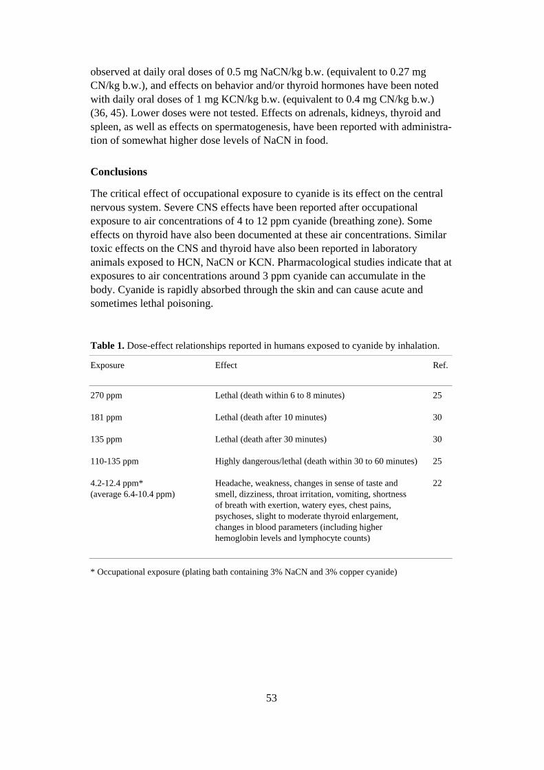
53
observed at daily oral doses of 0.5 mg NaCN/kg b.w. (equivalent to 0.27 mgCN/kg b.w.), and effects on behavior and/or thyroid hormones have been notedwith daily oral doses of 1 mg KCN/kg b.w. (equivalent to 0.4 mg CN/kg b.w.)(36, 45). Lower doses were not tested. Effects on adrenals, kidneys, thyroid andspleen, as well as effects on spermatogenesis, have been reported with administra-tion of somewhat higher dose levels of NaCN in food.
Conclusions
The critical effect of occupational exposure to cyanide is its effect on the centralnervous system. Severe CNS effects have been reported after occupationalexposure to air concentrations of 4 to 12 ppm cyanide (breathing zone). Someeffects on thyroid have also been documented at these air concentrations. Similartoxic effects on the CNS and thyroid have also been reported in laboratoryanimals exposed to HCN, NaCN or KCN. Pharmacological studies indicate that atexposures to air concentrations around 3 ppm cyanide can accumulate in thebody. Cyanide is rapidly absorbed through the skin and can cause acute andsometimes lethal poisoning.
Table 1. Dose-effect relationships reported in humans exposed to cyanide by inhalation.
Exposure Effect Ref.
270 ppm Lethal (death within 6 to 8 minutes) 25
181 ppm Lethal (death after 10 minutes) 30
135 ppm Lethal (death after 30 minutes) 30
110-135 ppm Highly dangerous/lethal (death within 30 to 60 minutes) 25
4.2-12.4 ppm*(average 6.4-10.4 ppm)
Headache, weakness, changes in sense of taste andsmell, dizziness, throat irritation, vomiting, shortnessof breath with exertion, watery eyes, chest pains,psychoses, slight to moderate thyroid enlargement,changes in blood parameters (including higherhemoglobin levels and lymphocyte counts)
22
* Occupational exposure (plating bath containing 3% NaCN and 3% copper cyanide)
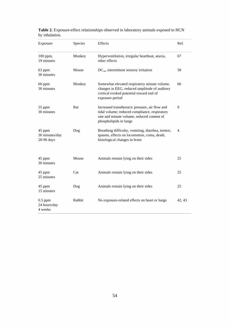
54
Table 2. Exposure-effect relationships observed in laboratory animals exposed to HCNby inhalation.
Exposure Species Effects Ref.
100 ppm,19 minutes
Monkey Hyperventilation, irregular heartbeat, ataxia,other effects
67
63 ppm30 minutes
Mouse DC50, intermittent sensory irritation 58
60 ppm30 minutes
Monkey Somewhat elevated respiratory minute volume,changes in EEG, reduced amplitude of auditorycortical evoked potential toward end ofexposure period
66
55 ppm30 minutes
Rat Increased transthoracic pressure, air flow andtidal volume; reduced compliance, respiratoryrate and minute volume, reduced content ofphospholipids in lungs
9
45 ppm30 minutes/day28-96 days
Dog Breathing difficulty, vomiting, diarrhea, tremor,spasms, effects on locomotion, coma, death,histological changes in brain
4
45 ppm30 minutes
Mouse Animals remain lying on their sides 25
45 ppm25 minutes
Cat Animals remain lying on their sides 25
45 ppm15 minutes
Dog Animals remain lying on their sides 25
0.5 ppm24 hours/day4 weeks
Rabbit No exposure-related effects on heart or lungs 42, 43
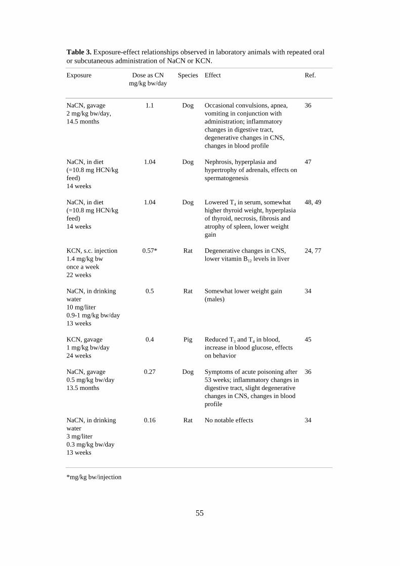
55
Table 3. Exposure-effect relationships observed in laboratory animals with repeated oralor subcutaneous administration of NaCN or KCN.
Exposure Dose as CNmg/kg bw/day
Species Effect Ref.
NaCN, gavage2 mg/kg bw/day,14.5 months
1.1 Dog Occasional convulsions, apnea,vomiting in conjunction withadministration; inflammatorychanges in digestive tract,degenerative changes in CNS,changes in blood profile
36
NaCN, in diet(=10.8 mg HCN/kgfeed)14 weeks
1.04 Dog Nephrosis, hyperplasia andhypertrophy of adrenals, effects onspermatogenesis
47
NaCN, in diet(=10.8 mg HCN/kgfeed)14 weeks
1.04 Dog Lowered T4 in serum, somewhathigher thyroid weight, hyperplasiaof thyroid, necrosis, fibrosis andatrophy of spleen, lower weightgain
48, 49
KCN, s.c. injection1.4 mg/kg bwonce a week22 weeks
0.57* Rat Degenerative changes in CNS,lower vitamin B12 levels in liver
24, 77
NaCN, in drinkingwater10 mg/liter0.9-1 mg/kg bw/day13 weeks
0.5 Rat Somewhat lower weight gain(males)
34
KCN, gavage1 mg/kg bw/day24 weeks
0.4 Pig Reduced T3 and T4 in blood,increase in blood glucose, effectson behavior
45
NaCN, gavage0.5 mg/kg bw/day13.5 months
0.27 Dog Symptoms of acute poisoning after53 weeks; inflammatory changes indigestive tract, slight degenerativechanges in CNS, changes in bloodprofile
36
NaCN, in drinkingwater3 mg/liter0.3 mg/kg bw/day13 weeks
0.16 Rat No notable effects 34
*mg/kg bw/injection
56
References
1. Abuye C, Kelbessa U, Wolde-Gebriel S. Health effects of cassava consumption i southEthiopia. East Afric Med J 1998;75:166-170.
2. Adewusi SRA, Akindahunsi AA. Cassava processing, consumption, and cyanide toxicity. JToxicol Environ Health 1994;43:13-23.
3. Ansell M, Lewis FAS. A review of cyanide concentrations found in human organs. JForensic Med 1970;17:148-155.
4. ATSDR. Toxicological profile for cyanide (update). PB98-101207. Agency for ToxicSubstances and Disease Registry, Atlanta, Georgia, USA, 1997.
5. Ballantyne B. Acute systemic toxicity of cyanides by topical application to the eye. JToxicol-Cut Ocular Toxicol 1983;2:119-129.
6. Ballantyne B. Toxicology of cyanides. In: Ballantyne B, Marrs TC, eds. Clinical andExperimental Toxicology of Cyanides. Bristol: Wright, 1987:41-126.
7. Banerjee KK, Bishayee A, Marimuthu P. Evaluation of cyanide exposure and its effect onthyroid function of workers in a cable industry. J Occup Environ Med 1997;39:258-260.
8. Beasley DMG, Glass WI. Cyanide poisoning: pathophysiology and treatmentrecommendations. Occup Med 1998;48:427-431.
9. Bhattacharya R, Kumar P, Sachan AS. Cyanide induced changes in dynamic pulmonarymechanics in rats. Indian J Physiol Pharmacol 1994;38:281-284.
10. Bhattacharya R, Lakshmana Rao PV. Cyanide induced DNA fragmentation in mammaliancell cultures. Toxicology 1997;123:207-215.
11. Blanc P. Hogan M, Mallin K, Hryhorczuk D, Hessl S, Bernard B. Cyanide intoxicationamong silver-reclaiming workers. J Am Med Assoc 1985;253:367-371.
12. Cagianut B, Schnebli HP, Rhyner K, Furrer J. Decreased thiosulfate sulfur transferase(rhodanese) in Leber´s hereditary optic atrophy. Klin Wochenschr 1984;62:850-854.
13. Calabrese EJ. Possible adverse side effects from treatment with laetrile. Med Hypotheses1979;5:1045-1049.
14. Carlsson L, Mlingi N, Juma A, Ronquist G, Rosling H. Metabolic fates in humans oflinamarin in cassava flour ingested as stiff porridge. Food Chem Toxicol 1999;37:307-312.
15. Chandra H, Gupta BN, Bhargava SK, Clerk SH, Mahendra PN. Chronic cyanide exposure - abiochemical and industrial hygiene study. J Anal Toxicol 1980;4:161-165.
16. Cliff J, Lundquist P, Rosling H, Sörbo B, Wide L. Thyroid function in cassava-eatingpopulation affected by epidemic spastic paraparesis. Acta Endocrinol 1986;113:523-528.
17. De Flora S. Study of 106 organic and inorganic compounds in the Salmonella/microsometest. Carcinogenesis 1981;2:283-298.
18. De Flora S, Zanacchi P, Camoirano A, Bennicelli C, Badolati GS. Genotoxic activity andpotency of 135 compounds in the Ames reversion test and in a bacterial DNA-repair test.Mutat Res 1984;133:161-198.
19. Doherty PA, Ferm VH, Smith RP. Congenital malformations induced by infusion of sodiumcyanide in the golden hamster. Toxicol Appl Pharmacol 1982;64:456-464.
20. Dugard PH. The absorption of cyanide through human skin in vitro from solutions of sodiumcyanide and gaseous HCN. In: Ballantyne B, Marrs TC, eds. Clinical and ExperimentalToxicology of Cyanides. Bristol: Wright, 1987:127-137.
21. Einhorn IN. Physiological and toxicological aspects of smoke produced during thecombustion of polymeric materials. Environ Health Persp 1975;11:163-189.
22. El Ghawabi SH, Gaafar MA, El-Saharti AA, Ahmed SH, Malash KK, Fares R. Chroniccyanide exposure: a clinical, radioisotope, and laboratory study. Br J Ind Med 1975;32:215-219.
57
23. Elkins HB. The Chemistry of Industrial Toxicology. New York: John Wiley & Sons,1959:94-95.
24. EPA. Drinking water criteria document for cyanide. PB92-173319. US EnvironmentalProtection Agency, Springfield VA, USA: NTIS, 1992.
25. Flury F, Zernik F. Schädliche Gase. Berlin: Verlag von Julius Springer, 1931:400-409.
26. Foulds WS, Chisholm IA, Bronte-Stewart J, Wilson T. Cyanide induced optic neuropathy.Ophthalmologica 1969;158:350-358.
27. Freeman AG. Optic neuropathy and chronic cyanide intoxication: a review. J R Soc Med1988;81:103-106.
28. Friedman MA, Staub J. Inhibition of mouse testicular DNA synthesis by mutagens andcarcinogens as a potential simple mammalian assay for mutagenesis. Mutat Res 1976;37:67-76.
29. Gettler AO, Baine JO. The toxicology of cyanide. Am J Med Sci 1938;195:182-198.
30. Hall AH, Rumack BH. Clinical toxicology of cyanide. Ann Emerg Med 1986;15:1067-1074.
31. Hall VA, Guest JM. Sodium nitroprusside-induced cyanide intoxication and prevention withsodium thiosulfate prophylaxis. Am J Crit Care 1992;1:19-27.
32. Hardy HL, Jeffries WM, Wasserman MM, Waddell WR. Thiocyanate effect followingindustrial cyanide exposure. N Engl J Med 1950;242:968-972.
33. Hartung R. Cyanides and nitriles. In: Clayton GD, Clayton FE, eds. Patty´s IndustrialHygiene and Toxicology, 4th ed. New York: John Wiley & Sons, 1994:3119-3172.
34. Hébert CD. NTP technical report on toxicity studies of sodium cyanide. NTP Toxicity ReportSeries 37. NIH Publication 94-3386. National Toxicology Program, Research Triangle Park,NC, USA, 1993.
35. Henderson L, Wolfreys A, Fedyk J, Bourner C, Windebank S. The ability of the Comet assayto discriminate between genotoxins and cytotoxins. Mutagenesis 1998;13:89-94.
36. Hertting G, Kraupp O, Schnetz E, Wuketich S. Untersuchungen über die Folgen einerchronischen Verabreichung akut toxischer Dosen von Natriumcyanid an Hunden. ActaPharmacol Toxicol 1960;17:27-43.
37. Hly´nczak JA, Kersten E, Fokt M, Wysocki K, Raczynski A, Michaliszyn J. Über den Gehalteiniger Metalle im Serum beruflich HCN-exponierter Frauen. Z ärztl Fortbild 1980;74:589-591.
38. Hly´nczak JA, Kersten E, Wysocki K, Stamm E, Fokt M, Raczynski A, Untersuchungen zurAktivität einiger Enzyme im Serum HCN-exponierter Frauen. Z ärztl Fortbild 1980;74:591-593.
39. Holland MA, Kozlowski LM. Clinical features and management of cyanide poisoning. ClinPharmacy 1986;5:737-741.
40. Homan ER. Reactions, processes and materials with potential for cyanide exposure. In:Ballantyne B, Marrs TC, eds. Clinical and Experimental Toxicology of Cyanides. Bristol:Wright, 1987:1-21.
41. Howlett WP, Brubaker GR, Mlingi N, Rosling H. Konzo, an epidemic upper motor neurondisease studied in Tanzania. Brain 1990;113:223-235.
42. Hugod C. Effect of exposure to 0,5 ppm hydrogen cyanide singly or combined with 200 ppmcarbon monoxide and/or 5 ppm nitric oxide on coronary arteries, aorta, pulmonary artery, andlungs in the rabbit. Int Arch Occup Environ Health 1979;44:13-23.
43. Hugod C. Myocardial morphology in rabbits exposed to various gas-phase constituents oftobacco smoke. Atherosclerosis 1981;40:181-190.
44. Hägg G. Allmän och oorganisk kemi, 5th ed. Stockholm: Almqvist & Wiksell, 1963:564.
45. Jackson LC. Behavioral effects of chronic sublethal dietary cyanide in an animal model:implications for humans consuming cassava. Hum Biol 1988;60:597-614.
46. Jones DA. Why are so many food plants cyanogenic? Phytochem 1998;47:155-162.
58
47. Kamalu BP. Pathological changes in growing dogs fed on a balanced cassava (Manihotesculenta Crantz) diet. Brit J Nutr 1993:69:921-934.
48. Kamalu BP. The effect of a nutritionally-balanced cassava (Manihot esculenta Crantz) dieton endocrine function using the dog as a model 1. Pancreas. Br J Nutr 1991;65:365-372.
49. Kamalu BP. The effect of a nutritionally-balanced cassava (Manihot esculenta Crantz) dieton endocrine function using the dog as a model 2. Thyroid. Br J Nutr 1991;65:373-379.
50. Kleinhofs A, Smith JA. Effect of excision repair on azide-induced mutagenesis. Mutat Res1976;41:233-240.
51. Kushi A, Matsumoto T, Yoshida D. Mutagen from the gaseous phase of protein pyrolyzate.Agric Biol Chem 1983;47:1979-1982.
52. Landahl HD, Herrmann RG. Retention of vapors and gases in the human nose and lung. ArchInd Hyg Occup Med 1950;1:36-45.
53. Leuschner J, Winkler A, Leuschner F. Toxicokinetic aspects of chronic cyanide exposure inthe rat. Toxicol Lett 1991;57:195-201.
54. Levin BC, Paabo M, Gurman JL, Harris SE. Effects of exposure to single or multiplecombinations of the predominant toxic gases and low oxygen atmospheres produced in fires.Fundam Appl Toxicol 1987;9:236-250.
55. Lewis JL, Rhoades CE, Gervasi PG, Griffith WC, Dahl AR. The cyanide-metabolizingenzyme rhodanese in human nasal respiratory mucosa. Toxicol Appl Pharmacol1991;108:114-120.
56. Lundquist P, Rosling H, Sörbo B. The origin of hydrogen cyanide in breath. Arch Toxicol1988;61:270-274.
57. Maehly AC, Swensson Å. Cyanide and thiocyanate levels in blood and urine of workers withlow-grade exposure to cyanide. Int Arch Arbeitsmed 1970;27:195-209.
58. Matijak-Schaper M, Alarie Y. Toxicity of carbon monoxide, hydrogen cyanide and lowoxygen. J Combust Toxicol 1982;9:21-61.
59. NIOSH. Criteria for a Recommended Standard. Occupational exposure to hydrogen cyanideand cyanide salts. US Department of Health, Education and Welfare. DHEW Publication No77-108. Washington DC: US Government Printing Office, 1976.
60. Okoh PN. Excretion of 14C-labeled cyanide in rats exposed to chronic intake of potassiumcyanide. Toxicol Appl Pharmacol 1983;70:335-339.
61. Olusi SO, Oke OL, Odusote A. Effects of cyanogenic agents on reproduction and neonataldevelopment in rats. Biol Neonate 1979;36:233-243.
62. Osuntokun BO, Monekosso GL. Degenerative tropical neuropathy and diet. Br Med J1969;3:178-179.
63. Painter RB, Howard R. The HeLa DNA-synthesis inhibition test as a rapid screen formutagenic carcinogens. Mutat Res 1982;92:427-437.
64. Peden NR, Taha A, McSorley PD, Bryden GT, Murdoch IB, Anderson JM. Industrialexposure to hydrogen cyanide: implications for treatment. Br Med J 1986;293:538.
65. Potter L. The successful treatment of two recent cases of cyanide poisoning. Br J Ind Med1950;7:125-130.
66. Purser DA. A bioassay model for testing the incapacitating effects of exposure to combustionproduct atmospheres using cynomolgus monkeys. J Fire Sci 1984;2:20-36.
67. Purser DA, Grimshaw P, Berrill KR. Intoxication by cyanide in fires: a study in monkeysusing polyacrylonitrile. Arch Environ Health 1984;39:394-400.
68. Riedorf F. Noxious gases and vapors. In: Di Palma JR, ed. Drill´s Pharmacology inMedicine. New York: McGraw-Hill, 1971:1189-1194.
69. Rietveld EC, Delbressine LPC, Waegemaekers THJM, Seutter-Berlage F. 2-Chlorobenzylmercapturic acid, a metabolite of the riot control agent 2-chlorobenzylidenemalononitrile (CS) in the rat. Arch Toxicol 1983;54:139-144.
59
70. Rizzo JF, Lessell S. Tobacco amblyopia. Am J Ophthalmol 1993;116:84-87.
71. Saillenfait AM, Sabaté JP. Comparative developmental toxicities of aliphatic nitriles: in vivoand in vitro observations. Toxicol Appl Pharmacol 2000;163:149-163.
72. Sandberg CG. A case of chronic poisoning with potassium cyanide? Acta Med Scand1967;181:233-236.
73. Schulz V. Clinical pharmacokinetics of nitroprusside, cyanide, thiosulphate and thiocyanate.Clin Pharmacokinet 1984;9:239-251.
74. Schulz V, Gross R, Pasch T, Busse J, Loeschcke G. Cyanide toxicity of sodium nitroprussidein therapeutic use with and without sodium thiosulphate. Klin Wochenschr 1982;60:1393-1400.
75. Schütze W. Über die Gefährdung von Mensch und Tier durch grosse Konzentrationen einigergiftiger Gase von der Haut aus. Arch Hyg 1927;98:70-83.
76. Singh JD. The lethality and teratogenicity of potassium cyanide in rat. Teratology1982;25:84A.
77. Smith ADM, Duckett S, Waters AH. Neuropathological changes in chronic cyanideintoxication. Nature 1963;200:179-181.
78. Solberg Y, Rosner M, Belkin M. The association between cigarette smoking and oculardiseases. Surv Ophthalmol 1998;42:535-547.
79. Spencer PS. Food toxins, AMPA receptors, and motor neuron diseases. Drug Metab Rev1999;31:561-587.
80. Tewe OO, Maner JH. Long-term and carry-over effect of dietary inorganic cyanide (KCN) inthe life cycle performance and metabolism of rats. Toxicol Appl Pharmacol 1981;58:1-7.
81. Tewe OO, Maner JH. Performance and pathophysiological changes in pregnant pigs fedcassava diets containing different levels of cyanide. Res Vet Sci 1981;30:147-151.
82. Tor-Agbidye J, Palmer VS, Lasarev MR, Craig AM, Blythe LL, Sabri MI, Spencer PS.Bioactivation of cyanide to cyanate in sulfur amino acid deficiency: relevance to neurologicaldisease in humans subsisting on cassava. Toxicol Sci 1999;50:228-235.
83. Tylleskär T, Howlett WP, Rwiza HT, Aquilonius SM, Stålberg E, Lindén B, Mandahl A,Larsen HC, Brubaker GR, Rosling H. Konzo: a distinct disease entity with selective uppermotor neuron damage. J Neurol Neurosurg Psychiatry 1993;56:638-643.
84. Umeda M, Nishimura M. Inducibility of chromosomal aberrations by metal compounds incultured mammalian cells. Mutat Res 1979;67:221-229.
85. Valade MP. Lésions du système nerveux central dans les intoxications chroniquesexpérimentales par l’acide cyanhydrique gazeux. Bull Acad Natl Med 1952;136:280-285.
86. Vernot EH, MacEwen JD, Haun CC, Kinkead ER. Acute toxicity and skin corrosion data forsome organic and inorganic compounds and aqueous solutions. Toxicol Appl Pharmacol1977;42:417-423.
87. Vock EH, Lutz WK, Hormes P, Hoffmann HD, Vamvakas S. Discrimination betweengenotoxicity and cytotoxicity in the induction of DNA double-strand breaks in cells treatedwith etoposide, melphalan, cisplatin, potassium cyanide, Triton X-100, and γ-irradiation.Mutat Res 1998;413:83-94.
88. Wilson J. Cyanide in human disease. In: Ballantyne B, Marrs TC, eds. Clinical andExperimental Toxicology of Cyanides. Bristol: Wright, 1987:292-311.
89. Wolfsie JH. Treatment of cyanide poisoning in industry. Arch Ind Hyg Occup Med1951;4:417-425.
90. Wood JL. Biochemistry. In: Newman AA. ed. Chemistry and Biochemistry of ThiocyanicAcid and its Derivatives. New York: Academic Press, 1975:156-221.
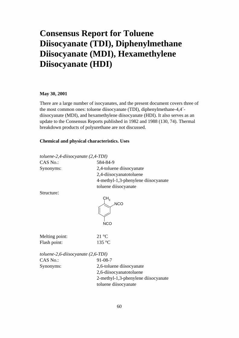
60
Consensus Report for TolueneDiisocyanate (TDI), DiphenylmethaneDiisocyanate (MDI), HexamethyleneDiisocyanate (HDI)
May 30, 2001
There are a large number of isocyanates, and the present document covers three ofthe most common ones: toluene diisocyanate (TDI), diphenylmethane-4,4´-diisocyanate (MDI), and hexamethylene diisocyanate (HDI). It also serves as anupdate to the Consensus Reports published in 1982 and 1988 (130, 74). Thermalbreakdown products of polyurethane are not discussed.
Chemical and physical characteristics. Uses
toluene-2,4-diisocyanate (2,4-TDI)CAS No.: 584-84-9Synonyms: 2,4-toluene diisocyanate
2,4-diisocyanatotoluene4-methyl-1,3-phenylene diisocyanatetoluene diisocyanate
Structure:CH3
NCO
NCO
Melting point: 21 °CFlash point: 135 °C
toluene-2,6-diisocyanate (2,6-TDI)CAS No.: 91-08-7Synonyms: 2,6-toluene diisocyanate
2,6-diisocyanatotoluene2-methyl-1,3-phenylene diisocyanatetoluene diisocyanate

61
Structure:CH3
NCOOCN
2,4-TDI:2,6-TDI (4:1)CAS No.: 26471-62-5Melting point: 12.5-13.5 °CFlash point: 132 °C
For all of the above:Formula: C9H6N2O2
Molecular weight: 174.16Density (liquid): 1.22 g/cm3 (25 °C)Density (vapor): 6.0 (air = 1)Boiling point: 251 °CVapor pressure: 2.7 Pa (25 °C)Saturation concentration: 27 ppmConversion factors: 1 µg/m3 = 0.138 ppb
1 ppb = 7.239 µg/m3
diphenylmethane-4,4´-diisocyanate (MDI)CAS No.: 101-68-8Synonyms: methylenebis(phenylisocyanate)
4,4´-diphenyl methane diisocyanatebis(1,4-isocyanatophenyl)methane4,4´-methylenediphenyl diisocyanate4,4´-diisocyanato-diphenyl methanediphenylmethane diisocyanate4,4´-methylphenyl diisocyanate
Formula: C15H10N2O2
Structure:
CH2OCN NCO
Molecular weight: 250.3Density (liquid): 1.23 g/cm3 (25 °C)Density (vapor): 8.5 (air = 1)Boiling point: 314 °CMelting point: 39.5 °C
62
Flash point: 196 °CVapor pressure: 6.7 x 10⁻⁴ Pa (25 °C)Saturation concentration: 6.6 ppbConversion factors: 1 µg/m3 = 0.096 ppb
1 ppb = 10.40 µg/m3
hexamethylene-1,6-diisocyanate (HDI)CAS No.: 822-06-0Synonyms: 1,6-diisocyanatohexane
1,6-hexamethylene diisocyanateFormula: C8H12N2O2
Structure: OCN–(CH2)6–NCOMolecular weight: 168Density (liquid): 1.04 g/cm3
Boiling point: 212.8 °CMelting point: - 67 °CFlash point: 140 °CVapor pressure: 1.3 Pa (20 °C)Saturation concentration: 13 ppmConversion factors: 1 µg/m3 = 0.143 ppb
1 ppb = 6.991 µg/m3
Isocyanates are characterized by their reactive -N=C=O group. Some diiso-cyanates (TDI, HDI) are volatile at room temperature and others can not beinhaled unless they are heated or in aerosol form (MDI). MDI and TDI usuallyoccur industrially as mixtures of different isomers and/or in prepolymerized form.Industrial grade TDI is usually a mixture of 2,4-TDI and 2,6-TDI in a ratio of 4:1or 65:35. MDI usually occurs as a mixture of several isomers and oligomers: onecommon mixture is approximately 30-40% diphenylmethane-4,4´-diisocyanate,2.5-4.0% diphenylmethane-2,4´-diisocyanate, 0.1-0.2 % diphenylmethane-2,2´-diisocyanate, and the remaining 50-60% oligomers. HDI rarely occurs in mono-meric form and is usually in adducts.
Isocyanates are used in the production of polyurethane, a common material forsoft and hard foam plastics, insulation materials, two-component adhesives, foamrubber, and various types of paints and hardeners. TDI is used primarily in theproduction of low-viscosity polyurethane foam, and MDI is used for the produc-tion of harder polyurethane items (in catalyzers, for example). TDI and MDI areused in surface coatings (e.g. for chutes used in mining and agriculture), in theshoe industry for shoe soles, in the automobile industry for shock absorbers, andin a wide variety of other industries. MDI occurs in foundries in binders forcasting forms, and in orthopedic surgery in casts for broken bones.
The reported odor threshold for TDI is 360-920 µg/m3 (53). MDI is reported tobe odorless (52). There is no information regarding the odor theshold for HDI.
63
Measuring air concentrations of TDI, MDI and HDI
Measuring isocyanates in air is complicated by several factors (97, 129).Isocyanates are extremely reactive compounds. They may occur as gases or asparticles of various sizes in aerosols. The monitoring method must be specific andextremely sensitive, since the threshold values are low. Because of the highreactivity of isocyanates, samples must be taken using chemosorption, i.e. byderivitization using a reagent in solution or a reagent-impregnated solid medium,and this must be followed by chromatographic analysis. The method in generaluse before the introduction of chromatography was the spectrophotometricMarcali method (86). Measurements made with the Marcali method must beregarded with considerable reservation today, since the sensitivity and specificityof the method do not meet present standards. In simple exposure situations,however, the Marcali method may yield reliable values for TDI.
The reagents most widely used during the past 20 years for isocyanatederivatization in conjunction with chromatographic analysis are the following:
N-[(4-nitrophenyl)methyl]propylamine (‘NITRO reagent’)9-(N-methylamino methyl)-anthracene (MAMA)1-(2-pyridyl)-piperazine (PP)1-(2-methoxyphenyl)-piperazine (MP)1-(9-anthracenylmethyl)-piperazine (MAP)tryptamine (TRYP)di-n-butylamine (DBA)
In monitoring TDI, MDI and HDI, the most important parameters are thegas:particle ratio and the size and reactivity of any isocyanate particles that maybe present. It is probable that under certain conditions (such as spraying of quick-setting MDI products) the particles react too slowly with the reagent whensamples are taken on reagent-impregnated filters, and samples must therefore betaken in an impinger (4). On the other hand, particles smaller than 2 µm are notcaptured effectively in an impinger. It is therefore recommended that, in environ-ments containing both quick-drying particles larger than 2 µm and particlessmaller than 2 µm, sampling in an impinger be followed by sampling on areagent-coated fiberglass filter (47, 128). In one of the methods commonly used inother countries, samples are taken in an impinger containing MP in toluene or on afiberglass filter coated with MP (47). MAMA in toluene has been widely used inSweden (11). For monitoring monomeric TDI, MDI and HDI in environmentswhere these substances are generated by polyurethane production, the choice ofreagent is generally not of critical importance. DBA in toluene has severaladvantages over other reagents (135), but disadvantages have also been found(67). As a rule, reagent-coated filters should be extracted in the field right afterthe sample is taken. With regard to measuring TDI, MDI and HDI in complexenvironments – around thermal breakdown of polyurethane materials, for example– our knowledge of the efficiency of the various reagents and sampling methods isstill quite limited.
64
Liquid chromatography is the only method used to separate isocyanates priorto analysis. One or a combination of detectors – UV, fluorescence, electro-chemical – may be used for identification and quantification (97, 129). Toidentify and quantify samples from complex environments it is necessary touse mass spectrometry. DBA is the reagent that has been most thoroughlyinvestigated in conjunction with mass spectrometric analysis (54, 57, 136).International standards are being drawn up for methods based on MP, MAP,DBA, various combinations of reagents, and different sampling methods (54).
In summary, we now know that earlier measurements of isocyanates, partic-ularly in complex environments (with thermal breakdown, for example), can beoff by a wide margin – especially if they were made before the introduction ofchromatography. Further, sampling in an impinger may yield measurements thatare too low because the particles smaller than 2 µm were not captured. Chromato-graphic analysis of derivatized isocyanates without confirmation by mass spectro-metry may lead to concentration estimates that are too high. When the derivatiza-tion reaction is slow, competing reactions may lead to estimates that are too low.In assessing the results of epidemiological studies, therefore, the monitoringmethod must always be assessed as well. If a study is to be accepted as reliable,the exposure levels must be determined by modern chromatographic methods. Anexception may be made for measurements made in simple exposure situations,where determination with the Marcali method may be acceptable.
Uptake, biotransformation, excretion
In studies with rats and guinea pigs it has been shown that inhaled TDI isabsorbed in the central respiratory passages and far out in the bronchioles (61).In general, isocyanates bind to proteins quite rapidly (62). Skin uptake of TDIhas been demonstrated in rats after exposure 3 hours/day for 4 consecutive days(115).
Due to their –N=C=O group, isocyanates are extremely reactive substances thatform adducts. Urine samples taken after exposure to isocyanates contain thecorresponding amines, which can be identified if the sample is prepared byhydrolysis. Thus, toluene diamines (TDA) can be found after exposure to TDI(115), 4,4´-methylenedianiline (MDA) and N-acetyl-4,4´-methylenedianiline(AcMDA) after exposure to MDI (124), and 1,6-hexamethylene diamine (HDA)after exposure to HDI (15). It has been assumed on the evidence of in vitro studiesthat the toxicity of isocyanates is largely due to the distribution of isocyanate-glutathione conjugates to various organs via blood and the release of freeisocyanate in peripheral tissues. This occurs particularly with low local concen-trations of glutathione (GSH) or elevated pH (14, 102). The distribution ofisocyanates in the human body, however, is largely unknown.
Data on metabolism of isocyanates are also sparse. In experiments in which ratswere given 14C-TDI in diet, insoluble polymers were formed in the stomach athigh doses (900 mg/kg b.w./day) in feed, but not at lower doses (60 mg/kg
65
b.w./day) (53). The metabolites of 2,6-TDI are excreted primarily in feces. Oraladministration of 2,6-TDI in oil (900 mg/kg b.w.) resulted in formation ofpolymers in the digestive tract, and 2,6-TDI polymers were still in the digestivetract 72 hours later. The half time for 2,6-TDI in aqueous solution is less than 2minutes in the ventricle and less than 30 seconds in serum (53). MDI and TDIhave been found in and below the respiratory epithelium of exposed laboratoryanimals from the nose to the terminal bronchioles. TDI-protein complexes havebeen found in pulmonary tissue of guinea pigs after inhalation of TDI (60).Bronchoalveolar lavage fluid from TDI-exposed guinea pigs contained five TDI-protein complexes, of which TDI-albumin was the most prevalent (56). In humansexposed to airborne 2,4-TDI and 2,6-TDI, the substances are bound mostly toalbumin in plasma (70). TDI-hemoglobin complexes have been identified inguinea pigs after inhalation of TDI (27). After rats had been exposed by inhalationto 14C-TDI, the highest concentrations of radioactivity were found in respiratorypassages, digestive tract and blood, in that order. The radioactivity in plasma waslinearly related to the dose of inhaled 14C-TDI, and the 14C-labeled compoundswere almost entirely in the form of conjugates (63).
Biological measures of exposure
Volunteers exposed to HDI in a test chamber (3.6 ppb for 7.5 hours) had thecorresponding amine (1,6-hexamethylene diamine, HDA) in hydrolyzed urine(15). The biological half time was about 1 hour. In a similar study with lowerexposures (1.7 to 3.1 ppb for 2 hours), HDA levels in urine were proportional toHDI levels in air, and the half time was 2.5 hours (134). HDA could not be foundin unhydrolyzed urine, which suggests that this metabolite occurs as an adduct.HDA was not detected in plasma in either of these studies (15, 134). HDA couldbe identified in hydrolyzed urine from car painters exposed to prepolymerizedHDI (116) and production workers making HDI monomers (80). Of 22 carpainters who used HDI-based paint and wore face masks with air filters, 4 hadHDA in hydrolyzed urine; none of seven controls had detectable amounts (149).
Subjects exposed to 2,4-TDI and 2,6-TDI in a test chamber (5.5 ppb for 7.5hours) had the corresponding amines in plasma and urine – identified by gaschromatography-mass spectrometry (GC-MS) after hydrolysis (125). Eliminationfrom plasma was slow. Elimination in urine had a slow and a fast phase, the latterwith a biological half time of one to two hours. Two persons were exposed tothree concentrations in the range 3.4-9.7 ppb for 4 hours, and there was acorrelation between TDI levels in air and TDA levels in plasma (16). Eliminationfrom plasma had two phases. The biological half time was two to five hours forthe fast phase and more than six days for the slow phase (16).
In a cross-sectional study of employees making automobile upholstery in aworkplace where there was low exposure to both aerosols from spraying of MDIand HDI adhesives and thermal breakdown products from the adhesives and fromTDI foam (air concentrations of MDI <0.76 ppb, HDI <0.1 ppb, TDI <0.01 ppb),
66
toluene-2,4-diamine (2,4-TDA) and toluene-2,6-diamine (2,6-TDA) were detectedin hydrolyzed plasma from respectively 16% and 7% of the production workers,but none of the office workers (72). Some TDA isomer was found in the urine of48% of the production workers and 21% of the office workers.
A TDI metabolite (2,6-TDA), but not TDI, could be identified in hydrolyzedurine from workers exposed to 2,6-TDI (79, 117). There was some correlation toair concentrations. Workers exposed to TDI (≤ 0.5 ppb) in a foam plastic factorywere monitored for 5 weeks: the urine content of TDA fluctuated, as did the ratiobetween 2,4-TDA and 2,6-TDA (103). There was some correlation with airconcentrations. The amounts of TDA in plasma were relatively constant, and notcorrelated to amounts in urine. In another study of workers in two foam plasticfactories where air concentrations of TDI were 0.05-0.5 ppb and 1.4-16.6 ppbrespectively, the TDA levels in plasma reflected the difference in air concen-trations (69). While the workers were on vacation the levels in plasma declinedwith an average half time of 21 days. The levels of TDA in urine dropped also,with a half time of 5.8 to 11 days and some indication of two phases.
Workers exposed to TDI (average 4.1 ppb) in a foam plastic factory had TDAin hydrolyzed plasma and urine (135). TDA in plasma of some of the workersexperimentally exposed for a short time had a biological half time of about 10days. The amount of TDA in urine from the workers varied quite a bit, and washighest right after work. There was no clear correlation between TDI levels in airand TDA levels in plasma or urine. The levels in plasma and urine were higherand the half time in plasma longer than they were for the briefly exposed subjectsin the test chamber studies (16), which lends support to the hypothesis that thereis a slow compartment. In plasma from a worker in the same factory, most of themetabolite was covalently bound to albumin (70).
Methylenedianiline (MDA) could be identified in pooled samples ofhydrolyzed plasma and urine from 10 workers exposed to MDI (it is unclearwhether thermal breakdown was involved) (126). MDA could be identified inhydrolyzed hemoglobin from 10 of 26 workers exposed to MDI (all but three <0.3ppb; values for the other three were 1.0, 1.8 and 2.9 ppb) (122). After alkalineextraction there were measurable amounts of acetyl-MDA (AcMDA) and lesseramounts of MDA in urine from 18 of the 26, MDA alone in 4 samples, andneither substance in 4. After acid hydrolysis the MDA levels were on averageabout 1/3 higher that the total of AcMDA and MDA in the previous analysis. Thelevels of hemoglobin adducts had no correlation to metabolites in urine (122).
In a polyurethane production facility where air concentrations of MDI wereusually below the detection limit, measurable amounts of 4,4´-MDA (0.035-0.83pmol/ml) and AcMDA (0.13-7.61 pmol/ml) could be found in urine in 15 of 20workers after alkaline extraction, and MDA values were 6.5 times higher afteracid hydrolysis. MDA was found as hemoglobin adducts in all the examinedworkers, and one worker also had adducts of AcMDA. Plasma levels of4,4´-MDA ranged from 0.25 to 5.4 pmol/ml, up to 120 fmol/mg of which wascovalently bound to albumin (124).
67
2,4-TDA, 2,6-TDA and 4,4´-MDA could be found in hydrolyzed urine from 15workers at a workplace where TDI- and MDI-based polyurethane was heated(26). The levels fluctuated widely from day to day. Levels of these metabolites inplasma were much more stable. In four of the monitored workers the levels ofMDA declined during an exposure-free period, with biological half times of 2.5-3.4 days in urine and 10-22 days in plasma.
It has long been known that some persons exposed to isocyanates form anti-bodies specific to conjugates of the isocyanate and serum albumin (40, 147).These are of doubtful pathogenic relevance, but may be used as biomarkers ofexposure (for those persons who develop antibodies). Three percent of workersexposed to spray aerosols of glue based on MDI or HDI had specific IgEantibodies, while 33% had IgG specific to MDI, 32% to TDI and 12% to HDI(72).
After exposure stops, the titer of specific antibodies declines (22, 81), but itmay remain elevated for as long as five years (71).
To sum up, it seems that in principle conditions exist for biological monitoringof exposure to isocyanates. With regard to biomarkers for exposure, it is possibleto analyze metabolites in plasma and urine, although it involves considerablework with sample treatment, determination by chromatography- mass spectro-metry, and quality control. Levels of metabolites in urine samples taken soon afteran exposure following a period without exposure reflect the exposure of theprevious hours, whereas levels in plasma reflect more long-term exposure.
However, there is much that is not known – both generally and about theindividual monomers. In all cases the relative importance of gas and particles inexposures is far from clear. The relevance of skin uptake is still largely unstudied.A special problem with biomonitoring is the difficulty of differentiating exposureto a diisocyanate from exposure to its amines and aminoisocyanates. For HDI theanalytical problems are such that only high exposures can be detected.
Specific IgG in serum increases with exposure to HDI, TDI and MDI, but inonly some exposed persons. The temporal relationship to exposure is not clear.The concentrations persist for months and even years after exposure has stopped.Virtually nothing is known about quantitative relationships between air concentra-tions and specific IgG. Similarly, very little is known about the relation betweenantibody concentration and risk of health problems. The only existing data pertainto respiratory symptoms and exposure to HDI, TDI and MDI in environmentswith thermal breakdown. Specific IgE is probably of very limited value forestimating exposure and risk.
Toxic effects
Animal data
For rats, the calculated LD50 for a single oral dose of TDI is 5.8 g/kg (152). TheLC50 for exposure via inhaled air, 4 hours/day for 2 weeks, was 9.7 ppm formice, 12.4 ppm for guinea pigs, and 13.8 ppm for rats. Watery eyes, salivation,

68
agitation and hyperactivity were noted in the animals during the exposures (31).Inhalation of up to 18 ppb 2,4-TDI for 3 hours caused no change in the respiratoryrate of mice, either after a single exposure or after the exposure was repeated forseveral days. Inhalation of 23 ppb for 3 hours did reduce the respiratory rate,however, and the effect was enhanced when the exposure was repeated thefollowing day (120). Acute inhalation exposure to high concentrations of TDIcauses extensive damage and necrosis in pulmonary epithelium, and leads todeath of the animals by occlusion of bronchioles with necrotic tissue, edema inmucous membranes, and the severe inflammatory reaction (31). Mice that inhaledTDI (time-weighted average 400 ppb) 6 hours/day for 5 days had squamousmetaplasias, exfoliative changes, erosion and ulceration in nasal epithelium (18).Exposure to 98 ppb TDI 6 hours/day for 4 days caused inflammation and necrosisin respiratory epithelium of mice (51). MDI and TDI present about the same toxicpicture in experimental animals. In an inhalation toxicity study (113), the acute (4hours) LC50 for rats exposed to an aerosol mixture of respirable MDI monomersand polymers (with ≤ 0.005% w/w phenyl isocyanate) was 490 mg/m3 (95%confidence interval 376-638 mg/m3). Two weeks of exposure to a mixture ofMDI polymers with 44.8-50.2% monomer at a concentration of 13.6 mg/m3resulted in severely retarded growth and some deaths, and 13 weeks of exposureto 12.3 mg/m3 also caused elevated mortality and retarded growth (113).
For mice, inhalation of 50 or 150 ppb TDI (2,4-TDI:2,6-TDI, 4:1) 6 hours/dayfor 104 weeks resulted in significantly lower body weights in the high-dose groupand elevated mortality among females in both exposure groups (survival incontrols 40%, low-dose group 23%, high-dose group 26%). No such increase inmortality was seen for the males (49). Interstitial pneumonitis and necroticchanges in nasal mucosa were observed at both exposure levels (49). Rats wereexposed to 50 or 150 ppb airborne TDI 6 hours/day, 5 days/week for 108 weeks(females) or 110 weeks (males): they initially lost weight, but weightdevelopment was normal after 12 weeks of exposure. There were no effects onsurvival and no observed changes in mucous membranes in the upper respiratorypassages (49).
In rabbits and guinea pigs, toluene diisocyanate induces bronchial hyper-reactivity to acetylcholine, with a dose-response relationship (37, 38, 89, 120).Increased bronchial response to acetylcholine was observed in guinea pigs after4 x 1 hours of exposure to TDI (unspecified isomer) in concentrations of 10 or30 ppb, but not 5 ppb (90). In some experiments the animals were pre-treated withcapsaicin, and it was found that this counteracted the isocyanate-induced increasein bronchial reactivity. This probably indicates that neuropeptides are involved inthe pathophysiological sequence (90, 109). Guinea pigs developed respiratorytract hypersensitivity to TDI after dermal exposure (59).
Wistar rats were exposed to an MDI aerosol (a mixture of polymers with 44.8-50.2% monomer) 6 hours/day, 5 days/week for up to two years: exposure to576 ppb resulted in hyperplasia of basal cells in olfactory epithelium and
69
accumulation of alveolar macrophages with surrounding fibroses in the lungs(112).
Bronchial reactivity in guinea pigs was increased more by dermal application ofMDI (intradermal 0.0003-0.3%; epidermal 10-100%) than by inhalation exposure(2775 or 3390 ppb) (110). Bronchoalveolar lavage fluid from guinea pigs withTDI-induced bronchial hyperreactivity contained elevated numbers of eosino-philic granular leukocytes (eosinophils) and elevated concentrations of cysteinylleukotrienes, leukotriene B4 (LTB4) and prostaglandin F2_ (PGF2_) (111).Several polyisocyanate prepolymers were assessed for their potential to inducecontact allergy (delayed dermal hypersensitivity) in a study with guinea pigs,using the method described by Buehler (154). TDI and HDI were among thesubstances tested, and both were strongly allergenic. Eighteen of 20 animals weresensitized to TDI, which was ranked as a grade V allergen on the Magnusson-Kligman scale (78); the induction concentration was 5% and the test concentra-tion was 1%. Fourteen of 20 animals were sensitized to HDI, which was ranked asa grade IV allergen on the Magnusson-Kligman scale (78); the inductionconcentration was 1% and the test concentration was 0.1%. The control animalstested negative. Isocyanates have also been tested on mice for skin sensitization,measured as ear swelling. TDI (131, 133, 137), MDI (131, 133) and HDI (133)were found to be contact allergens. HDI was observed to be more potent thanMDI, which in turn was more potent than TDI (133). The SD50 (the dose thatsensitized 50% of animals) was 0.088 mg/kg for HDI, 0.73 mg/kg for MDI and5.3 mg/kg for TDI. It was also demonstrated in this study that the differentisocyanates cross-reacted and that TDI, which was the least potent sensitizingagent, also had the lowest tendency to cross-react (133). A 5% (but not 1%)solution of TDI (2,4-TDI:2,6-TDI, 4:1) caused ear swelling in previouslyunexposed mice. After sensitization, the 1% solution also caused ear swelling(131). Sensitization was observed in 7 of 8 guinea pigs seven days after dermalapplication of TDI (2,4-TDI:2,6-TDI, 4:1) (59). A number of other studies withguinea pigs, which were made by methods other than those prescribed in theOECD Guideline (99), have also shown that MDI and TDI are medium-strong tostrong contact allergens (59, 66, 110).
Human data
Irritation of respiratory passagesVid TDI-koncentrationer (exponeringen vanligtvis ej närmare karakteriserad menSeven men exposed to TDI (the substance is usually not more closely specified inthese studies, but in most cases is probably a 4:1 mixture of 2,4-TDI and 2,6-TDI)concentrations exceeding 100 ppb (724 _g/m3) immediately showed symptomsof respiratory irritation (cough, nasal congestion, throat irritation) (91). It has longbeen known that exposure to TDI in concentrations exceeding 500 ppb results inirritation of the nose and throat (152). In a study of 379 TDI-exposed workers at14 workplaces, 30% (n = 115) had symptoms that could with some confidence be
70
attributed to their exposure (32). All 12 persons exposed to (unspecified) iso-cyanates in the concentration range 30-70 ppb had symptoms in the form ofcough, dyspnea and/or irritation of mucous membranes in nose and throat (43).In this study, at exposures below 30 ppb no immediate symptoms of respiratoryirritation were seen in persons who were not hypersensitive to isocyanate (43). AWHO report published in 1987 states that exposure to TDI concentrationsexceeding 50 ppb causes irritation of eyes and upper and lower respiratorypassages (53). This report does not mention the source of the original data. Theexposure measurements in most of the studies reviewed here were made bymethods that do not meet present standards.
Exposure to MDI is irritating to skin, eyes and respiratory passages (52). ForMDI, the relationship between symptoms and exposure levels is still insufficientlyknown. In one study, nose and throat irritation was observed in about half of 13employees who had been transferred because of their reactions to isocyanateexposure and in a few of the 20 who had stayed in the same jobs and were stillbeing exposed. Precise exposure levels are not given in this study, but it isreported that the exposure limit for MDI (96 ppb) was reached during someoperations and that higher peaks may have occurred (65).
Asthma and asthma-like symptomsPeople who become hypersensitive to isocyanates develop symptoms such ascoughing, rales and dyspnea at exposures below 20 ppb (10, 13, 98, 151).Isocyanate-induced asthma often begins with coughing and breathing difficulty inconjunction with exposure, but sometimes bronchial obstruction with exertion oron exposure to other bronchoconstricting stimuli is the only symptom of incipientisocyanate asthma. The asthmatic reaction may be of either the immediate (within30 minutes of exposure) or delayed type (3 to 6 hours after exposure), and mayalso be biphasic. Persons with isocyanate asthma sometimes have rhinitis and/orconjunctivitis, and may have urticarial reactions as well (8).
In a prospective cohort study, 89 previously unexposed workers who wereemployed in the manufacture of TDI were followed for 2.5 years (20). TDI levelsregistered by stationary monitors (8-hour time-weighted averages, 10 occasions)were in the range 3-54 ppb, median 6.5 ppb; personal monitors showed 1-25 ppb,median 5 ppb. The TDI-exposed workers had a significantly higher occurrence ofsymptoms involving the lower respiratory passages (cough, wheezing, chest tight-ness, dyspnea etc.) than unexposed controls (20).
A review article by Musk et al. addresses the problems of defining arelationship between isocyanate exposure and effects on health and determiningthe relative importance of brief exposures to high concentrations and continuousexposure to low concentrations (96). It has been proposed that, for healthypersons, brief episodes of high exposure are more likely to lead to isocyanateasthma than long-term exposure to lower concentrations. The relative importanceof high, short-term exposures and low, long-term exposures in the development ofisocyanate asthma is still unclear, however.
71
White et al. (148) examined 203 women who were occupationally exposed toTDI while sewing automobile seat covers of polyurethane plastic. In some partsof the factory there was exposure to both TDI and fibrous dust. In an initial sub-study of 68 women who worked in the factory, 48 of whom worked with thepolyurethane material, 17 (25%) were found to have respiratory symptoms. Ten ofthese had developed symptoms and three had become worse after they beganwork (and thus exposure) at the factory. It is not clear from the report how manyof the 48 upholstery workers reported periods of breathing difficulty andwheezing on the questionnaire the subjects filled in for a medical interview.Another sub-study reports periods of dyspnea and wheezing in 30% of thewomen: 24% worked with the upholstery material and 11% had other jobs wherethey were not exposed to TDI. The difference between these groups was notstatistically significant, however. Furthermore, the group exposed to isocyanatescontained more smokers (55%; 42% in controls), and there was no attempt toadjust for this difference. The levels of TDI were monitored in the breathing zoneof the women while they were working, and also near the needles of the sewingmachines and the scissors used to cut the plastic. Air samples were collected for5 to 29 minutes (= 5 to 29 liters of air) and analyzed with HPLC. One short-coming of this study is that it is not clear how many measurements were used tocalculate the exposure levels. The measured air concentrations of TDI werebetween 0.3 and 3 ppb, and in the opinion of the authors there were no exposurepeaks that deviated significantly from these values. Dust concentrations reportedlydid not exceed 1 mg/l (1000 mg/m3) (148). The most serious shortcomings of thisstudy are inadequately reported exposure data and flawed data analysis.
In an English study (93), exposure conditions for 27 workers whose isocyanateasthma had been reported to a register were compared with conditions for 51persons in the same factory who did not have asthma. Individual exposures to TDIwere measured with the paper tape method (described in Reference 29) during theentire workshift, and reported as 8-hour time-weighted averages (TWA) andexposure peaks. The 8-hour TWA was somewhat higher for the asthma cases(1.5 ppb; 95% confidence interval 1.2-1.8 ppb) than for controls (1.2 ppb; 95%confidence interval 1.0-1.4 ppb). Individual top exposures were between 1 and50 ppb, the same for cases and controls. For persons whose exposure was higherthan the median value for the control group (1.125 ppb), the odds ratio fordeveloping asthma was 3.2 (95% confidence interval 0.96-10.6). An exposureincrease of 0.1 ppb corresponded to an 8% increase in risk of developing asthma.Time from hiring to the onset of asthma symptoms ranged from less than 1 monthto 23 years (median 21 months). The authors describe the study as a case-referentstudy, but it is a dubious description since the cases are defined by both theirillness and their exposure. Exposure conditions for the asthma cases and forcontrols were estimated later by monitoring workers doing similar jobs. Aprerequisite for the study to be valid is that workers with low and high exposurewithin in the same area had the same chance to be diagnosed as having asthma.The authors do not make clear whether this requirement was met. The study, for
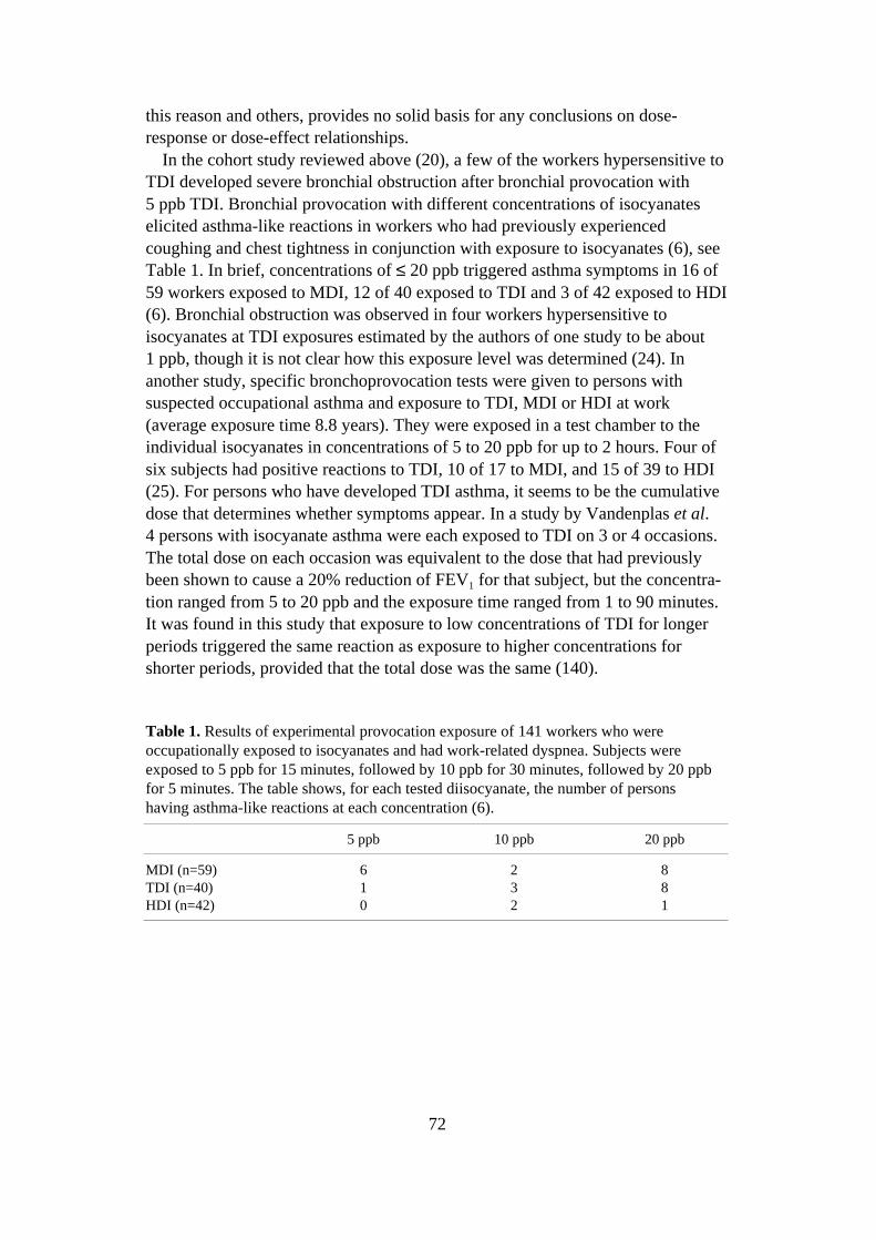
72
this reason and others, provides no solid basis for any conclusions on dose-response or dose-effect relationships.
In the cohort study reviewed above (20), a few of the workers hypersensitive toTDI developed severe bronchial obstruction after bronchial provocation with5 ppb TDI. Bronchial provocation with different concentrations of isocyanateselicited asthma-like reactions in workers who had previously experiencedcoughing and chest tightness in conjunction with exposure to isocyanates (6), seeTable 1. In brief, concentrations of ≤ 20 ppb triggered asthma symptoms in 16 of59 workers exposed to MDI, 12 of 40 exposed to TDI and 3 of 42 exposed to HDI(6). Bronchial obstruction was observed in four workers hypersensitive toisocyanates at TDI exposures estimated by the authors of one study to be about1 ppb, though it is not clear how this exposure level was determined (24). Inanother study, specific bronchoprovocation tests were given to persons withsuspected occupational asthma and exposure to TDI, MDI or HDI at work(average exposure time 8.8 years). They were exposed in a test chamber to theindividual isocyanates in concentrations of 5 to 20 ppb for up to 2 hours. Four ofsix subjects had positive reactions to TDI, 10 of 17 to MDI, and 15 of 39 to HDI(25). For persons who have developed TDI asthma, it seems to be the cumulativedose that determines whether symptoms appear. In a study by Vandenplas et al.4 persons with isocyanate asthma were each exposed to TDI on 3 or 4 occasions.The total dose on each occasion was equivalent to the dose that had previouslybeen shown to cause a 20% reduction of FEV1 for that subject, but the concentra-tion ranged from 5 to 20 ppb and the exposure time ranged from 1 to 90 minutes.It was found in this study that exposure to low concentrations of TDI for longerperiods triggered the same reaction as exposure to higher concentrations forshorter periods, provided that the total dose was the same (140).
Table 1. Results of experimental provocation exposure of 141 workers who wereoccupationally exposed to isocyanates and had work-related dyspnea. Subjects wereexposed to 5 ppb for 15 minutes, followed by 10 ppb for 30 minutes, followed by 20 ppbfor 5 minutes. The table shows, for each tested diisocyanate, the number of personshaving asthma-like reactions at each concentration (6).
5 ppb 10 ppb 20 ppb
MDI (n=59) 6 2 8TDI (n=40) 1 3 8HDI (n=42) 0 2 1
73
Six cases of occupational asthma were identified in a group of 48 spray painterswho were exposed to TDI, MDI and HDI at work. Exposure levels were notdetermined in this study (123). Foundry workers who had developed asthma withhypersensitivity to isocyanates had positive reactions to one hour of exposure toMDI (bronchial provocation in a test chamber). The exposure levels in this studywere on average 12 ppb and never higher than 20 ppb (151).
Bronchial inflammation due to isocyanate asthma, regardless of whichisocyanate is the cause, resembles that observed with other types of asthma.Bronchial biopsies from patients with isocyanate asthma have elevated levels ofactivated eosinophils in mucosa and submucosa and higher numbers of mast cellsin epithelium (35). Bronchoalveolar lavage fluid and biopsies of respiratorymucosa from persons with isocyanate asthma contain elevated numbers ofeosinophils and activated lymphocytes (83, 84, 119). Induced sputum frompatients with isocyanate asthma contains elevated numbers of eosinophils (76).Indications of eosinophil activation (eosinophil cationic protein (ECP)) in bloodincrease after provocation with isocyanate (85). In patients with isocyanateasthma, acute exposure to TDI (unspecified isomer) leads to a temporary increasein the number of lymphocytes containing interleukin 4 in respiratory mucosa,possibly indicating a preponderance of Th2 lymphocytes (77). Early in thereaction to TDI there is a migration of neutrophilic granular leukocytes (neutro-phils) to the respiratory passages. This has been observed with TDI provocation inpersons occupationally exposed to TDI (34) and also in animal studies (46, 111).It resembles the early phase of the allergic asthma reaction, when an invasion ofneutrophils can be observed in airways (28, 30, 94). Asthmatics with delayedreactions triggered by TDI (unspecified isomer) also have elevated numbers ofeosinophils and CD8-positive lymphocytes in blood (36). Stimulating mono-nuclear blood cells with antigens to TDI-, MDI- and HDI-albumin complexesliberates more histamine releasing factors in patients with isocyanate asthma thanin asymptomatic controls exposed to diisocyanate (45, 73). The authors suggestthat liberation of histamine releasing factors on specific provocation might serveas a biological marker for isocyanate asthma.
Some studies report a correlation between certain HLA types and risk ofdeveloping TDI-induced asthma. There was an elevated risk of developing asthmaas a result of TDI exposure for persons who had the allele DQB1*0503 or theallele combination DQB1*0201-0301, whereas the risk was reduced for personswith the allele pair DQB1*501 or the combination DQA1*0101-DQB1*0501(12). Other researchers, however, have not been able to confirm that special HLAclass II alleles are relevant in this context (114) and no definite conclusions canyet be drawn regarding a relationship between HLA type and risk of isocyanateasthma.
In a retrospective study (17-year follow-up) of 300 exposed workers, acorrelation was found between hypersensitivity to TDI and exposure to highconcentrations of TDI (above 50 ppb, usually a 4:1 mixture of 2,4-TDI and 2,6-TDI ) (108). The exposure was brought down below 20 ppb, and no new cases of
74
TDI hypersensitivity were subsequently seen in a 3-year retrospective follow-upof workers who worked with TDI daily (108). In another study, IgE antibodiesspecific to isocyanates – TDI (unspecified isomer), MDI, HDI – were found in 20of 94 exposed workers. There was no significant correlation between specific andtotal IgE (92). This study includes no analysis of a relationship between symp-toms and the presence of IgE antibodies. In various other studies, 10 to 30% ofthose with isocyanate asthma have been found to have circulating IgE antibodiesspecific to albumin-bound isocyanate and/or positive reactions to isocyanates inprick tests. (21, 25, 58, 64, 143, 151). Despite the fact that the isocyanates arequite dissimilar in chemical structure, cross-reactions have often been observedboth in vitro (IgE, IgG) and in tests of specific bronchial reactivity (5, 25, 132).This is believed to be due to the high reactivity of isocyanates and their rapidformation of complexes with more high-molecular substances, with resultingsensitization to these new complexes rather than to the isocyanate itself (39). Thecorrelation between the presence of isocyanate-specific IgE antibodies in bloodand positive reactions to specific bronchial provocation (25) or respiratory symp-toms (9) is weak, which may indicate that isocyanate-specific IgE antibodies haveonly a minor role in isocyanate asthma. In most persons who have isocyanateasthma it is impossible to find circulating IgE antibodies specific to isocyanate.
In car painters exposed to vapors and aerosols containing HDI (prepolymersand monomer), titers of IgG specific to HDI prepolymers (but not the monomer)were significantly higher than in unexposed controls (147). No HDI-specific IgEantibodies were found in this study.
For many people, the asthma persists despite breaking off the exposure, withboth symptoms and elevated bronchial reactivity to direct stimuli (82, 101). It isimportant to diagnose isocyanate asthma early and stop the exposure of personswho develop it, since early intervention will alleviate the asthma symptoms andmay eliminate the asthma completely (107).
AlveolitisThere are a few reported cases of alveolitis caused by exposure to airborne TDI,MDI and HDI. This alveolitis is characterized by restrictive reduction in lungfunction, interstitial fibrosis, increase of CD8-positive cells in bronchoalveolarlavage fluid (CD4/CD8 < 1.0) and IgG antibodies specific to albumin-boundisocyanate (7, 139, 150, 153). In a total of 1,780 isocyanate-exposed workers,Baur (7) identified 14 cases of dyspnea and fever associated with exposure toisocyanates. These persons had signs of alveolitis on lung x-rays and/or restrictivereduction in lung function and/or reduced diffusion capacity and/or IgG anti-bodies against albumin-bound TDI, MDI or HDI in serum. Bronchoalveolarlavage and biopsies from the respiratory tract showed inflammatory changes, butno isocyanate-specific IgE antibodies were found in serum. The average exposuretime was 6 years (0.5-20 years) but cumulative exposures were neither calculatednor estimated. Baur (7) found the occurrence of alveolitis to be about 1%,whereas Vandenplas et al. (141) found a prevalence of 4.7% among workers
75
occupationally exposed to resins containing MDI or MDI prepolymers. In boththese studies it was remarked that exposure to MDI was more commonlyassociated with isocyanate-induced alveolitis than exposure to TDI or HDI. Mostof the alveolitis cases in the Vandenplas study (141) had symptoms so severe thatthey had been forced to quit their jobs soon after the symptoms appeared, leadingthe authors to postulate that the ‘healthy worker’ effect for isocyanate-inducedalveolitis may be quite large, and that alveolitis caused by isocyanate exposure isprobably more common than these studies indicate. Isocyanate-induced alveolitisseems to affect non-smokers more than smokers (7).
Other effects on lung functionLong-term exposure to TDI in concentrations below 20 ppb usually causes noacute symptoms, but it has been argued that it may lead to reduced lung function(105, 144, 145). A cross-sectional study of workers exposed to MDI (usuallybelow 20 ppb; a few measurements showing concentrations up to 87 ppb)revealed that their lung function was lower than that of an unexposed controlgroup (106). Workers (n = 65) occupationally exposed to air concentrations ofHDI too low to cause symptoms (below the detection limit in 92% of measure-ments), as well as various organic solvents, showed a small but statisticallysignificant decline of lung function (FEV1 , FVC) in comparisons with unexposedcontrols (n = 68) and workers occupationally exposed to organic solvents alone(n = 40). This 2.5-year prospective study, however, does not include a groupexposed to HDI alone (2).
The above findings are contradicted by the results of several other studies. In a9-year study of asymptomatic workers exposed to TDI, their decline in lungfunction was no different from that in an unexposed control group (1). TDI-exposed workers with symptoms, however, showed a more rapid decline in lungfunction than the unexposed controls (1). A study by Butcher et al. (20) revealedno effect on lung function after 2 years of exposure to TDI in the concentrationrange 3-54 ppb (average values for 8-hour samples). In a study by Musk et al.(95), 107 workers in a factory producing polyurethane foam were followed for5 years. During this period a total of 2,573 monitoring measurements (20 to 60minutes) of TDI and MDI were made in the breathing zone of the workers. Theaverage TDI concentration was 1.2 ppb, and 90% of the measurements werebelow 5 ppb. The average MDI concentration was 0.6 ppb, with 90% of measure-ments below 2.2 ppb. No information on exposure peaks is given. Changes inlung function during the 5 years of the study were the same for exposed indi-viduals as for controls. No increase in respiratory symptoms and no decline inlung function were found in the exposed group (95). In a 4-year follow-up ofTDI-exposed workers, lung function changes in 57 exposed workers were thesame as those in 24 workers in a control group. When the exposed workers weredivided into three exposure groups, however, it was found that the 15 workers inthe high-exposure group (TWA 8.2 ppb, with individual top exposures brieflyexceeding 30 ppb) had a more rapid decline in lung function (measured as
76
average mid-expiratory flow, FEV1/FVC, and end-expiratory flow) during theperiod than the 14 workers in the medium-exposure group (average TDI exposure1.7 ppb, individual top exposures 3-14 ppb), the 28 workers in the low-exposuregroup (average 0.1 ppb, top exposures below 1 ppb), and the control group (100).
In a study by Hathaway et al., no effect on lung function was found in 43workers after 6 years of occupational exposure to HDI, HDI biuret and HDItrimer (HDI adducts), when they were compared with 42 controls. HDI concentra-tions in this study were about 0.5 ppb (2 hours of exposure), and the calculated12-hour TWA was 0.1 ppb. Daily top exposures averaged 2.9 ppb (44).
Tornling et al. found no difference in lung function changes in a 6-year follow-up study of non-smoking car painters exposed to HDI and HDI biurettrimer whenthey were compared with non-smoking controls who were repair shop metal-workers and mechanics (138). Exposed smokers, however, had a greater annualloss of lung function (FVC, VC, FEV1) than smokers in the control group. Themeaning of this is difficult to interpret, since total tobacco exposure is not takeninto account – the only information is whether the subject was a smoker, ex-smoker or non-smoker. Average exposure in this study was 0.2 ppb, but concen-trations exceeding 286 ppb were not rare. A significant correlation was foundbetween decline in FEV over time and the number of occasions with high peakexposure. This may possibly indicate that, for smokers, decline of lung functionresulting from HDI exposure depends more on brief episodes of high exposurethan on the average long-term exposure. The number of non-smokers in this studywas too small to support any general conclusions drawn from observed effects inthis group. Further, there may have been some exposure of the controls(especially the metalworkers).
Pham et al. examined 318 workers: 83 not exposed to isocyanates, 117 in-directly exposed and 118 directly exposed to MDI (106). The concentration ofairborne MDI was on most occasions below 20 ppb, but there were a fewexposure peaks up to 87 ppb. The exposed groups had a slight restrictivereduction in ventilation capacity when compared with unexposed controls.
Effects on skinIn several case reports of isocyanate-exposed workers with allergic contacteczema on hands, arms and face, contact allergy (delayed dermal hypersensitivity)to MDI, TDI and/or HDI has been diagnosed by patch tests (17, 23, 33). Of 15workers exposed to TDI in the range 70-170 ppb, 5 had a positive patch test forTDI and 3 had contact eczema caused by TDI (48). Rothe described 12 cases ofcontact allergy and allergic contact eczema caused by occupational exposure toMDI (118). In some cases patients have positive test reactions to several iso-cyanates, and occasionally to methylene dianiline (MDA) as well – often despitethe fact that they had not previously been exposed to the particular substance thatinduced the reaction (33, 68). This has been interpreted as evidence of cross-reactivity. The patients had been sensitized in jobs such as mold sprayer, carpainter, spray painter and medical technician. Several of them had become
77
sensitized because of inadequate protection against skin exposure (work withworn-out gloves or no gloves, for example), although working conditions wereotherwise good (33, 68). In several cases sensitization occurred after a few weeksor months of exposure at work (33). In some reported cases the patient had MDI-or HDI-induced asthma in addition to the contact eczema (33).
Teratogenicity, mutagenicity, carcinogenicity
Animal data, in vitro studiesTDI (2,4-TDI:2,6-TDI, 4:1) and MDI, but not HDI, were mutagenic in Salmonellatyphimurium strains TA1538, TA98 and TA100 with metabolic activation (3).Changes in DNA conformation were observed in DNA from calf thymus after invitro exposure to TDI (2,4-TDI:2,6-TDI, 4:1), but no such effect was obtainedwith exposure to MDI or HDI (104). Diisocyanates and their metabolites(including diamines) form adducts with DNA (142). In vitro incubation with TDI(2,4-TDI:2,6-TDI, 4:1) caused double strand breaks in leukocytes (88), althoughin vitro exposure to pure TDI caused no DNA damage. It was concluded that thedamage is caused by metabolites. On contact with water isocyanates formaromatic amines, many of which are carcinogenic. TDI (2,4-TDI:2,6-TDI, 4:1)and a mixture of 45% MDI, 25% 4,4´-methylenediphenyl triisocyanate and 30%unspecified agent caused chromosome aberrations in human lymphocytes after24 hours of incubation, without metabolic activation (75).
TDI (86% 2,4-TDI, 14% 2,6-TDI) given orally to mice 5 days/week for105 weeks (120 or 240 mg/kg/day to males, 60 or 120 mg/kg/day to females)increased the occurrence (significant dose-response trend) of hemangiomas,hemangiosarcomas and hepatic adenomas in the females, but not in the males(49). Oral doses of the same substance given to rats 5 days/week for 106 weeks(30 or 60 mg/kg/day to males, 60 or 120 mg/kg/day to females) led to significantincreases in the occurrence of subcutaneous fibromas and fibrosarcomas in themales. The males in the high-dose group also had significantly more pancreaticadenomas than controls (p = 0.034) (49).
Long-term inhalation exposure to 50 or 150 ppb TDI (2,4-TDI:2,6-TDI, 4:1)6 hours/day did not increase the occurrence of tumors in either mice (104 weeksof exposure) or rats (108-110 weeks of exposure) (49).
In a long-term toxicity and carcinogenicity study with Wistar rats, inhalation of19, 96 or 576 ppb MDI (a mixture of polymers with 44.8-50.2% MDI monomer),6 hours/day, 5 days/week for 2 years, resulted in lung adenomas in 10% of themales and 3% of the females in the highest exposure group. One of 60 males inthe high-exposure group also had a lung adenocarcinoma. No lung tumors werefound in controls. In this study, two years of exposure to 576 ppb MDI wasassociated with an elevated occurrence of lung tumors, whereas concentrations of96 ppb or less did not increase the frequency of tumors (112).
2,4-TDA given to mice in feed (100 or 200 mg/kg feed) for 101 weeksincreased the occurrence of hepatocellular carcinomas and lymphomas in the
78
females (49). 2,4-TDA given to rats in feed (0.1%) for 36 weeks resulted in livercarcinomas (55). Rats were given (ad libitum) 2,4-TDA in diet, 125 or 250 mg/kgfeed for 40 weeks followed by 50 or 100 mg/kg feed for 63 weeks. An elevatedoccurrence of hepatocellular cancers was seen in both sexes (49). No such effectswere seen with oral administration of 2,6-diaminotoluene. 2,6-TDA in feed givento mice (50 or 100 mg/kg feed) and rats (250 or 500 mg/kg feed) for 103 weeksdid not increase the frequency of tumors (49).
MDA (4,4´-methylenedianiline ) was tested for carcinogenicity in an NTPstudy (146). There was a dose-dependent increase of hepatocellular nodules inexposed rats. Mice developed hepatocellular cancer. Thyroid adenomas andcarcinomas were seen in the highest dose groups in both species. Smaller orpoorly documented studies also indicate a carcinogenic effect. According to theIARC, there is ‘sufficient evidence’ that MDA is carcinogenic to experimentalanimals (50).
According to the IARC, there is ‘sufficient evidence’ that TDI is carcinogenicto experimental animals (51), and ‘limited evidence’ that MDI is carcinogenic toexperimental animals (52).
Wistar rats were exposed to MDI 6 hours/day on days 6 to 15 of gestation.A slight increase in the occurrence of asymmetrical sternums was observed infetuses at 864 ppb, but not at 288 ppb, and no other anomalies were noted infetuses (19). Food intake by the mothers dropped during the exposures, but noindications of toxicity were observed and their weight development was nodifferent from that of controls (19).
Human dataInhalation of a mixture of MDI (60%), various triisocyanates (30%) andundefined isocyanates (10%) in concentrations of 5 to 20 ppb increased theoccurrence of double strand breaks in the leukocytes of a 51-year-old workeroccupationally exposed to MDI and MDI oligomers (87). Analyses were madeboth before and two hours after an exposure, after the subject had been away fromwork for 5 days. As a further control, the blood of a healthy unexposed personwas also examined (87).
Epidemiological studies have revealed no increase in cancer risk for peopleexposed to isocyanates. Hagmar et al. made a study of cancer incidence among4,145 employees who were occupationally exposed to TDI (unspecified isomer)and MDI at nine polyurethane production facilities. No increase in cancerincidence could be related to the exposure (42). No increase in the occurrence ofmalignant tumors was found in a case-referent study of 7,023 employees at theseworkplaces (an expansion of the group covered in Reference 42). In this studyexposure levels were 3.6-414 ppb for TDI and less than 1 ppb for MDI, andexposures ranged from a few days to more than 10 years (41). Sorahan et al., in acohort study of 8,288 workers at 11 polyurethane production facilities in the U.K.,found no increase in occurrence of malignant tumors (127). In this study theexposure time was at least 6 months and the exposures were estimated from
79
historic data on the jobs done and the associated exposure level. A slight elevationin the occurrence of pancreatic and lung cancers could be observed among thewomen, and was interpreted by the authors as an effect of smoking, possibly incombination with the occupational exposure (127). No increase in the incidenceof malignant tumors was seen in a cohort study of 4,611 employees at fourdifferent polyurethane production plants in the U.S.A. In this study, estimatedexposure to TDI was below 5.5 ppb during the later part of the observation periodbut had previously been higher. The studied cohort had a low average age, andaverage duration of employment was only 2.4 years – circumstances which, in theexpressed opinion of the authors, make the results of the study inconclusive (121).
In its updated summary evaluations of carcinogenic risks to humans, the IARChas placed TDI in Group 2B: ‘possibly carcinogenic to humans,’ and MDI inGroup 3: ‘not classifiable with regard to carcinogenicity to humans’ (51, 52). ForTDI, MDI and HDI, no studies were found of toxic effects on human reproductionor embryotoxic effects on humans (51, 52)
Dose-response / dose-effect relationships
More pronounced bronchial reactivity to acetylcholine was noted in guinea pigsafter repeated exposure (4 x 1 hour) to TDI in concentrations of 10 or 30 ppb, butnot 5 ppb. Mice exposed by inhalation to 23 ppb 2,4-TDI for 3 hours had a slowerrespiratory rate, and the effect was enhanced when exposure was repeated thefollowing day.
In the studies in which irritation of eyes and respiratory passages was ex-amined, persons hypersensitive to isocyanates could not be differentiated fromthose with normal sensitivity. It was also impossible to differentiate the effects ofirritation, often accompanied by a cough, from the asthma symptoms of those whohad isocyanate asthma.
Asthma-like symptoms (periods of dyspnea and wheezing) were observed ina rather poorly controlled study in which it was concluded that 2.5 years ofoccupational exposure to 0.3-3 ppb TDI increased the risk of developing suchsymptoms. This study is flawed by inadequate data presentation and data analysis.In another study with higher exposures, it was found that long-term exposure toTDI concentrations in the range 1-25 ppb (median 5 ppb) may lead to asthmasymptoms in previously healthy persons. Persons hypersensitive to isocyanatesmay develop asthma symptoms on provocation with a TDI concentrationestimated at 1 ppb.
Too little is known of the relationship between exposure levels and thedevelopment of alveolitis. Six years of occupational exposure to HDI (TWA0.5 ppb, daily top exposures on average 2.9 ppb) had no effect on lung functionchanges, which were the same in exposed workers and controls.
Several studies were excluded from this description of dose-response relation-ships because they contain exposure measurements made with methods that donot meet present standards.
80
Conclusion
The critical effect of exposure to diisocyanates is development of asthma.Isocyanate asthma has been observed in persons occupationally exposed to TDI atworkplaces where air concentrations ranged from 1 to 25 ppb (median 5 ppb). Inone study of substandard quality, the development of asthma-like symptoms suchas dyspnea and wheezing was correlated to exposure levels of 0.3 to 3 ppb TDI.
Asthma symptoms have been observed in individuals hypersensitive to iso-cyanates in conjunction with TDI levels of 5 ppb and estimated TDI levels of1 ppb.
TDI, MDI and HDI have been shown to be sensitizing to skin.TDI and a mixture of MDI and its polymers have been shown to induce cancer
in experimental animals. Both substances are considered genotoxic. For HDI,there are no data on cancer or genotoxicity.
References
1. Adams WGF. Long-term effects on the health of men engaged in the manufacture of toluenediisocyanate (TDI). Br J Ind Med 1975;32:72-78.
2. Akbar-Khanzadeh F, Rivas RD. Exposure to isocyanates and organic solvents, andpulmonary-function changes in workers in a polyurethane molding process. J Occup EnvironMed 1996;38:1205-1212.
3. Andersen M, Binderup M-L, Kiel P, Larsen H, Maxild J. Mutagenic action of isocyanatesused in the production of polyurethanes. Scand J Work Environ Health 1980;6:221-226.
4. Andersson K, Gudéhn A, Levin JO, Nilsson CA. A comparative study of solvent and solvent-free sampling methods for airborne 4,4´-diphenylmethane diisocyanate (MDI) generated inpolyurethane production. Am Ind Hyg Assoc J 1983;44:802-808.
5. Baur X. Immunologic cross-reactivity between different albumin-bound isocyanates. JAllergy Clin Immunol 1983;71:197-205.
6. Baur X, Marek W, Ammon J, Czuppon AB, Marczynski B, Raulf-Heimsoth M, Roemmelt H,Fruhmann G. Respiratory and other hazards of isocyanates. Int Arch Occup Environ Health1994;66:141-152.
7. Baur X. Hypersensitivity pneumonitis (extrinsic allergic alveolitis) induced by isocyanates. JAllergy Clin Immunol 1995;95:1004-1010.
8. Baur X. Occupational asthma due to isocyanates. Lung 1996;174:23-30.
9. Baur X, Chen Z, Flagge A, Posch A, Raulf-Heimsoth M. EAST and CAP specificity for theevaluation of IgE and IgG antibodies to diisocyanate-HSA conjugates. Int Arch AllergyImmunol 1996;110:332-338.
10. Bernstein H. Isocyanate-induced pulmonary diseases: a current perspective. J Allergy ClinImmunol 1982;70:24-31.
11. Bestämning av isocyanater i luft. Metodserien 1023. Stockholm: The Swedish NationalBoard of Occupational Safety and Health, 1980. (In Swedish)
12. Bignon JS, Aron Y, Ju LY, Kopferschmitt MC, Garnier R, Mapp C, Fabbri LM, Pauli G,Lockhart A, Charron D, Swierczewski E. HLA class II alleles in isocyanate-induced asthma.Am J Respir Crit Care Med 1994;149:71-75.
13. Brooks SM, McKay RT. Epidemiologic methods for identifying occupational asthma due toisocyanates. Am Rev Respir Dis 1981;123:133.
14. Bruggemann IM, Temmink JHM, van Bladeren PJ. GSH- and cysteine-mediated cytotoxicityof allyl and benzyl isothiocyanate. Toxicol Appl Pharmacol 1986;83:349-359.
81
15. Brorson T, Skarping G, Nielsen J. Biological monitoring of isocyanates and related amines.II. Test chamber exposure of humans to 1,6-hexamethylene diisocyanate (HDI). Int ArchOccup Environ Health 1990;62:385-389.
16. Brorson T, Skarping G, Sangö C. Biological monitoring of isocyanates and related amines.IV. 2,4- and 2,6-toluenediamine in hydrolysed plasma and urine after test-chamber exposureof humans to 2,4- and 2,6-toluene diisocyanate. Int Arch Occup Environ Health1991;63:253-259.
17. Brugsch HG, Elkins HB. Toluene di-isocyanate (TDI) toxicity. New Engl J Med1963;268:353-357.
18. Buckley LA, Jiang XZ, James RA, Morgan KT, Barrow CS. Respiratory tract lesionsinduced by sensory irritants at the RD50 concentration. Toxicol Appl Pharmacol 1984;74:417-429.
19. Buschmann J, Koch W, Fuhst R, Heinrich U. Embryotoxicity study of monomeric 4,4´-methylenediphenyl diisocyanate (MDI) aerosol after inhalation exposure in Wistar rats.Fundam Appl Toxicol 1996;32:96-101.
20. Butcher BT, Jones RN, O’Neil CE, Glindmeyer HW, Diem JE, Dharmarajan V, Weill H,Salvaggio JE. Longitudinal study of workers employed in the manufacture of toluenediisocyanate. Am Rev Respir Dis 1977;116:411-421.
21. Butcher BT, O’Neil CE, Reed MA, Salvaggio JE. Radioallergosorbent testing of toluenediisocyanate-reactive individuals using p-totyl isocyanate antigen. J Allergy Clin Immunol1980;66:213-216.
22. Butcher BT, O’Neil CE, Reed MA, Salvaggio JE, Weill H. Development and loss of toluenediisocyanate reactivity: immunologic, pharmacologic, and provocative challenge studies. JAllergy Clin Immunol 1982;70:231-235.
23. Calas E, Castelain PY, Lapointe HR, Ducos P, Cavelier C, Duprat P, Poitou P. Allergiccontact dermatitis to a photopolymerizable resin used in printing. Contact Dermatitis1977;3:186-194.
24. Carroll KB, Secombe CJP, Pepys J. Asthma due to non-occupational exposure to toluenediisocyanate. Clin Allergy 1976;6:99-104.
25. Cartier A, Grammer L, Malo J-L, Lagier F, Ghezzo H, Harris K, Patterson R. Specific serumantibodies against isocyanates: association with occupational asthma. J Allergy Clin Immunol1989;84:507-514.
26. Dalene M, Skarping G, Lind P. Workers exposed to thermal degradation products of TDI-and MDI-based polyurethane: Biomonitoring of 2,4-TDA, 2,6-TDA, and 4,4´-MDA inhydrolyzed urine and plasma. Am Ind Hyg Assoc J 1997;58:587-591.
27. Day B, Jin R, Karol MH. In vivo and in vitro reactions of toluene diisocyanate isomers withguinea pig hemoglobin. Chem Res Toxicol 1996;9:568-573.
28. De Monchy GR, Kauffman HF, Venge P, Koeter GH, Jansen HM, Sluiter HJ, De Vries K.Bronchoalveolar eosinophilia following allergen-induced delayed asthmatic reactions. AmRev Respir Dis 1985;131:272-276.
29. Dharmarajan V, Rando JR. Critical evaluation of continuous monitors for toluenediisocyanate. Am Ind Hyg Assoc J 1980;41:869-878.
30. Diaz P, Gonzales C, Galleguillos F, Ancic P, Kay AB. Eosinophils and macrophages inbronchial mucus and bronchoalveolar lavage during allergen-induced late-phase asthmaticreactions. J Allergy Clin Immunol 1986;77:244-249.
31. Duncan B, Scheel LD, Fairchild EJ, Killens R, Graham S. Toluene diisocyanate inhalationtoxicology: Pathology and mortality. Am Ind Hyg Assoc J 1962;19:447-456.
32. Elkins HB, McCarl GW, Brugch HG, Fahy JP. Massachusetts experience with toluenediisocyanates. Am Ind Hyg Assoc J 1962;23:265-272.
33. Estlander T, Keskinen H, Jolanki R, Kanerva L. Occupational dermatitis from exposure topolyurethane chemicals. Contact Dermatitis 1992;27:161-165.
82
34. Fabbri L, Boschetto P, Zocca E, Milani G, Pivirotto F, Plebani M, Burlina A, Licata B, MappCE. Bronchoalveolar neutrophilia during late asthmatic reactions induced by toluenediisocyanate. Am Rev Respir Dis 1987;136:36-42.
35. Fabbri LM, Ciaccia A, Maestrelli P, Saetta M, Mapp CE. Pathophysiology of occupationalasthma. In: Bernstein IL, Chan-Yeung M, Malo J-L, Bernstein DI, eds. Asthma in theWorkplace. New York: Marcel Dekker, 1993;61-92.
36. Finotto S, Fabbri L, Rado LM, Mapp CE, Maestrelli P. Increase in numbers of CD8 positivelymphocytes and eosinophils in peripheral blood of subjects with late asthmatic reactionsinduced by toluene diisocyanates. Br J Ind Med 1991;48:116-121.
37. Gagnaire F, Micillino JC, Bonnet P, Simon P, de Ceaurriz I. Toluene diisocyanate-inducedairway hyperresponsiveness in intravenous ACH: a study on single and repeated exposure inguinea pigs. Toxicol Lett 1988;44:273-280.
38. Gordon T, Sheppard D, McDonald DM, Di Stefano S, Scypinski L. Airwayhyperresponsiveness and inflammation induced by toluene diisocyanate in guinea pigs. AmRev Respir Dis 1985;132:1106-1112.
39. Grammer LC, Harris KE, Malo J-L, Cartier A, Patterson R. The use of an immunoassayindex for antibodies against isocyanate human protein conjugates and application to humanisocyanate disease. J Allergy Clin Immunol 1990;86:94-98.
40. Hagmar L, Nielsen J, Skerfving S. Clinical features and epidemiology of occupationalobstructive respiratory disease caused by small molecular weight organic chemicals. MonogrAllergy 1987;21:42-58.
41. Hagmar L, Strömberg U, Welinder H, Mikoczy Z. Incidence of cancer and exposure totoluene diisocyanate and methylene diphenyldiisocyanate: a cohort based case-referent studyin the polyurethane foam manufacturing industry. Br J Ind Med 1993;50:1003-1007.
42. Hagmar L, Welinder H, Mikoczy Z. Cancer incidence and mortality in the Swedishpolyurethane foam manufacturing industry. Br J Ind Med 1993;50:537-543.
43. Hama GM. Symptoms in workers exposed to isocyanates – suggested exposureconcentrations. Arch Ind Health 1957;16:232-233.
44. Hathaway JA, DeWilde A, Shepperly DC, Nguyen LT, Johnson JE. Evaluation of pulmonaryfunction in workers exposed to hexamethylene diisocyanate. J Occup Environ Med1999;41:378-383.
45. Herd ZL, Bernstein DI. Antigen-specific stimulation of histamine releasing factors indiisocyanate-induced occupational asthma. Am J Respir Crit Care Med 1994;150:988-994.
46. Hesbert A, Ban M, Bonnet P, Simon P, Bottin MC, Lemonnier M, de Ceaurriz J.Interdependence of polymorphonuclear neutrophils and macrophages stained for N-acetyl-b-glucosaminidase in lavage effluents from toluene diisocyanate-exposed rat lung. Toxicol Lett1991;56:53-59.
47. HSE (Health and Safety Executive). Methods for the determination of hazardous substances:organic isocyanates in air. Health and Safety Laboratory 1999; MDHS 25/3.
48. Huang J, Wang XP, Ueda A, Aoyama K, Chen BM, Matsushita T. Allergologic evaluationfor workers exposed to toluene diisocyanate. Ind Health 1991;29:85-92.
49. IARC. Some chemicals used in plastic and elastomers. Toluene diisocyanate. IARCMonographs on the Evaluation of Carcinogenic Risks to Humans. Lyon: InternationalAgency for Research on Cancer, 1986;39:287-323.
50. IARC. Some chemicals used in plastic and elastomers. 4,4´-Methylenedianiline and itsdihydrochloride. IARC Monographs on the Evaluation of Carcinogenic Risks to Humans.Lyon: International Agency for Research on Cancer, 1986;39:347-365.
51. IARC. Re-evaluation of some organic chemicals, hydrazine and hydrogen peroxide (parttwo). Toluene diisocyanates. IARC Monographs on the Evaluation of Carcinogenic Risks toHumans. Lyon: International Agency for Research on Cancer, 1999;71:865-879.
83
52. IARC. Re-evaluation of some organic chemicals, hydrazine and hydrogen peroxide (partthree). 4,4´-Methylenediphenyl diisocyanate and polymeric 4,4´-methylenediphenyldiisocyanate. IARC Monographs on the Evaluation of Carcinogenic Risks to Humans. Lyon:International Agency for Research on Cancer, 1999;71:1049-1058.
53. IPCS. Environmental health criteria 75. Toluene diisocyanates. Geneva: InternationalProgramme on Chemical Safety, WHO, 1987.
54. ISO. TCI46/SC2/WG4. Document under preparation (http://www.iso.ch/).
55. Ito N, Hiasa X, Konishi Y, Marugami M. The development of carcinoma in the liver of ratstreated with m-toluenediamine and the synergistic and antagonistic effects of otherchemicals. Cancer Res 1969;29:1137-1145.
56. Jin R, Day BW, Karol MH. Toluene diisocyanate protein adducts in the bronchoalveolarlavage of guinea pigs exposed to vapors of the chemical. Chem Res Toxicol 1993;6:906-912.
57. Karlsson D, Dalene M, Skarping G. Determination of complex mixtures of airborneisocyanates and amines. Part 4. Determination of aliphatic isocyanates as dibutylaminederivatives using liquid chromatography and mass spectrometry. Analyst 1998;123:117-123.
58. Karol MH, Alarie Y. Antigens which detect IgE antibodies in workers sensitive to toluenediisocyanate. Clin Allergy 1980;10:101-109.
59. Karol MH, Hauth BA, Riley EJ, Magreni CM. Dermal contact with toluene diisocyanate(TDI) produces respiratory tract hypersensitivity in guinea pigs. Toxicol Appl Pharmacol1981;58:221-230.
60. Karol MH, Jin R, Lantz RC. Immunohistochemical detection of toluene diisocyanate (TDI)adduct in pulmonary tissue of guinea pigs following inhalation exposure. Inhal Toxicol1997;9:63-83.
61. Kennedy AL, Stock MF, Alarie Y, Brown WE. Uptake and distribution of 14C during andfollowing exposure to radioactive toluene diisocyanate. Toxicol Appl Pharmacol1989;100:280-292.
62. Kennedy AL, Brown WE. Isocyanates and lung disease: experimental approaches tomolecular mechanisms. Occup Med 1992;7:301-329.
63. Kennedy AL, Wilson TR, Stock MF, Alarie Y, Brown WE. Distribution and reactivity ofinhaled 14C-labeled toluene diisocyanate (TDI) in rats. Arch Toxicol 1994;68:434-443.
64. Keskinen H, Tupasela O, Tikkainen U, Nordman H. Experiences of specific IgE in asthmadue to diisocyanates. Clin Allergy 1998;18:597-604.
65. Kolmodin-Hedman B, Alexandersson R, Hedenstierna G. Diisocyanates – MDI. Lungphysiology studies on personel from plastic industry. Arbete och Hälsa 1980;10:1-18. (inSwedish, English abstract)
66. Koschier FJ, Burden EJ, Brunkhorst CS, Friedman MA. Concentration-dependent elicitationof dermal sensitization in guinea pigs treated with 2,4-toluene diisocyanate. Toxicol ApplPharmacol 1983;67:401-407.
67. Kuck M, Balle G, Slawyk W. Sampling of diisocyanates (HDI, TDI) in air by derivatisationwith secondary amines as reagents. Part 1. Partial rate factors (PRF) of reagents. Analyst1999;124:933-939.
68. Lidén C. Allergic contact dermatitis from 4,4´-diisocyanato-diphenyl methane (MDI) in amolder. Contact Dermatitis 1980;6:301-302.
69. Lind P, Skarping G, Dalene M. Biomarkers of toluene diisocyanate and thermal degradationproducts of polyurethane, with special reference to the sample preparation. AnalyticaChimica Acta 1996,333:277-283.
70. Lind P, Dalene M, Lindström V, Grubb A, Skarping G. Albumin adducts in plasma fromworkers exposed to toluene diisocyanate. Analyst 1997;122:151-154.
71. Littorin M, Truedsson L, Welinder H, Skarping G, Mårtensson U, Sjöholm AG. Acuterespiratory disorder, rhinoconjunctivitis and fever associated with the pyrolysis of
84
polyurethane derived from diphenylmethane diisocyanate. Scand J Work Environ Health1994;20:216-222.
72. Littorin M, Rylander L, Skarping G, Dalene M, Welinder H, Strömberg U, Skerfving S.Exposure biomarkers and risk at gluing and heating of polyurethane – a cross-sectional studyof respiratory symptoms. Occup Environ Med 2000;57:396-405.
73. Lummus ZL, Alam R, Bernstein JA, Bernstein DI. Characterization of histamine releasingfactors in diisocyanate-induced occupational asthma. Toxicology 1996;111:191-206.
74. Lundberg P (ed). Scientific Basis for Swedish Occupational Standards. IX. Arbete och Hälsa1988;32:107-121. Arbetsmiljöinstiutet, Solna.
75. Maeki-Paakanen J, Norppa H. Chromosome aberrations and sister chromatid exchangesinduced by technical grade toluene diisocyanate and methylenediphenyl diisocyanate incultured human lymphocytes. Toxicol Lett 1987;36:37-43.
76. Maestrelli P, Calcagni PG, Saetta M, Di Stefano A, Hosselet JJ, Santonastaso A, Fabbri LM,Mapp CE. Sputum eosinophilia after asthmatic responses induced by isocyanates insensitized subjects. Clin Exp Allergy 1994;24:29-34.
77. Maestrelli P, Accari P, Turato G, Papiris SA, Di Stefano A, Mapp CE, Milani GF, Fabbri L,Saetta M. Expression of IL-4 and IL-5 in asthma induced by toluene diisocyanate. Clin ExpAllergy 1997;27:1292-1298.
78. Magnusson B, Kligman AM. The identification of contact allergens by animal assay, theguinea pig maximization test method. J Invest Derm 1969;52:268-276.
79. Maitre A, Berode M, Perdrix A, Romazini S, Savolainen H. Biological monitoring ofoccupational exposure to toluene diisocyanate. Int Arch Occup Environ Health 1993;65:97-100.
80. Maitre A, Berode M, Perdrix A, Stoklov M, Mallion JM, Savolainen H. Urinary hexanediamine as an indicator of occupational exposure to hexamethylene diisocyanate. Int ArchOccup Environ Health 1996;69:65-68.
81. Malo JL, Ouimet G, Cartier A, Levitz D, Zeiss CR. Combined alveolitis and asthma due tohexamethylene diisocyanate (HDI), with demonstration of crossed respiratory andimmunologic reactivities to diphenylmethane diisocyanate (MDI). J Allergy Clin Immunol1983;72:413-419.
82. Mapp C, Corona P, Marzo N, Fabbri L. Persistent asthma due to isocyanates. Am Rev RespirDis 1988;137:1327-1329.
83. Mapp CE, Saetta M, Maestrelli P, Ciaccia A, Fabbri LM. Low molecular weight pollutantsand asthma: pathogenetic mechanisms and genetic factor. Eur Respir J 1994;7:1559-1563.
84. Mapp CE, Saetta M, Maestrelli P, Di Stefano A, Chitano P, Boschetto P, Ciaccia A, FabbriLM. Mechanisms and pathology of occupational asthma. Eur Respir J 1994;7:544-554.
85. Mapp CE, Plebani M, Faggian D, Maestrelli P, Saetta M, Calcani P, Borghesan F, Fabbri L.Eosinophil cationic protein (ECP), histamine and tryptase in peripheral blood before andduring inhalation challenge with toluene diisocyanate (TDI) in sensitized subjects. Clin ExpAllergy 1994;24:730-736.
86. Marcali K. Microdetermination of toulenediisocyanates in atmosphere. Anal Chem1957;29:552-558.
87. Marczynski B, Czuppon AB, Hoffarth HP, Marek W, Baur X. DNA damage in human whiteblood cells after inhalative exposure to methylenediphenyl diisocyanate (MDI). Toxicol Lett1992;60:131-138.
88. Marczynski B, Czuppon AB, Marek W, Baur X. Indication of DNA strand breaks in humanwhite blood cells after in vitro exposure to toluene diisocyanate (TDI). Toxicol Ind Health1992;8:157-169.
89. Marek W, Potthast J, Marczynski B, Baur X. Unspecific airway hyperreactivity induced bytoluene diisocyanate in an animal model of occupational lung disease. Pfluegers Arch1991;419 Suppl. 1:95.
85
90. Marek W, Potthast J, Marczynski B, Baur X. Alteration of smooth muscles responses toacetylcholine induced by toluene diisocyanate (TDI) and neuropeptides in guinea pigs.Cahiers de médecine du travail / Cahiers voor Arbeidsgeneeskunde 1994;31:42-44.
91. Maxon FC. Respiratory irritation from toluene diisocyanate. Arch Environ Health1964;8:755-758.
92. Mazur G, Pethran A. Detection of specific IgE in isocyanate and phthalic anhydride exposedworkers: comparison of RAST RIA, Immuno CAP System FEIA, and Magic Lite SQ.Allergy 1993;48:627-630.
93. Meredith SK, Bugler J, Clark RL. Isocyanate exposure and occupational asthma: a case-referent study. Occup Environ Med 2000;57:830-836.
94. Metzger WJ, Richerson HB, Worden K, Monick M, Hunninghake GW. Bronchoalveolarlavage of allergic asthmatic patients following allergen bronchoprovocation. Chest1986;89:477-483.
95. Musk AW, Peters JM, Deberardinis L, Murphy RLH. Absence of respiratory effects insubjects exposed to low concentrations of TDI and MDI. J Occup Med 1982;24:746-749.
96. Musk AW, Peters JM, Wegman DH. Isocyanate and respiratory disease: current status. Am JInd Med 1988;13:331-349.
97. NIOSH (National Institute for Occupational Safety and Health). Determination of airborneisocyanate exposure. In: Cassinelli ME, O’Connor PF, eds. NIOSH Manual of AnalyticalMethods. 4th ed., 2nd supplement. DHHS (NIOSH) Publication No. 98-119. Cincinnati OH:National Institute for Occupational Safety and Health 1998.
98. O’Brien IM, Harries MG, Burge PS, Pepys J. Toluene diisocyanate induced asthma.Reactions to TDI, MDI, HDI and histamine. Clin Allergy 1979;9:1-6.
99. OECD (Organization for Economic Co-operation and Development). OECD guideline fortesting of chemicals No. 406: Skin Sensitisation. July 1992.
100. Omae K, Higashi T, Nakadate T, Tsugane S, Nakaza M, Sakurai H. Four year follow-up ofeffects of toluene diisocyanate exposure on the respiratory system in polyurethane foammanufacturing workers. Four-year changes in the effects on the respiratory system. Int ArchOccup Health 1992;63:565-569.
101. Paggiaro PL, Vagagginin B, Bacci E, Bancalari L, Carrara M, Di Franco A, Giannini D,Dente FL, Giuntini C. Prognosis of occupational asthma. Eur Respir J 1994;7:761-767.
102. Pearson PG, Slatter JG, Rashed MS, Han DH, Baille TA. Carbamoylation of peptides andproteins in vitro by S-(N-methylcarbamoyl)GSH and S-(N-methylcarbamoyl)cysteine, twoelectrophilic S-linked conjugates of methyl isocyanate. Chem Res Toxicol 1991;4:436-444.
103. Persson P, Dalene M, Skarping G, Adamsson M, Hagmar L. Biological monitoring ofoccupational exposure to toluene diisocyanate: measurement of toluenediamine inhydrolysed urine and plasma by gas chromatography-mass spectrometry. Br J Ind Med1993;50:1111-1118.
104. Peel M, Marczynski B, Baur X. Comparison of the binding potential of various diisocyanateson DNA in vitro. J Toxicol Environ Health 1997;52:517-526.
105. Peters PM, Wegman DH. Epidemiology of toluene diisocyanate (TDI) induced respiratorydisease. Environ Health Perspect 1975;11:97-100.
106. Pham QT, Cavelier C, Mereau P, Mur JM, Cicolella A. Isocyanate and respiratory function.A study of workers producing polyurethane foam moulding. Ann Occup Hyg 1978;21:121-129.
107. Pisati G, Baruffini A, Zedda S. Toluene diisocyanate induced asthma: outcome according topersistence or cessation of exposure. Br J Ind Med 1993;50:60-64.
108. Porter CV, Higgins RL, Scheel LD. A retrospective study of clinical, physiologic andimmunologic changes in workers exposed to toluene diisocyanates. Am Ind Hyg Assoc J1975;36:159-163.
86
109. Potthast J, Marek W, Marczynski W, Baur X. Isocyanate-induced airway hyperreactivity:role of neuropeptides. Cahiers de médecine du travail / Cahiers voor Arbeidsgeneeskunde1994;31:47-49.
110. Rattray NJ, Botham PA, Hext PM, Woodcock DR, Fielding I, Dearman RJ, Kimber I.Induction of respiratory hypersensitivity to diphenylmethane-4,4´-diisocyanate (MDI) inguinea pigs. Influence of route of exposure. Toxicology 1994;88:15-30.
111. Raulf M, Tennie L, Marczynski B, Potthast J, Marek W, Baur X. Cellular and mediatorprofile in bronchoalveolar lavage of guinea pigs after toluene diisocyanate (TDI) exposure.Lung 1995;173:57-68.
112. Reuzel PGJ, Arts JHE, Lomax LG, Kuijpers MHM, Kuper CF, Gembardt C, Feron VJ, LoserE. Chronic inhalation toxicity and carcinogenicity study of respirable polymeric methylenediphenyl diisocyanate (polymeric MDI) aerosol in rats. Fundam Appl Toxicol 1994;22:195-210.
113. Reuzel PGJ, Kuper CF, Feron VJ, Appelman LM, Loser E. Acute, subacute, and subchronicinhalation toxicity studies of respirable polymeric methylene diphenyl diisocyanate(polymeric MDI) aerosol in rats. Fundam Appl Toxicol 1994;22:186-194.
114. Rihs H-P, Barbalho-Krölls T, Huber H, Baur X. No evidence for the influence of HLA classII in alleles in isocyanate-induced asthma. Am J Ind Med 1997;32:522-527.
115. Rosenberg C, Savolainen H. Detection of urinary amine metabolites in toluene diisocyanateexposed rats. Chromatography 1985;323:429-433.
116. Rosenberg C, Savolainen H. Determination in urine of diisocyanate-derived amines fromoccupational exposure by gas chromatography-mass fragmentography. Analyst1986;111:1069-1071.
117. Rosenberg C, Savolainen H. Determination of occupational exposure to toluene diisocyanateby biological monitoring. J Chromatogr 1986;367:385-392.
118. Rothe A. Zur Frage arbeitsbedingter Hautschädigungen durch Polyurethanchemikalien.Berufsdermatosen 1976;24:7-24.
119. Saetta M, Maestrelli P, Turato G, Mapp CE, Milani G, Pivirotto F, Fabbri LM, Di Stefano A.Airway wall remodeling after cessation of exposure to isocyanates in sensitized asthmaticsubjects. Am J Respir Crit Care Med 1995;151:489-494.
120. Sangha GK, Alarie Y. Sensory irritation by toluene diisocyanate in single and repeatedexposure. Toxicol Appl Pharmacol 1979;50:533-547.
121. Schnorr TM, Steenland K, Egeland GM, Boeniger M, Egilman D. Mortality of workersexposed to toluene diisocyanate in the polyurethane foam industry. Occup Environ Med1996;53:703-707.
122. Schutze D, Sepai O, Lewalter J, Miksche L, Henschler D, Sabbioni G. Biomonitoring ofworkers exposed to 4,4´-methylenedianiline or 4,4´-methylenediphenyl diisocyanate.Carcinogenesis 1995;16:573-582.
123. Seguin P, Allard A, Cartier A, Malo JL. Prevalence of occupational asthma in spray paintersexposed to several types of isocyanates, including polymethylene polyphenylisocyanates. JOccup Med 1987;29:340-344.
124. Sepai O, Henschler D, Sabbioni G. Albumin adducts, hemoglobin adducts and urinarymetabolites in workers exposed to 4,4´-methylenediphenyl diisocyanate. Carcinogenesis1995;16:2583-2587.
125. Skarping G, Brorson T, Sangö C. Biological monitoring of isocyanates and related amines.III. Test chamber exposure of humans to toluene diisocyanate. Int Arch Occup EnvironHealth 1991;63:83-88.
126. Skarping G, Dalene M. Determination of 4,4´-methylenediphenyldianiline (MDA) andidentification of isomers in technical-grade MDA in hydrolysed plasma and urine fromworkers exposed to methylene diphenyldiisocyanate by gas chromatography-massspectrometry. J Chromatogr B Biomed Appl 1995;663:209-216.
87
127. Sorahan R, Pope D. Mortality and cancer morbidity of production workers in the UnitedKingdom flexible polyurethane industry. Br J Ind Med 1993;50:528-536.
128. Streicher RP, Kennedy ER, Lorberau CD. Strategies for the simultaneous collection ofvapours and aerosols with emphasis on isocyanate sampling. Analyst 1994;119:89-97.
129. Streicher RP, Reh CM, Key-Schwartz RJ, Schlecht PC, Cassinelli ME, O’Connor P.Determination of airborne isocyanate exposure: Considerations in method selection. Am IndHyg Assoc J 2000;61:544-556.
130. Swedish Criteria Group for Occupational Exposure Limits. Scientific basis for Swedishoccupational standards. II. Arbete och Hälsa 1982;9:60-68. Arbetsmiljöinstiutet, Solna.
131. Tanaka KI. Contact sensitivity in mice induced by toluene diisocyanate (TDI). J Dermatol1980;7:277-280.
132. Tee RD, Cullinan P, Welch J, Sherwood Burge P, Newman-Taylor AJ. Specific IgE toisocyanates: A useful diagnostic role in occupational asthma. J Allergy Clin Immunol1998;101:709-715.
133. Thorne PS, Hillebrand JA, Lewis GR, Karol MH. Contact sensitivity by diisocyanates:potencies and cross-reactivities. Toxicol Appl Pharmacol 1987;87:155-165.
134. Tinnerberg H. Exposure of human volunteers to hexamethylene diisocyanate (HDI) andisophorone diisocyanate (IPDI), with special reference to the monitoring of biomarkers inurine hydrolysates using gas and liquid chromatography with mass spectrometry. Dept ofIndustrial Engineering, Division of Working Environment, Lund Institute of Technology,Lund University and Dept. of Occupational and Environmental Medicine, Lund University,Licentiate Thesis 1995.
135. Tinnerberg H, Spanne M, Dalene M, Skarping G. Determination of complex mixtures ofairborne isocyanates and amines. Part 2. Toluene diisocyanate and aminoisocyanate andtoluenediamine after thermal degradation of a toluenediisocyanate-polyurethane. Analyst1996;121:1101-1106.
136. Tinnerberg H, Spanne M, Dalene M, Skarping G. Determination of complex mixtures ofairborne isocyanates and amines. Part 3. Methylenediphenyl-diisocyanate-aminoisocyanate,and -diamine and structural analogues after thermal degradation of a polyurethane. Analyst1997;122:275-278.
137. Tominaga M, Kohno S, Tanaka K, Ohata K. Studies on toluene diisocyanate (TDI)-induceddelayed type hypersensitivity. Jpn J Pharmacol 1985;39:163-171.
138. Tornling G, Alexandersson R, Hedenstierna G, Plato N. Decreased lung function andexposure to diisocyanates (HDI and HDI-BT) in car repair painters: Observation on re-examination 6 years after initial study. Am J Ind Med 1990;17:299-310.
139. Vandenplas O, Malo J-L, Saetta M, Mapp CE, Fabbri LM. Occupational asthma and extrinsicalveolitis due to isocyanates: current status and perspective. Br J Ind Med 1993;50:213-228.
140. Vandenplas O, Cartier A, Ghezzo H, Cloutier Y, Malo J-L. Response to isocyanates: Effectof concentration, duration of exposure, and dose. Am Rev Respir Dis 1993;147:1287-1290.
141. Vandenplas O, Malo JL, Dugas M, Cartier A, Desjardins A, Levesque J, Shaughnessy MA,Grammer LC. Hypersensitivity pneumonitis-like reaction among workers exposed todiphenylmethane diisocyanate (MDI). Am Rev Respir Dis 1993;147:338-346.
142. Vock EH, Hoyman HG, Heinrich U, Lutz WK. 32P-postlabelling of DNA adduct derivedfrom 4,4´-methyldianiline in the olfactory epithelium of rats exposed by inhalation to 4,4´-methylphenyl diisocyanate. Carcinogenesis 1996;17:1069-1073.
143. Vogelmeier C, Baur X, Fruhmann G. Isocyanate-induced asthma: results of inhalation testswith TDI, MDI and methacholine. Int Arch Occup Environ Health 1991;63:9-13.
144. Wegman DH, Pagnotto LD, Fine LJ, Peters JM. A dose-response relationship in TDI-workers. J Occup Med 1974;16:258-260.
145. Wegman DH, Peters JM, Pagnotto L, Fine LJ. Chronic pulmonary function loss fromexposure to toluene diisocyanate. Br J Ind Med 1977;34:196-200.
88
146. Weisburger EK, Murthy ASK, Lilja HS, Lamb JC. Neoplastic response of F344 rats andB6C3F mice to the polymer and dyestuff intermediates 4,4´-methylenebis(N,N-dimethyl)-benzenamine, 4,4´-oxydianiline, and 4,4´-methylenedianiline. J Natl Cancer Inst1984;72:1457-1461.
147. Welinder H, Nielsen J, Bensryd I, Skerfving S. IgG antibodies against polyisocyanates in carpainters. Clin Allergy 1988;18:85-93.
148. White WG, Sugden E, Morris MJ, Zapata E. Isocyanate-induced asthma in a car factory.Lancet 1980;1:756-760.
149. Williams NR, Jones K, Cocker J. Biological monitoring to assess exposure from use ofisocyanates in motor vehicle repair. Occup Environ Med 1999;56:598-601.
150. Yoshizawa Y, Ohtsuka M, Noguchi K, Uchida Y, Suko M, Hasegawa S. Hypersensitivitypneumonitis induced by toluene diisocyanate: sequelae of continuous exposure. Ann InternMed 1989;110:31-34.
151. Zammit-Tabona M, Sherkin M, Kijek K, Chan H, Chan-Yeung M. Asthma caused bydiphenylmethane diisocyanate in foundry workers. Clinical, bronchial provocation andimmunologic studies. Am Rev Resp Dis 1983;128:226-230.
152. Zapp JA. Hazards of isocyanates in polyurethane foam plastic production. Arch Ind Health1957;15:324-330.
153. Zeiss CR, Kanellakes TM, Bellone JD, Levitz D, Pruzansky JJ, Patterson R. ImmunoglobulinE-mediated asthma and hypersensitivity pneumonitis with precipitating anti-haptenantibodies due to diphenylmethane diisocyanate (MDI) exposure. J Allergy Clin Immunol1980;65:346-352.
154. Zissu D, Binet S, Limasset J-C. Cutaneous sensitization to some polyisocyanate prepolymersin guinea pigs. Contact Dermatitis 1998,39:248-251.
89
Summary
Montelius J (ed). Scientific Basis for Swedish Occupational Standards. XXII.Arbete och Hälsa 2001:20, pp 1-96. National Institute for Working Life, Solna.
Critical review and evaluation of those scientific data which are relevant as a backgroundfor discussion of Swedish occupational exposure limits. This volume consists of theconsensus reports given by the Criteria Group at the Swedish National Institute forWorking Life from July, 2000 through June, 2001.
Key Words: Diphenylmethane diisocyanate (MDI), Ethylenethiourea, Hexamethylenediisocyanate (HDI), Hydrogen cyanide, α-Methylstyrene, Occupationalexposure limit (OEL), Potassium cyanide, Risk assessment, Scientificbasis, Sodium cyanide, Toluene-2,4-diamine, Toluene-2,6-diamine,Toluene diisocyanate (TDI), Toxicology.
Sammanfattning
Montelius J (ed). Vetenskapligt underlag för hygieniska gränsvärden. XXII. Arbete ochHälsa 2001:20, s 1-96. Arbetslivsinstitutet, Solna.
Sammanställningar baserade på kritisk genomgång och värdering av de vetenskapligafakta, vilka är relevanta som underlag för fastställande av hygieniskt gränsvärde.Volymen omfattar de underlag som avgivits från Kriteriegruppen för hygieniskagränsvärden under perioden juli 2000 - juni 2001.
Nyckelord: Cyanväte, Difenylmetandiisocyanat (MDI), Etylentiourinämne,Hexametylendiisocyanat (HDI), Hygieniskt gränsvärde, Kaliumcyanid,α–Metylstyren, Natriumcyanid, Riskvärdering, Toluen-2,4-diamin, Toluen-2,6-diamin, Toxikologi, Toluendiisocyanat (TDI), Vetenskapligt underlag.
En svensk version av dessa vetenskapliga underlag finns publicerad i Arbete och Hälsa2001:19.
90
APPENDIX
Consensus reports in this and previous volumes
Substance Consensus date Volume in Arbeteoch Hälsa (No.)
Acetaldehyde February 17, 1987 1987:39 (VIII)Acetamide December 11, 1991 1992:47 (XIII)Acetic acid June15, 1988 1988:32 (IX)Acetone October 20, 1987 1988:32 (IX)Acetonitrile September 12, 1989 1991:8 (XI)Acrylamide April 17, 1991 1992:6 (XII)Acrylates December 9, 1984 1985:32 (VI)Acrylonitrile April 28, 1987 1987:39 (VIII)Aliphatic amines August 25, 1982 1983:36 (IV)Aliphatic hydrocarbons, C10-C15 June 1, 1983 1983:36 (IV)Aliphatic monoketons September 5, 1990 1992:6 (XII)Allyl alcohol September 9, 1986 1987:39 (VIII)Allylamine August 25, 1982 1983:36 (IV)Allyl chloride June 6, 1989 1989:32 (X)Aluminum April 21, 1982 1982:24 (III)
revised September 14, 1994 1995:19 (XVI)p-Aminoazobenzene February 29, 1980 1981:21 (I)Ammonia April 28, 1987 1987:39 (VIII)Amylacetate March 23, 1983 1983:36 (IV)
revised June 14, 2000 2000:22 (XXI)Aniline October 26, 1988 1989:32 (X)Anthraquinone November 26,1987 1988:32 (IX)Antimony + compounds December 8, 1999 2000:22 (XXI)Arsenic, inorganic December 9, 1980 1982:9 (II)
revised February 15, 1984 1984:44 (V)Arsine October 20, 1987 1988:32 (IX)Asbestos October 21, 1981 1982:24 (III)
Barium June 16, 1987 1987:39 (VIII)revised January 26, 1994 1994:30 (XV)
Benzene March 4, 1981 1982:9 (II)revised February 24, 1988 1988:32 (IX)
Benzoyl peroxide February 13, 1985 1985:32 (VI)Beryllium April 25, 1984 1984:44 (V)Borax October 6, 1982 1983:36 (IV)Boric acid October 6, 1982 1983:36 (IV)Boron Nitride January 27 1993 1993:37 (XIV)Butadiene October 23, 1985 1986:35 (VII)1-Butanol June 17, 1981 1982:24 (III)Butanols June 6, 1984 1984:44 (V)Butyl acetate June 6, 1984 1984:44 (V)Butyl acetates February 11, 1998 1998:25 (XIX)Butylamine August 25, 1982 1983:36 (IV)Butyl glycol October 6, 1982 1983:36 (IV)
Cadmium January 18, 1980 1981:21 (I)revised February 15, 1984 1984:44 (V)revised May 13, 1992 1992:47 (XIII)
Calcium hydroxide February 24, 1999 1999:26 (XX)
91
Calcium nitride January 27, 1993 1993:37 (XIV)Calcium oxide February 24, 1999 1999:26 (XX)Caprolactam October 31, 1989 1991:8 (XI)Carbon monoxide December 9, 1981 1982:24 (III)Cathecol September 4, 1991 1992:47 (XIII)Chlorine December 9, 1980 1982:9 (II)Chlorine dioxide December 9, 1980 1982:9 (II)o-Chlorobenzylidene malononitrile June 1, 1994 1994:30 (XV)Chlorocresol December 12, 1990 1992:6 (XII)Chlorodifluoromethane June 2, 1982 1982: 24 (III)Chlorophenols September 4, 1985 1986:35 (VII)Chloroprene April 16, 1986 1986:35 (VII)Chromium December 14, 1979 1981:21 (I)
revised May 26, 1993 1993:37 (XIV)revised May 24, 2000 2000:22 (XXI)
Chromium trioxide May 24, 2000 2000:22 (XXI)Coal dust September 9, 1986 1987:39 (VIII)Cobalt October 27, 1982 1983:36 (IV)Copper October 21, 1981 1982:24 (III)Cotton dust February14, 1986 1986:35 (VII)Creosote October 26, 1988 1989:32 (X)Cresols February 11, 1998 1998:25 (XIX)Cumene June 2, 1982 1982:24 (III)Cyanamid September 30, 1998 1999:26 (XX)Cyanoacrylates March 5, 1997 1997:25 (XVIII)Cycloalkanes, C5-C15 April 25, 1984 1984:44 (V)Cyclohexanone March 10, 1982 1982:24 (III)
revised February 24 1999 1999:26 (XX)Cyclohexanone peroxide February 13, 1985 1985:32 (VI)Cyclohexylamine February 7, 1990 1991:8 (XI)
Desflurane May 27, 1998 1998:25 (XIX)Diacetone alcohol December 14, 1988 1989:32 (X)Dichlorobenzenes February 11, 1998 1998:25 (XIX)1,2-Dibromo-3-chloropropane May 30, 1979 1981:21 (I)Dichlorodifluoromethane June 2, 1982 1982:24 (III)1,2-Dichloroethane February 29, 1980 1981:21 (I)Dichloromethane February 29, 1980 1981:21 (I)Dicumyl peroxide February 13, 1985 1985:32 (VI)Dicyclopentadiene March 23, 1994 1994:30 (XV)Diethanolamine September 4, 1991 1992:47 (XIII)Diethylamine August 25, 1982 1983:36 (IV)2-Diethylaminoethanol January 25, 1995 1995:19 (XVI)Diethylene glycol September 16, 1992 1993:37 (XIV)Diethyleneglycol ethylether + acetate December 11, 1996 1997:25 (XVIII)Diethyleneglycol methylether + acetate March 13, 1996 1996:25 (XVII)Diethyleneglycol monobutylether January 25, 1995 1995:19 (XVI)Diethylenetriamine August 25, 1982 1983:36 (IV)
revised January 25, 1995 1995:19 (XVI)Diisocyanates April 8, 1981 1982:9 (II)
revised April 27, 1988 1988:32 (IX)Diisopropylamine February 7, 1990 1991:8 (XI)N,N-Dimethylacetamide March 23, 1994 1994:30 (XV)Dimethyl adipate December 9, 1998 1999:26 (XX)Dimethylamine December 10, 1997 1998:25 (XIX)N,N-Dimethylaniline December 12, 1989 1991:8 (XI)Dimethyldisulfide September 9, 1986 1987:39 (VIII)Dimethylether September 14, 1994 1995:19 (XVI)
92
Dimethylethylamine June 12, 1991 1992:6 (XII)Dimethylformamide March 23, 1983 1983:36 (IV)Dimethyl glutarate December 9, 1998 1999:26 (XX)Dimethylhydrazine January 27, 1993 1993:37 (XIV)Dimethyl succinate December 9, 1998 1999:26 (XX)Dimethylsulfide September 9, 1986 1987:39 (VIII)Dimethylsulfoxide, DMSO December 11, 1991 1992:47 (XIII)Dioxane August 25, 1982 1983:36 (IV)
revised March 4, 1992 1992:47 (XIII)Diphenylamine January 25, 1995 1995:19 (XVI)4,4'-Diphenylmethanediisocyanate (MDI) April 8, 1981 1982:9 (II)
reviderat May 30 2001 2001:20 (XXII)Dipropylene glycol May 26, 1993 1993:37 (XIV)Dipropyleneglycol monomethylether December 12, 1990 1992:6 (XII)Disulfiram October 31, 1989 1991:8 (XI)
Enzymes, industrial June 5, 1996 1996:25 (XVII)Ethanol May 30, 1990 1991:8 (XI)Ethanolamine September 4, 1991 1992:47 (XIII)Ethylacetate March 28, 1990 1991:8 (XI)Ethylamine August 25, 1982 1983:36 (IV)Ethylamylketone September 5, 1990 1992:6 (XII)Ethylbenzene December 16, 1986 1987:39 (VIII)Ethylchloride December 11, 1991 1992:47 (XIII)Ethylene December 11, 1996 1997:25 (XVIII)Ethylene chloride February 29, 1980 1981:21 (I)Ethylene diamine August 25, 1982 1983:36 (IV)Ethylene glycol October 21, 1981 1982:24 (III)Ethylene glycol methylether + acetate June 2, 1999 1999:26 (XX)Ethyleneglycol monoisopropylether November 16, 1994 1995:19 (XVI)Ethyleneglycol monopropylether + acetate September 15, 1993 1994:30 (XV)Ethylene oxide December 9, 1981 1982:24 (III)Ethylenethiourea September 27, 2000 2001:20 (XXII)Ethylether January 27, 1993 1993:37 (XIV)Ethylglycol October 6, 1982 1983:36 (IV)
Ferbam September 12, 1989 1991:8 (XI)Ferric dimethyldithiocarbamate September 12, 1989 1991:8 (XI)Flour dust December 10, 1997 1998:25 (XIX)Formaldehyde June 30, 1979 1981:21 (I)
revised August 25, 1982 1983:36 (IV)Formamide December 12, 1989 1991:8 (XI)Formic acid June 15, 1988 1988:32 (IX)Furfural April 25, 1984 1984:44 (V)Furfuryl alcohol February 13, 1985 1985:32 (VI)
Gallium + Gallium compounds January 25, 1995 1995:19 (XVI)Glutaraldehyde September 30 1998 1999:26 (XX)Glycol ethers October 6, 1982 1983:36 (IV)Glyoxal September 13, 1996 1996:25 (XVII)Grain dust December 14, 1988 1989:32 (X)Graphite December 10, 1997 1998:25 (XIX)
Halothane April 25, 1985 1985:32 (VI)2-Heptanone September 5, 1990 1992:6 (XII)3-Heptanone September 5, 1990 1992:6 (XII)Hexachloroethane September 15, 1993 1994:30 (XV)Hexamethylenediisocyanate (HDI) April 8, 1981 1982:9 (II)
93
revised May 30, 2001 2001:20 (XXII)Hexamethylenetetramine August 25, 1982 1983:36 (IV)n-Hexane January 27, 1982 1982:24 (III)2-Hexanone September 5, 1990 1992:6 (XII)Hexyleneglycol November 17, 1993 1994:30 (XV)Hydrazine May 13, 1992 1992:47 (XIII)Hydrogen bromide February 11, 1998 1998:25 (XIX)Hydrogen cyanide February 7 2001 2001:20 (XXII)Hydrogen fluoride April 25, 1984 1984:44 (V)Hydrogen peroxide April 4, 1989 1989:32 (X)Hydrogen sulfide May 4, 1983 1983:36 (IV)Hydroquinone October 21, 1989 1991:8 (XI)
Indium March 23, 1994 1994:30 (XV)Industrial enzymes June 5, 1996 1996:25 (XVII)Isophorone February 20, 1991 1992:6 (XII)Isopropanol December 9, 1981 1982:24 (III)Isopropylamine February 7, 1990 1991:8 (XI)Isopropylbenzene June 2, 1982 1982:24 (III)
Lactates March 29, 1995 1995:19 (XVI)Lactate esters June 2, 1999 1999:26 (XX)Lead, inorganic February 29, 1980 1981:21 (I)
revised September 5, 1990 1992:6 (XII)Lithium boron nitride January 27, 1993 1993:37 (XIV)Lithium nitride January 27, 1993 1993:37 (XIV)
Maleic anhydride September 12, 1989 1991:8 (XI)Manganese February 15, 1983 1983:36 (IV)
revised April 17, 1991 1992:6 (XII)revised June 4, 1997 1997:25 (XVIII)
Man made mineral fibers March 4, 1981 1982:9 (II)revised December 1, 1987 1988:32 (IX)
Mercury, inorganic April 25, 1984 1984:44 (V)Mesityl oxide May 4, 1983 1983:36 (IV)Metal stearates, some September 15, 1993 1994:30 (XV)Methacrylates September 12, 1984 1985:32 (VI)Methanol April 25, 1985 1985:32 (VI)Methyl acetate March 28 1990 1991:8 (XI)Methylamine August 25, 1982 1983:36 (IV)Methylamyl alcohol March 17, 1993 1993:37 (XIV)Methyl bromide April 27, 1988 1988:32 (IX)Methyl chloride March 4, 1992 1992:47 (XIII)Methyl chloroform March 4, 1981 1982:9 (II)Methylene chloride February 29, 1980 1981:21 (I)4,4'-Methylene dianiline June 16, 1987 1987:39 (VIII)Methyl ethyl ketone February 13, 1985 1985:32 (VI)Methyl ethyl ketone peroxide February 13, 1985 1985:32 (VI)Methyl formate December 12, 1989 1991:8 (XI)Methyl glycol October 6, 1982 1983:36 (IV)Methyl iodide June 30, 1979 1981:21 (I)Methylisoamylamine September 5, 1990 1992:6 (XII)Methyl mercaptane September 9, 1986 1987:39 (VIII)Methyl methacrylate March 17, 1993 1993:37 (XIV)Methyl pyrrolidone June 16, 1987 1987:39 (VIII)α-Methylstyrene November 1 2000 2001:20 (XXII)Methyl-t-butyl ether November 26, 1987 1988:32 (IX)
revised September 30, 1998 1999:26 (XX)
94
Mixed solvents, neurotoxicity April 25, 1985 1985:32 (VI)Molybdenum October 27, 1982 1983:36 (IV)Monochloroacetic acid February 20, 1991 1992:6 (XII)Monochlorobenzene September 16,1993 1993:37 (XIV)Monomethylhydrazine March 4, 1992 1992:47 (XIII)Mononitrotoluene February 20, 1991 1992:6 (XII)Monoterpenes February 17, 1987 1987:39 (VIII)Morpholine December 8, 1982 1983:36 (IV)
revised June 5, 1996 1996:25 (XVII)
Naphthalene May 27, 1998 1998:25 (XIX)Natural crystallinic fibers (except asbestos) June 12, 1991 1992:6 (XII)Nickel April 21, 1982 1982:24 (III)Nitroethane April 4, 1989 1989:32 (X)Nitrogen oxides December 11, 1985 1986:35 (VII)Nitroglycerin February 13, 1985 1985:32 (VI)Nitroglycol February 13, 1985 1985:32 (VI)Nitromethane January 6, 1989 1989:32 (X)Nitropropane October 28, 1986 1987:39 (VIII)2-Nitropropane March 29, 1995 1995:19 (XVI)Nitroso compounds December 12, 1990 1992:6 (XII)Nitrosomorpholine December 8, 1982 1983:36 (IV)Nitrotoluene February 20, 1991 1992:6 (XII)Nitrous oxide December 9, 1981 1982:24 (III)
Oil mist April 8, 1981 1982:9 (II)Organic acid anhydrides, some September 12, 1989 1991:8 (XI)Oxalic acid February 24, 1988 1988:32 (IX)Ozone April 28, 1987 1987:39 (VIII)
Paper dust February 7, 1990 1991:8 (XI)Pentaerythritol November 16, 1994 1995:19 (XVI)1,1,1,2,2-Pentafluoroethane February 24, 1999 1999:26 (XX)Pentyl acetate June 14, 2000 2000:22 (XXI)Peroxides, organic February 13, 1985 1985:32 (VI)Phenol February 13, 1985 1985:32 (VI)Phosphorous chlorides September 30, 1998 1999:26 (XX)Phosphorous oxides February 11, 1998 1998:25 (XIX)Phthalates December 8, 1982 1983:36 (IV)Phthalic anhydride September 12, 1989 1991:8 (XI)Piperazine September 12, 1984 1985:32 (VI)Plastic dusts December 16, 1986 1987:39 (VIII)Platinum June 4, 1997 1997:25 (XVIII)Polyaromatic hydrocarbons February 15, 1984 1984:44 (V)Polyisocyanates April 27, 1988 1988:32 (IX)Potassium aluminium fluoride June 4, 1997 1997:25 (XVIII)Potassium cyanide February 7 2001 2001:20 (XXII)Potassium dichromate May 24, 2000 2000:22 (XXI)Potassium hydroxide Marsh 15, 2000 2000:22 (XXI)2-Propanol December 9, 1981 1982:24 (III)Propene September 13, 1996 1996:25 (XVII)Propionic acid November 26, 1987 1988:32 (IX)Propylacetate September 14, 1994 1995:19 (XVI)Propylene glycol June 6, 1984 1984:44 (V)Propylene glycol-1,2-dinitrate May 4, 1983 1983:36 (IV)Propylene glycol monomethylether October 28, 1986 1987:39 (VIII)Propylene oxide June 11, 1986 1986:35 (VII)Pyridine May 13, 1992 1992:47 (XIII)
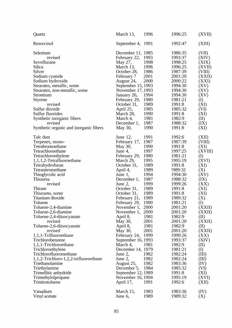
95
Quartz March 13, 1996 1996:25 (XVII)
Resorcinol September 4, 1991 1992:47 (XIII)
Selenium December 11, 1985 1986:35 (VII)revised February 22, 1993 1993:37 (XIV)
Sevoflurane May 27, 1998 1998:25 (XIX)Silica March 13, 1996 1996:25 (XVII)Silver October 28, 1986 1987:39 (VIII)Sodium cyanide February 7 2001 2001:20 (XXII)Sodium hydroxide August 24, 2000 2000:22 (XXI)Stearates, metallic, some September 15, 1993 1994:30 (XV)Stearates, non-metallic, some November 17, 1993 1994:30 (XV)Strontium January 26, 1994 1994:30 (XV)Styrene February 29, 1980 1981:21 (I)
revised October 31, 1989 1991:8 (XI)Sulfur dioxide April 25, 1985 1985:32 (VI)Sulfur fluorides March 28, 1990 1991:8 (XI)Synthetic inorganic fibers March 4, 1981 1982:9 (II)
revised December 1, 1987 1988:32 (IX)Synthetic organic and inorganic fibers May 30, 1990 1991:8 (XI)
Talc dust June 12, 1991 1992:6 (XII)Terpenes, mono- February 17, 1987 1987:39 (VIII)Tetrabromoethane May 30, 1990 1991:8 (XI)Tetrachloroethane June 4, 1997 1997:25 (XVIII)Tetrachloroethylene February 29, 1980 1981:21 (I)1,1,1,2-Tetrafluoroethane March 29, 1995 1995:19 (XVI)Tetrahydrofuran October 31, 1989 1991:8 (XI)Tetranitromethane April 4, 1989 !989:32 (X)Thioglycolic acid June 1, 1994 1994:30 (XV)Thiourea December 1, 1987 1988:32 (IX)
revised June 2, 1999 1999:26 (XX)Thiram October 31, 1989 1991:8 (XI)Thiurams, some October 31, 1989 1991:8 (XI)Titanium dioxide February 21, 1989 1989:32 (X)Toluene February 29, 1980 1981:21 (I)Toluene-2,4-diamine November 1, 2000 2001:20 (XXII)Toluene-2,6-diamine November 1, 2000 2001:20 (XXII)Toluene-2,4-diisocyanate April 8, 1981 1982:9 (II)
revised May 30, 2001 2001:20 (XXII)Toluene-2,6-diisocyanate April 8, 1981 1982:9 (II)
revised May 30, 2001 2001:20 (XXII)1,1,1-Trifluoroethane February 24, 1999 1999:26 (XX)Trichlorobenzene September 16, 1993 1993:37 (XIV)1,1,1-Trichloroethane March 4, 1981 1982:9 (II)Trichloroethylene December 14, 1979 1981:21 (I)Trichlorofluoromethane June 2, 1982 1982:24 (III)1,1,2-Trichloro-1,2,2-trifluoroethane June 2, 1982 1982:24 (III)Triethanolamine August 25, 1982 1983:36 (IV)Triethylamine December 5, 1984 1985:32 (VI)Trimellitic anhydride September 12, 1989 1991:8 (XI)Trimethylolpropane November 16, 1994 1995:19 (XVI)Trinitrotoluene April 17, 1991 1992:6 (XII)
Vanadium March 15, 1983 1983:36 (IV)Vinyl acetate June 6, 1989 1989:32 (X)

96
Vinyl toluene December 12, 1990 1992:6 (XII)
White spirit December 16, 1986 1987:39 (VIII)Wood dust June 17, 1981 1982:9 (II)
revised June 25, 2000 2000:22 (XXI)
Xylene February 29, 1980 1981:21 (I)
Zinc April 21, 1982 1982:24 (III)Zinc chromate May 24, 2000 2000:22 (XXI)Zinc dimethyl dithiocarbamate September 12, 1989 1991:8 (XI)Ziram September 12, 1989 1991:8 (XI)
Sent for publication December 2001