sample conference paper - brookhaven national laboratory
TRANSCRIPT
1
BNL-96182-2011-CP
Compact CdZnTe-based gamma camera for prostate cancer imaging
Y. Cui, T. Lall, B. Tsui, J. Yu, G. Mahler, A. Bolotnikov, P. Vaska, G. De Geronimo, P. O’Connor, G. Meinken, J. Joyal,
J. Barrett, G. Camarda, A. Hossain, K.H. Kim, G. Yang, M. Pomper, S. Cho, K. Weisman, Y. Seo, J. Babich, N. LaFrance,
and R.B. James
Presented at the 2011 IEEE Nuclear Science Symposium and Medical Imaging Conference (2011 NSS/MIC)
Valencia, Spain October 23-29, 2011
August 2011
Nonproliferation and National Security Department
Brookhaven National Laboratory
U.S. Department of Energy Office of Science, National Nuclear Security Administration Notice: This manuscript has been authored by employees of Brookhaven Science Associates, LLC under Contract No. DE-AC02-98CH10886 with the U.S. Department of Energy. The publisher by accepting the manuscript for publication acknowledges that the United States Government retains a non-exclusive, paid-up, irrevocable, world-wide license to publish or reproduce the published form of this manuscript, or allow others to do so, for United States Government purposes. This preprint is intended for publication in a journal or proceedings. Since changes may be made before publication, it may not be cited or reproduced without the author’s permission.
BROOK~ENNAT~/AL LABORATORY

2
DISCLAIMER
This report was prepared as an account of work sponsored by an agency of the United States Government. Neither the United States Government nor any agency thereof, nor any of their employees, nor any of their contractors, subcontractors, or their employees, makes any warranty, express or implied, or assumes any legal liability or responsibility for the accuracy, completeness, or any third party’s use or the results of such use of any information, apparatus, product, or process disclosed, or represents that its use would not infringe privately owned rights. Reference herein to any specific commercial product, process, or service by trade name, trademark, manufacturer, or otherwise, does not necessarily constitute or imply its endorsement, recommendation, or favoring by the United States Government or any agency thereof or its contractors or subcontractors. The views and opinions of authors expressed herein do not necessarily state or reflect those of the United States Government or any agency thereof.

Compact CdZnTe-based gamma camera for prostate cancer imaging
Yonggang Cuia*, Terry Lallb, Benjamin Tsuic, Jianhua Yuc
, George Mahlera, Aleksey Bolotnikova
,
Paul Vaskaa, Gianluigi De Geronimoa
, Paul O'Connora, George Meinkena
, John Joya( John Barrettf,
Giuseppe Camardaa, Anwar Hossaina
, Ki Hyun Kima, Ge Yanga
, Marty Pompel, Steve Choc, Ken
Weismand, Youngho Seoe, John Babichf
, Norman LaFrancef, and Ralph B. Jamesa
a Brookhaven National Laboratory, Upton, NY 11973, USAbHybridyne Imaging Technologies, Inc., Toronto, ON M2N 6K1, Canada
C Johns Hopkins University, Baltimore, MD 21205, USAd Midstate Hospital, Meriden, CT 06451, USA
eUniversity of California at San Francisco, San Francisco, CA 94107, USAfMolecular Insight Pharmaceuticals, Cambridge, MA 02142, USA
ABSTRACT
In this paper, we discuss the design of a compact gamma camera for high-resolution prostate cancer imaging usingCadmium Zinc Telluride (CdZnTe or CZT) radiation detectors. Prostate cancer is a common disease in men.Nowadays, a blood test measuring the level of prostate specific antigen (PSA) is widely used for screening for thedisease in males over 50, followed by (ultrasound) imaging-guided biopsy. However, PSA tests have a high falsepositive rate and ultrasound-guided biopsy has a high likelihood of missing small cancerous tissues. Commercialmethods of nuclear medical imaging, e.g. PET and SPECT, can functionally image the organs, and potentially findcancer tissues at early stages, but their applications in diagnosing prostate cancer has been limited by the smallness ofthe prostate gland and the long working distance between the organ and the detectors comprising these imaging systems.
CZT is a semiconductor material with wide band-gap and relatively high electron mobility, and thus can operate at roomtemperature without additional cooling. CZT detectors are photon-electron direct-conversion devices, thus offering highenergy-resolution in detecting gamma rays, enabling energy-resolved imaging, and reducing the background ofCompton-scattering events. In addition, CZT material has high stopping power for gamma rays; for medical imaging, afew-mm-thick CZT material provides adequate detection efficiency for many SPECT radiotracers. Because of theseadvantages, CZT detectors are becoming popular for several SPECT medical-imaging applications.
Most recently, we designed a compact gamma camera using CZT detectors coupled to an application-specific-integratedcircuit (ASIC). This camera functions as a trans-rectal probe to image the prostate gland from a distance of only 1-5 cm,thus offering higher detection efficiency and higher spatial resolution. Hence, it potentially can detect prostate cancers attheir early stages. The performance tests of this camera have been completed. The results show better than 6-mmresolution at a distance of I cm. Details of the test results are discussed in this paper.
Keywords: CdZnTe, Radiation Detectors, Prostate Cancer, Nuclear Medical Imaging, Gamma Camera
1. INTRODUCTIONProstate cancer is one of the most common cancers in the male population. According to the American Cancer Society,about I in 6 men will be diagnosed with prostate cancer during his lifetime; in 2010, about 217,730 new cases werediagnosed, and about 32,050 men died of the disease [I]. Furthermore, prostate cancer is the second leading course ofcancer death in America. Early detection of the prostate cancer plays an important role in the effectiveness of itstreatment. A blood test of the level of the prostate specific antigen (PSA) often is used as the initial screening marker inthis process. However, the PSA tests have limitations, as it is not cancer-specific but tissue-specific [2]. An elevatedPSA level normally triggers further examinations, e.g., a digital rectal exam to study abnormalities in the size and shapeof the gland, followed by (ultrasound) image-guided biopsy to verify the presence of cancer. During the biopsy
* [email protected]; phone 1 631 344-5351; fax 1 631 344-876; www.bnl.gov
procedure, the prostate is divided into several regions, and tissue samples are removed randomly from each forexamination. Small cancerous tissues are likely to be missed in this painful procedure.
Nuclear-medical imaging affords accurate information about anatomic- and metabolic-processes, increasingly playing animportant role in diagnosing cancers. Such imaging techniques include computed tomography (CT), magnetic resonanceimaging (MRI), positron emission tomography (PET), and single photon-emission computed tomography (SPECT).However, several factors limit the applications of commercial systems in clinical studies. CT has poor contrastresolution for soft tissues and cannot distinguish the boundary between cancerous- and normal-tissue [3]. MRI may notalways differentiate cancer tissue and edema fluid [5], and some regions of prostate gland have low intensity signals onMR images. Functional MRI systems are being developed, but the guidelines for using them have not been established[3]. PET and SPECT can image the distribution of radioactive tracers introduced into the patient's body, and monitorthe metabolism of specific organs. In principle, PET and SPECT imaging systems can detect small tumors at earlystages. However, most commercial systems are bulky, and they use scintillation detectors to detectgamma-ray photonsemitted from the tracers. During medical examinations, the detectors of the imaging system are far away (e.g., 50+ cm)from the imaged organs. At such a long working distance, the efficiency of detection (gamma-ray photon) and spatialresolution are low. The examination process can take several 10's of minutes. In addition, these systems are veryexpensive.
Because of these limitations in current imaging systems, patients and physicians are demanding new techniques toimprove the process of diagnosing prostate cancer. One possibility is based on nuclear medical-imaging technology thatis inexpensive and can provide high spatial- and detection-efficiency. Recently, we developed a compact gamma camera(ProxiScan™) using Cadmium Zinc Telluride (CdZnTe or CZT) radiation detectors. CZT is a semiconductor materialwith wide band-gap and relatively high electron mobility, and thus it can operate at room temperature without additionalcooling. The CZT detector is a photon-electron direct-conversion device, thus potentially offering higher energyresolution in gamma-ray detection; it enables energy-resolved imaging, allowing the subtraction of the background dueCompton scattering-events. In addition, CZT material has high stopping power for gamma rays. For medical imaging, afew-mm-thick CZT material affords sufficient detection efficiency. Because of these advantages, CZT detectors arebecoming popular for several SPECT medical imaging applications. In this project, we developed a compact gammacamera, ProxiScan™, for imaging prostate cancer. Basically, we integrated the CZT detectors, together with applicationspecific-integrated-circuits (ASICs) for signal readout into a stainless-steel sheath with an outer diameter of 25 mm.This camera works as a trans-rectal probe to image the prostate gland at a short distance of 1-5 cm, so offering highdetection- efficiency and high spatial-resolution. In this paper, we discuss the detailed design of the camera and presentthe results from performance tests and preclinical animal tests.
2. SYSTEM DESIGN
2.1 Detector development
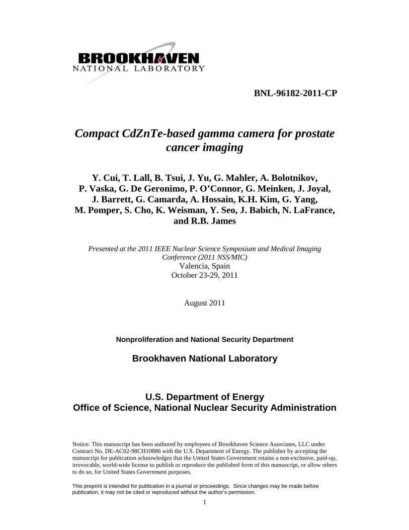
Figure 1. Pixilated CZT detectors employed in the ProxScan™ gamma camera system.
The detectors for the gamma camera are pixilated CZT detectors. We optimized the detectors' thickness for highdetection-efficiency and to fit within limited space inside the probe. Based on our calculations, we selected 5-mm-thick

CZT crystals that ensure 76% detection efficiency for Tc-99m gamma-rays at 140.5 keY. The pixel pitch of thedetectors was 2.46 mm in the first prototype system. Each system used two detectors, and each detector had a 6x8 pixelarray.
2.2 Readout ASIC
A multiple energy window ASIC (MWASIC) [6] developed at Brookhaven National Laboratory (BNL) was employed inthe gamma camera to read out signals from the CZT detectors. Each ASIC has 64 readout channels, and each of themhas a dedicated charge-sensitive amplifier (CSA), shaping circuits, discriminators, and digital counters for each energywindow (Figure 2). The low-noise CSA was optimized for CZT pixilated detectors. The shaping circuits have a 9th
_
order semi-Gaussian architecture to ensure a high counting rate [6].
,----------------------------------------------------- -----------------~I I
! :-:::::::__ --:::::::::::::::::::::::::::::::::::::::::::::::::J_,: : : ,-------- --------------------------------------------------~: : : :I I I
: : :I I II I I
.-- I II I II .-- II I II 1-"I I II I II I ,I I ,I I ,I I II , ,"-I I
I ,
"-L ~60 MHz, 16-bitoutput bus20 IJs readout
Figure 2. Block diagram of multiple energy window ASIC (MWASIC) [6].
2.3 System design
Figure 3 is a block diagram of the gamma camera system. The CZT detectors and the appropriate ASICs are attachedtogether on two sides of the PCB board. A Cypress microcontroller, FX2LP, controls the ASIC and read-out data. Toensure the highly compact design, we also integrated power-management circuits, low-voltage regulation, and highvoltage generation, into the probe. Externally, we provided a single DC power supply, and a USB port forcommunication with the computer. Figure 4 is a photograph of the fully integrated gamma-camera.
Readout and Control
PowerSupply
H.V.
Low VoltageRegulator
andHigh Voltage
Generator
ReadoutControlLogic
Front-endElectronics
Probe
Figure 3. Block diagram of ProxScan™ gamma camera. The compact design has gamma-ray detectors, readoutelectronics, control logic and voltage generator integrated into a hand-held probe.

Figure 4. Photo of ProxScan™ compact gamma camera.
3. PERFORMANCE CHARACTERIZATION
The performance of the camera has been tested in the lab; some ofthe important features are discussed below.
3.1 Energy resolution
200150100Energy (keV)
50
Am.h~l 59.5 keV I\ Tc·99m 140 keV
8.3% FW -1M \\ 5% FWHM
• •\}- ~ IIJvV" ~o
o
500
2500
1000
3000
2000
¥!c5 1500u
Figure 5. Spectra of Am-241 and Tc-99m isotopes coilected by ProxiScan™ gamma camera. Good energyresolution of 5% (FWHM) was obtained for the Tc-99m 140.5-keV energy line.
The energy resolution of the camera was measured by exposing the camera to different sealed sources (Am-241 and Tc99m) and scanning the threshold of a specific energy-window. Figure 5 shows the findings exhibiting a 5% full-widthhalf-maximum (FWHM) for the Tc-99m 140.5-keV line. The result is better than scintillator detectors, which normallyare used in nuclear-medical imaging systems (FWHM is ~ 15% for many commonly used scintillators).

3.2 Energy linearity
//
//
/
350
>' 300~
~ 250(I)0.£2 2000."'0
~ 150(I)Ul
<3 100
50
50 100 150 200 250
Gamma ray energy (keV)
300 350
Figure 6. Energy linearity of ProxiScan™ gamma camera. The results demonstrated good linearity from 59.5 to303 keY covering the energies of gamma-rays emitted by Tc-99m, 1-123 and In- 1II tracers, which aremostly used as radiopharmaceuticals for SPECT.
The energy linearity of the camera was tested by measuring peak positions of several sealed sources including Am-241(59.5 keY), Tc-99m (140.5 keY), and Ba-133 (303 keY). As shown in Figure 6, the camera has good linearity in theenergy range up to 303 keY, covering the photon peaks of Tc-99m, 1-123 and In-Ill, which are three frequently usedradioisotopes used today for nuclear medical imaging.
3.3 Counting rate
~
//
//
//
Voo 50,000 100.000 150.000 200.000 250,000 300.000 350,000 400,000
ICR (cps)
50.000
100,000
250,000~~ 200,000uo
400.000
300.000
350.000
150.000
Figure 7. Counting rate characteristic of the gamma camera.
The counting rate characteristic was tested using a procedure similar to that described in the NEMA standard [8].Because the readout electronics and the MWASIC was optimized for a low-noise, high rate application, it ensured agood counting rate, as shown in Figure 7.
4. IMAGING TESTS
In order to verify the imaging performance of the gamma camera, several tests were undertaken, including a spatialresolution test, a contrast test, and one for the camera's response to shaped phantoms. We report the results in thefollowing section.
4.1 Spatial resolution
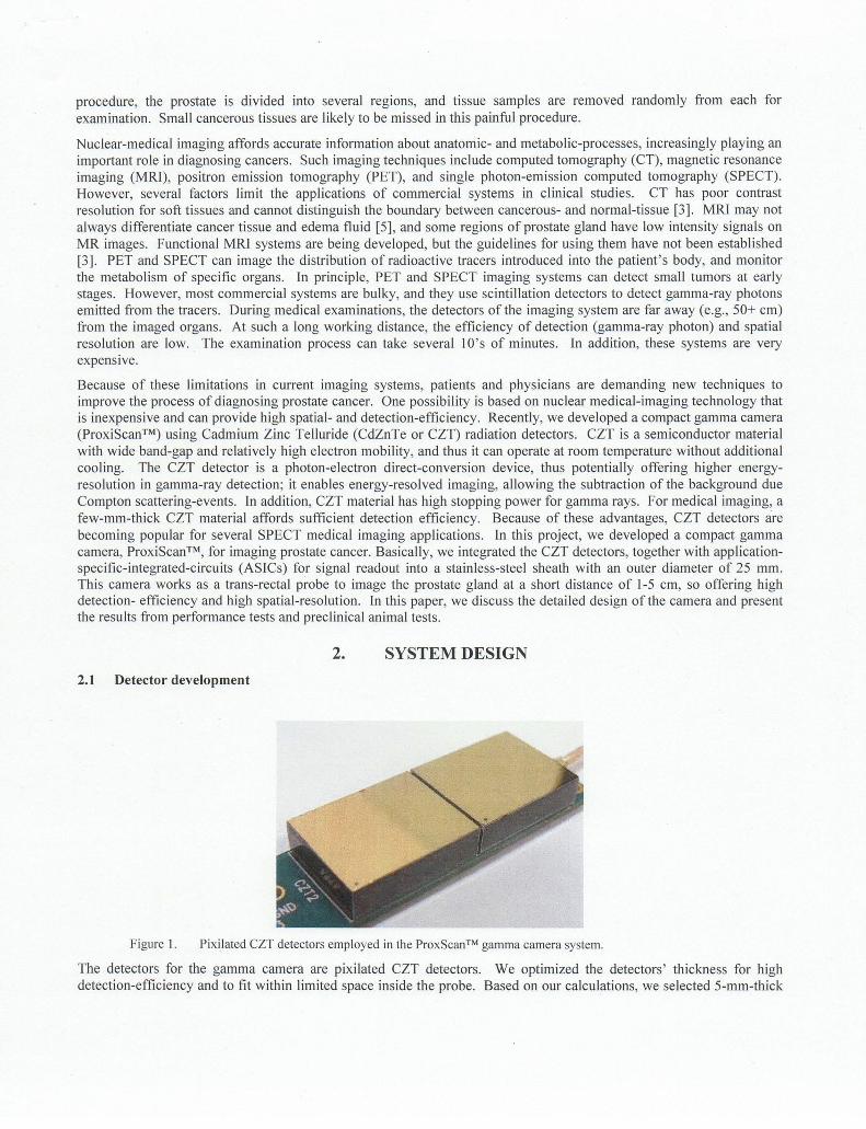
For the spatial-resolution test we used a Tc-99m point-source with a diameter of 0.3 nun and an activity of 6.6 /-lCi at thetime of the experiment. The source was placed above the camera's detector plane, and at different distances trom it (d),viz., 0, 13-, 23-, 43-, 63- and 83-mm. At each position, we acquired the point-spread function (PSF) for a 2-mincollection time. Figure 8 illustrates the raw images. The green grid in the images indicates the size of the pixel,2.46x2.46 nun2
. To calculate the spatial resolution, we fit a Gaussian function into each image and extracted the FWHMvalue. We repeated this calculation for images of d = 0, 13,23, and 43 nun, covering the distance range from the camerato the prostate gland in patients. The results show the camera has 5.7 nun resolution at a distance of 23 nun, whichapproximates the distance from the camera to the center of the prostate gland.
dFWHM
Omm
2.3 mm
13mm
4.4mm
23 mm
5.7mm
43mm
9mm
63mm 83mm
Figure 8. Response of the ProxiScan™ gamma camera to a point source at different distances (d). The green gridindicates the size of the pixels. The spatial resolution was calculated by fitting a Gaussian distributioninto each image.
(1) (2) (3) (4)
(10)
(5) (6)
(12)
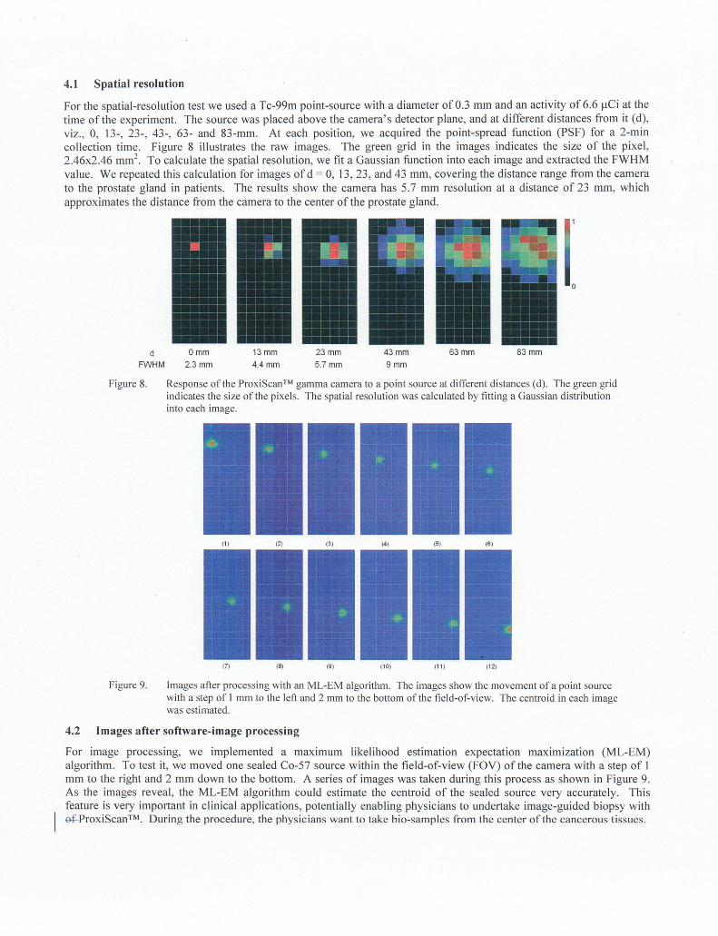
Figure 9. Images after processing with an ML-EM algorithm. The images show the movement ofa point sourcewith a step of I mm to the left and 2 mm to the bottom ofthefield-of-view. The centroid in each imagewas estimated.
4.2 Images after software-image processing
For image processing, we implemented a maximum likelihood estimation expectation maxlmlzation (ML-EM)algorithm. To test it, we moved one sealed Co-57 source within the field-of-view (FOY) of the camera with a step of 1mm to the right and 2 mm down to the bottom. A series of images was taken during this process as shown in Figure 9.As the images reveal, the ML-EM algorithm could estimate the centroid of the sealed source very accurately. Thisfeature is very important in clinical applications, potentially enabling physicians to undertake image-guided biopsy withef.ProxiScan™ During the procedure, the physicians want to take bio-samples from the center of the cancerous tissues.

4.3 Shaped source test
x y
Figure 10. Response of ProxiScan™ gamma camera to a shaped source. The cylindrical phantom for this test was7.3 mm in length and 3.6 mm in diameter. It was placed at two different orientations, horizontally (X)and vertically (Y). In both cases, the shape of the phantom was resolved.
For this test, we used a cylindrical phantom (Tc-99m), 7.3 mm in length and 3.6 mm in diameter. Its activity was 22 IJ.Ciat the time of the experiment. The phantom was placed at two different positions during the testing: X-position, whereinthe axis of the phantom was parallel to the shorter edge of the FOY of the camera, and in the Y-position, where the axiswas parallel to the longer edge of the FOY. In both cases, the phantom was placed 13 mm above the camera. As shownin Figure 10, the shape of the phantom was resolved in both orientations.
4.4 Contrast test
In addition to the dependence of the image quality on the system performance, it also depends on the performance of theradiopharmaceutical used in the examination; The purpose of the contrast test was to establish the lower boundary ofactivity ratio of the hot spot (cancerous tissue) to the background (normal tissue and surrounding organs) that the cameracan detect. For this test, we used two shaped sources, one flood source and one point source. The flood source is a 10mm tall acrylic plate container with a 3-mm thick active region. The container was filled with 230 IJ.Ci/ml ofTc-99m forthe experiment. The point source is a sphere container with a 3.9-mrn diameter; it was filled with Tc-99m of differentconcentrations to obtain ratios of 8: 1,4: I and 2: I radioactivity to the flood source. For 2: I ratio, we tested two differentsource positions in the test: For position A, the sphere SOUtce is above the flood source; and for position B, the sphere isunderneath the flood source (Figure II). Due to photon scattering in the acrylic material, position A has lower contrastthan position B. For the 8: I and 4: I ratios, only position A was used. For each setup, we collected images for 2minutes. Figure 12 shows the images. In addition to the high contrast images for the 8: I and 4: I configurations, thecamera detected the hot spot in a low contrast (2: I) as well. Even in the 2: I position-A configuration (lower contrastthan 2: I), the hot spot still was visible because of the camera's high sensitivity.
Sphere source (position A)
---;=======°=======:::;-1 Plate flood source
; Sphere source (position B)---------=---) Probe
Figure II. Experimental setup for contrast tests. The flood source was in a IO-mm thick acrylic container with 3mm thick active volume. The sphere source had a diameter of 3.9 mm. The activity ratio of the spheresource to the flood source was set as 2: 1,4: I, and 8: 1. Two different positions of the sphere source, Aand S, were tested for the 2: 1 ratio. For other ratios, only position A was used.

2:1 (A)
4:1 (A)
2: 1 (B)
o
Figure 12. Images acquired from contrast tests with different activity ratios. For 2: I ratio setting and position A, theactual ratio is less than 2: 1, but the source was still resolved in the image.
S. CONCLUSIONS
We developed a gamma camera for imaging prostate cancer. Because of several advantages of CZT detectors overscintillation detectors and the development of highly integrated ASICs, the camera is compact and can function as atrans-rectal probe to image the prostate gland. Such a short working distance greatly improves the efficiency ofdetection and the spatial resolution. The prototype system demonstrated very promising imaging performance. Thecamera has high sensitivity, and it can detect low-contrast objects in a high background. Because of its high sensitivity,the camera potentially can work with a wide range of SPECT radiopharmaceuticals. Although the current prototypesystem does not have very high spatial resolution, it will be improved considerably after we have completed reducing thepixel pitch trom 2.46 mm down to 1.6 mm or less. Our new results will be reported later.
6. ACKNOWLEDGEMENT
The authors would like to thank the U.S. Department of Energy's Office of Nonproliferation Research and Development(NA-22) as the principal sponsor funding the research on CZT detector development. The authors are also grateful toHybridyne Imaging Technologies, Inc. for the funding support on the design, engineering and testing of the ProxiScan™gamma camera.
REFERENCES
[1] www.cancer.org[2] Per-Anders Abrahamsson and Martina Tinzl, "Do We Need PSA and Early Detection of Prostate Cancer?",
European Urology Supplements 7, 2008, pp. 393-395.[3] S. Jeschke, E. Schweigreiter and G. Janetschek, "Role of Imaging in Prostate Cancer: A Review", Imaging
Decisions MR!, no. 13, 2010, pp. 68-87.[4] Mohsen Beheshti, Werner Langsteger and Ignac Fogelman, "Prostate Cancer: Role of SPECT and PET in Imaging
Bone Metastases", Semin. Nucl. Med. 39,2009, pp. 396-407.[5] www.radiologyinfo.org[6] G. De Geronimo, A. Dragone, 1. Grosholz, P. O'Connor and E. Vernon, "ASIC with Multiple Energy
Discrimination for High-rate Photon Counting Applications", IEEE Trans. Nucl. Sci., vol. 54, issue 2, pp. 303-312,2007.
[7] www.cypress.com[8] "Performance measurements of scintillation cameras", NEMA Standards Publication No. NU 1, National Electrical
Manufacturers Association, 2007.