rysavy data paper-sept2005-final - 4g americas
TRANSCRIPT
Isolation of Bacillus thuringiensis and Investigation of
Its Crystal Protein Genes
By
Fatoş Tuba ÇETİNKAYA
A Dissertation Submitted to the
Graduate School in Partial Fulfillment of the
Requirements for the Degree of
MASTER OF SCIENCE
Department: Biotechnology and Bioengineering
Major: Biotechnology
İzmir Institute of Technology
İzmir, Turkey
October, 2002

i
ABSTRACT
Bacillus thuringiensis is a ubiquitous, gram-positive and spore-forming
bacterium. During sporulation, it produces intracellular crystal proteins (cry proteins),
which are toxic to insects. Because of its insecticidal activity, it has been used for nearly
fifty years to control certain insect species among the orders Lepidoptera, Coloeptera,
and Diptera. However, it is still necessary to search for more toxins to control other
insect orders and to provide alternatives for coping with the problem of insect
resistance. The genetic diversity of B. thuringiensis strains shows differences according
to the regions where they were isolated. Thus, each habitat may contain novel B.
thuringiensis strains, which have some toxic effects on target spectra of insects.
The aim of this study was to isolate B. thuringiensis strains from different
environments and to identify the crystalline protein gene content of the isolates. Sixty
five samples including soil, stored product dust, insect cadavers, and dry leaf residues
were collected from Akhisar/Manisa, İzmir, and Ereğli/Konya. Three approaches were
applied for the isolation of B. thuringiensis: sodium acetate selection, heat treatment,
and endospore staining. Polymerase Chain Reaction (PCR) method was used for the
characterization of cry gene content of B. thuringiensis strains. The universal primers
specific to cry 1, cry2, cry 3, and cry 9 genes were used to detect the type of cry gene
carried by each environmental isolate of B. thuringiensis strains. In addition, 16S rRNA
based PCR-restriction fragment length polymorphism (RFLP) was carried out to
confirm B. thuringiensis strains. Finally, SDS-PAGE analysis was optimized to detect
protein profiles of crystal proteins obtained from B. thuringiensis isolates.
It was found that, 136 of 359 isolates showed B. thuringiensis-like colony
morphology and subterminal endospore position. One hundred isolates were screened
by PCR and 18 of them were found to contain cry genes (5 cry 1, 3 cry3, and 10 cry 9).
However, the cry 2 gene was not detected from any isolates. 16S rRNA based PCR-
RFLP for 18 isolates gave the same restriction pattern as positive controls, indicating
that all 18 isolates were B. thuringiensis. SDS-PAGE studies for Cry 9 proteins of the
isolates exhibited different protein profile from positive control of B thuringiensis
strain.

ii
ÖZ
Bacillus thuringiensis yaygõn olarak yeryüzünün her kõsmõnda bulunabilen,
gram-pozitif ve spor oluşturan bir bakteridir. Spor oluşumu sõrasõnda, böceklere karşõ
toksik, hücre içi kristal proteinleri (cry proteinleri) sentezler. İnsektisidal aktivitesinden
dolayõ, yaklaşõk olarak elli yõldan beri Lepidopteralarõn, Coloepteralarõn, ve Dipteralarõn
arasõnda bulunduğu böcek gruplarõnõ kontrol altõnda tutmak amacõyla kullanõlmaktadõr.
Bununla birlikte, başka böcek gruplarõnõ kontrol altõnda tutmak ve mevcut böceklerin bu
tür ilaçlara karşõ geliştirdiği dirence alternatif olarak, yeni kristal proteinlerinin
araştõrõlmasõ gereklidir. B. thuringiensis türlerinin genetiksel çeşitliliği izole edildikleri
bölgelere bağlõ olarak farklõlõk göstermektedir. Böylelikle, her bir habitat amaçlanan
böcek gruplarõna toksik etki gösteren yeni B. thuringiensis türlerini barõndõrabilir.
Bu çalõşmanõn amacõ, farklõ ortamlardan B. thuringiensis türlerinin izole
edilmesi ve bu izolatlarõn içerdikleri kristal protein genlerinin saptanmasõydõ.
Topraktan, depo tozlarõndan, böcek ölülerinden ve kuru yapraklardan oluşan altmõş beş
örnek, Akhisar/Manisa, İzmir, ve Ereğli/Konya�dan toplandõ. B. thuringiensis
izolasyonu için sodyum asetat seleksiyonu, sõcaklõk uygulamasõ ve endospore boyama
yöntemlerine başvuruldu. B. thuringiensis türlerinin içerdikleri kristal protein genlerinin
karakterizasyonu için Polimeraz Zincir Reaksiyonu (PCR) uygulandõ. Farklõ
ortamlardan izole edilen B. thuringiensis türlerinin hangi tip kristal geni taşõdõğõnõn
belirlenmesi için cry 1, cry 2, cry 3, ve cry 9 kristal genleri için spesifik üniversal
primerler kullanõldõ. Buna ek olarak, elde edilen izolatlarõn B. thuringienis olduğunu
teyit etmek için 16S rRNA based PCR-restriction length polymorphisim (RFLP)
metodu kullanõldõ. Son olarak, elde edilen izolatlarõn kristal protein profillerinin tespiti
için SDS-PAGE metodu optimize edildi.
349 izolattan 136 tanesinin B. thuringiensis�e benzer koloni morfolojisi
gösterdiği ve subterminal pozisyonda endospore içerdiği bulundu. 100 izolat PCR
metodu ile tarandõ ve 18 tanesinin cry geni (5 cry 1, 3 cry 3, ve 10 cry 9) taşõdõğõ
belirlendi. Bununla birlikte, hiçbir izolatta cry 2 geni tespit edilmedi. Pozitif 18 izolatõn
16S rRNA based PCR-RFLP ile taranmasõ sonucunda, tip türlerle birlikte aynõ
restriksiyon profilleri elde edildi ki, bu da 18 izolatõn B. thuringiensis olduğunu
gösterdi. Bu izolatlarõn Cry 9 proteinleri için SDS-PAGE ile yapõlan çalõşmalarda B.
thuringiensis tip türünden farklõ kristal proteini profili elde edildi.

iii
TABLE OF CONTENTS
LIST OF FIGURES v
LIST OF TABLES vi
Chapter 1 INTRODUCTION 1
1.1.Developments of Bacillus thuringiensis Research 2
1.2. Conventional Bacillus thuringiensis Preparations 2
1.3. General Characteristics of Bacillus thuringiensis 4
1.4. Ecology and Prevalence of Bacillus thuringiensis 5
1.5. Insecticidal Crystal Proteins of Bacillus thuringiensis 6
1.5.1. Mode of Action 6
1.5.2. Structural Features of Crystal Proteins 6
1.5.3. Other Pathogenic Factors of Bacillus thuringiensis 8
1.6. Genetics of Bacillus thuringiensis 9
1.6.1. Bacillus thuringiensis Genome 9
1.6.2. The cry Genes 10
1.6.3. Cry Gene Expression 11
1.7. Strain Collections of Bacillus thuringiensis 12
1.7.1. Isolation Methods to Establish
Bacillus thuringiensis Strain Collections 12
1.7.2. Morphological Properties of Bacillus thuringiensis 13
1.7.3. Characterization Methods to Establish
Bacillus thuringiensis Strain Collections 14
1.8. Objectives 16
Chapter 2 MATERIALS AND METHODS 17
2.1. Materials 17
2.2. Methods 17
2.2.1. Sample Collection 17
2.2.2. Isolation of Bacillus thuringiensis 18
2.2.3. Bacillus thuringiensis Strains 19
2.2.4. DNA Isolation 20
iv
2.2.5. Oligonucleotide Primers for PCR 20
2.2.6. PCR Analysis of cry Genes 21
2.2.7. 16S rRNA based PCR-RFLP 22
2.2.8. Protein Profiling 23
Chapter 3 RESULTS AND DISCUSSION 24
3.1. Sample Collection and Isolation 24
3.2. Identification of cry-type Genes From Environmental
Bacillus thuringiensis Isolates 26
3.2.1. cry 1 Gene Analysis of Bacillus thuringiensis 27
3.2.2. cry 3 Gene Analysis of Bacillus thuringiensis 28
3.2.3. cry 9 Gene Analysis of Bacillus thuringiensis 29
3.3. Distribution of cry Genes 31
3.4. 16S rRNA based PCR-RFLP 34
3.5. Crystal Morphology of Bacillus thuringiensis Isolates 36
3.6. Crystal Protein Profiles of Bacillus thuringiensis Isolates 38
Chapter 4 CONCLUSIONS AND FUTURE EXPERIMENTS 41
REFERENCES 43

v
LIST OF FIGURES
Figure 1.1. Formation of the toxic parasporal crystalin B. thuringiensis 4
Figure 1.2. The structure of Cry 3A 7
Figure 1.3. The structure of Cyt 2A 8
Figure 3.1 Agarose gel (1.2%) electrophoresis of PCR products from
positive controls 27
Figure 3.2. Agarose gel (1.2%) electrophoresis of PCR products for cry 1 genes 28
Figure 3.3. Agarose gel (1.2%) electrophoresis of PCR products for cry3 genes 29
Figure 3.4. Agarose gel (1.2%) electrophoresis for PCR products of cry 9 30
Figure 3.5. Agarose gel (1%) electrophoresis for 16s rRNA �PCR 34
Figure 3.6. Agarose gel (1%) electrophoresis for Hae III 35
Figure 3.7. Agarose gel ( %1) electrophoresis for Taq I 36
Figure 3.8. Photomicrograph of sporangia, crystals and vegetative cells of cry 1
positive 7-0.25-C isolate 37
Figure 3.9. Photomicrograph of sporangia, crystals and vegetative cells of cry 3
positive 21-K-B isolate 37
Figure 3.10. SDS-PAGE (%10) with reference strains of B. thuringiensis 39
Figure 3.11. SDS-PAGE (%10) of Cry 9 proteins 40
vi
LIST OF TABLES
Table 1.1. Examples of products incorporating Bacillus thuringiensis
δ-endotoxins 3
Table 2.1. Types, locations, and numbers of collected samples 18
Table 2.2. Reference strains of B. thuringiensis 20
Table 2.3. Universal primers 21
Table 3.1. Colony morphologies of Bacillus isolates 25
Table 3.2. Type and distribution of B. thuringiensis isolates
determined by morphological and PCR analysis 33
1
Chapter 1
INTRODUCTION
The competition for crops between human and insects is as old as agriculture.
The use of chemical substances to control pests was started in the mid-1800s. Early
insecticides were some inorganic chemicals and organic arsenic compounds.
Organochloride compounds, organophosphates, carbamatespyrethroids and formamides
followed them. Many of these chemicals are also being used today. Certain properties
made these chemicals useful, such as long residual action and toxicity to a wide
spectrum of organisms. However, chemical pesticide applications have caused many
environmental problems including insect resistance, toxicity to humans and to beneficial
insects (Glazer and Nikaido, 1995).
Like all organisms, insects are susceptible to infection by pathogenic
microorganisms. Many of these infectious agents have a narrow host range and,
therefore, do not cause uncontrolled destruction of beneficial insects and are not toxic to
vertebrates. Bacillus thuringiensis is a major microorganism, which shows
entamopathogenic activity (Glazer and Nikaido, 1995; Schnepf et al.,1998). The
organism is a ubiquitous, gram-positive and spore-forming bacteria that forms
parasporal crystals during the stationary phase of its growth cycle. Its insecticidal
activity depends on parasporal crystals encoded by cry genes and this insecticidal
activity varies according to insect type. Natural isolates of B. thuringiensis have been
used as a biological pesticide since the 1950s for the control of certain insect species
among the orders Lepidoptera, Coloeptera and Diptera. The genes of B. thuringiensis
coding parasporal crystals are also a key source for transgenic expression which provide
pest resistance in plants (Schnepf et al., 1998). This feature makes B. thuringiensis the
most important biopesticide on the world market (Bernhard et al., 1997). In 1995,
worldwide sales of B. thuringiensis based insecticides were estimated at $90 million
representing about 2% of the total global insecticide market (Lambert and Pereron,
1992; Schnepf et al.,1998).
2
1.1. Developments of Bacillus thuringiensis Research
B. thuringiensis was first isolated by S. Ishiwata in 1901 from a diseased
silkworm larvae (Bombyx mori ) and named the isolate as Bacillus sotto. B.
thuringiensis was not characterized until a decade later. E. Berliner isolated a similar
Bacillus from a diseased Mediterranean flour moth larvae (Anagasta kuehniella), and
named his isolate as B. thuringiensis (Connon, 1995). B. thuringiensis was first to be
used as an insecticide in the 1950s in the USA. The first commercial name was
Thurincide, which was prepared from B. thuringiensis subsp. kurstaki (Beegle and
Yamamoto, 1992). Dulmage discovered more active B. thuringiensis var. kurstaki
(HD1), which was commercialized in the USA as Dipel (Glazer and Nikaido, 1995).
The demand of B. thuringiensis based insecticides in agriculture sector declined, in the
mid 1970s, because of more effective chemical pesticides. In the 1980s, B. thuringiensis
research was stimulated by progress in biotechnology. First, Schnepf and Whiteley
(1981) cloned a crystal toxin gene from B. thuringiensis subsp. kurstaki into E.coli,
since then much research has been performed to improve target spectra and to find out
more infectious strains of B. thuringiensis.
1.2. Conventional Bacillus thuringiensis Preparations
Most B.thuringiensis preparations available on the market contain spores with
parasporal inclusion bodies composed of δ-endotoxins. In commercial production, the
crystals and spores obtained from fermentation are concentrated and formulated for
spray-on application according to conventional agriculture practices (Baum et al.,
1996). Although, there are numerous strains of B.thuringiensis having insecticidal
activity against insect orders (e.g. Lepidoptera, Diptera, Coloptera, Homoptera,
Mollaphoga), nematodes and aphids, only a few of them have been commercially
developed.
B. thuringiensis based insecticides are divided into three groups, which are
summarized on Table 1.1. Group I has been used for the control of Lepidopterans.
These groups of insecticides are formulated with B. thuringiensis subsp. kurstaki. Group
II contains the sandiego and tenebrionis strains of B. thuringiensis and has been applied
for the control of certain Coloepterans and their larvae. Group III contains the

3
israelensis strain of B. thuringiensis, which has been used to control black flies and
mosquitoes
Table 1.1. Examples of products incorporating Bacillus thuringiensis δ-endotoxins (Cannon, 1995, Biological Control: Benefits and Risks, Chapter 17, pp 192).
Product Bt variety Manufacturer Products for the control of Lepidopteran larvae DIPEL kustaki ( HD 1 ) Abbott Laboratories THURICIDE kustaki Sandoz AG JAVELIN/DELFIN kustaki ( NRD-12) Sandoz AG BACTEC BERNAN I, III, V kustaki Bactec Corporation CATERPILLER ATTACT kustaki Ringer Coporation BIOBIT kustaki Novo Nordisk BACTOSPEINE kustaki Novo Nordisk TOAROW CT kustaki Taogosei Chem TUREX/AGREEa kustaki/aizawai Ciba Geigy CUTLASSa kustaki Ecogen Inc MVb kustaki Mycogen Coporation M PERILb kustaki Mycogen Coporation CERTAN PLUS aizawai Sandoz AG FLORBAC aizawai Novo Nordisk XENTARI aizawai Abbott Laboratories BACTEC BERNAN II morrisoni Bactec Coporation Products for the control of Dipterous (mosquito and blackfly ) larvae VECTOBAC israelensis Abbot Laboraties TECHNAR israelensis Sandoz AG ACROBE israelensis American Cyanamid MOSQUITO ATTACK israelensis Ringer Coporation SKEETAL israelensis Novo Nordisk BACTIMOS israelensis Novo Nordisk Products for the control of Coloepteran larvae NOVADOR tenebrionis Novo Nordisk BK-100 tenebrionis Novo Nordisk FOILa kurstaki/tenebrionis Ecogen Inc M-TRAKb san diego Mycogen Coporation Products for the control of forest defoliating caterpillers THURICIDE kurstaki Sandoz AG FORAY kurstaki Novo Nordisk BIODART kurstaki ICI (Zenece) CONDOR kurstaki Ecogen Inc a- transconjugant b-killed microbial (transgenic)

4
1.3. General Characteristics of Bacillus thuringiensis
B. thuringiensis is a member of the genus Bacillus and like the other members
of the taxon has the ability to form endospores that are resistant to inactivation by heat,
desiccation and organic solvents. The spore formation of the organism varies from
terminal to subterminal in sporangia that are not swollen, therefore, B. thuringiensis
resembles other Bacillus species in morphology and shape (Stahly et al., 1991). The
organism is a gram-positive and facultative anaerobe. The shape of the cells of the
organism is rod. The width of the rod varies 3-5 µm in size when grown in standard
liquid media. The most distinguishing feature of B. thuringiensis from closely related
bacillus species (e.g. B. cereus, B. anthracis ) is the presence of a parasporal crystal
body that is near to the spore, outside the exosporangium during the endospore
formation, which is shown in Figure 1.1 (Andrews et al., 1985; Andrews et al., 1987;
Bulla et al.,1995).
Figure 1.1. Formation of the toxic parasporal crystal in B. thuringiensis (Madigan et al., 2000,
Brock Biology of Microorganisms, Chapter 12, pp 509).
It is thought that B. thuringiensis is an insecticide-producing variant of B.cereus
(Gordon et al., 1973). Several B. thuringiensis strains also produce B .cereus type
enterotoxin (Carson and Kolstø, 1993). Plasmids coding for the insecticidal toxin of B.
thuringiensis have been transferred into B. cereus to make it a crystal producing variant
5
of B. thuringiensis (Gonzales et al., 1982). Molecular methods including genomic
restriction digestion analysis and 16S rRNA sequence comparison support that B.
thuringiensis, B. anthracis, and B. cereus are closely related species and they should be
considered as a single species (Carson, 1994, 1996; Ash et al., 1991, Bourque et al.,
1995; Helgason et al., 2000).
1.4. Ecology and Prevalence of Bacillus thuringiensis
B. thuringiensis occurs naturally and it can also be added to an ecosystem
artificially to achieve insect control. For this reason, the prevalence of B. thuringiensis
in nature can be defined as “natural” and “artificial”. The habitat is considered as
natural when B. thuringiensis can be isolated when there is no previous record of
application of the organism for insect control. The artificial habitats of B. thuringiensis
are areas sprayed with B. thuringiensis based insecticides (usually a mixture of spores
and crystals) (Stahly et al., 1991).
B.thuringiensis is indigenous to many environments including soil (Martin and
Travers, 1989; Bernard et al., 1997), insect cadavers (Corazzi et al., 1991; Kaelin et al.,
1994; Itaqou-Apoyolo et al., 1995; Lopez-Meza and Ibarra, 1996; Cadavos et al., 2001),
stored product dust (Chambers et al., 1991; Meadows et al., 1992; Hongyu et al., 2000),
leaves of plants (Smith and Couche, 1991; Bel et al., 1997; Mizuki et al., 1999), and
aquatic environments (Iriarte et al., 2000; Ichimatsu et al., 2000). Moreover, B.
thuringiensis has recently been isolated from marine sediments (Maeda et al., 2000),
and also from the soils of Antarctica (Forsty and Logan, 2000). Thus, it is obvious that
B. thuringiensis is widespread in nature. However, the normal habitat of the organism is
soil. The organism grows naturally as a saprophyte, feeding on dead-organic matter,
therefore, the spores of B. thuringiensis persist in soil and vegetative growth occurs
when nutrients are available. Because of this, B. thuringiensis can also be found in dead
insects.
Meadows (1993) suggested three prevailing hypothetical niches of B.
thuringiensis in the environment: as an entomopathogen, as a phylloplane inhabitant,
and as a soil microorganism. However, the true role of the bacteria is not clear.
Although, it produces parasporal crystal inclusions that are toxic to many orders of
insects, many B. thuringiensis strains obtained from diverse environments show no
insecticidal activity. For example, Maeda et al. (2000) has found that B. thuringiensis

6
strains obtained from marine environments of Japan exhibit no insecticidal activities.
The insecticidal activity of B. thuringiensis are rare in nature. For instance, Iriarte et al.
(2000) reported that there is no relationship between mosquito breeding sites and
pathogenic action level of B. thuringiensis in the surveyed aquatic habitats. However,
another study suggests that habitats with a high density of insect mortality were
originated by the pathogenic action of this bacterium (Itoqou-Apoyolo et al., 1995).
1.5. Insecticidal Crystal Proteins of Bacillus thuringiensis
1.5.1. Mode of Action
The crystal proteins of B. thuringiensis show host specificity. For this reason,
each type of Cry protein can be toxic to one or more specific insect species. Because of
this specific toxicity, they do not affect many beneficial insects, plants and animals
including humans. The specificity of these insecticidal crystal proteins (ICPs) derives
from their mode of action (Adang, 1991; Gill et al., 1992).
The parasporal crystals of B. thuringiensis contain the ICPs in the form of
protoxins. After ingestion of parasporal crystals by the susceptible insect, the crystals
are dissolved in alkaline conditions (pH 10-12) in the insect mid-gut, generating 130 to
135 kDa protein chains called protoxin. These proteins are then processed to the actual
toxic fragments of 60-65 kDa by the gut proteases (Gill et al., 1992; Höfte and
Whiteley, 1989; Knowles, 1994). Finally, these activated toxins bind to specific
receptors present in the larval mid-gut epithelia. The activated toxin binding to the
specific receptors on the cell membrane creates ion channels or pores. The pore
formation causes osmotic shock. As a result of this process, the cell membrane lyses,
paralysis occurs and consequently, the insect stops feeding and dies from starvation
(Knowless, 1994).
1.5.2. Structural Features of Crystal Proteins
B. thuringiensis produce one or more crystalline inclusion (parasporal crystal)
bodies during the sporulation of its growth cycle and these can be seen under the phase-
contrast microscope. Several terminologies are used for the crystalline inclusions, for
example, insecticidal crystal proteins (ICPs), cry toxins or δ-endotoxin. These
7
parasporal crystals consist of proteins, which exhibit highly toxic insecticidal activity.
On the other hand, actively growing cells lack the crystalline inclusions, so that, they
are not toxic.
The δ-endotoxins fall into two categories; Cyt and Cry. These two types of δ-
endotoxins do not share significant sequence homology, although, both seem to work
through pore formation that leads to cell lysis and irreversible damage of the insect mid-
gut (Gill et al., 1987; Thomas and Ellar, 1983; Chang et al., 1993; Guerchcoff et al.,
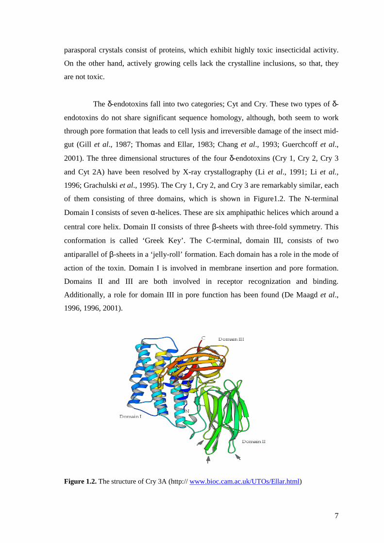
2001). The three dimensional structures of the four δ-endotoxins (Cry 1, Cry 2, Cry 3
and Cyt 2A) have been resolved by X-ray crystallography (Li et al., 1991; Li et al.,
1996; Grachulski et al., 1995). The Cry 1, Cry 2, and Cry 3 are remarkably similar, each
of them consisting of three domains, which is shown in Figure1.2. The N-terminal
Domain I consists of seven α-helices. These are six amphipathic helices which around a
central core helix. Domain II consists of three β-sheets with three-fold symmetry. This
conformation is called ‘Greek Key’. The C-terminal, domain III, consists of two
antiparallel of β-sheets in a ‘jelly-roll’ formation. Each domain has a role in the mode of
action of the toxin. Domain I is involved in membrane insertion and pore formation.
Domains II and III are both involved in receptor recognization and binding.
Additionally, a role for domain III in pore function has been found (De Maagd et al.,
1996, 1996, 2001).
Figure 1.2. The structure of Cry 3A (http:// www.bioc.cam.ac.uk/UTOs/Ellar.html)
8
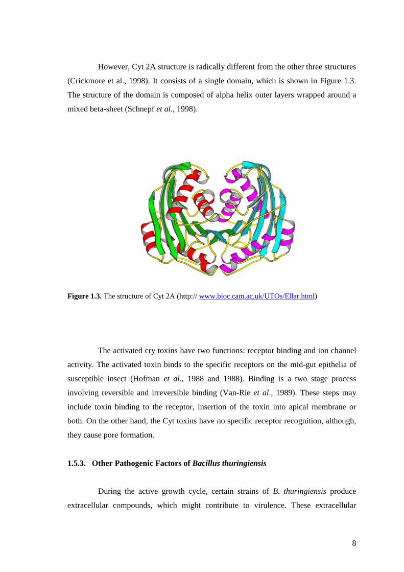
However, Cyt 2A structure is radically different from the other three structures
(Crickmore et al., 1998). It consists of a single domain, which is shown in Figure 1.3.
The structure of the domain is composed of alpha helix outer layers wrapped around a
mixed beta-sheet (Schnepf et al., 1998).
Figure 1.3. The structure of Cyt 2A (http:// www.bioc.cam.ac.uk/UTOs/Ellar.html)
The activated cry toxins have two functions: receptor binding and ion channel
activity. The activated toxin binds to the specific receptors on the mid-gut epithelia of
susceptible insect (Hofman et al., 1988 and 1988). Binding is a two stage process
involving reversible and irreversible binding (Van-Rie et al., 1989). These steps may
include toxin binding to the receptor, insertion of the toxin into apical membrane or
both. On the other hand, the Cyt toxins have no specific receptor recognition, although,
they cause pore formation.
1.5.3. Other Pathogenic Factors of Bacillus thuringiensis
During the active growth cycle, certain strains of B. thuringiensis produce
extracellular compounds, which might contribute to virulence. These extracellular
9
compounds include phospholipases, β-exotoxins, proteases, chitinases and vegetative
insecticidal proteins (VIPs) (Zhang et al., 1993; Levinson, 1990; Estruch, 1996;
Lövgren, 1990; Schnepf et al., 1998). B. thuringiensis also produces antibiotic
compounds having antifungal activity (Stabb et al., 1994). However, the cry toxins are
more effective than these extracelluler compounds and allow the development of the
bacteria in dead or weakened insect larvae.
Some strains of B. thuringiensis produce a low molecular weight, heat stable
toxin called β-exotoxin, which has a nucleotide-like structure. Because of its nucleotide-
like structure it inhibits the activity of DNA-dependent RNA polymerase of both
bacterial and mammalian cells (Glazer and Nikaido, 1995). B. thuringiensis strains also
produce a protease, which is called inhibitor A. This protein attacks and selectively
destroys cecropins and attacins which are antibacterial proteins in insect. As a result of
this, the defense response of the insect collapses. The protease activity is specific,
because it attacks an open hydrophobic region near the C-terminus of the cecropin and
it does not attacks the globular proteins (Dalhambar and Steiner, 1984).
Other important insecticidal proteins, unrelated to Cry proteins, are vegetative
insecticidal proteins (VIPs). These proteins are produced by some strains of B.
thuringiensis during vegetative growth. These VIPs do not form parasporal crystals and
are secreted from the cell. For this reason, they are not included in the Cry protein
nomenclature. For example, the VIP 1A gene encodes a 100 kDa protein which is
processed from its N-terminus. This processing produces an 80 kDa product, which has
been shown to be toxic to western corn root warp larvae (Schnepf, 1998).
1.6. Genetics of Bacillus thuringiensis
1.6.1. Bacillus thuringiensis Genome
B. thuringiensis strains have a genome size of about 2.4 to 5.7 Mb (Carson et
al., 1994). A physical map has been constructed for B. thuringiensis (Carson and
Kolstø, 1993). Comparison with the B. cereus chromosomal map suggested that all of
these chromosomes have a similar organization in the half near the replication origin
while displaying greater variability in the terminal half (Carson et al., 1996). Most B.
thuringiensis isolates have several extra-chromosomal elements (plasmids) ranging in

10
size from 2 to >200 kb. Some of these plasmids are circular and some are linear. The
parasporal crystal proteins are generally encoded by large plasmids. Sequence
hybridizing studies with cry gene probes have been shown that cry genes are also found
in the bacterial chromosome (Carson et al., 1994). B. thuringiensis also contains large
variety of transposable elements (Mahillon et al., 1994). These transposable elements
are thought to be involved in the amplification of the cry genes in the bacterial cell.
Another possible role of these elements could be mediating the transfer of plasmid by a
conduction process involving the formations of cointegrate structures between self-
conjugative plasmids and chromosomal DNA or nonconjugative plasmids. The last
function of these elements may be the horizontal dissemination of genetic material,
including cry genes, within B. cereus and B. thuringiensis species (Schnepf et al.,
1998).
1.6.2. The cry Genes
The genes coding for the insecticidal crystal proteins are normally associated
with plasmid of large molecular mass (Gonzales and Carlton, 1980). Many Cry protein
genes have been cloned, sequenced, and named cry and cyt genes. To date, over 100 cry
gene sequences have been organized into 32 groups and different subgroups on the basis
of their nucleotide similarities and range of specificity (Crickmore et al., 1998; Bravo et
al., 1998). For example, the proteins toxic for lepidopteran insects belong to the Cry 1,
Cry 9, and Cry 2 groups. The toxins against coleopteran insects are the Cry 3, Cry 7,
and Cry 8 proteins and Cry1Ia1, which is a subgroup of Cry 1 proteins. The Cry 5, Cry
12, Cry 13 and Cry 14 proteins are nematocidal, and the Cry 2Aa1, which is a subgroup
of Cry 2 proteins, Cry 4, Cry 10, Cry 11, Cry 16, Cry 17, Cry 19, and Cyt proteins are
toxic to dipteran insects (Zeigler, 1999).
Each of the B. thuringiensis strains can carry one or more crystal toxin genes,
and therefore, strains of the organism may synthesize one or more crystal protein.
Transfer of plasmids among B. thuringiensis strains is the main mechanism for
generating diversity in toxin genes (Thomas et al., 2001).
11
1.6.3. Cry Gene Expression
The insecticidal crystal proteins are synthesized during the stationary phase of
the bacterial life cycle growth. These proteins generally accumulate in the mother cell.
The dry weight of the proteins account for up to 25% in sporulated cells of B.
thuringiensis. The high level of crystal protein synthesis in B. thuringiensis is controlled
by a variety of mechanisms. These mechanisms may occur at the transcriptional, post-
transcriptional and post-translational levels (Agassie and Lereclus, 1995).
The sporulation-specific genes control cry gene expression. However, some of
cry gene expression occurs during the vegetative growth. Thus, the expression of cry
gene mechanisms have been grouped in two groups, sporulation-dependent and
sporulation independent. The cry 1Aa gene, encoding toxins active against lepidoptera,
is a typical example of a sporulation-dependent cry gene. This gene is only expressed
during the sporulation phase. On the other hand, cry 3Aa gene, isolated from the
coloepteran-active B. thuringiensis var. tenebrionis, is expressed during the vegetative
growth and also during the stationary phase. In the stationary phase, the expression of
this gene has been found to be less than the vegetative phase (Sekar et al., 1988; De
Souza et al., 1993).
The stability of cry mRNA is an important contributor to high levels toxin
production at the post-transcriptional level. The half-life of cry mRNA is about 10
minutes which is at least fivefold greater than the half-life of an average bacterial
mRNA (Glathorn and Rapoport, 1973). The putative transcriptional terminator of the
cry1Aa (a stem loop structure) acts as a positive retroregulator (Wong and Chang,
1986). The fusion of a DNA fragment carrying this terminator to the 3′ end of the
heterogonous genes increases the half-life of their transcripts by two to threefold. The
stability of cry mRNA is also increased by the 3′-stem loop structures. Three-fold
structure reduces the movement of 3′-5′ exoribonucleases. For example, the cry 1Aa
transcriptional terminator sequence increases cry mRNA stability by protecting it from
exonucleotytic degradation at the 3’ end (Schnepf et al., 1998).
The crystal proteins are generally found in the form of crystalline inclusion in
the mother cell compartment. The crystal shape depends on the protoxin composition.
This ability of the protoxins to crystallize may decrease their susceptibility to premature
proteolytic degradation. The factors, including the secondary structure of the protoxin,

12
the energy of the disulphide bonds and the presence of additional B. thuringiensis
specific components affect the structure and the solubility characteristics of cry proteins
(Schnepf et al., 1998)
1.7. Strain Collections of Bacillus thuringiensis
Intensive screening programs have identified B. thuringiensis strains from soil,
plant surfaces, dead insects, and stored grain samples. The screening for novel isolates
has led to the discovery of strains with toxic activity against a broad range of insect
orders, including Lepidoptera, Coloeptera, Diptera, Hymenoptera, Homoptera,
Molophoga, and Acari (Feitelson et al., 1992). Furthermore, B. thuringiensis strains
able to control other insect orders such as Nemalthelmintes, Platyhelmintes, and
Sarcomastigophora have been found (Feitelson, 1993). Some strains of B. thuringiensis
have also been found to be toxic to nematodes, mites and protozoa (Feitelson et al.,
1992; Feitelson, 1993; Edwards et al., 1988) It is still necessary to search for more
toxins, since a significant number of pests remain to be uncontrolled with the available
Cry proteins. It is also very important to provide alternatives to overcome the problem
of insect resistance, especially, with regard to the expression of B. thuringiensis genes
encoding insecticidal proteins in transgenic plants (Van-Rie, 1991).
The genetic diversity of B. thuringiensis strains shows differences according to
the regions where they were isolated. In fact, each habitat may contain novel B.
thuringiensis which may have some toxic effects on a target spectra of insects. The
characterization of B. thuringiensis strain collections may help in the understanding of
the role of B. thuringiensis in the environment and the distribution of cry genes. Several
B. thuringiensis strain collections have been described in the literature (Chak et al.,
1994; Ben-Dov et al., 1997; Juárez-Pérez et al., 1997; Bravo et al., 1998; Thenius et al.,
1998; Iriarte et al., 2000; Kaelin et al., 1994; Smith and Couche et al., 1991; Bernard et
al., 1997).
1.7.1. Isolation Methods to Establish Bacillus thuringiensis Strain Collections
Isolation of B. thuringiensis from soil and other natural environments is
greatly facilitated by the use of selective techniques. These techniques improve the
isolation rate of the organism from environmental samples. There are many selective
13
enrichment methods described in the literature (Travers et al., 1987; Johnson and
Bishop, 1996). The sodium acetate selection method has been used routinely for the
isolation of B. thuringiensis from environmental samples (Martin and Travers, 1989;
Carozzi et al., 1991; Ben-Dov et al., 1997; Bravo et al., 1998; Hongyu et al., 2000). The
germination of spores in crystal forming bacilli, including both B. thuringiensis and B.
sphaericus, is inhibited by sodium acetate concentrations of approximately 0.25M. Soil
containing up to 109 bacteria/g is inoculated into a nutrient medium containing the
sodium acetate. After a period of growth, the vegetative cells are eliminated by heat
treatment and remaining spores are isolated on a nutrient medium without acetate. The
survivors from this treatment range from 20-96% B. thuringiensis and B. sphaericus.
These two species are easily differentiated by observation of colonial and cellular
morphology (Travers et al., 1987).
B. thuringiensis is usually isolated together with B. cereus from the
environment. Selective mediums for B. cereus can also be used for the isolation of B.
thuringiensis from environmental samples (Mizuki et al., 1999; Ichimatsu et al., 2000).
B. thuringiensis can be differentiated from B. cereus by the observation of parasporal
crystals under phase contrast microscope. Recently, B. thuringiensis has also been
isolated from extreme environments (Maeda et al., 2000; Forsty and Logan, 2000),
therefore, isolation methods have been adapted according to the requirements of B.
thuringiensis strains that isolated from such extreme conditions. For example, Maeda et
al. (2000) isolated B. thuringiensis strains from marine sediments in Japan by the use of
B. cereus selective medium containing 3% NaCl.
1.7.2. Morphological Properties of Bacillus thuringiensis
Colony morphology can help to distinguish B. thuringiensis colonies from
other Bacillus species. The organism forms white, rough clonies, which spread out and
can expand over the plate very quickly. B. thuringiensis strains have unswollen and
ellipsoidal spores that lie in the subterminal position. The presence of parasporal
crystals that are adjacent to the spore in the mother cell is the best criteria to distinguish
B. thuringiensis from other closely related Bacillus species. The morphology, size, and
number of parasporal inclusions may vary among B. thuringiensis strains. However,
four distinct crystal morphologies are apparent: the typical bipyramidal crystal, related
to Cry 1 proteins (Aronson et al., 1976); cuboidal inclusions related to Cry 2 proteins
14
and usually associated with bipyramidal crystals (Ohba and Aizawi, 1986); amorphous
and composite crystals releated to Cry 4 and Cyt proteins (Federici et al., 1990); and
flat, square crystals, related to Cry 3 proteins (Hernstadt et al., 1986 ; Lopez-Meza and
Ibarra, 1996). Spherical and irregular pointed crystal morphologies can also be observed
in B. thuringiensis strains.
There is a relationship between toxic activity and crystal shape, so that the
observation of crystal morphology by phase contrast microscopy can provide important
clues. For instance, Maeda et al. (2000), collected 22 isolates of B. thuringiensis from
marine sediments in Japan. Two isolates of B. thuringiensis subsp. kurstaki, which are
toxic to lepidopteran larvae formed typically bipyramidal inclusions, whereas isolate
higo, which is toxic to mosquitoes, formed spherical crystals.
The observation of crystal morphology is the first step for establishing B.
thuringiensis strain collections. Ohba and Aizawa (1986) isolated 189 isolates of B.
thuringiensis from 136 soil samples from nonagricultural areas of Japan. The
classification was based in part on the possession of parasporal bodies. Bernard et al.
(1997) isolated 5303 B. thuringiensis from 80 different countries and 2793 of them were
classified according to their crystal shape. They reported that the proportion with
bipyramidal shaped crystals was 45.9%, while 14 % were spherical and 4 %
rectangular.
1.7.3. Characterization Methods to Establish Bacillus thuringiensis
Strain Collections
An important aspects for establishing B. thuringiensis strain collections is to
have a method which allows for rapid and exact characterization. Many methods have
been described for characterization of B. thuringiensis strains, such as bioassay,
serotyping (De Barjac and Franchon, 1990), southern blot analysis for search of known
homologous genes (Kongstad and Whiteley, 1986), analysis for reactivity to different
monoclonal antibodies (Höfte et al., 1988), and electrophoretic analysis of PCR
products using specific primers (Corazzi et al., 1991). Analysis of δ-endotoxin genes by
bioassays is an exhaustive and time consuming process, since it is necessary to screen
all target insect isolates. Serotyping is also an impractical method, it does not reflect the
specific cry gene classes of strains . The main disadvantage of the analysis of reactivity
15
to different monoclonal antibodies is cross-reaction. Because of this, Polymerase Chain
Reaction (PCR) is the best alternative of such methods.
PCR is a highly sensitive and rapid method to detect and identify the target
DNA sequences (cry genes). It requires very little DNA and allows quick, simultaneous
screening of B. thuringiensis strains isolated from the environment. PCR has also been
exploited to predict insecticidal activities of B. thuringiensis strains, to determine the
distribution of cry genes, and to detect new genes. Corazzi et al. (1991) reported the
sequence of twelve PCR primers, which distinguish three major classes of cry genes
(cry I, cry III, and cry IV). These primers were exploited to predict insecticidal activities
of B. thuringiensis strains collected from soil samples and insect cadavers.
Each cry gene group is divided into different subgroups, which can show toxic
effect to different species of an insect order. Multiplex-PCR method has been used to
detect subgroups of a cry gene family. This method is based on two sets of primers. The
first set of primers, the universal primer set is applied to detect the related cry gene
family, and the other set of primer called the specific primer set is used for the detection
of the subgroup of the cry gene family. The universal primers are chosen from highly
conserved regions of a cry gene family. The specific primers, on the other hand, are
designed from variable regions of these gene family sequences. For instance, Ceron et
al. (1994, 1995) exploited this method to detect subgroups of cry I and cry III genes
from soil collected B. thuringiensis strains. Detection of novel cry genes has also been
achieved by this method. For example, Juarez-Perez et al. (1997) applied PCR
technology to detect new cry genes. For this purpose, degenerate primers, which exist
between two conserved regions of cry 1 gene family, and specific primers were used
together to detect cry 1 type of novel cry genes. By this method, a novel cry 1B was
reported.
The genetic diversity and distribution of cry genes varies according to the
regions where they were isolated, so that each habitat can contain a novel cry gene
group that has a different insecticidal activity. Because of this, PCR-based methods
have been used extensively to establish B. thuringiensis strain collections. For instance,
it was reported that the distribution of cry 1-type genes in the Taiwan strain collection is
dependent on geographic location and some of these isolates may contain novel cry IC-
type genes (Chak et al., 1994). In Russian strain collection, six pairs of universal
primers (cry 1, cry 2, cry 3, cry 4, cry 7/8, and cry 9) were used to determine the
distribution of cry genes, and also specific primers (20 cry 1, 3 cry 2, 4 cry 3, 2 cry 4, 2
16
cry 7, 3 cry 8, and 4 cry 9) were applied to detect new cry genes from 124 soil isolates
of B. thuringiensis. Strains containing cry1 type genes were found to be most abundant,
while strains with cry 3 were absent and it was also reported that 43 of 124 field
collected strains of B. thuringiensis might contain new cry gene or genes (Ben-Dov et
al., 1997, 1999). A similar study was done by Bravo et al. (1998). They isolated 496 B.
thuringiensis strains from Mexico. The analysis of strains was based on Multiplex-PCR
with general and specific primers that could detect cry 1, cry 2, cry 3, cry 5, cry 7, cry 8,
cry 9, cry 11, cry 12, cry 13, cry 14, cry 21, and cyt genes. They reported that cry 5, cry
12, cry 13, cry 14, and cry 21 carrying strains were absent in their collection.
The main drawbacks of the PCR approaches are the detection of already-
known genes and the failure to detect and identify novel cry genes. Kuo and Chank
(1996) have proposed a method to increase the efficiency of PCR- based methods. This
method is PCR-based Restriction Length Polymorphism (PCR- based RFLP). PCR-
RFLP is essentially based on the amplification of cry-type gene family with known
universal primers and PCR products are then cut with restriction enzymes to detect the
differences in a cry gene group.
1.8. Objectives
Because the genetic diversity and distribution of cry genes in B. thuringiensis
strains vary based on geographical location. Each habitat may contain novel B.
thuringiensis isolate that have more toxic effects on target spectra of insects. Intensive
screening programs have been identified B. thuringiensis strains from soil, plant
surfaces and stored product dust samples. The screening for novel isolates of B.
thuringiensis is a worldwide project. Therefore, many strain collections have been
described in the litrature, such as Assian (Chak et al., 1994; Ben-Dov et al., 1997, 1999)
and Mexican (Bravo et al., 1998) strain collections. Therefore, the main goals of this
study were:
1) To isolate B. thuringiensis strains from different environments in Turkey.
2) To identify crystalline protein gene content of the isolates by PCR analysis with
universal primers specific to cry 1, cry 2, cry 3, and cry 9 genes.
3) To confirm the B. thuringiensis isolates based on 16S rRNA based PCR-RFLP
analysis.
4) To determine crystal protein profiles of isolated B. thuringiensis strains by SDS-
PAGE analysis.
17
Chapter 2
MATERIALS AND METHODS
2.1. Materials
Nutrient broth was purchased from Oxoid. Bacteriological agar, mineral oil,
safranin, malachite green, methylene blue, commassie brillant blue, ethidium bromide
were from Merck Chemical Company. Bacteriological plates were obtained from
Greiner. Sodium acetate (monobasic), crystal violet, ammonium oxalate, ammonium per
sulfate, iodine, potassium iodide, EDTA, Tris-base, agarose, and Direct Load Range
DNA Molecular Weight Marker, acrylamide solution (40%), bromophenol blue,
TEMED were purchased from Sigma Chemical Company. Glycerol was from
Applichem Chemical Company. Taq DNA Polymerase kit and dNTPs were obtained
from Promega. Restriction enzymes, Taq I and Hae III, Protein Molecular Weight
Marker were from Fermentas. Primers were synthesized by Integrated DNA
Technologies, INC. Reference strains of Bacillus thuringiensis; B. thuringiensis subsp.
kurstaki, B. thuringiensis subsp. aizawai, B. thuringiensis biovar. tenebrionis, B.
thuringiensis biovar. israelensis, and B. thuringiensis subsp. kumamotoensis, were
kindly supplied by Bacillus Genetic Stock Center, Ohio State University, USA.
2.2. Methods
2.2.1. Sample Collection
Sixty five samples including soil, marshy areas, dead insects, dry leaf residues,
animal feces, and stored product dust samples were collected from Akhisar/ Manisa,
İzmir, and Ereğli/ Konya, where there is no previous record of application of B.
thuringiensis based insecticides. The collected samples are summarized on Table 2.1.
The soil samples were taken 2 to 5 cm below the surface, after scraping of the surface
18
material with sterile spatula. Finally, collected samples were stored in sterile plastic
bags at 4 °C
Table 2.1. Types, locations, and numbers of collected samples.
Sample Type Location Sample Number
Soil İzmir
Ereğli/ Konya
30
14
Stored product dust
Akhisar/ Manisa
İzmir
Ereğli/ Konya
3
3
2
Dead insect Akhisar/ Manisa
İzmir
1
2
Animal feces İzmir 3
Dry leaf residues İzmir 3
Marshy Areas İzmir 4
2.2.2. Isolation of Bacillus thuringiensis
The sodium acetate/ heat treatment method was applied to isolate B.
thuringiensis from environmental samples. Approximately, 0.25 g of each sample were
suspended in 18×180 mm test tubes containing 10 ml nutrient broth with concentrations
of sodium acetate 0.12M and 0.25M [pH: 6.8]. The samples were also suspended in
nutrient broth without sodium acetate as negative control. Next, suspensions were
vortexed vigorously and incubated overnight at 37 °C in a shaking water bath.
Afterwards, the samples were pasteurized for 5 minute at 80 °C in order to kill
vegetative bacterial cells and to eliminate non-sporeforming bacterial cells. Following
heat treatment, the samples were plated on nutrient agar plates, which were incubated
overnight at 35 °C. Finally, bacterial colonies were separated by their colony
morphology. The colonies, which showed B. thuringiensis-like colony morphology
were rough, white and spread out over the plate. These colonies were subcultured on
nutrient agar plates and incubated for 48h at 35 °C to check the position of the spore in
19
the bacterial cell by light microscopy. For this purpose, endospore and simple staining
methods were carried out.
Simple staining means that one dye and a one step procedure was used to stain
microbial cells. Spores of Bacillus species do not stain, and they may be seen as
unstained bodies within bacterial cells stained with methylene blue. Smears of Bacillus
isolates were prepared and they were fixed by heat. The bacterial smears were then
floded with methylene blue. Staining lasted for 5 min. Finally, destaining was
performed by washing under the tap water and stained bacterial colonies were observed
under an oil�immersion objective. In addition, endospore staining with malatchite green
was performed for a better observation of Bacillus spores. This staining procedure
involved primary staining with malatchite green for 5 min and steam heat to drive the
stain into spores. This stain was retained by endospores but washed out of the rest cells
with water. Cells were then counterstained with the red dye safranin. The spores
appeared green and cells appeared red after staining by this procedure.
Isolates having ellipsoidal and subterminal spores in unswollen bacterial cells
were identified as B. thuringiensis and stored in nutrient broth containing 50 % glycerol
at �80 °C.
2.2.3. Bacillus thuringiensis Strains
Reference strains of B. thuringiensis, which are shown in Table 2.2, were
received absorbed onto paper disks. The disks were aseptically plated on nutrient agar.
Two drops of nutrient broth were used to hydrate disks. Then, the plates were incubated
overnight at 35 °C. A single colony was then subcultured on the nutrient agar plate.
Finally, subcultured strains were stored in nutrient broth containing 50 % glycerol, at
�80 °C.
20
Table 2.2. Reference strains of B. thuringiensis
Strains BGCS Code
Original
Code Genotype Genes
B. thuringiensis subsp.kurstaki 4D1 HD1 serotype
3a3b
cry 1,2
B. thuringiensis subsp.aizawai 4J3 HD133 serotype 7 cry 1,2,9
cry 7/8
B. thuringiensis biovar.tenebrionis 4AA1 tenebrionis serovar
tenebrionis
cry 3
B. thuringiensis biovar.israelensis
ONR60A
4Q2 HD500 serotype 14 cry 4,11
B.thuringiensis subsp.kumamotoensis 4W1 HD867(3-11) wildtype cry 7/8
2.2.4 DNA Isolation
DNA extraction was performed as described by Bravo et al. (1998). Reference
B. thuringiensis strains, which were used as positive controls, and the environmental
isolates were grown overnight on nutrient agar plates at 35 °C. A loopfull of cells were
transferred into 0.1 ml of sterile distilled water. The mixture was then frozen for 20
minutes at �80 °C. Thereafter, the mixture was immediately transferred into a boiling
waterbath for 10 minutes to lyse the cells. Finally, the resulting cell lysate was
centrifuged (Henttich, Micro 12-24 Ependorf Model) for 10 seconds at 10 000 rpm. 15
µl of the supernatant were used as the DNA template.
2.2.5. Oligonucleotide Primers for PCR
The primers used in this study have been described by Ben-Dov et al. (1998,
1999). One pair of universal primers (e.g., Un1 direct and reverse) for each four
homology groups was applied to amplify a specific fragment. Their sequences and the
expected sizes of their PCR products are shown in Table 2.3.

21
Table 2.3. Universal Primers
Universal Primers Expected PCR
Product Size
For cry 1 genes
Un1, D1 5�- CATGATTCATGCGGCAGATAAAC-3�
R1 5�- TTGTGACACTTCTGCTTCCCATT-3�
274-277 bp
For cry 2 genes
Un2, D2 5�- GTTATTCTTAATGCAGATGAATGGG-3�
R2 5�- CGGATAAAATAATCTGGGAAATAGT-3�
689-701 bp
For cry 3 genes
Un3, D3 5�- CGTTATCGCAGAGAGATGACATTAAC-3�
R3 5�- CATCTGTTGTTTCTGGAGGCAAT-3�
589-604 bp
For cry 9 genes
Un9, D6 5�- CGGTGTTACTATTAGCGAGGGCGG-3�
R6 5�- GTTTGAGCCGCTTCACAGCAATCC-3�
351-354 bp
2.2.6. PCR Analysis of cry Genes
All PCR reactions were carried out in 50 µl reaction volumes. 15 µl of
template DNA was mixed with reaction buffer containing 200 µM deoxynucleoside
triphosphate mix, 0.5 to 1 µM (reverse and direct) primers, 3mM magnesium chloride,
and 2U of Taq DNA polymerase. Amplifications were carried out in a DNA thermal
cycler (Techno-Progen). The conditions for PCR were as follows: a single denaturation
step for 3 min at 95 °C, a step cycle program set for 35 cycles with a cycle of
denaturation step for 1 min at 95 °C, annealing for 1 min at 52 °C, and extension for 1
min at 72 °C. Finally, an extra extension step for 10 min at 72 °C was used. Each
experiment was performed with positive (a standard template) controls.
Following the amplification, 10 µl of each PCR sample was electrophoresed
on 1.2 % agarose-ethidium bromide gel in Tris-Actate/ EDTA (TAE) electrophoresis
buffer (0.04M Tris-Acetate, 0.001M EDTA [pH 8]) at 100V for 40 min.

22
2.2.7. 16S rRNA based PCR-RFLPs
PCR amplifications were routinely carried out in a 50 µl reaction volume that
contained 15µl of DNA template; primers EGE 1: (5′-agagtttgatcctggctcag-3′) and EGE
2: (5′-ctacggctaccttgttacga-3′) at 10 pmol each; 1.5 mM magnesium chloride; dNTP mix
at 200 µM; and 1.5 U Taq DNA polymerase. Two drops of mineral oil was used to
cover PCR mixture. The conditions for PCR were as following: a step program set for
40 cycles with a cycle of denauration for 1 min at 94 °C, annealing for 1 min at 56 °C,
and extension for 1 min at 72 °C. An extra extension step for 10 min at 72 °C was also
used. After amplification, 10 µl of each PCR product was electrophoresed on 1%
agarose-ethidium bromide gel in TAE buffer (0.04M Tris-Acetate, 0.001M EDTA [pH
8]) at 100V for 40 min. Finally, the PCR products were extracted for RFLP analysis.
Chloroform extraction method was carried out to PCR products. 60 µl of TE
buffer were added to adjust the reaction volumes to 100 µl. Mineral oil was then
removed from the mixture by centrifugation for 5 min at 10 000 rpm and PCR samples
were transferred into sterile 1.5 ml eppendorf tubes. Two volumes of chloroform (200
µl) were added and mixed thoroughly. The samples were then centrifuged for 6 min at
10 000 rpm. The upper phase of samples was taken and 200 µl of chloroform was
added. Thereafter, the mixtures were vortexed thoroughly and centrifuged for 2 min at
10 000 rpm. Upper phase of the 100 µl samples were transferred into eppendorf tubes
containing 10 µl of 3M sodium acetate (pH 5.2) and vortexed thoroughly. Two volumes
of 95% ethanol was added. The samples were then incubated for 30 min at �20 °C.
After that, the samples were centrifuged for 15 min at 10 000 rpm and supernatants
were discarded without disturbing the pellets. The DNA pellets were washed in 300 µl
of 70% ethanol. The samples were then centrifuged for 5 min at 10 000 rpm and
supernatants were removed. Washing step was repeated once more. Finally, dried
pellets were dissolved in 10 µl of 1* TE and the resuspended samples were centrifuged
at 6000 rpm for 3 second. 5 µl of each sample were transferred into 0.5 ml of PCR tubes
in order to be used for restriction enzyme digestion.
Restriction enzyme digestions were carried out in a 20 µl reaction volume that
contained 2U of restriction enzyme. The samples were digested with Taq I (at 65 °C)
and Hae III (at 37°C) restriction enzymes. The samples were then incubated overnight.
Additionally, mineral oil was used in order to avoid evaporation for Taq I. Finally, the

23
samples were electrophoresed on 1 % agarose-ethidium bromide gel in TAE buffer at
100 V for 40 min.
2.2.8. Protein Profiling
Purification of crystal proteins was done according to the method of Bel et al.
(1997). B. thuringiensis isolates were cultured and allowed to sporulate on nutrient agar
plates. When sporulation was assumed to be completed, two loops of the colony
material were removed from the plate and transferred to sterile eppendorf tubes
containing 1 ml of ice-cold sodium hydroxide. Resuspended samples were centrifuged
for 5 min at 13 000 rpm and supernatants were discarded. After that, pellets were
resuspended in 140 µl of 1 % SDS-0.01 % β-mercaptoethanol, and boiled for 10 min to
dissolve the crystals. Samples were then centrifuged for 10 min at 13 000 rpm and
supernatants were removed to be applied to TCA precipitation. For protein
precipitation, 140 µl of 20 % TCA (thricholoroaceticacid) was added onto protein pellet
and incubated for 10 min on ice. Thereafter, the resuspended pellets were centrifuged
for 15 min at 10 000 rpm and supernatants carefully removed. An equal volume of cold
acetone was then added and centrifuged for 5 min at 12 000 rpm. Finally, supernatants
were removed to be used for electrophoresis by resuspending the pellets in SDS-PAGE
loading buffer (0.15M Tris/Cl pH 8.8, 3.75mM EDTA, 0.75M sucrose, 0.075 %
bromophenol blue, 2.5 % SDS, and 7.4 mM dithiothreitol) in equal volumes. The
samples were then boiled for 10 min. Electrophoresis was carried out in 10 %
polyacrylamide gel, at 150V.
After electrophoresis, the gel was fixed with a solution containing 10 ml of
acetic acid, 20 ml of methanol, and 70 ml of sterile distilled water. The coomassie
brilliant blue R250 was used for the staining. The stain was prepared by dissolving 1.25
g of dye in 450 ml of methanol: H20 (1:1 v/v) and 50 ml of glacial acetic acid. The
staining was carried out overnight in a glass tray containing 500 ml of staining solution
at room temperature on an orbital shaker. Finally, the gel was destained by soaking it in
the methanol/ acetic acid solution containing 90 ml of methanol: H2O (1:1 v/v) and 10
ml of acetic acid on an orbital shaker for 4h. After destaining, the gel was dried in
gelatin.
24
Chapter 3
RESULTS AND DISCUSSION
3.1. Sample Collection and Isolation
Sixty five samples comprising soil, soil from marshy places, dead insects, dry
leaf residues, animal feces, and stored product dust samples, were collected from
Akhisar/ Manisa, İzmir and Ereğli / Konya. Locations of samples are shown in Table
2.1 of the material and method section.
Travers et al., (1987) tested 37 strains of spore-forming bacteria in four sodium
acetate concentrations (0.06M, 0.12M, 0.25M, and 0.5M) in order to determine their
ability to germinate in acetate buffered medium. They reported that the germination of
B. thuringiensis strains was usually inhibited by 0.25M sodium acetate concentration,
while other spore-formers germinated. Several B. thuringiensis isolates lacked the
ability to germinate in the presence of 0.12 M acetate buffer and, while other B.
thuringiensis (non-acetate selected isolates) strains could germinate in high sodium
acetate concentrations. Similar to their isolation study, in this study, two different
sodium acetate concentrations (0.12M and 0.25M) and also nutrient broth without
sodium acetate (negative control) were used to increase the efficiency of the isolation
rate of B. thuringiensis strains from environmental samples and to compare sodium
acetate selection results with negative control. Pasteurization of samples at 80°C for 5
min was performed to kill vegetative cells of other spore-formers and to eliminate non-
spore formers. Both sodium acetate/heat treatment and negative control with heat
treatment gave similar isolation rate of B. thuringiensis, however, more of the other
spore-formers were isolated with negative control, and overall 359 Bacillus species
were isolated from 65 environmental samples.
Based on literature, the colony morphology of B. thuringiensis is rough, white
and spread out over the plate, and the cellular morphology of the organism is a rod-like
shaped. The bacteria have ellipsoidal spores located at subterminal or paracentral
position in the unswollen mothercell (Thiery and E. Frachon, 1997). In this study, the
discrimination of Bacillus isolates were done according to their colony morphology and
ten different morphologies were observed for 359 Bacillus species, which are displayed
25
in Table 3.1. After that, the cellular morphology of the isolates, which was randomly
chosen, was observed to identify B. thuringiensis strains by a light microscopy. Two
methods were used for the microscopic examinations; simple staining and endospore
staining. According to the colonial and cellular observations, 136 of 359 (38%) isolates
were provisionally identified as B. thuringiensis strains. The isolates exhibiting A, C, I,
and D type colonies were considered to resemblance B. thuringiensis.
The isolates were named according to the sample number, where they were
isolated; the sodium acetate concentration; and colony morphology such as 2-K-C and
2-0.12-A. The �2� shows sample number, �K� represents negative control, �0.12�
displays sodium acetate concentration, �C, and A� shows colony morphology.
Table 3.1. The colony morphologies of Bacillus isolates
Colony Code Morphology Number of
Isolates
% of Total
Isolate
Number
A Spread, white, and wavy 97 27%
B Small, yellow, smooth and bright 91 25%
C Round, white and rough 31 8%
D Spread, white and round with raised
margin 25 6%
E Yellow, irregular and spreading 21 5%
F Yellow, and round with radiating margin 9 2%
G Resembles to colony C, but brighter than C 22 6%
H Resembles to colony D, but the color is
flue 3 0.8%
I Spread, white, wavy and round with
scalloped margin 32 8%
J Drop like and runny 28 7%

26
Martin and Travers (1989) reported that the normal habitat of B. thuringiensis
is soil, since the spores of the organism persist in soil. Therefore, 44 soil samples were
collected from agricultural areas in this study and the most of the B. thuringiensis
strains were isolated from these soil samples. Hongyu et al., (2000) and Meadows et al.,
(1992) reported that stored product dust samples are rich with B. thuringiensis strains.
However, in our study, among the 8 stored product samples, only one B. thuringiensis
strain was isolated. Meadows et al., (1992) also isolated B. thuringiensis from samples
of bird and mammalian feces and suggested that B. thuringiensis multiplied in the
cadavers of insects, and these cadavers were ingested by birds and mammals. For this
reason, three animal feces samples were used in our study and four B. thuringiensis
strains were isolated from them.
3.2. Identification of cry-type Genes From Environmental Bacillus thuringiensis
Isolates
The cry gene contents of 100 B. thuringiensis isolates were determined by
PCR analysis of cry 1, cry 2, cry 3, and cry 9 genes. Universal primers and their
expected PCR products are shown in Table 2.2. of material and method section.
Genomic DNA from each B. thuringiensis isolate served as template in PCR reactions.
Reactions without any DNA template served as negative control in each PCR
experiment. Reactions with Un 1 (direct and reverse), Un 2, Un 3, Un 9 primers were
carried out to detect cry 1, cry 2, cry 3, and cry 9 genes, respectively. A similar PCR
analysis for cry-type genes was previously reported by Ben-Dov et al.,(1997, 1999).
Each PCR analysis was checked with the appropriate positive control strains of B.
thuringiensis. These strains were B. thuringiensis subsp. kurstaki for cry 1 and cry 2
gene groups, B. thuringiensis biovar. tenebrionis for cry 3 gene groups, and B.
thuringiensis subsp. aizawai for cry 9 gene group. PCR conditions were optimized
using the control strains of B. thuringiensis before screening the isolates. All positive
controls gave the expected PCR products (Figure 3.1.A, and B).

27
M 1 2 3 4
750 bp 500 bp 300 bp 200 bp
B
750 bp
500 bp 300 bp 200 bp
M 1 M 1 2 3 4
A
Figure 3.1. Agarose gel (1%) electrophoresis of PCR products from positive controls. (A):
Lane M: 0.5-10kb DNA MW marker, Lane 1: B. thuringiensis subsp.kurstaki as cry 2 positive
control; (B): Lane 1: B. thuringiensis biovar.tenebrionis as cry 3 positive control, Lane 2: B.
thuringiensis biovar.tenebrionis cry 3 positive control, Lane 3: B. thuringiensis subsp.kurstaki
as cry 1 positive control, Lane 4: B. thuringiensis subsp.aizawai as cry 9 positive control.
3.2.1. cry 1 Gene Analysis of Bacillus thuringiensis
When the template DNA from environmental isolates of 100 B. thuringiensis
samples was amplified with PCR in the presence of primers for cry 1 gene, five isolate
were shown to contain cry 1 gene (Figure 3.2.A, lane 2; B, lanes 2,3,4,5) as in the
positive control (Figure 3.2., A, lane 1, B, lane 1). Universal primers produced PCR
products of expected size at around 270bp for the isolates 7-K-A, 7-0.12-C, 7-0.12-D,
and 10-0.12-D (Figure 3.2., A, lane 2, B, lanes 2, 3, 5). However, isolate 8-0.25-C
produced a strong band at 350bp, which was larger than the expected size (Figure 3.2.,
B, lane 4). Because we used universal primers to detect each cry gene group, it was not
possible to determine the cry gene sub-type in this present study. Therefore, the band at
350bp could correspond to a different sub-type of cry 1 gene. Corazzi et al. (1991)
suggested that novel isolates containing novel cry genes may give PCR products
different in size relative to the standard or may completely lack PCR products.
Therefore, homology to known cry genes for these strains can be analyzed by using
additional primers. In this present study, some B. thuringiensis isolates (8-0.25-C)
containing cry 1 gene produced PCR product profile that was different from that of
reference strain. This B. thuringiensis isolate most probably contains subgroups of cry 1

28
750 bp 500 bp
300 bp 200 bp
750 bp
500 bp
300 bp 200 bp
M 1 2 M 1 2 3 4 5 A B
gene. Future experiments with specific primers for cry 1 gene subtypes is expected to
reveal the type of cry gene subgroup and also new cry gene carried by this isolate.
Figure 3.2. Agarose gel (1.2%) electrophoresis of PCR products for cry 1 genes. (A): Lane M:
0.5-10kb DNA MW marker, Lane 1: B. thuringiensis subsp. kurstaki as cry 1 positive control,
Lane 2: 7-0.12-D; (B): Lane M: 0.5-10kb DNA MW marker, Lane 1: B. thuringiensis subsp.
kurstaki as cry 1 positive control, Lane 2: 7-K-A, Lane 3: 7-0.25-C, Lane 4: 8-0. 25-D, Lane 5:
10-0.12-D
3.2.2. cry 3 Gene Analysis of Bacillus thuringiensis
One hundred environmental isolates of B. thuringiensis were screened with PCR
for the presence of cry 3 gene. Three of them produced PCR product at about 600bp in
length, the size as the band obtained in positive strains of B. thuringiensis (Figure 3.3.,
A and B, lane 1). These isolates, which were cry 3 positive, were 21-K-B, 46-K-A, and
46-0.25-A (Figure 3.3., A, lane 2, B, lanes 4 and 7 respectively). The environmental
isolates, 46-K-A and 46-0.25-A produced 500 bp PCR product. These strains may
contain a few cry-gene subgroups, which might have homology with the cry 3 gene
universal primer-binding region. In every PCR reaction, a negative control without
template DNA was included in order to see if there was any cross-contamination in
PCR reagents. As it can be seen in Figure 3.3., B lane 8, there was no cross
29
M 1 2 A B
M 1 2 3 4 5 6 7 8 9
750bp 500bp
300bp
750bp 500bp
300bp
contamination in the reagents, because no PCR band was observed in the absence of
DNA template.
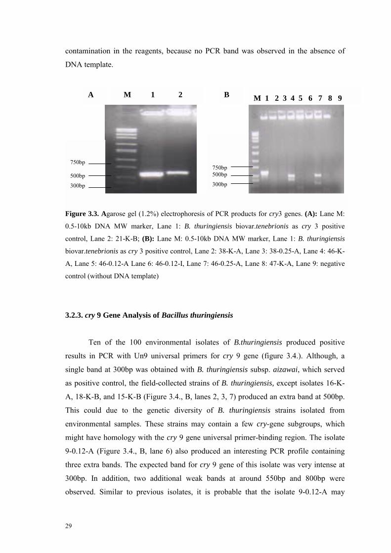
Figure 3.3. Agarose gel (1.2%) electrophoresis of PCR products for cry3 genes. (A): Lane M:
0.5-10kb DNA MW marker, Lane 1: B. thuringiensis biovar.tenebrionis as cry 3 positive
control, Lane 2: 21-K-B; (B): Lane M: 0.5-10kb DNA MW marker, Lane 1: B. thuringiensis
biovar.tenebrionis as cry 3 positive control, Lane 2: 38-K-A, Lane 3: 38-0.25-A, Lane 4: 46-K-
A, Lane 5: 46-0.12-A Lane 6: 46-0.12-I, Lane 7: 46-0.25-A, Lane 8: 47-K-A, Lane 9: negative
control (without DNA template)
3.2.3. cry 9 Gene Analysis of Bacillus thuringiensis
Ten of the 100 environmental isolates of B.thuringiensis produced positive
results in PCR with Un9 universal primers for cry 9 gene (figure 3.4.). Although, a
single band at 300bp was obtained with B. thuringiensis subsp. aizawai, which served
as positive control, the field-collected strains of B. thuringiensis, except isolates 16-K-
A, 18-K-B, and 15-K-B (Figure 3.4., B, lanes 2, 3, 7) produced an extra band at 500bp.
This could due to the genetic diversity of B. thuringiensis strains isolated from
environmental samples. These strains may contain a few cry-gene subgroups, which
might have homology with the cry 9 gene universal primer-binding region. The isolate
9-0.12-A (Figure 3.4., B, lane 6) also produced an interesting PCR profile containing
three extra bands. The expected band for cry 9 gene of this isolate was very intense at
300bp. In addition, two additional weak bands at around 550bp and 800bp were
observed. Similar to previous isolates, it is probable that the isolate 9-0.12-A may

30
C
contain other cry 9 subgroups which have certain degree of homology to the primers
chosen for cry 9 gene amplification. Similar to this present study, Kuo and Chak (1996)
reported such extra PCR products with cry 1 universal primers. They also reported that
this might be due to the non-specific priming of the oligonucleotide primers or the
difference between the sizes of an unknown cry gene and predicted size of the known
cry gene. Brown and Whiteley (1992) reported that two or three cry genes might be
positioned next to each other, forming an operon. In this case, priming of an
oligonucleotide primer to the neighbor cry gene may produce an extra PCR product
larger than expected size. In the studies of others (Ben-Dov et al., 1997; 1999), a set of
bands for a specific cry gene group was shown in multiplex PCR analysis. In order to
specifically identify such cry gene groups, these isolates will be examined with primers
specific for subgroups of the each cry genes.
Figure 3.4. agarose gel (1.2%) electrophoresis for PCR products of cry 9. (A): Lane M: 0.5-
10kb DNA MW marker, Lane 1: B. thuringiensis subsp. aizawai as cry 9 positive control, 33-
750 bp
500 bp
300 bp 200 bp
M 1 2 3 4 A
M 1 2 3 4 5 6 7 B
750 bp
500 bp
300 bp 200 bp
M 1 2
750 bp 500 bp 400 bp 300 bp

31
0.12-A, Lane 3: 37-K-A, Lane 4: 37-K-D; (B): Lane M: 0.5-10kb DNA MW marker,Lane 1: B.
thuringiensis subsp. aizawai as cry 9 positive control, Lane 2: 16-K-A, Lane 3: 18-K-B, Lane 4:
20-0.12-B, Lane 5: 23-K-A, Lane 6: 9-0.12-A, Lane 7: 15-K-B., (C): Lane M: 0.5-10kb DNA
MW marker, Lane 1: B. thuringiensis subsp. aizawai as cry 9 positive control, Lane 2: 13-K
Overall, 359 Bacillus members were isolated from 65 environmental samples.
One hundred and thirty of them (38%) were identified as B. thuringiensis strains based
on microscopic observation of spore position in the cell. One hundred of these 136
isolates were examined by PCR in order to identify the type of cry gene content of these
isolates. Universal primers only for cry 1, cry 2, cry 3, and cry 9 were used for PCR.
Among the 100 isolates, only 18 of them were found to be positive for the cry genes
tested. The reason only 18% of isolates were positive with PCR is most probably due to
the use of limited cry gene primers. In fact, 32 different cry gene groups were reported
by Crickmore et al., (1998). Similar to the results of this study, Ben-Dov et al., (1997)
found that 89 of 215 field-collected strains of them did not produce PCR products with
these universal primers. Therefore, we expect to see more cry gene positive isolates by
using different sets of cry gene primers in the future.
3.3. The Distribution of cry Genes
Diversity, distribution and abundance of cry gene type are dependent on the
geographical area where B. thuringiensis strains were collected. In this study, cry 9 was
found in 10 % of B. thuringiensis isolates; therefore, this gene group was the most
abundant one. According to the literature, Bravo et al., (1998) screened 496 field-
isolated B. thuringiensis by PCR and detected cry 9 genes in 2.6 % of their Mexican
strain collection. In addition, Ben-Dov et al., (1999) detected cry 9 genes in 10.2% of
their 215 soil isolated B. thuringiensis strains from Israel, Kazakhstan, and Uzbekistan.
The cry 3 and cry 1 containing strains were detected in 3% and 5 % of all B.
thuringiensis isolates, respectively, in this study. Ben-Dov et al., (1997) reported that
strains containing cry 1 genes were most abundant, while strains with cry 3 were absent
in soil isolated B. thuringiensis strains from Israel, Kazakhstan, and Uzbekistan.
Another study (Chak et al., 1994) reported that soil isolated 225 B. thuringiensis strains

32
from Taiwan did not harbor cry 3 genes; however, cry 1A gene containing strains were
the most abundant of them. Although, these two studies suggested that cry 3 genes were
absent in Asia, three isolates containing cry 3 were isolated in this study. In addition,
among the one hundred isolates screened by PCR, none of them was positive with cry 2
primers. The distribution of B. thuringiensis isolates in this study is summarized in
Table 3.2.

33
Table 3.2. Type and distribution of B. thuringiensis isolates determined by morphological and PCR analysis.
IsolateCode cry Gene Morphology Spore Position Crystal Shape Sample Type Place % of Isolates Tested by
PCR
7-K cry 1 A Subterminal ND Dried leaf Kalkõç /İzmir
7-0.12 cry 1 D Terminal IP Dried leaf Kalkõç /İzmir
7-0.25 cry 1 C Subterminal B, R, IP Dried leaf Kalkõç /İzmir
8-0.25 cry 1 D Subterminal ND Soil Kalkõç /İzmir
10-0.12 cry 1 D Terminal A, IP Soil Kalkõç /İzmir
5 %
21-K-B* cry 3 A Subterminal S Insect cadaver Urla /İzmir
46-K cry 3 A Subterminal ND Soil Ereğli /Konya
46-0.25 cry 3 A Subterminal ND Soil Ereğli /Konya
3 %
9-0.12 cry 9 A Subterminal IP Soil Kuşcenneti /İzmir
13-K cry 9 A Subterminal A Stored Product Dust Bayraklõ/İzmir
15-K-B* cry 9 A Subterminal IP Soil İzmir
16-K cry 9 A Subterminal IP Soil Kuşcenneti /İzmir
18-K-B* cry 9 A Subterminal S, IP Soil Kuşcenneti /İzmir
20-0.12-B* cry 9 A Subterminal IP Soil Çiğli /İzmir
23-K cry 9 A Subterminal IP Soil Çiğli /İzmir
33-0.12 cry 9 A Subterminal IP Soil Dikili /İzmir
37-K cry 9 A Subterminal IP Soil Dikili /İzmir
37-K cry 9 D Terminal ND Soil Dikili /İzmir
10%
ND: Not determined; B: Bipyramidal, R: Rectangular, S: Spherical, A: Amorphous, IR: Irregularly Pointed
34
3.4. 16S rRNA based PCR-RFLP
Because 16S rRNA ribotyping is one of the most reliable methods for the
identification of bacteria at the species level, ribotyping of 16S rRNA from 18 B.
thuringiensis isolates was carried out. DNA isolated from each B. thuringiensis isolates
was amplified in the presence of primers for variable regions of 16S rRNA. As it can be
seen from Figure 3.5, each environmental isolates gave only one band at expected size,
which is same as the band produced from positive control of B. thuringiensis strains
(Lanes 7, 8, 9, 10, 11, 12).
Figure 3.5. Agarose gel (1%) electrophoresis for 16s rRNA �PCR. Lane M: DNA MW Marker,
Lane 1: B. thuringiensis subsp. kurstaki, Lane 2: B. thrungiensis biovar. tenebrionis, Lane 3: B.
thuringiensis subsp. kumamotoensis, Lane 4: B. thuringiensis subsp. aizawai, Lane 5: B.
thuringiensis biovar. israelensis, Lane 6: B. thuringiensis subsp. kurstaki, Lane 7: 7-K-A, Lane
8: 7-0.12-D, Lane 9: 7-0.25-C, Lane 10: 10-0.12-D, Lane 11: B. thuringiensis subsp.
tenebrionis, Lane 12: 21-K-B
Two restriction enzymes, Taq I and Hae III, were used to cut PCR products in
order to see if they will give the same restriction pattern in all isolates (Figure 3.6).
After cutting the PCR products with restriction enzyme Hae III, three bands were
obtained in all B. thuringiensis positive samples (lanes 1, 2, 3), and in all environmental
M 1 2 3 4 5 6 7 8 9 10 11 12
1400bp 1000bp 750bp
35
isolates (Lanes from 5 to 15). Some weak bands were obtained in lanes 4. 6. and 11.
This was because of insufficient chloroform extraction of the restriction products.
Restriction of 16S rRNA PCR products with Taq I enzyme resulted in the same
restriction pattern with two bands in both positive control (Figure 3.7., lanes 1, 2, 3) and
environmental isolates (Lanes from 4 to15). Therefore, 16S rRNA based PCR-RFLP
results indicated that 18 environmental B. thuringiensis isolates characterized by
morphological and PCR analysis were B. thuringiensis. In other words, 18 isolates were
strongly confirmed by ribotyping experiments.
Figure 3.6. Agarose gel (1%) electrophoresis for Hae III. Lane M: DNA MWM Marker, Lane
1: B. thuringiensis subsp. kumamotoensis,Lane2: B. thuringiensis biovar. israelensis,Lane 3: B.
thuringiensis subsp. kurstaki,Lane 4: 7-K-A, Lane 5: 7-0.12-D, Lane 6: 7-0.25-C, Lane 7:10-
0.12-D, Lane 8: B. thrungiensis biovar. tenebrionis, Lane 9: 21-K-B, Lane 10: B. thuringiensis
subsp. aizawai, Lane 11: 15-K-B, Lane 12: 37-K-A, Lane 13: 13-K, Lane 14: 23-K-A, Lane 15:
18-K-B
M 1 2 3 4 5 6 7 8 9 10 11 12 13 14 15
1000bp 750bp 500bp
250bp

36
Figure 3.7. Agarose gel ( %1) electrophoresis for Taq I. Lane M: DNA MW Marker, Lane 1: B.
thuringiensis subsp. kumamotoensis , Lane 2: B. thuringiensis biovar. israelensis, Lane 3: B.
thuringiensis subsp. kurstaki, Lane 4: 7-K-A, Lane 5: 7-0.12-D, Lane 6: 7-0.25-C, Lane 7:10-
0.12-D, Lane 8: B. thrungiensis biovar. tenebrionis, Lane 9: 21-K-B, Lane 10: B. thuringiensis
subsp. aizawai, Lane 11: 15-K-B, Lane 12: 37-K-A, Lane 13: 13-K, Lane 14: 23-K-A, Lane 15:
18-K-B
3.5. Crystal Morphology of Bacillus thuringiensis Isolates
The morphology, size, and the number of parasporal inclusions may vary among
the B. thuringiensis strains. However, some distinct morphologies are apparent: the
typical bipyramidal crystal, related to Cry 1 protein, (Aronson et al., 1976); cuboidal
inclusions related to Cry 2 proteins (Ohba and Aizawi, 1986); and flat, square, and
spherical crystals related to Cry 3 proteins (Hernstadt et al., 1986). Therefore, the
crystal morphologies of thirty of the 18 positive isolate were observed by phase contrast
microscopy in this study. Five distinct crystal morphologies were observed:
bipyramidal, rectangular, amorphous, spherical and irregularly pointed (Table 3.2). All
of the isolates collected from soil produced irregularly pointed parasporal inclusion.
M 1 2 3 4 5 6 7
M 8 9 10 11 12 13 14 15
1000bp 750bp
1000bp 750bp
37
Based on the litrature, Cry 1 type of crystal proteins encoded by cry 1 gene group
usually associated with the bipyramidal crystals, so that, cry 1 gene carrying isolate 7-
0.25-C produced the biypiramidal crystal protein and also contained rectangular and
irregular pointed secondary crystals in this study(Figure 3.8.). Similar to our study,
Maeda et al. (2000) reported that crystal inclusions of two non-insecticidal B.
thuringiensis isolates were the mixture of two morphotypes: bipyramidal and spherical.
Baum and Malvar (1995) reported that the different cry genes are with in operons which
include open reading frames (orf�s) encoding proteins with undefined functions.
Therefore, the presence of these secondary crystals may be due to the different cry
genes expressed during the sporulation phase. In addition, other crystal types were
detected in the isolates. For example, the spherical crystal morphology was observed
only in isolate 21-K-B, which was isolated from insect cadaver (Figure3.9). Most of the
cry 9 containing isolates produced one type crystal protein which was irregularly
pointed crystal protein (data not shown).
Figure 3.8. Photomicrograph of sporangia, crystals and vegetative cells of cry 1 positive 7-0.25-
C isolate. Bar represents 10µm.
Figure 3.9. Photomicrograph of sporangia, crystals and vegetative cells of cry 3 positive 21-K-
B isolate. Bar represents 10µm.
ICPs
Spore
ICPs
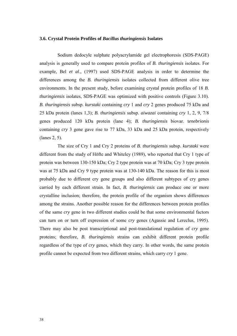
38
3.6. Crystal Protein Profiles of Bacillus thuringiensis Isolates
Sodium dedocyle sulphate polyacrylamide gel electrophoresis (SDS-PAGE)
analysis is generally used to compare protein profiles of B. thuringiensis isolates. For
example, Bel et al., (1997) used SDS-PAGE analysis in order to determine the
differences among the B. thuringiensis isolates collected from different olive tree
environments. In the present study, before examining crystal protein profiles of 18 B.
thuringiensis isolates, SDS-PAGE was optimized with positive controls (Figure 3.10).
B. thuringiensis subsp. kurstaki containing cry 1 and cry 2 genes produced 75 kDa and
25 kDa protein (lanes 1,3); B. thuringiensis subsp. aiwazai containing cry 1, 2, 9, 7/8
genes produced 120 kDa protein (lane 4); B. thuringiensis biovar. tenebrionis
containing cry 3 gene gave rise to 77 kDa, 33 kDa and 25 kDa protein, respectively
(lanes 2, 5).
The size of Cry 1 and Cry 2 proteins of B. thuringiensis subsp. kurstaki were
different from the study of Höfte and Whiteley (1989), who reported that Cry 1 type of
protein was between 130-150 kDa; Cry 2 type protein was at 70 kDa; Cry 3 type protein
was at 75 kDa and Cry 9 type protein was at 130-140 kDa. The reason for this is most
probably due to different cry gene groups and also different subtypes of cry genes
carried by each different strain. In fact, B. thuringiensis can produce one or more
crystalline inclusion; therefore, the protein profile of the organism shows differences
among the strains. Another possible reason for the differences between protein profiles
of the same cry gene in two different studies could be that some environmental factors
can turn on or turn off expression of some cry genes (Agassie and Lereclus, 1995).
There may also be post transcriptional and post-translational regulation of cry gene
proteins; therefore, B. thuringiensis strains can exhibit different protein profile
regardless of the type of cry genes, which they carry. In other words, the same protein
profile cannot be expected from two different strains, which carry cry 1 gene.
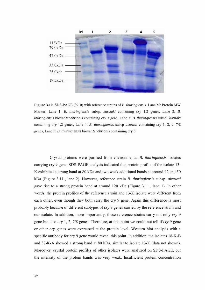
39
Figure 3.10. SDS-PAGE (%10) with reference strains of B. thuringiensis. Lane M: Protein MW
Marker, Lane 1: B. thuringiensis subsp. kurstaki containing cry 1,2 genes, Lane 2: B.
thuringiensis biovar.tenebrionis containing cry 3 gene, Lane 3: B. thuringiensis subsp. kurstaki
containing cry 1,2 genes, Lane 4: B. thuringiensis subsp aizawai containing cry 1, 2, 9, 7/8
genes, Lane 5: B. thuringiensis biovar.tenebrionis containing cry 3
Crystal proteins were purified from environmental B. thuringiensis isolates
carrying cry 9 gene. SDS-PAGE analysis indicated that protein profile of the isolate 13-
K exhibited a strong band at 80 kDa and two weak additional bands at around 42 and 50
kDa (Figure 3.11., lane 2). However, reference strain B. thuringiensis subsp. aizawai
gave rise to a strong protein band at around 120 kDa (Figure 3.11., lane 1). In other
words, the protein profiles of the reference strain and 13-K isolate were different from
each other, even though they both carry the cry 9 gene. Again this difference is most
probably because of different subtypes of cry 9 genes carried by the reference strain and
our isolate. In addition, more importantly, these reference strains carry not only cry 9
gene but also cry 1, 2, 7/8 genes. Therefore, at this point we could not tell if cry 9 gene
or other cry genes were expressed at the protein level. Western blot analysis with a
specific antibody for cry 9 gene would reveal this point. In addition, the isolates 18-K-B
and 37-K-A showed a strong band at 80 kDa, similar to isolate 13-K (data not shown).
Moreover, crystal protein profiles of other isolates were analyzed on SDS-PAGE, but
the intensity of the protein bands was very weak. Insufficient protein concentration
118kDa 79.0kDa 47.0kDa 33.0kDa
25.0kda 19.5kDa
M 1 2 3 4 5
40
M 1 2
118kDa
79.0kDa 47.0kDa 33.0kDa
25.0kda
could be the reason for that; and therefore, protein bands were not detectable with
coomassie brilliant blue staining. More sensitive staining techniques such as silver
staining kit could be alternative to visualize the bands. For example, Iriarte et al. (2000)
found that a strain of B. thuringiensis have 66 kDa protein, which was not visible on
SDS-PAGE gel staining with commassie brilliant blue 250, but it was detectable after
silver staining. The main reason for less protein amount in environmental samples than
that of reference strains could probably be due to the differences in sporulation time of
the strains. Because the time for completion of sporulation varies from strain to strain
and depends on growth medium conditions, parasporal crystal formation was always
checked with phase contrast microscope before isolating crystal proteins from B.
thuringiensis isolates in the literature. Therefore, crystal protein isolation will be carried
out after verification of crystal formation by phase contrast microscope in future studies.
Figure 3.11. SDS-PAGE (%10) of Cry 9 proteins: Lane M: Protein MW Marker, Lane 1: B.
thuringiensis subsp. aizawai containing cry 9 gene, Lane 2: 13-K
41
Chapter 4
CONCLUSION AND FUTURE EXPERIMENTS
Bacillus thuringiensis is a ubiquitous, gram-positive and spore-forming
bacterium. The organism produces intracellular crystal proteins, which are toxic to
insects, during the stationary phase of its growth cycle. Because of its insecticidal
activity, B. thuringiensis has been used for a long time as a biopesticide. However, it is
still necessary to search for more toxins to control insect orders, which have the ability
to develop resistance against such pesticides, and to also provide alternatives for
chemical insecticides. The genetic diversity of B. thuringiensis is directly related to
geographical areas. Thus, toxic activity of the bacteria varies according to regions
where the organism was isolated. Therefore, the purpose of this study was to isolate B.
thuringiensis strains that are collected from Turkey, and to identify the cry gene content
of these isolates, which may carry new crystal genes coding effective crystal proteins.
As a result of the study, 359 Bacillus species were isolated from 65
environmental samples collected from agricultural areas, warehouses and marshy
places. One hundred and thirty six of them (38%) were identified as B. thuringiensis
strains based on colony morphology and microscopic observation of spore position in
the cell. One hundred of these 136 isolates were examined by PCR using for cry 1, cry
2, cry 3, and cry 9 universal primers in order to identify the type of cry gene content of
these isolates. Among the 100 isolates, only 18 of them were found to be positive for
the cry genes tested. In addition, these 18 isolates were confirmed to be B. thuringienis
strains by carrying out 16S rRNA-based PCR-PFLP experiments. The reason that only
18% of isolates were positive with PCR could most probably be due to the use of
limited cry gene primers. Therefore, use of different sets of cry gene universal primers
will enable us the identification of more B. thuringiensis strains containing other type of
cry gene groups. Furthermore, these 18 PCR positive isolates may contain novel cry
genes coding for more effective insecticidal crystal proteins. In the future, specific

42
primers will be used to screen these isolates again in order to detect subgroups of these
genes.
Five distinct crystal morphologies were observed for 13 isolates of 18 PCR
positive strains by phase contrast microscopy. These were spherical, amorphous,
rectangular, bipyramidal, and irregularly pointed. All of the observed isolates produced
irregularly pointed crystals; on the other hand, one of the isolates contained three
parasporal crystals, which were bipyramidal, rectangular and irregular pointed. Among
the 13 isolates, only one isolate produced spherical crystal. Finally, SDS-PAGE analysis
was carried out to determine crystal protein profiles of B. thuringiensis isolates and to
discriminate these bacterial strains from each other. First of all, SDS-PAGE technique
was optimized for crystal proteins of B. thuringiensis. SDS-PAGE analysis of reference
strains resulted in protein bands which were distinct from each other. Similarly, protein
profiles of our isolates carrying cry 9 gene produced a major protein band at around
80kDa, indicating that B. thuringiensis isolates of this study had different subgroups of
cry 9 gene from reference cry 9strains, or reference strain is expressing another cry
gene. Moreover, protein profiles of other B. thuringiensis isolates in this study were
examined but the intensity of protein bands was not strong enough. In a future study,
after conformation of crystal protein formation by phase contrast microscopy, crystal
protein isolation will be carried out in order to allow sufficient protein isolation for
SDS-PAGE analysis.
In a future study, the bioactivity of crystal proteins purified from the 18 B.
thuringiensis isolates will be examined on different insect groups. After that,
preliminary studies on culture conditions of B. thuringiensis strains with higher
insecticidal activity will be carried out for large scale production of crystal protein.
Because, crystalline genes are mostly carried on the plasmids, plasmid profiles will also
be prepared and the cry genes, they contain, will be cloned.

43
REFERENCES
Adang, M. J., �Bacillus thuringiensis insecticidal crystal proteins: gene structure, action
and utilization.� 1991; in Biotechnology for Biological Control of Pest and
Vectors, edited by K. Maramorosh, (CRC Press, Boca Raton Fla, 1991), 3-24.
Agaisse, H., D. Lereclus, �How does Bacillus thuringiensis produce so much
insecticidal crystal protein?�, J. Bacteriol., 177, (1995), 6027-6032.
Andrews, R. E., Jr. R. M. Faust, H. Wabiko, K. C. Raymond, and L. B. Bulla, �The
biotechnology of Bacillus thuringiensis”, CRC Critical Reviews of
Biotechnology, 6, (1987), 163-232.
Andrews, R. E., M. M. Bibilops, and L. A. Bulla, �Protease activation of the
entomocidal protoxin of Bacillus thuringiensis subsp. kurstaki�, Appl. Environ.
Microbiol., 50, (1985), 737-742.
Aronson, A.I., and P. Fritz-James, �Structures and morphogenesis of the bacterial pore
coat�, Bacteriol. Rev., 40, (1976), 360-402
Ash, C., J. A. E. Farrow, M. Darsh, E. Stackebrant, and M. D. Collins, �Comparative
analysis of Bacillus anthracis, Bacillus cereus, and related species on the basis
of reverse transcriptase sequencing of 16s rRNA.�, International Jounal of
Systemic Bacteriology, 41,(1991), 343-346.
Baum,J.A, and T. Malvar, �Regulation of insecticidal crystal protein production in
Bacillus thuringiensis�, Mol. Microbiol., 18, (1995), 1-12.
Baum, J. A., M. Kakefuda, and C. Gawron-Burke, �Engineering Bacillus thuringiensis
bioinsecticides with an indigenous site-specific recombination system�, Appl.
Environ. Microbiol, 62, (1996), 4367-4373.
44
Beegle, C. C. and T. Yamamoto, �Invitation paper ( C. P. Alexander fund): History of
Bacillus thuringiensis Berliner research and development�, Canadian
Entomologist, 124, (1992), 587-616.
Bel, Y., F. Granedo, T. M. Alberola, M. J. Martinez-Sebastian, and J. Ferrė,
�Distribution, frequency and diversity of Bacillus thuringiensis in olive tree
environments in Spain.�, Systematic of Applied Microbiology, 20, (1997), 652-
658.
Ben-Dov, E., A Zaritsky, E. Dahan, Z. Barak, R. Sinai, R. Manasherob, A. Khamraev,
E. Troittskaya, A. Dubitsky, N. Berezine, and Y. Margalith, �Extended
screening by PCR for seven cry-group genes from field-collected strains of
Bacillus thuringiensis� Appl. Environ. Microbiol., 63, (1997), 4883-4890.
Ben-Dov, E., E.Q. Wang, A Zaritsky, R. Manasherob, Z. Barak, B. Schneider, A.
Khamraev, M. Baizhanov, G. Glupov, and Y. Margalith, �Multiplex PCR
screening to detect cry9 genes in Bacillus thuringiensis strains�, Appl. Environ.
Microbiol. 65, (1999), 3714-3716.
Bernhard, K., P. Jarrett , M. Meadows, J. Butt, D. J. Ellis, G. M. Roberts, S.Pauli, P.
Rodgers, and H. D. Burges, �Natural isolates of Bacillus thuringiensis:
worldwide distribution, characterization, and activity against insect pests�,
Journal of InvertebratePathology, 70, (1997), 59-68.
Bourque, S. N., J. R. Valero, M. C. Lavoie, and R. C. Levesque, �Comparative analysis
of the 16S to 23S ribozomal õntergenic spacer sequences of Bacillus
thuringiensis strains and subspecies and of closely releated species�, Applied
and Environmental Microbiolgy, 61, (1995), 1623-1626.
Bravo, A., S. Sarabia, L. Lopez, H. Ontiveros, C. Abarca, A. Ortiz, M. Ortiz, L. Lina, F.
J. Villalobos, G. Pena, M.-E. Nunez-Valdez, M. Soberon, R. Quintero,
�Characterization of cry genes in a Mexican Bacillus thuringiensis strain
collection�. Appl. Environ. Microbiol.,64, (1998), 4965-4972.
45
Brown, K. L., and H. R. Whiteley, �Molecular characterization of two novel crystal
protein genes from Bacillus thuringiensis subsp. thompsoni. �, J. Bacteriol,
174, (1992), 549-557.
Bulla, L. A., Jr. R. M. Faust, R. Andrews, and N. Goodman, �Insecticidal bacilli�, in
The Moleculer Biology of the Bacilli, vol 2, (Academic Press, Inc., New
York,1985), 185-209.
Cadavos, C. F. G., R. N. Fonseca, J. Q. Chaves, L. Rabinovitch, C. J. P. C. Araứjo-
Coutinho, �Identification of entomopathogenic Bacillus isolated from Simulium
(Diptera, Simuliidae) larvae and adults.�, Mem Inst Oswaldo Cruz, Rio de
Janerio, 96, (2001), 1017-1021.
Carozzi, N. B., V. C. Kramer, G. W. Warren, S. Evola, and M. G. Koziel, �Prediction of
insecticidal activity of Bacillus thuringiensis strains by Polymerase Chain
Reaction product profiles.�, Applied and Environmental Microbiolgy, 57,
(1991), 3057-3061.
Carozzi, N.B., V.C. Kramer, G.W. Warren, S. Evola, and M.G. Koziel, �Prediction of
insecticidal activity of Bacillus thuringiensis strains by polymerase chain
reaction product profiles�, Appl. Environ. Microbiol. 57, (1991), 3057-3061.
Carson, C. R., D. A. Caugant, and A.-B. Kolstǿ, �Genotypic diversity among Bacillus
cereus and Bacillus thuringiensis strains.�, Applied and Environmental
Microbiolgy, 60, (1994), 1719- 1725.
Carson, C. R., T. Johansen, M.-M. Lecadet, and A.-B. Kolstǿ, �Genomic organization
of the entomopathogenic bacterium Bacillus thuringiensis subsp. berliner
1715�, Microbiology, 142, (1996), 1625-1634.
Carson, C. R.., and A.-B. Kolstǿ, �A complete physical map of Bacillus thuringiensis
chromosome�, Journal of Bacteriology, 175, (1993), 1053-1060.
46
Ceron, J., A. Ortiz, R. Quintero, L. Güereca and A Bravo, �Specific PCR pimers
directed to identify cryI and cry III genes within a Bacillus thuringiensis strain
collection�, Appl. Environ. Microbiol. 61, (1995), 3826-3831.
Ceron, J., L. Covarrubias, R. Quintero, A. Ortiz, M. Ortiz, E. Aranda, L. Lina, and A
Bravo, �PCR analysis of the cryI insecticidal rytal family genes from Bacillus
thuringiensis�, Appl. Environ. Microbiol., 60, (1994), 353-356.
Chak, K.-F., D.-C. Chao, M.-Y. Tseng, S.-S. Tuan, and T.-Y. Feng, �Determination and
distribution of cry-type genes of Bacillus thuringiensis isolates from Taiwan�,
Appl. Environ. Microbiol., 60, (1994), 2415-2420.
Chambers, J. A., A. Jelen, M. P. Gilbert, C. S. Jany, T. M. Johnson, and C. Gawron-
Burke, � Isolation and characterization of a novel insecticidal crystal protein
gene from Bacillus thuringiensis subsp. aizawai�, Journal of Bacteriology,
173, (1991), 3966-3976.
Chang C., Y.-M. Yu, S.-M. Dai, S. K. Law, and S. S. Gill, � High-level cryIVD and cyt
A gene expression does not require the 20-kilodalton protein, and the
coexpressed gene products are synergistic in their toxicity to mosquitoes.�,
Applied and Environmental Microbiology, 59, (1993), 815-821.
Cannon, R. J. C., �Bacillus thuringiensis in pest control�, in Biological Control:
Benefits and Risks, edited by H. M. T. Hokkanen and J. M. Lynch (Cambridge
University Press, 1995), 190-197.
Crickmore, N., D.R. Zeigler, J. Feitelson, E. Schnepf, J. Van Rie, D. Lereclus, J. Baum,
and D. H. Dean, � Revison of nomenclature for the Bacillus thuringiensis
pesticidal crystal proteins.�, Microbiol. Mol. Biol. Rev., 62, (1998), 807-813.
Dalhambar, G., and H. Steiner, �Characterization of inhibitor A, a protease from
Bacillus thuringiensis which degrades attacins and cecropins, two classes of
antibacterial proteins in insects.� Eur. J. Biochem., 139, (1984), 247-252.

47
De Barjac, H., and E. Franchon, �Classification of B.t. strains�, Entomophaga, 35,
(1990), 233-240.
De Maagd, R. A., M. Weemen-Hendriks, W. Stiekema, and D. Bosch, �Bacillus
thuringiensis delta-endotoxin Cry1C domain III can function as a specificity
determinant for Spodeptera exigua in different, but not all, Cry1-Cry1C
hybrids.�, Appl. Environ. Microbiol., 66, (1996), 1559-1563.
De Maagd, R., H. Van Der Klei, P. L. Bakker, W. J. Stiekema, and D. Bosch, �
Different domains of Bacillus thuringiensis δ-endotoxins can bind to insect
midgut membrane protens on ligand blots.�, Appl. Environ. Microbiol., 62,
(1996), 2753-2757.
De Maagd, R.A., A. Bravo, and N. Crickmore, �How Bacillus thuringiensis has evolved
specific toxins to colonize the insect world�, Trends in Genetic, 17, (2001),
193-200. De Souza, M.-T., M.-M. Lecaded, and D. Lereclus, �Full expression of the Cry IIIA
toxin gene of Bacillus thuringiensis requires a distant upstream DNA sequence
affecting transcription�, J. Bacteriol., 175, (1993), 2952-2960.
Edwards, D. L., J. Payne, and G. G. Soares, � Novel isolates of Bacillus thuringiensis
having activity against nematodes.�, Europian Patent Application, (1988), EP
0 303 426 A2.
Estruch, J. J., G. W. Warren, M. A. Mullins, G. Y. Nye, J. A. Craig, and M. G. Koziel, �
Vip3, a novel Bacillus thuringiensis vegetative insecticidal protein with a wide
spectrum of activities against lepidopteraninsects.�, Proceeding National
Academic Science USA, 93, (1996), 5389-5394.
Federici, B.A., P. Lüthy, and J. E. Ibarra, �The parasporal body of Bacillus
thuringiensis subsp. israelensis: structure, protein composition and toxicity�, in
Bacterial control of mosquitos and blackflies: biochemistry, genetics and
applications of Bacillus thuringiensis and Bacillus sphaericus, edited by H. De
48
Barjac and S. Sutherland, (Rutgers University Press, New Brunswick, N.J.,
1990)
Feitelson, J. S., � The Bacillus thuringiensis family tree.�, in Advanced Engineered
Pesticides, edited by, L. Kim, (Marcel Dekker, Inc., New York, NY, 1993), 63-
72.
Feitelson, J. S., J. Payne, and L.Kim, �Bacillus thuringiensis: insects and beyond.�,
Bio/Technology, 10, (1992), 271-275.
Forsty, G., and N: A: Logan, � Isolation of Bacillus thuringiensis from Northern
Victoria Land, Antartica.�, Letters in Applied Microbiology, 30, (2000), 263-
266.
Frachon, E, and T. Frachon, �Identification, isolation, culture and presevation of
entamopathogenic bacteria�, in Biological Techniques, Manual of Techniques
in Insect Pathology, edited by Lawrence H. Lacey, (Academic Press, 1997),
55-72.
Gill, S .S., G. J. P. Sing, and J. M. Hornung, � Cell membrane activaiton of Bacillus
thuringiensis subsp. israelensis cytolytic toxins.�, Infections and Immunity, 55,
(1987), 1300-1308.
Gill, S. S., E. A Cowles, and P. V. Pietranto, � The mode of action of Bacillus
thuringiensis endotoxins.�, Annu. Rev. Entomol., 37, (1992), 615�636.
Glathorn, M. F., and G. Rapoport, � Biosynthesis of of the parasporal inclusion of
Bacillus thuringienis: half-life of its corresponding mesenger RNA.�,
Biochimie, 54, (1973), 1291-1301.
Glazer, A. N., H. Nikaido, "Microbial insecticides", in Microbial Biotechnology
Fundamentals of Applied Microbiology, (W.H. Freeman and Company, New
York, 1995), p. 209-229.
49
Gonzales, J. M., and B. C. Carlton, �Patterns of plasmid DNA in crystalliferous strains
of B thuringiensis.�. Plasmid, 3, (1980), 92-98.
Gonzales, J. M., Jr., B. J. Brown, and B. C. Carlton, �Transfer of Bacillus thuringiensis
plasmids coding for δ-endotoxin among the strains of Bacillus thuringiensis
and Bacillus cereus., Proceedings of National Academic Science USA, 79,
(1982), 6951-6955.
Gordon, R. E., W. C. Haynes, and C. H.-P. Pang, �The genus bacillus�, Department of
Agriculture Handbook No. 427., ( U.S. Department of Agriculture,
Washington, D. C., 1973)
Grachulski, P., L. Masson, S. Borisova, M. Puzstai-Carey, J.-L. Schwartz, R.
Brousseau, and M. Cygler, �Bacillus thuringiensis CryIA(a) insecticidal toxin:
crystal structure and channel formation.� J. Mol. Biol., 254, (1995), 447-464.
Guerchicoff, A., A. Delécluse, and C. P. Rubinstein, � The Bacillus thuringiensis cyt
genes for the hemolytic endotoxins constitude a gene family.� Applied and
Environmental Microbiology, 67, (2001), 1090-1096.
Helgason, E., O. A. Økstad, D. A. Caugant, H. A. Johansen, A. Fouet, M. Mock, I.
Hegna, and A.-B. Kolstǿ, � Bacillus anthracis, Bacillus cereus, and Bacillus
thuringiensis- one species on the basis of genetic events�, Applied and
Environmental Microbiolgy, 66, (2000), 2627-2630.
Herrnstand, C., G.G. Soares, E.R. Wilcox, and D.I. Edwards, �A new strain of Bacillus
thuringiensis with activity against coleopteran insects�, Bio/Technology, 4,
(1986), 305-308.
Hofman, C., H. Vanderbruggen, H. Höfte, J. Van-Rie, S. Jansen, and H. Van Melleart, �
Specificity of Bacillus thuringiensis delta-endotoxins is correlateted with the
presence of high affinity binding sites in the brush border membrane of target
insect midguts.� Proc. Natl. Acad. Sci USA, 85, (1988), 7844-7848.
50
Hofman, C., P. Lüthy, and V. Pliska, � Binding of the of the delta endotoxin from
Bacillus thuringiensis to brush border membrane vesicles of cabbage butterfly (
Pieris brassicae ).� Eur. J. Biochem., 173, (1988), 85-91.
Hongyu, Z., Y. Ziniu, D. Wangxi, �Isolation, distribution and toxicity of Bacillus
thuringiensis from warehauses in China.�, Crop Protection, 19, (2000), 449-
454.
Höfte, H., and H. R. Whiteley, �Insecticidal crystal proteins of Bacillus thuringiensis.�,
Microbiological Reviews, 53, (1989), 242-255.
Höfte, H., J. Van-Rie, S. Jansens, A. Van Houtven, H. Vanderbruggen, and M. Vaeck,
�Monoclonal antibody analysis and insecticidal spectrum of three types of
lepidopteran-specific insecticidal crystal proteins of Bacillus thuringiensis�,
Appl. Environ. Microbiol., 54, (1988), 2010-2017.
Ichimatsu, T., E. Mizuki, K. Nishimura, T. Akao, H. Saitoh, K. Higuchi, and M. Ohba,
�Occurrence of Bacillus thuringiensis in fresh waters of Japan�, Current
Microbiol. 40, (2000), 217-220.
Ichimatsu,T., E. Mizuki, K. Nishimura, T. Akao, and H. Saitoh, � Ocurance of Bacillus
thuringiensis in fresh waters of Japan.�, Current Microbiology, 40, (2000),
217-220.
Iriarte, M., M.Porcar, M.-M. Lecadet, and P. Caballero, � Isolation and characterization
of Bacillus thuringiensis strains from aquatic environments in Spain.� Current
Microbiology, 40, (2000), 402-408.
Iriarte, J., V. C. Dumanoir, Y. Bel, M. Porcar, M. D. Ferrandis, M.-M. Lecadet, J. Ferré,
P. Caballero, " Characterization of Bacillus thuringiensis ser. balearica
(Serotype H48) and ser. Navarrensis (Serotype H50): two novel serovars in
Spain.", Current Microbiology, 40, (2000), 17-22

51
Itoqou-Apoyolo, C., L. Drift, J. M. Vassal, H. DeBarjac, J. P. Bossy, F. Leclant, and R.
Frutos, �Isolation multiple subspecies of Bacillus thuringiensis from a
population of the europian sunflower moth, Homoesoma nebulella.�, Applied
and Environmental Microbiolgy, 61, (1995), 4343-4347.
Johnson, C., A. H. Bishop, �A technique for the enrichment and isolation of Bacillus
thuringiensis.�, FEMS Microbiology Letters, 142, (1996), 173-177.
Juarez-Perez, V.M., M.D. Ferrandis, and R. Fructos, �PCR-based approach for detection
of novel Bacillus thuringiensis cry genes�, Appl. Environ. Microbiol, 63,
(1997), 2997-3002.
Kaelin, P., P. Morel, and F. Gadani, �Isolation of Bacillus thuringiensis from stored
tobacco and Lasioderma serricorne (F.)�, Applied and Environmental
Microbiolgy, 60, (1994), 19-25.
Knowles,B. H., �Mechanism of action of Bacillus thuringiensis insecticidal deta-
endotoxins.� Adv. Insect. Physiol, 24, (1994), 27-308.
Kronstad, J.W., and H.R: Whiteley, �Three classes of homologous Bacillus
thuringiensis crystal protein genes�, Gene,43, (1986), 29-40.
Kuo, W.-S., K.-F. Chak., � Identification of novel cry � type genes from Bacillus
thuringiensis strains on the basis of restriction fragment length polymorphisim
of the PCR-Amplified DNA�, Appl. Environ. Microbiol. 62, (1996), 1369-
1377.
Lambert, B., M. Peferoen, �Insecticidal promise of Bacillus thuringiensis. Facts and
mysteries about a successful biopesticide�, 42, (1992), 112-122.
Levinson, B. L., � High performance liquid charamotography analysis of two β-
exotoxins produced by some Bacillus thuringiensis strains�, in Analytical
Chemistry of Bacillus thuringiensis, edited by L. A. Hickle and W. L. Fitch,
(American Chemical Society, Washington, D.C. 1990), 115-136.
52
Li, J., J. Carroll, and D. J. Ellar, � Crystal structure of insecticidal δ-endotoxin from
Bacillus thuringiensis at 2.5 A0 resolution.�, Nature, 353, (1991), 81-821.
Li, J., P. A. Koni, and D. J. Ellar, �Structure of the mosquitocidal δ-endotoxin cyt B
from Bacillus thuringiensis.”, Journal of Moleculer Biology, 257, (1996), 129-
152.
Lopez-Meza, J. E., J. E. Ibarra, �Characterization of a novel strain of Bacillus
thuringiensis.�, Applied and Environmental Microbiolgy, 62, (1996), 1306-
1310.
Lopez-Meza, J.E., J.E. Ibarra, �Characterization of a novel strain of Bacillus
thuringiensis�, Appl. Environ. Microbiol., 62, (1996), 1306-1310. Lövgren, A, M.-Y. Zang, A. Engstöm, G. Dalhammar, and R. Landen, � Molecular
characterization of immune inhibitor A, a secreted virulence protease from
Bacillus thuringiensis.�, Moleculer Microbiology, 4, (1990), 2137-2146.
Madigan, M. T., J. M. Martinco, J. Parker, �Endospore-forming, low GC, gram-positive
bacteria�, in Brock Biology of Microorganisms, (Prentice-Hall, Inc., 2000),
509.
Mahillon, J., R. Rezsöhazy, B. Ballet, and J. Delcour, �IS231 and other Bacillus
thuringiensis transposable elements: a review.�, Genetica, 93, (1994), 13-26.
Martin, P. W, R. S. Travers, �Worldwide abundance and distribution of Bacillus
thuringiensis isolates.�, Applied and Environmental Microbiolgy, 55, (1989),
2437-2442.
Maeda, M. E. Mizuki, Y. Nakamura, T. Hatano, and M. Ohba, � Recovery of Bacillus
thuringiensis from marine sediments of Japan.�, Current Microbiology, 40,
(2000), 418-422.

53
Meadows, M. P., �Bacillus thuringiensis in the environments: ecology and risk
assessment.�, in Bacillus thuringiensis, an environmental biopesticide: theory
and practice, edited by P. F. Entwistle, J. S. Cory, M. J. Bailey, (Wiley, New
York, N.Y., 1993), 193-220.
Meadows, M.P., D. J. Ellis, J. Butt, P. Jarret, and H. D. Burges, �Distribution,
frequency, and diversity of Bacillus thuringiensis in an animal feed mill.�,
Applied and Environmental Microbiolgy, 58, (1992), 1344-1350.
Mizuki,E., T. Ichimatsu, S.-H. Hwang, Y. S. Park, H. Saitoh, K. Higuchi, and M. Ohba,
� Ubiquity of Bacillus thuringiensis on phylloplanes of arboreous and
herbaceous plants in Japan.�, Journal of AppliedMicrobiology, 86, (1999), 979-
984.
Ohba, M., and K. Aizawa, �Distribution of Bacillus thuringiensis in soils of Japan.�, J.
Invertebrate. Pathol, 47, (1986), 277-282.
Schnepf, E., N. Crickmore, J. Van-Rie, D. Lereclus, J. Baum, J. Feitelson, D. R.
Zeigler, and D. H. Dean, �Bacillus thuringiensis and its insecticidal proteins.�
Microbiology and Moleculer Biology Reviews, 62, (1998), 774-806.
Schnepf, H. E. and H. R. Whiteley, �Cloning and expression of the Bacillus
thuringiensis crystal protein gene in Escherichia coli�, Proceeding of the
National Academy of Sciences USA, 57, (1981), 2893-2897.
Sekar, V., �The insecticidal crystal protein gene is expressed in vegetative cells of
Bacillus thuringiensis subsp. tenebrionis�, Current Microbiol., 17, (1988),
347-349.
Smith, R. A., and G. A. Couche, �The phylloplane as a source of Bacillus thuringiensis
variants.�, Applied and Environmental Microbiology, 57, (1991), 311-315.

54
Stabb, E. V., L. M. Jcobson, and J. Handelsman, �Zwittermycin A-producing strains of
B. cereus from diverse soils.�, Appl. Environ. Microbiol., 60, (1994), 4404-
4412.
Stahly, D. P., R. E. Andrews, A. A. Yousten, �The genus Bacillus- õnsect pathogens�, in
The Prokaryotes 2nd eddition, edited by A. Ballows, (Springer Verlag, New
York, 1991), 1097-1745.
Thenius, W., R. M. Aguda, W.T. Cruz, C. Decock, M. Peferon, B. Lambert, D.G.
Gould, J.A. Litsinger, M.B. Cohen, �Bacillus thuringiensis isolates from the
Philippines: habitat, distribution, diversity, δ-endotoxin diversity, and toxicity
to rice stem boreres (Lepidoptera: Pyralidae)�, Bultein of Entomological
Research, 88, (1998), 335-342.
Thomas, D. J., A. W. Morgan, J. M. Whipps, and J. R. Saunders, �Plasmid transfer
between Bacillus thuringiensis subsp. israelensis strains in labrotory culture,
river water, and dipteran larvae.�, Appl. Environ. Microbiol, 67, (2001), 330-
338.
Thomas, W. E., and D: J: Ellar, � Mechanism of action of Bacillus thuringiensis var
israelensis insecticidal delta-endotoxins.�, FEBS Letter, 154, (1983), 362-368.
Travers, R. S., P. A. W. Martin, and C. F. Reichelderfer, �Selective process for efficient
isolation of Bacillus spp.�, Appl Environ. Microbiol., 53, (1987), 1263-1266.
Van Rie; J., �Insect control with transgenic plants: resistance proof?.�, Trens in
Microbiology, 9, (1991), 177-179.
Van-Rie, J., S. Jansens, H. Höfte, D. Degheele, and H. Van Mellaert, �Specificity of
Bacillus thuringiensis delta-endotoxin: importance of specific receptors on the
brush border of the mid-gut of target insect.� Eur. J. Biochem., 186, (1989),
239-247.
55
Wong, H.C., and S. Chang, �Identification of positive retroregulator that stabilizes
mRNAs in bacteria�, Proc. Natl. Acad. Sci. USA, 83, (1986), 3233-3237.
Zeigler, D.R., (1999), Bacillus Genetic Stock Center Catalog of Strains, Seventh
Edition,Part 2: Bacillus thuringiensis and Bacillus cereus, 37
Zhang, M.-Y., A. Lövgren, M. G. Low, and R. Landen, � Characterization of an a
virulent pleiotropic mutant of the insect pathogen Bacillus thuringiensis:
reduced expression of flagellin and phospholipases.�, Infections and Immunity,
61, (1993), 4947-4954.