rsk2 phosphorylates t-bet to attenuate colon cancer ... · immune suppression, accelerating colon...
TRANSCRIPT
RSK2 phosphorylates T-bet to attenuate colon cancermetastasis and growthKe Yaoa,b,1, Cong Penga,c,1, Yuwen Zhanga,1, Tatyana A. Zykovaa,1, Mee-Hyun Leea,b, Sung-Young Leea, Enyu Raoa,Hanyong Chena, Joohyun Ryua, Lei Wanga, Yi Zhanga,b,d, Ge Gaoa,b,d, Wei Hea,b, Wei-Ya Maa, Kangdong Liua,b,d,Ann M. Bodea, Ziming Dongd, Bing Lia,2, and Zigang Donga,2
aThe Hormel Institute, University of Minnesota, Austin, MN 55912; bChina-US (Henan) Hormel Cancer Institute, Zhengzhou 450008, China; cDepartment ofDermatology, Xiangya Hospital, Central South University, Changsha 410011, China; and dSchool of Basic Medical Sciences, Zhengzhou University,Zhengzhou 450001, China
Edited by Peter K. Vogt, The Scripps Research Institute, La Jolla, CA, and approved October 20, 2017 (received for review June 14, 2017)
Metastasis is a major cause of cancer-related deaths. Approximately80% of patients with colorectal cancer develop liver metastasis and20% develop lung metastasis. We found that at different stages ofcolon cancer, IFNγ secretion from peripheral blood mononuclear cellswas decreased compared with healthy controls. The ribosomalS6 kinase (RSK) family of kinases has multiple cellular functions, andwe examined their roles in this observed IFNγ decrease. Flow cytom-etry analysis of wild-type (WT) and RSK2 knockout (KO) mice revealedsignificantly lower levels of IFNγ in the RSK2 KO mice compared withthe WT mice. Since IFNγ is a component of immunity, which contrib-utes to protection against metastatic carcinomas, we conducted acolon cancer liver metastasis experiment. We found significantlygreater metastasis in RSK2 KO mice compared with WT mice. Tran-scription factor T-bet can directly activate Ifnγ gene transcription. Invitro kinase assay results showed that RSK2 phosphorylated T-bet atserines 498 and 502. We show that phosphorylation of T-bet byRSK2 is required for IFNγ expression, because knockdown ofRSK2 expression or overexpression of mutant T-bet reduces IFNγmRNA expression. To verify the function of the phosphorylation sites,we overexpressed a constitutively active mutant T-bet (S498E/S502E)in bone marrow. Mutant T-bet restored the IFNγ mRNA levels anddramatically reduced the metastasis rate in these mice. Overall, theseresults indicate that phosphorylation of T-bet is required for the in-hibition of colon cancer metastasis and growth through a positiveregulation of RSK2/T-bet/IFNγ signaling.
RSK2 | T-bet | IFN gamma | metastasis | growth
Metastasis is a major cause of death for cancer patients (1).Liver metastases are cancerous tumors that have spread
from another part of the body to the liver. Most cases of livermetastases develop from colon or rectal cancers; in fact, ∼80%and 20% of patients with colorectal cancer develop liver and lungmetastasis, respectively (2, 3). Cancer metastasis is acceleratedthrough immunosuppression (4); however, the interaction be-tween immune cells and cancer cells in the context of metastasisremains incompletely understood.Ribosomal S6 kinase 2 (RSK2) is a serine/threonine kinase
with two catalytic domains, and simultaneous mutation of bothadenosine triphosphate (ATP)-binding sites abrogates kinaseactivity (5). RSK family kinases exhibit multiple cellular func-tions when activated by growth factors, peptide hormones, orneurotransmitters (6). They can regulate gene expression byphosphorylating transcription factors (7). Finally, loss-of-functionmutations of RSK2 in humans causes Coffin–Lowry syndrome(CLS), an X-linked form of mental retardation associated withdelayed bone age, belated closure of cranial fontanels, and shortstature (8–10). Although the RSK family of proteins has beenextensively studied, little is known regarding their role in theimmune system.T-bet (encoded by Tbx21) is an immune cell-specific member
of the T-box family of transcription factors (11). T-bet undergoesposttranslational protein modifications and can determine cell
fate by exerting direct stimulatory or indirect inhibitory activityon target gene expression (12). T-bet can directly regulate andactivate Ifnγ gene transcription (13). IFNγ is produced inCD4, CD8, and natural killer (NK) cells (14). In response toIFNγ, T-bet is induced and is involved in remodeling thechromatin of the Ifnγ gene and stabilization of the IFNγprotein (15). Among the many factors produced by Th1 andCD8+ T cells, IFNγ is the most significant cytokine, with a rolein preventing and suppressing the development of cancers(16). In addition, IFNγ produced in CD4+ and CD8+ T cellsplays an important role in inhibiting and killing tumor cellsand impeding growth (17).In this study, we have demonstrated that RSK2 phosphory-
lates T-bet and promotes T-bet regulation of IFNγ mRNAexpression levels. Moreover, our results indicate that RSK2knockout (KO) mice exhibit down-regulation of IFNγ and ahigher rate of colon cancer liver metastasis compared withwild-type (WT) mice. Overall, these findings suggest thatT-bet is phosphorylated by RSK2, and that phosphorylation ofserines 498 and 502 of T-bet is required for inhibition of coloncancer metastasis and growth through a positive regulation ofRSK2/T-bet/IFNγ signaling.
Significance
Many patients with colorectal cancer die because of metastases indistant organs such as the liver and lungs, rather than from theprimary tumor. A better molecular understanding of colorectalcancer has allowed for improved patient prognosis and thelaunching of precision medicine for treating metastatic colorectalcancer. Here we demonstrate that a deficiency of ribosomalS6 kinase 2 (RSK2) can result in dramatically decreased IFNγ se-cretion through an inappropriate phosphorylation status of T-bet,a modulator of IFNγ expression. Decreased IFNγ levels can lead toimmune suppression, accelerating colon cancer-mediated liver andlung metastasis. We found that RSK2-mediated phosphorylationof T-bet at serines 498 and 502 is required for the inhibition ofcolon cancer metastasis and growth, through a positive regulationof RSK2/T-bet/IFNγ signaling.
Author contributions: K.Y., C.P., Ziming Dong, B.L., and Zigang Dong designed research;K.Y., C.P., Yuwen Zhang, T.A.Z., M.-H.L., S.-Y.L., E.R., H.C., J.R., L.W., Yi Zhang, G.G., W.H.,W.-Y.M., and K.L. performed research; K.Y., C.P., T.A.Z., and H.C. analyzed data; and K.Y.,A.M.B., and Zigang Dong wrote the paper.
The authors declare no conflict of interest.
This article is a PNAS Direct Submission.
Published under the PNAS license.1K.Y., C.P., Yuwen Zhang, and T.A.Z. contributed equally to this work.2To whom correspondence may be addressed. Email: [email protected] or [email protected].
This article contains supporting information online at www.pnas.org/lookup/suppl/doi:10.1073/pnas.1710756114/-/DCSupplemental.
www.pnas.org/cgi/doi/10.1073/pnas.1710756114 PNAS | November 28, 2017 | vol. 114 | no. 48 | 12791–12796
IMMUNOLO
GYAND
INFLAMMATION
Dow
nloa
ded
by g
uest
on
Janu
ary
10, 2
020
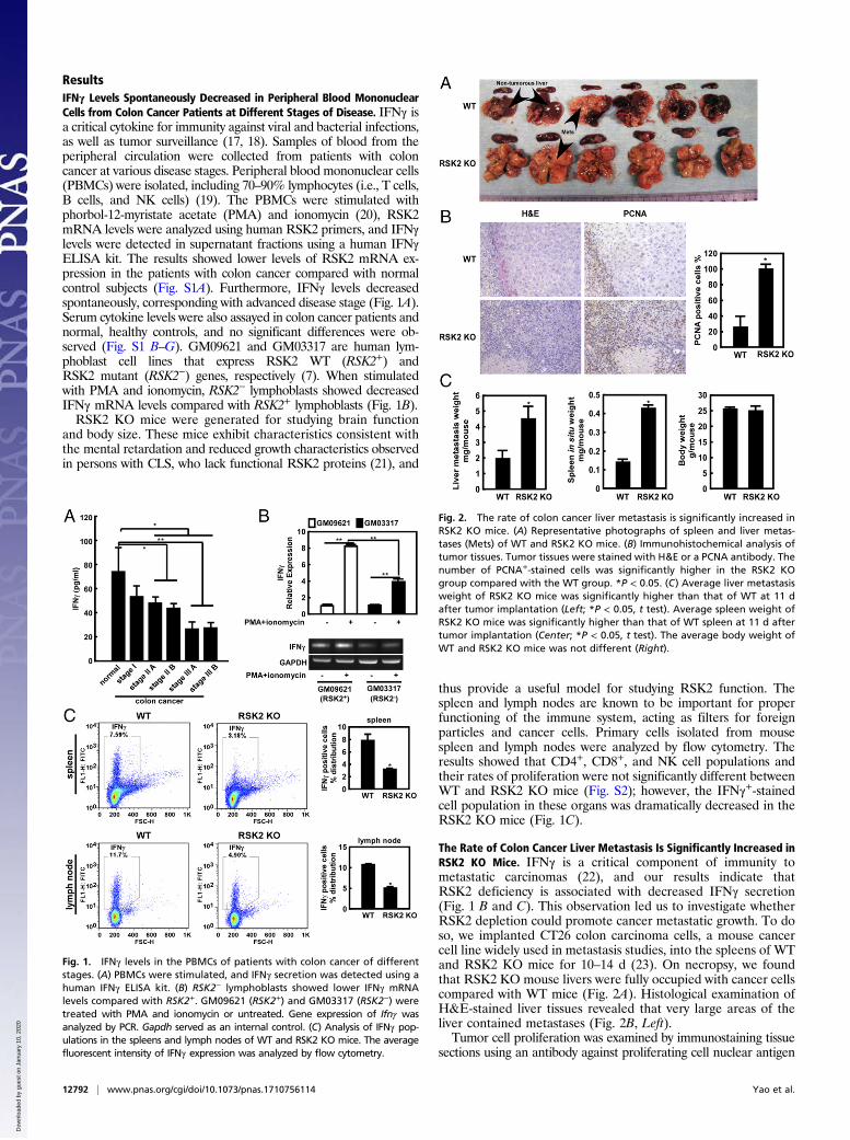
ResultsIFNγ Levels Spontaneously Decreased in Peripheral Blood MononuclearCells from Colon Cancer Patients at Different Stages of Disease. IFNγ isa critical cytokine for immunity against viral and bacterial infections,as well as tumor surveillance (17, 18). Samples of blood from theperipheral circulation were collected from patients with coloncancer at various disease stages. Peripheral blood mononuclear cells(PBMCs) were isolated, including 70–90% lymphocytes (i.e., T cells,B cells, and NK cells) (19). The PBMCs were stimulated withphorbol-12-myristate acetate (PMA) and ionomycin (20), RSK2mRNA levels were analyzed using human RSK2 primers, and IFNγlevels were detected in supernatant fractions using a human IFNγELISA kit. The results showed lower levels of RSK2 mRNA ex-pression in the patients with colon cancer compared with normalcontrol subjects (Fig. S1A). Furthermore, IFNγ levels decreasedspontaneously, corresponding with advanced disease stage (Fig. 1A).Serum cytokine levels were also assayed in colon cancer patients andnormal, healthy controls, and no significant differences were ob-served (Fig. S1 B–G). GM09621 and GM03317 are human lym-phoblast cell lines that express RSK2 WT (RSK2+) andRSK2 mutant (RSK2−) genes, respectively (7). When stimulatedwith PMA and ionomycin, RSK2− lymphoblasts showed decreasedIFNγ mRNA levels compared with RSK2+ lymphoblasts (Fig. 1B).RSK2 KO mice were generated for studying brain function
and body size. These mice exhibit characteristics consistent withthe mental retardation and reduced growth characteristics observedin persons with CLS, who lack functional RSK2 proteins (21), and
thus provide a useful model for studying RSK2 function. Thespleen and lymph nodes are known to be important for properfunctioning of the immune system, acting as filters for foreignparticles and cancer cells. Primary cells isolated from mousespleen and lymph nodes were analyzed by flow cytometry. Theresults showed that CD4+, CD8+, and NK cell populations andtheir rates of proliferation were not significantly different betweenWT and RSK2 KO mice (Fig. S2); however, the IFNγ+-stainedcell population in these organs was dramatically decreased in theRSK2 KO mice (Fig. 1C).
The Rate of Colon Cancer Liver Metastasis Is Significantly Increased inRSK2 KO Mice. IFNγ is a critical component of immunity tometastatic carcinomas (22), and our results indicate thatRSK2 deficiency is associated with decreased IFNγ secretion(Fig. 1 B and C). This observation led us to investigate whetherRSK2 depletion could promote cancer metastatic growth. To doso, we implanted CT26 colon carcinoma cells, a mouse cancercell line widely used in metastasis studies, into the spleens of WTand RSK2 KO mice for 10–14 d (23). On necropsy, we foundthat RSK2 KO mouse livers were fully occupied with cancer cellscompared with WT mice (Fig. 2A). Histological examination ofH&E-stained liver tissues revealed that very large areas of theliver contained metastases (Fig. 2B, Left).Tumor cell proliferation was examined by immunostaining tissue
sections using an antibody against proliferating cell nuclear antigen
Fig. 1. IFNγ levels in the PBMCs of patients with colon cancer of differentstages. (A) PBMCs were stimulated, and IFNγ secretion was detected using ahuman IFNγ ELISA kit. (B) RSK2− lymphoblasts showed lower IFNγ mRNAlevels compared with RSK2+. GM09621 (RSK2+) and GM03317 (RSK2−) weretreated with PMA and ionomycin or untreated. Gene expression of Ifnγ wasanalyzed by PCR. Gapdh served as an internal control. (C) Analysis of IFNγ pop-ulations in the spleens and lymph nodes of WT and RSK2 KO mice. The averagefluorescent intensity of IFNγ expression was analyzed by flow cytometry.
Fig. 2. The rate of colon cancer liver metastasis is significantly increased inRSK2 KO mice. (A) Representative photographs of spleen and liver metas-tases (Mets) of WT and RSK2 KO mice. (B) Immunohistochemical analysis oftumor tissues. Tumor tissues were stained with H&E or a PCNA antibody. Thenumber of PCNA+-stained cells was significantly higher in the RSK2 KOgroup compared with the WT group. *P < 0.05. (C) Average liver metastasisweight of RSK2 KO mice was significantly higher than that of WT at 11 dafter tumor implantation (Left; *P < 0.05, t test). Average spleen weight ofRSK2 KO mice was significantly higher than that of WT spleen at 11 d aftertumor implantation (Center; *P < 0.05, t test). The average body weight ofWT and RSK2 KO mice was not different (Right).
12792 | www.pnas.org/cgi/doi/10.1073/pnas.1710756114 Yao et al.
Dow
nloa
ded
by g
uest
on
Janu
ary
10, 2
020
(PCNA), a proliferation marker. Staining results showed that theaverage level of PCNA in the liver metastases of RSK2 KO micewas more than five times higher than that of WT mice (Fig. 2B,Right). In addition, the average tumor weight in the liver was morethan twofold greater in the RSK2 KO mice compared with the WTmice at 11 d after implantation (Fig. 2C, Left), indicating thatRSK2 deficiency promotes liver metastatic growth in mice. More-over, the average weight of the spleen was more than threefoldgreater in the RSK2 KO mice (Fig. 2C, Center), but average bodyweight did not differ between the RSK2 KO andWTmice (Fig. 2C,Right). Consistent with our ex vivo data, the liver metastasis resultsdemonstrate that the absence of RSK2 promotes both tumorgrowth and metastasis in mice.
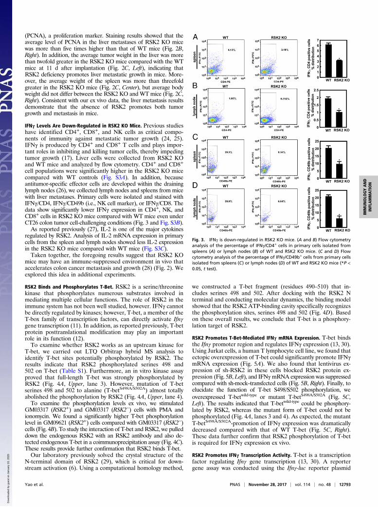
IFNγ Levels Are Down-Regulated in RSK2 KO Mice. Previous studieshave identified CD4+, CD8+, and NK cells as critical compo-nents of immunity against metastatic tumor growth (24, 25).IFNγ is produced by CD4+ and CD8+ T cells and plays impor-tant roles in inhibiting and killing tumor cells, thereby impedingtumor growth (17). Liver cells were collected from RSK2 KOand WT mice and analyzed by flow cytometry. CD4+ and CD8+
cell populations were significantly higher in the RSK2 KO micecompared with WT controls (Fig. S3A). In addition, becauseantitumor-specific effector cells are developed within the draininglymph nodes (26), we collected lymph nodes and spleens from micewith liver metastases. Primary cells were isolated and stained withIFNγ/CD4, IFNγ/CD49b (i.e., NK cell marker), or IFNγ/CD8. Thedata show significantly lower IFNγ expression in CD4+, NK, andCD8+ cells in RSK2 KO mice compared with WT mice even underCT26 colon tumor cell-challenging conditions (Fig. 3 and Fig. S3B).As reported previously (27), IL-2 is one of the major cytokines
regulated by RSK2. Analysis of IL-2 mRNA expression in primarycells from the spleen and lymph nodes showed less IL-2 expressionin the RSK2 KO mice compared with WT mice (Fig. S3C).Taken together, the foregoing results suggest that RSK2 KO
mice may have an immune-suppressed environment in vivo thataccelerates colon cancer metastasis and growth (28) (Fig. 2). Weexplored this idea in additional experiments.
RSK2 Binds and Phosphorylates T-Bet. RSK2 is a serine/threoninekinase that phosphorylates numerous substrates involved inmediating multiple cellular functions. The role of RSK2 in theimmune system has not been well studied, however. IFNγ cannotbe directly regulated by kinases; however, T-bet, a member of theT-box family of transcription factors, can directly activate Ifnγgene transcription (11). In addition, as reported previously, T-betprotein posttranslational modification may play an importantrole in its function (12).To examine whether RSK2 works as an upstream kinase for
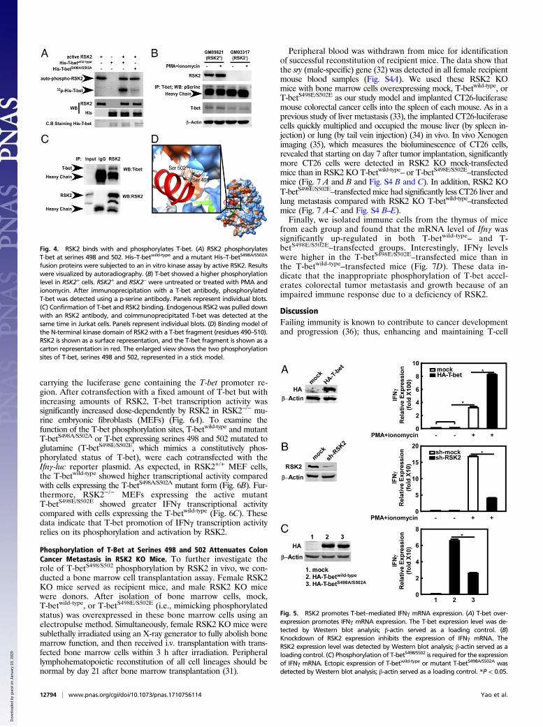
T-bet, we carried out LTQ Orbitrap hybrid MS analysis toidentify T-bet sites potentially phosphorylated by RSK2. Theresults indicate that RSK2 phosphorylated serines 498 and502 on T-bet (Table S1). Furthermore, an in vitro kinase assayproved that full-length T-bet was strongly phosphorylated byRSK2 (Fig. 4A, Upper, lane 3). However, mutation of T-betserines 498 and 502 to alanine (T-betS498A/S502A) almost totallyabolished the phosphorylation by RSK2 (Fig. 4A, Upper, lane 4).To examine the phosphorylation levels ex vivo, we stimulated
GM03317 (RSK2+) and GM03317 (RSK2−) cells with PMA andionomycin. We found a significantly higher T-bet phosphorylationlevel in GM09621 (RSK2+) cells compared with GM03317 (RSK2−)cells (Fig. 4B). To study the interaction of T-bet and RSK2, we pulleddown the endogenous RSK2 with an RSK2 antibody and also de-tected endogenous T-bet in a coimmunoprecipitation assay (Fig. 4C).These results provide further confirmation that RSK2 binds T-bet.Our laboratory previously solved the crystal structure of the
N-terminal domain of RSK2 (29), which is critical for down-stream activation (6). Using a computational homology method,
we constructed a T-bet fragment (residues 490–510) that in-cludes serines 498 and 502. After docking with the RSK2 Nterminal and conducting molecular dynamics, the binding modelshowed that the RSK2 ATP-binding cavity specifically recognizesthe phosphorylation sites, serines 498 and 502 (Fig. 4D). Basedon these overall results, we conclude that T-bet is a phosphory-lation target of RSK2.
RSK2 Promotes T-Bet-Mediated IFNγ mRNA Expression. T-bet bindsthe Ifnγ promoter region and regulates IFNγ expression (13, 30).Using Jurkat cells, a human T lymphocyte cell line, we found thatectopic overexpression of T-bet could significantly promote IFNγmRNA expression (Fig. 5A). We also found that lentivirus ex-pression of sh-RSK2 in these cells blocked RSK2 protein ex-pression (Fig. 5B, Left), and IFNγmRNA expression was suppressedcompared with sh-mock–transfected cells (Fig. 5B, Right). Finally, toelucidate the function of T-bet S498/S502 phosphorylation, weoverexpressed T-betwild-type or mutant T-betS498A/S502A (Fig. 5C,Left). The results indicated that T-betwild-type could be phosphory-lated by RSK2, whereas the mutant form of T-bet could not bephosphorylated (Fig. 4A, lanes 3 and 4). As expected, the mutantT-betS498A/S502A-promotion of IFNγ expression was dramaticallydecreased compared with that of WT T-bet (Fig. 5C, Right).These data further confirm that RSK2 phosphorylation of T-betis required for IFNγ expression ex vivo.
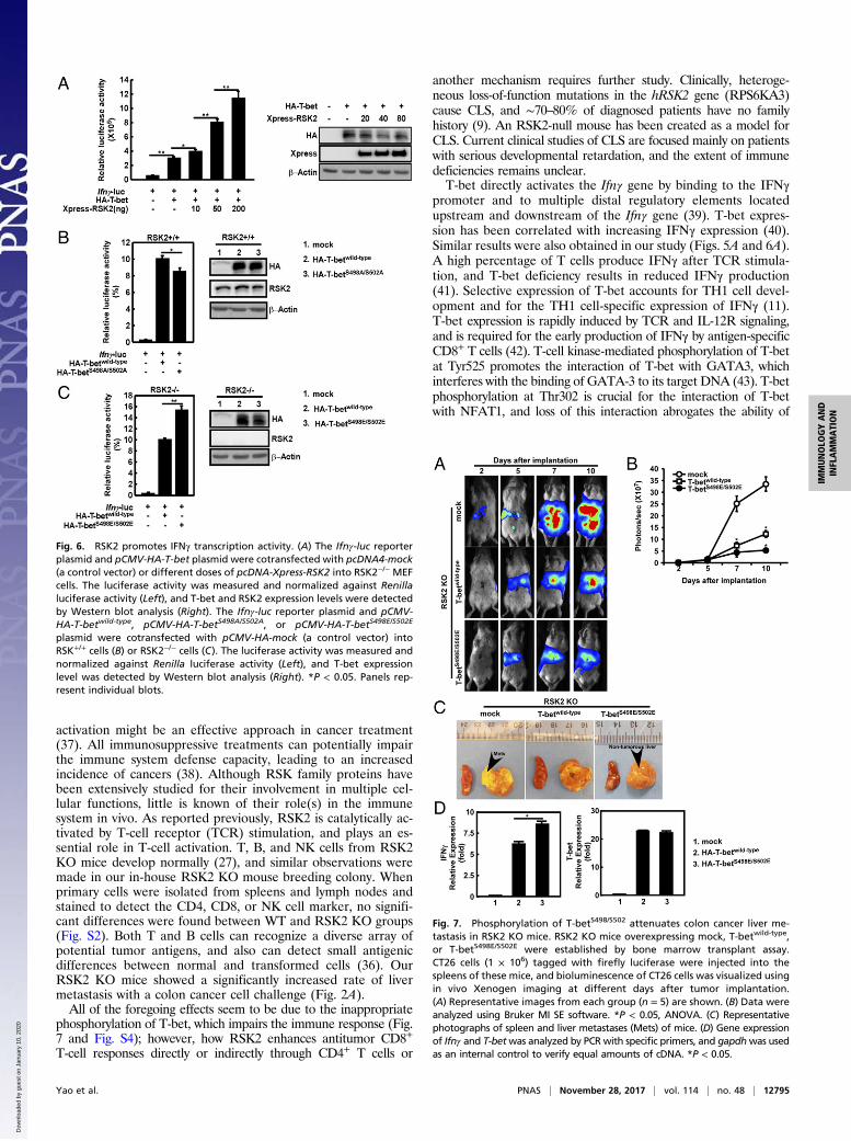
RSK2 Promotes IFNγ Transcription Activity. T-bet is a transcriptionfactor regulating Ifnγ gene transcription (13, 30). A reportergene assay was conducted using the Ifnγ-luc reporter plasmid
Fig. 3. IFNγ is down-regulated in RSK2 KO mice. (A and B) Flow cytometryanalysis of the percentage of IFNγ/CD4+ cells in primary cells isolated fromspleens (A) or lymph nodes (B) of WT and RSK2 KO mice. (C and D) Flowcytometry analysis of the percentage of IFNγ/CD49b+ cells from primary cellsisolated from spleens (C) or lymph nodes (D) of WT and RSK2 KO mice (*P <0.05, t test).
Yao et al. PNAS | November 28, 2017 | vol. 114 | no. 48 | 12793
IMMUNOLO
GYAND
INFLAMMATION
Dow
nloa
ded
by g
uest
on
Janu
ary
10, 2
020
carrying the luciferase gene containing the T-bet promoter re-gion. After cotransfection with a fixed amount of T-bet but withincreasing amounts of RSK2, T-bet transcription activity wassignificantly increased dose-dependently by RSK2 in RSK2−/− mu-rine embryonic fibroblasts (MEFs) (Fig. 6A). To examine thefunction of the T-bet phosphorylation sites, T-betwild-type and mutantT-betS498A/S502A or T-bet expressing serines 498 and 502 mutated toglutamine (T-betS498E/S502E, which mimics a constitutively phos-phorylated status of T-bet), were each cotransfected with theIfnγ-luc reporter plasmid. As expected, in RSK2+/+ MEF cells,the T-betwild-type showed higher transcriptional activity comparedwith cells expressing the T-betS498A/S502A mutant form (Fig. 6B). Fur-thermore, RSK2−/− MEFs expressing the active mutantT-betS498E/S502E showed greater IFNγ transcriptional activitycompared with cells expressing the T-betwild-type (Fig. 6C). Thesedata indicate that T-bet promotion of IFNγ transcription activityrelies on its phosphorylation and activation by RSK2.
Phosphorylation of T-Bet at Serines 498 and 502 Attenuates ColonCancer Metastasis in RSK2 KO Mice. To further investigate therole of T-betS498/S502 phosphorylation by RSK2 in vivo, we con-ducted a bone marrow cell transplantation assay. Female RSK2KO mice served as recipient mice, and male RSK2 KO micewere donors. After isolation of bone marrow cells, mock,T-betwild-type, or T-betS498E/S502E (i.e., mimicking phosphorylatedstatus) was overexpressed in these bone marrow cells using anelectropulse method. Simultaneously, female RSK2 KO mice weresublethally irradiated using an X-ray generator to fully abolish bonemarrow function, and then received i.v. transplantation with trans-fected bone marrow cells within 3 h after irradiation. Peripherallymphohematopoietic reconstitution of all cell lineages should benormal by day 21 after bone marrow transplantation (31).
Peripheral blood was withdrawn from mice for identificationof successful reconstitution of recipient mice. The data show thatthe sry (male-specific) gene (32) was detected in all female recipientmouse blood samples (Fig. S4A). We used these RSK2 KOmice with bone marrow cells overexpressing mock, T-betwild-type, orT-betS498E/S502E as our study model and implanted CT26-luciferasemouse colorectal cancer cells into the spleen of each mouse. As in aprevious study of liver metastasis (33), the implanted CT26-luciferasecells quickly multiplied and occupied the mouse liver (by spleen in-jection) or lung (by tail vein injection) (34) in vivo. In vivo Xenogenimaging (35), which measures the bioluminescence of CT26 cells,revealed that starting on day 7 after tumor implantation, significantlymore CT26 cells were detected in RSK2 KO mock-transfectedmice than in RSK2 KO T-betwild-type– or T-betS498E/S502E–transfectedmice (Fig. 7 A and B and Fig. S4 B and C). In addition, RSK2 KOT-betS498E/S502E–transfected mice had significantly less CT26 liver andlung metastasis compared with RSK2 KO T-betwild-type–transfectedmice (Fig. 7 A–C and Fig. S4 B–E).Finally, we isolated immune cells from the thymus of mice
from each group and found that the mRNA level of Ifnγ wassignificantly up-regulated in both T-betwild-type– and T-betS498E/S502E–transfected groups. Interestingly, IFNγ levelswere higher in the T-betS498E/S502E–transfected mice than inthe T-betwild-type–transfected mice (Fig. 7D). These data in-dicate that the inappropriate phosphorylation of T-bet accel-erates colorectal tumor metastasis and growth because of animpaired immune response due to a deficiency of RSK2.
DiscussionFailing immunity is known to contribute to cancer developmentand progression (36); thus, enhancing and maintaining T-cell
Fig. 4. RSK2 binds with and phosphorylates T-bet. (A) RSK2 phosphorylatesT-bet at serines 498 and 502. His–T-betwild-type and a mutant His–T-betS498A/S502A
fusion proteins were subjected to an in vitro kinase assay by active RSK2. Resultswere visualized by autoradiography. (B) T-bet showed a higher phosphorylationlevel in RSK2+ cells. RSK2+ and RSK2− were untreated or treated with PMA andionomycin. After immunoprecipitation with a T-bet antibody, phosphorylatedT-bet was detected using a p-serine antibody. Panels represent individual blots.(C) Confirmation of T-bet and RSK2 binding. Endogenous RSK2 was pulled downwith an RSK2 antibody, and coimmunoprecipitated T-bet was detected at thesame time in Jurkat cells. Panels represent individual blots. (D) Binding model ofthe N-terminal kinase domain of RSK2 with a T-bet fragment (residues 490–510).RSK2 is shown as a surface representation, and the T-bet fragment is shown as acarton representation in red. The enlarged view shows the two phosphorylationsites of T-bet, serines 498 and 502, represented in a stick model.
Fig. 5. RSK2 promotes T-bet–mediated IFNγ mRNA expression. (A) T-bet over-expression promotes IFNγ mRNA expression. The T-bet expression level was de-tected by Western blot analysis; β-actin served as a loading control. (B)Knockdown of RSK2 expression inhibits the expression of IFNγ mRNA. TheRSK2 expression level was detected by Western blot analysis; β-actin served as aloading control. (C) Phosphorylation of T-betS498/S502 is required for the expressionof IFNγ mRNA. Ectopic expression of T-betwild-type or mutant T-betS498A/S502A wasdetected by Western blot analysis; β-actin served as a loading control. *P < 0.05.
12794 | www.pnas.org/cgi/doi/10.1073/pnas.1710756114 Yao et al.
Dow
nloa
ded
by g
uest
on
Janu
ary
10, 2
020
activation might be an effective approach in cancer treatment(37). All immunosuppressive treatments can potentially impairthe immune system defense capacity, leading to an increasedincidence of cancers (38). Although RSK family proteins havebeen extensively studied for their involvement in multiple cel-lular functions, little is known of their role(s) in the immunesystem in vivo. As reported previously, RSK2 is catalytically ac-tivated by T-cell receptor (TCR) stimulation, and plays an es-sential role in T-cell activation. T, B, and NK cells from RSK2KO mice develop normally (27), and similar observations weremade in our in-house RSK2 KO mouse breeding colony. Whenprimary cells were isolated from spleens and lymph nodes andstained to detect the CD4, CD8, or NK cell marker, no signifi-cant differences were found between WT and RSK2 KO groups(Fig. S2). Both T and B cells can recognize a diverse array ofpotential tumor antigens, and also can detect small antigenicdifferences between normal and transformed cells (36). OurRSK2 KO mice showed a significantly increased rate of livermetastasis with a colon cancer cell challenge (Fig. 2A).All of the foregoing effects seem to be due to the inappropriate
phosphorylation of T-bet, which impairs the immune response (Fig.7 and Fig. S4); however, how RSK2 enhances antitumor CD8+
T-cell responses directly or indirectly through CD4+ T cells or
another mechanism requires further study. Clinically, heteroge-neous loss-of-function mutations in the hRSK2 gene (RPS6KA3)cause CLS, and ∼70–80% of diagnosed patients have no familyhistory (9). An RSK2-null mouse has been created as a model forCLS. Current clinical studies of CLS are focused mainly on patientswith serious developmental retardation, and the extent of immunedeficiencies remains unclear.T-bet directly activates the Ifnγ gene by binding to the IFNγ
promoter and to multiple distal regulatory elements locatedupstream and downstream of the Ifnγ gene (39). T-bet expres-sion has been correlated with increasing IFNγ expression (40).Similar results were also obtained in our study (Figs. 5A and 6A).A high percentage of T cells produce IFNγ after TCR stimula-tion, and T-bet deficiency results in reduced IFNγ production(41). Selective expression of T-bet accounts for TH1 cell devel-opment and for the TH1 cell-specific expression of IFNγ (11).T-bet expression is rapidly induced by TCR and IL-12R signaling,and is required for the early production of IFNγ by antigen-specificCD8+ T cells (42). T-cell kinase-mediated phosphorylation of T-betat Tyr525 promotes the interaction of T-bet with GATA3, whichinterferes with the binding of GATA-3 to its target DNA (43). T-betphosphorylation at Thr302 is crucial for the interaction of T-betwith NFAT1, and loss of this interaction abrogates the ability of
Fig. 6. RSK2 promotes IFNγ transcription activity. (A) The Ifnγ-luc reporterplasmid and pCMV-HA-T-bet plasmid were cotransfected with pcDNA4-mock(a control vector) or different doses of pcDNA-Xpress-RSK2 into RSK2−/− MEFcells. The luciferase activity was measured and normalized against Renillaluciferase activity (Left), and T-bet and RSK2 expression levels were detectedby Western blot analysis (Right). The Ifnγ-luc reporter plasmid and pCMV-HA-T-betwild-type, pCMV-HA-T-betS498A/S502A, or pCMV-HA-T-betS498E/S502E
plasmid were cotransfected with pCMV-HA-mock (a control vector) intoRSK+/+ cells (B) or RSK2−/− cells (C). The luciferase activity was measured andnormalized against Renilla luciferase activity (Left), and T-bet expressionlevel was detected by Western blot analysis (Right). *P < 0.05. Panels rep-resent individual blots.
Fig. 7. Phosphorylation of T-betS498/S502 attenuates colon cancer liver me-tastasis in RSK2 KO mice. RSK2 KO mice overexpressing mock, T-betwild-type,or T-betS498E/S502E were established by bone marrow transplant assay.CT26 cells (1 × 10⁶) tagged with firefly luciferase were injected into thespleens of these mice, and bioluminescence of CT26 cells was visualized usingin vivo Xenogen imaging at different days after tumor implantation.(A) Representative images from each group (n = 5) are shown. (B) Data wereanalyzed using Bruker MI SE software. *P < 0.05, ANOVA. (C) Representativephotographs of spleen and liver metastases (Mets) of mice. (D) Gene expressionof Ifnγ and T-betwas analyzed by PCR with specific primers, and gapdhwas usedas an internal control to verify equal amounts of cDNA. *P < 0.05.
Yao et al. PNAS | November 28, 2017 | vol. 114 | no. 48 | 12795
IMMUNOLO
GYAND
INFLAMMATION
Dow
nloa
ded
by g
uest
on
Janu
ary
10, 2
020
T-bet to suppress NFAT1-dependent cytokine expression (44).T-bet phosphorylation at Ser508 by casein kinase I and glycogensynthase kinase 3 (GSK3) promotes the interaction of T-bet withRelA, which impairs RelA binding to the Il2 promoter and thesubsequent transcriptional activation of the Il2 gene (45). Mutationof lysine 313 (K313) decreases ubiquitination-mediated T-bet deg-radation and completely abrogates T-bet functions involving DNAbinding and transcriptional activation of IFNγ (44). Based on recentstudies, we hypothesized that T-bet activity might be regulated byposttranslational modification, specifically phosphorylation. Wefound that T-bet is phosphorylated by RSK2 at serines 498 and 502,and plays an important role in regulating IFNγ mRNA (Figs. 5Cand 7D) and transcription level (Fig. 6 B and C).Overall, our results show that RSK2 deficiency can result in
dramatically decreased IFNγ secretion through inappropriatephosphorylation of T-bet. This can lead to immune suppression,which accelerates colon cancer metastasis and growth. The clinical
relevance of these findings requires additional study. In addition,analysis of the cancer incidence and immune function in CLS pa-tients could provide valuable information.
Materials and MethodsThe in vivo Xenogen imaging of mice was performed using the XtremeImaging system (CareStream Health), and bioluminescence was quantifiedusing Bruker MI software. The materials and methods used in this study aredescribed in detail in SI Materials and Methods.
ACKNOWLEDGMENTS. We thank Dr. Rebecca Morris and Kelly Johnson forassisting with the bone marrow transplant assay, Drs. Dan Li, Xuejiao Liu,Haitao Li, Young Jin Jeon, and Do Young Lim and Todd Schuster forsupporting experiments; and Dr. Tia Rai and Nicki Brickman for assistancewith manuscript submission. This work was supported by The HormelFoundation; National Institutes of Health (Grants CA166001, CA172457,CA196639, and CA187027, to Zigang Dong), and the National ScienceFoundation of Henan Province, China (Grant 162300410337).
1. Mehlen P, Puisieux A (2006) Metastasis: A question of life or death. Nat Rev Cancer 6:449–458.
2. Misiakos EP, Karidis NP, Kouraklis G (2011) Current treatment for colorectal livermetastases. World J Gastroenterol 17:4067–4075.
3. Hess KR, et al. (2006) Metastatic patterns in adenocarcinoma. Cancer 106:1624–1633.4. Kudo-Saito C, Shirako H, Takeuchi T, Kawakami Y (2009) Cancer metastasis is
accelerated through immunosuppression during Snail-induced EMT of cancer cells.Cancer Cell 15:195–206.
5. Chen H, et al. (2015) Computational and biochemical discovery of RSK2 as a noveltarget for epigallocatechin gallate (EGCG). PLoS One 10:e0130049.
6. Cho YY, et al. (2009) A regulatory mechanism for RSK2 NH(2)-terminal kinase activity.Cancer Res 69:4398–4406.
7. Zhang Y, et al. (2003) Ataxia telangiectasia mutated proteins, MAPKs, and RSK2 areinvolved in the phosphorylation of STAT3. J Biol Chem 278:12650–12659.
8. Jacquot S, Zeniou M, Touraine R, Hanauer A (2002) X-linked Coffin-Lowry syndrome(CLS, MIM 303600, RPS6KA3 gene, protein product known under various names:pp90(rsk2), RSK2, ISPK, MAPKAP1). Eur J Hum Genet 10:2–5.
9. Pereira PM, Schneider A, Pannetier S, Heron D, Hanauer A (2010) Coffin-Lowry syn-drome. Eur J Hum Genet 18:627–633.
10. Trivier E, et al. (1996) Mutations in the kinase Rsk-2 associated with Coffin-Lowrysyndrome. Nature 384:567–570.
11. Szabo SJ, et al. (2000) A novel transcription factor, T-bet, directs Th1 lineage com-mitment. Cell 100:655–669.
12. Lazarevic V, Glimcher LH, Lord GM (2013) T-bet: A bridge between innate andadaptive immunity. Nat Rev Immunol 13:777–789.
13. Lugo-Villarino G, Maldonado-Lopez R, Possemato R, Penaranda C, Glimcher LH (2003)T-bet is required for optimal production of IFN-gamma and antigen-specific T cellactivation by dendritic cells. Proc Natl Acad Sci USA 100:7749–7754.
14. Schoenborn JR, Wilson CB (2007) Regulation of interferon-gamma during innate andadaptive immune responses. Adv Immunol 96:41–101.
15. Mullen AC, et al. (2001) Role of T-bet in commitment of TH1 cells before IL-12-dependent selection. Science 292:1907–1910.
16. Zamarron BF, Chen W (2011) Dual roles of immune cells and their factors in cancerdevelopment and progression. Int J Biol Sci 7:651–658.
17. Ikeda H, Old LJ, Schreiber RD (2002) The roles of IFN gamma in protection against tumordevelopment and cancer immunoediting. Cytokine Growth Factor Rev 13:95–109.
18. Perry AK, Chen G, Zheng D, Tang H, Cheng G (2005) The host type I interferon re-sponse to viral and bacterial infections. Cell Res 15:407–422.
19. Du X, et al. (2006) Genomic profiles for human peripheral blood T cells, B cells, naturalkiller cells, monocytes, and polymorphonuclear cells: Comparisons to ischemic stroke,migraine, and Tourette syndrome. Genomics 87:693–703.
20. Ai W, Li H, Song N, Li L, Chen H (2013) Optimal method to stimulate cytokine pro-duction and its use in immunotoxicity assessment. Int J Environ Res Public Health 10:3834–3842.
21. Dufresne SD, et al. (2001) Altered extracellular signal-regulated kinase signaling andglycogen metabolism in skeletal muscle from p90 ribosomal S6 kinase 2 knockoutmice. Mol Cell Biol 21:81–87.
22. Pulaski BA, Smyth MJ, Ostrand-Rosenberg S (2002) Interferon-gamma-dependentphagocytic cells are a critical component of innate immunity against metastaticmammary carcinoma. Cancer Res 62:4406–4412.
23. Gorden DL, et al. (2007) Resident stromal cell-derived MMP-9 promotes the growth ofcolorectal metastases in the liver microenvironment. Int J Cancer 121:495–500.
24. Cheng M, Chen Y, Xiao W, Sun R, Tian Z (2013) NK cell-based immunotherapy formalignant diseases. Cell Mol Immunol 10:230–252.
25. Hadrup S, Donia M, Thor Straten P (2013) Effector CD4 and CD8 T cells and their rolein the tumor microenvironment. Cancer Microenviron 6:123–133.
26. Yu J, et al. (2009) Antitumor activity of T cells generated from lymph nodes drainingthe SEA-expressing murine B16 melanoma and secondarily activated with dendriticcells. Int J Biol Sci 5:135–146.
27. Lin JX, Spolski R, Leonard WJ (2008) Critical role for Rsk2 in T-lymphocyte activation.Blood 111:525–533.
28. Kakuta S, Tagawa Y, Shibata S, Nanno M, Iwakura Y (2002) Inhibition of B16melanoma experimental metastasis by interferon-gamma through direct inhibitionof cell proliferation and activation of antitumour host mechanisms. Immunology105:92–100.
29. Malakhova M, et al. (2009) Structural diversity of the active N-terminal kinase domainof p90 ribosomal S6 kinase 2. PLoS One 4:e8044.
30. Oh S, Hwang ES (2014) The role of protein modifications of T-bet in cytokine pro-duction and differentiation of T helper cells. J Immunol Res 2014:589672.
31. Duran-Struuck R, Dysko RC (2009) Principles of bone marrow transplantation (BMT):Providing optimal veterinary and husbandry care to irradiated mice in BMT studies.J Am Assoc Lab Anim Sci 48:11–22.
32. Clapcote SJ, Roder JC (2005) Simplex PCR assay for sex determination in mice.Biotechniques 38:702–706.
33. Ohana G, et al. (2003) Inhibition of primary colon carcinoma growth and liver me-tastasis by the A3 adenosine receptor agonist CF101. Br J Cancer 89:1552–1558.
34. Weng YL, Liao HF, Li AF, Chang JC, Chiou RY (2010) Oral administration of resveratrolin suppression of pulmonary metastasis of BALB/c mice challenged withCT26 colorectal adenocarcinoma cells. Mol Nutr Food Res 54:259–267.
35. Liu C, et al. (2013) IQGAP1 suppresses TβRII-mediated myofibroblastic activation andmetastatic growth in liver. J Clin Invest 123:1138–1156.
36. Ghirelli C, Hagemann T (2013) Targeting immunosuppression for cancer therapy.J Clin Invest 123:2355–2357.
37. Disis ML, Bernhard H, Jaffee EM (2009) Use of tumour-responsive T cells as cancertreatment. Lancet 373:673–683.
38. Gerlini G, Romagnoli P, Pimpinelli N (2005) Skin cancer and immunosuppression. CritRev Oncol Hematol 56:127–136.
39. Hatton RD, et al. (2006) A distal conserved sequence element controls Ifng gene ex-pression by T cells and NK cells. Immunity 25:717–729.
40. Klose CS, et al. (2013) A T-bet gradient controls the fate and function of CCR6-RORγt+innate lymphoid cells. Nature 494:261–265.
41. Chen L, et al. (2007) Epigenetic and transcriptional programs lead to default IFN-gamma production by gammadelta T cells. J Immunol 178:2730–2736.
42. Takemoto N, Intlekofer AM, Northrup JT, Wherry EJ, Reiner SL (2006) Cutting edge:IL-12 inversely regulates T-bet and eomesodermin expression during pathogen-induced CD8+ T cell differentiation. J Immunol 177:7515–7519.
43. Hwang ES, Szabo SJ, Schwartzberg PL, Glimcher LH (2005) T helper cell fate specifiedby kinase-mediated interaction of T-bet with GATA-3. Science 307:430–433.
44. Jang EJ, Park HR, Hong JH, Hwang ES (2013) Lysine 313 of T-box is crucial for mod-ulation of protein stability, DNA binding, and threonine phosphorylation of T-bet.J Immunol 190:5764–5770.
45. Hwang ES, Hong JH, Glimcher LH (2005) IL-2 production in developing Th1 cells isregulated by heterodimerization of RelA and T-bet and requires T-bet serine residue508. J Exp Med 202:1289–1300.
12796 | www.pnas.org/cgi/doi/10.1073/pnas.1710756114 Yao et al.
Dow
nloa
ded
by g
uest
on
Janu
ary
10, 2
020