review trends in cell biology vol.12 no.4 april 2002...
TRANSCRIPT

TRENDS in Cell Biology Vol.12 No.4 April 2002
http://tcb.trends.com 0962-8924/02/$ – see front matter © 2002 Elsevier Science Ltd. All rights reserved. PII: S0962-8924(02)02263-8
185Review
Catherin Niemann
Fiona M. Watt*
Keratinocyte Laboratory,Cancer Research UK, 44 Lincoln’s Inn Fields,London, UK WC2A 3PX.*e-mail: [email protected]
The epidermis forms the protective outer covering ofmammalian skin. It comprises a multilayeredepithelium, the interfollicular epidermis (IFE),associated hair follicles and sebaceous and sweatglands (Fig. 1). The epidermis is maintainedthroughout adult life by stem cells that not onlyself-renew but also generate daughter cells thatundergo terminal differentiation (a process ofdifferentiation resulting in a highly specialized cell that cannot divide). There is a single terminaldifferentiation pathway within the IFE, albeit one that varies slightly between sites in the body. This culminates in the production of the outermost,cornified epidermal layers that provide a protectivecovering for the skin. The terminally differentiatedcells of the sebaceous gland are lipid-filled sebocytesthat eventually burst, releasing their contents onto the surface of the skin. The hair follicle is more complex, comprising eight different cell lineages (Fig. 2).
The availability of promoters to target transgenesto specific compartments of mouse epidermis or to generate epidermal-specific knockouts has resultedin a wealth of data about the factors that regulateepidermal growth, differentiation and homeostasis[1,2]. Our purpose here is to highlight one aspect ofepidermal differentiation, namely the regulation oflineage commitment (the decision to undergo aspecific programme of terminal differentiation) inpostnatal life. Put simply, an understanding oflineage commitment provides the prospect ofengineering the epidermis to produce more hairfollicles for balding men, to banish excess sebumproduction for teenagers and to ensure smooth, hair-free legs for women. More importantly, it is becoming clear that the diversity of epidermaltumour types reflects the activity of molecules thatregulate lineage commitment and this knowledgemight be exploited to design tumour-specific anti-cancer drugs.
Epidermal stem cells, transit amplifying cells and
committed progenitors
It is well known that epidermal stem cells aremultipotent [3,4]. Stem cells in the hair follicle cangive rise not only to the hair lineages but also tosebocytes and IFE [5,6]. Conversely, with appropriatemesenchymal stimuli, cells of the IFE can be inducedto differentiate into hair and sebocyte lineages [7,8].Less is known about the origin of the cells of the sweat glands, although they too are probably formedby the differentiated progeny of multipotentepidermal stem cells and, under some conditions,cells from sweat glands can reconstitute a stratifiedsquamous epithelium [9].
It has been proposed that the stem cells that lie ina specialized region of the hair follicle outer rootsheath (ORS), known as the bulge, are ultimatelyresponsible for replenishing the differentiated cells of the IFE and sebaceous gland in addition togenerating all the hair lineages [5,6]. The bulge liesbeneath the sebaceous glands at the point of insertionof the arrector pili muscle (Fig. 1). However, recentlong-term lineage marking has demonstrated that, inundamaged epidermis, there are distinct stem-cellpopulations in the IFE, sebaceous gland and hair [10].Thus, although bulge stem cells have the ability togenerate all the different epidermal lineages, it islikely that, under steady-state conditions, stem cells
The epidermis is populated by stem cells that produce daughters that
differentiate to form the interfollicular epidermis, hair follicles and sebaceous
glands. Diffusible factors, cell–cell contact and extracellular matrix proteins are
all important components of the microenvironment of individual stem cells and
profoundly affect the differentiation pathways selected by their progeny. Here,
we summarize what is known about stem-cell populations and lineage
relationships within the epidermis. We also present evidence that postnatal
epidermis can be reprogrammed, altering the number and location of cells that
differentiate along specific epidermal lineages.
Designer skin: lineage commitment in
postnatal epidermis
Catherin Niemann and Fiona M. Watt
IFE
SGB
HF
Fig. 1. Haematoxylin- and eosin-stained section through the dorsal skinof an adult mouse during the anagen (growth) phase of the hair folliclecycle. Skin at this body site lacks sweat glands. Abbreviations: B,approximate location of the bulge; HF, hair follicle; IFE, interfollicularepidermis; SG, sebaceous gland. Bar, 200 µm.

feed only one or a subset of lineages, the choice oflineage being determined by the specific location, ormicroenvironment, of the cells.
Our view of the stem cell compartment isillustrated in Fig. 3. Separate stem cells for IFE, hair lineages and sebaceous gland are shown as beinginterconvertible, to emphasize that each populationhas multipotent differentiation capacity. Constituents of the microenvironment that coulddictate the differentiation pathways selected bystem-cell progeny include the extracellular matrix,diffusible factors (growth factors, cytokines andmorphogens), direct contact with neighbouring cells,the availability of oxygen and nutrients, and evenmechanical stimuli such as stretch forces [11,12]. It has long been appreciated that communicationwith the underlying dermis plays an important role in regulating epidermal differentiation [13], withthe specialized mesenchymal cells of the dermalpapilla at the base of the hair follicle (Fig. 2) providingthe best-characterized microenvironment (oftenreferred to as a niche) both in vivo [1,4] and in vitro[7,14]. Growth and differentiation of postnatal hairfollicles are controlled by reciprocal interactionsbetween the dermal papilla and the cells of the hair matrix (Fig. 2).
How are epidermal stem cells identified? Inundamaged adult epidermis, the stem cells divide
infrequently, although they have a high capacity for self-renewal [15]. In the mouse, they can thereforebe identified by injecting neonatal animals repeatedlywith 3H-thymidine or bromodeoxyuridine (BrdU) tolabel all the epidermal cells at a time when the tissueis maximally proliferative and then finding the cellsthat retain label in adulthood. In human IFE, thestem cells are found among those basal cells that areneither actively cycling nor committed to differentiate[16,17]. Although stem cells divide infrequently inadult epidermis, they can found actively proliferatingclones in culture and, in rodents, there is a goodcorrelation between the populations of keratinocytesidentified as stem cells by label retention and byclonal analysis [6,18]. In addition, several cell surface markers can be used to enrich for clonogenicor label-retaining keratinocytes directly from theepidermis [18].
If stem cells are cells that divide infrequently, what are the actively dividing cells withinmammalian epidermis? The most widely acceptedmodel is that, in the IFE, they are the non-stemdaughters of stem cells and that they divide a fewtimes before the onset of terminal differentiation.Such dividing cells are known as transit [19], ortransient [20], amplifying cells (Fig. 3). As the nameimplies, their role is to increase the number ofterminally differentiated cells produced by eachstem-cell division. Thus, even though stem cells havea high self-renewal capacity, they actually divideinfrequently in normal undamaged epidermis.
The significance of the transit amplifyingpopulation in terms of lineage commitment is lessclear. By analogy with haematopoiesis [21], eachtransit amplifying cell might be committed todifferentiate along only one or a few lineages; that is,transit amplifying cells might be ‘committedprogenitors’ (Fig. 3, brown). Alternatively, transitamplifying cells might be multipotent and differ fromstem cells only in their self-renewal ability. There isno fixed relationship between the timing ofirreversible exit from the cell cycle and the onset ofepidermal terminal differentiation [22]. Therefore,within a given lineage, there might be more than one class of transit amplifying cell, differing from oneanother in their expression of early differentiationmarkers, as proposed recently for human oesophagealepithelium [23]. Although it is convenient to think of stem and transit amplifying cells as discretepopulations, there might instead be gradients of cell behaviour [24], ranging from a cell that has maximum self-renewal capacity and zerodifferentiation probability (Fig. 3, red) to one that hascompleted terminal differentiation and can neverdivide again (Fig. 3, green); transit amplifying cellswould then lie somewhere in the middle of eachgradient [25].
The epidermis has not been ignored in the recentflurry of interest in adult stem cell plasticity. It hasnow been shown that haematopoietic stem cells can
TRENDS in Cell Biology Vol.12 No.4 April 2002
http://tcb.trends.com
186 Review
TRENDS in Cell Biology
IRS
Precortex
ORS
MatrixDermalpapilla
Hair shaft
Hair shaft IRS
(a) (b) Hai
r cu
ticle
Cor
tex
Med
ulla
OR
SC
ompa
nion
laye
r
IRS
cut
icle
Hux
ley
laye
rH
enle
laye
r
Fig. 2. Hair follicle lineages. (a) Haematoxylin- and eosin-stained section through the base of agrowing follicle. (b) The different hair follicle lineages; the dermal papilla is shown in dark grey andthe hair matrix in light grey. Abbreviations: IRS, inner root sheath; ORS, outer root sheath. Bar, 50 µm.
give rise to cells within the hair follicle and IFE [26].Conversely, stem cells from neonatal mouseepidermis injected into mouse blastocysts contributeto ectoderm-, mesenchyme- and neural-crest-derivedtissues in adulthood, including neurons,haematopoietic cells and endothelium [27]. The transit amplifying compartment in adult
rabbit and mouse corneal epithelium can bereprogrammed by exposure to embryonic mousedermis to generate hair, sebocytes, sweat glands and IFE, showing not only differentiation plasticitybut also, potentially, differentiation reversal tomultipotent stem cells [28]. By contrast, epidermaltransit amplifying cells cannot contribute to any tissues after mouse blastocyst injection [27].Although these observations reveal that epidermalstem and transit amplifying cells can bereprogrammed under some circumstances, they do not imply that reprogramming occurs undernormal conditions. Environmental cues are ofparamount importance in regulating cell fate [11,12] and, because keratinocytes do not normallyleave the epidermis, it would be surprising ifdifferentiation into non-epithelial cell types occurswith any measurable frequency.
Stem-cell locations and lineage relationships
Where does lineage commitment take place? For theIFE, this question is relatively easy to answerbecause cells that have begun to express terminaldifferentiation markers can be readily identified in the epidermal basal layer. In human epidermis ofnon-hair-bearing skin, basal cells that are not activelycycling and express several putative stem-cellmarkers are found in clusters that have a specifictopology with respect to the underlying connectivetissue [16,17,29]. The stem-cell clusters aresurrounded by basal cells that are actively cycling and express markers of transit amplifying cells, and itis from within this latter compartment that cellscommitted to terminal differentiation move upwardsinto the layers above.
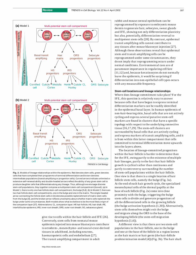
The location of lineage-committed progenitorswithin the hair follicle is harder to determine than for the IFE, owing partly to the existence of multiplehair lineages, partly to the fact that hair folliclegrowth is cyclical rather than continuous and partly to controversy surrounding the number of stem-cell populations within the hair follicle. One view is that there is a single location of hairfollicle stem cells, namely the bulge (Fig. 3a). At the end of each hair growth cycle, the specializedmesenchymal cells of the dermal papilla at the base of each follicle (Fig. 2a) come into close proximity with the bulge, triggering the bulge stem cells to divide and generate progenitors of all the differentiated cells in the growing follicle (the bulge activation hypothesis [1,30]). Alternatively,stem cells themselves might exit the bulge and migrate along the ORS to the base of thedeveloping follicle (the stem-cell migration hypothesis [1,6]).
A different view is that there are two stem-cellpopulations in the hair follicle, one in the bulge and one at the base of the follicle in a region known as the hair matrix or hair germ (the hair-folliclepredetermination model [4]) (Fig. 3b). The hair shaft
TRENDS in Cell Biology Vol.12 No.4 April 2002
http://tcb.trends.com
187Review
HSprogenitors
IRSprogenitors
HSprogenitors
IRSprogenitors
TRENDS in Cell Biology
SebocyteHenleHuxley
CLCuticleCuticleCornifiedcell
Medulla ORS
Sel
f-re
new
al c
apac
ity
Term
inal
diff
eren
tiatio
n
Sel
f-re
new
al c
apac
ity
Term
inal
diff
eren
tiatio
nMulti-potential stem cell compartmentModel 1(a)
Multi-potential stem cell compartmentModel 2(b)
IFEstemcell
Bulgestemcell
SGstemcell
IFEstemcell
Matrixstemcell
Bulgestemcell
SGstemcell
Cortex
Basal transitamplifying
Hairmatrix
ORSprogenitors
Sebocyteprogenitors
Basal transitamplifying
ORSprogenitors
Sebocyteprogenitors
SebocyteHenleHuxley
CLCuticleCuticleCornifiedcell
Medulla ORSCortex
Fig. 3. Models of lineage relationships within the epidermis. Red denotes stem cells, green denotescells that have completed their programme of terminal differentiation and brown denotesintermediate populations (transit amplifying or progenitor cells). Curved arrows indicate unlimited orextensive self-renewal ability and double-headed arrows reflect the ability of any given stem cell toproduce daughter cells that differentiate along any lineage. Thus, although we envisage discretestem-cell populations, they together compose a multipotent stem-cell compartment (boxed). (a) InModel 1, there is only one hair-follicle stem-cell compartment, the bulge [5,6]. (b) In Model 2, there aretwo hair-follicle stem-cell compartments, one in the bulge and one in the matrix. The single-headedarrow connecting the follicle stem cells in (b) denotes possible replenishment of matrix stem cellsfrom the bulge [4], and the broken arrow reflects uncertainty about whether matrix cells replenish thebulge under some circumstances. Both models show what we believe to be the most likely origin ofthe companion layer [37]. Abbreviations: CL, companion layer; HF, hair follicle; HS, hair shaft; IFE,interfollicular epidermis; IRS, inner root sheath; ORS, outer root sheath; SG, sebaceous gland.

and inner root sheath (IRS) lineages would be derivedfrom the matrix stem-cell population, whereas thebulge stem cells would give rise to the ORS (Fig. 3b).This model proposes that, once the downward growthphase of the hair follicle is complete, stem cells fromthe bulge move down the ORS to form a distinctpopulation of cells at the periphery of the hair bulb.These cells survive the apoptosis that leads to loss ofthe lower part of the hair follicle at the end of hairgrowth and form a reservoir of stem cells that respondto signals from the dermal papilla to replenish theIRS and hair shaft when the new hair cycle begins. In support of this model, lineage marking studieshave established the existence of long-lived cells with multilineage differentiation capacity in the hair matrix [10,31].
Irrespective of whether there are one or tworeservoirs of stem cells in the hair follicle, clusteringof stem cells at specific sites of the IFE and follicleimplies lateral migration of their progeny. There isnow good evidence for this both within the hair follicle [6,31] and in human epidermis reconstitutedin vitro [18]. In both situations, there is an inverserelationship between stem-cell motility and in vitroclonogenicity [6,17]. Some of the proposed markers of the human epidermal stem cell compartment, suchas β1 integrins and the Notch ligand Delta1, mightpromote stem-cell clustering by restricting motility or stimulating intercellular cohesiveness and thus act as dual regulators of cell adhesion anddifferentiation [17,32,33].
Commitment to the three hair-shaft lineages(medulla, cortex, cuticle) and three IRS lineages (cuticle, Huxley’s layer and Henle’s layer)takes place within the hair matrix (Figs 2,3). A subcompartment of the matrix known as theprecortex is the origin of the hair-shaft lineages (Fig. 2). Analysis of chimeric follicles expressingdifferent genotypic markers has shown that, inaddition to cells whose progeny contribute to all sixIRS and hair-shaft lineages [31] (putative stem cells),there is a common matrix progenitor cell for thehair-shaft lineages and one for the IRS lineages[10,34] (Fig. 3). The ORS represents a separatelineage [34] (Fig. 3).
Whereas the matrix is agreed to be the origin of the IRS and hair-shaft lineages, the location of thecompanion layer (Fig. 2) progenitors has been more of a mystery. The companion layer has been variouslydescribed as being a component of the IRS or the ORS, or as being a completely distinct lineage[4,35,36]. Analysis of keratin production patternswithin the hair follicle lineages suggests that the companion layer is not derived from ORS progenitors [36]. Rather, the observation ofcoexpression of the transcriptional regulator CCAAT displacement protein (CDP) in both the IRSand companion cells suggests that the companioncells constitute the fourth and outermost layer of theIRS [37] (Fig. 3).
Proliferation versus terminal differentiation
Stem-cell progeny must make two decisions. The first is whether or not to remain a stem cell. Havingdecided to leave the stem compartment, a cell mustthen select a particular differentiation pathway,leading ultimately to irreversible loss of proliferativecapacity. In regulating the balance betweenproliferation and differentiation in postnatal hairfollicles, the secreted signalling molecules Sonichedgehog (Shh) and bone morphogenetic proteins(Bmps) are of central importance. Shh promotesproliferation and Bmps promote differentiation. In adult epidermis, Shh expression is restricted tocells at the distal portion of the growing follicle[38,39]; mice treated with anti-Shh antibodies showimpaired epithelial growth at sites of folliculogenesis[40]. Several Bmp family members are produced inthe postnatal hair-shaft precursor cells and in thedermal papilla [41]. Inhibition of Bmp signalling bythe Bmp antagonist Noggin is required for new hairgrowth in postnatal skin and the growth-inducingeffect of Noggin is mediated, at least in part, by Shh [42]. In transgenic mice that produce Noggin in proliferating hair matrix cells and earlydifferentiating precursors of the IRS and hair shaft,differentiation of the hair shaft but not the IRS isseverely impaired; cells in the precortex and hairshaft continue to proliferate and to express genes thatare normally downregulated during differentiation[43]. There is evidence that the homeodomaintranscription factors Msx1 and Msx2 are Bmp targets [43] and, consistent with this, overproductionof Msx2 in the hair matrix reduces proliferation andinduces premature differentiation [44].
Other factors believed to regulate the balancebetween epidermal proliferation and differentiationinclude the transcription factors Forkhead-box n1(Foxn1) [45] and nuclear factor κB (NF-κB) [46]. Inpostnatal mouse epidermis, Foxn1 is produced indifferentiating cells of the IFE and in postmitoticprecursor cells of the hair shaft and IRS, as well as incells undergoing the transition from a proliferative toa postmitotic state [45]. Loss of Foxn1 function,resulting in the nude mouse phenotype, leads to afailure to form the cortex and causes defects in theIRS and hair cuticle; the IFE also undergoes aberrantdifferentiation. Manipulation of the NF-κB pathwayby deletion or overproduction of different componentshas established roles in immune responses and inregulating the decision of epidermal cells toproliferate or differentiate [46]. NF-κB might also beinvolved in lineage commitment, because it isrequired for normal morphogenesis of hair folliclesand sweat glands downstream of the tumour necrosisfactor receptor family member EDAR [47].
Lineage selection within the hair follicle
Many transgenic and knockout mice have strikinghair abnormalities, such as hair loss or a ruffled coat[2]. These can provide clues to the identities of
TRENDS in Cell Biology Vol.12 No.4 April 2002
http://tcb.trends.com
188 Review

molecules that regulate the decision to embark along different hair lineages and of molecules that are required for execution of a specificdifferentiation programme. An example of the secondclass is the homeodomain transcription factorHoxc13, which is produced in cells of the hair matrixand the lower shaft of growing follicles [48,49]. In Hoxc13-null mice, hair follicle formation duringdevelopment is normal but the hair shafts fracture atthe skin surface [50]. This phenotype appears to occurbecause Hoxc13 activates the expression of severalgenes for hair keratins during early hair-lineagedifferentiation [48,49].
One molecule for which the consequences of loss ormisexpression have been well worked out is Notch, areceptor that is activated by transmembrane proteinligands such as Delta and Jagged. The research onNotch highlights the importance of direct cell–cellcontact in regulating epidermal cell fate. In mouseepidermis, Notch1 is produced in matrix cells,whereas Notch3 is produced in the precursor anddifferentiating cortex and cuticle of the hair follicle[51]. Expression of a constitutively active Notch1transgene in the cortex of the hair follicle results inabnormal differentiation of the medulla and cuticle,two neighbouring cell types that are transgenenegative [51]. Notch might either direct thedifferentiation of follicular cells or assist those cells tointerpret signals emanating from the dermal papilla[51]. Notch1 is also found in all the viable layers ofhuman IFE [32] and keratinocyte-specific inducibledeletion of Notch1 results in IFE hyperproliferation[52]. This is consistent with the observation that, incultured human IFE, activation of Notch at the edgesof stem-cell clusters promotes neighbouring cells toenter the transit amplifying compartment [32].
Another key regulator of lineage choice in the hairfollicle is Cutl1, which encodes mammalian CDP.CDP is produced in the hair bulb, IRS progenitor cellsof the matrix, the three differentiating layers of theIRS and the companion cell layer [37]. Mice with anull mutation in Cutl1 have disrupted hair folliclemorphogenesis [37]: differentiation of IRS cells isimpaired, the cell layers of the IRS are reduced andthe ORS is enlarged, possibly as a result of ectopicShh signalling. The absence of CDP does not interferewith cell proliferation within the matrix but leads toderegulated expression of IRS-specific genes in thehair shaft.
Lineage choice within the hair follicle is alsocontrolled by secreted factors and these include Wnt3and Bmp4. In growing hair follicles, Wnt3 is producedin the precursor cells of the hair-shaft medulla andalso in the IFE [53]. Overproduction of Wnt3 in theORS via the keratin14 promoter causes defects inhair-shaft structure and elevated expression ofseveral hair-shaft proteins, suggesting that one of the functions of Wnt3 is to regulate hair-shaftdifferentiation [53]. Bmp4 is activated in hair-shaftprecursor cells and in the dermal papilla [41]. Ectopic
production of Bmp4 in the ORS inhibits proliferationin the hair matrix and activates hair keratin genes inthe ORS [54].
Sebocyte differentiation
Sebaceous glands are intimately associated with hair follicles and the two structures are often referred to collectively as the pilosebaceous unit.Nevertheless, there are conditions in which hairfollicles are induced without associated sebaceousglands [7] and there are molecules that promotesebocyte differentiation independent of induction of hair lineages. One of these is the product of theproto-oncogene c-Myc, which is expressed in several subpopulations of epidermal cells, including the basal layer of the IFE, the bulge, the proliferating cells of the hair bulb and thedifferentiating matrix cells that give rise to the hair fibre [55].
c-Myc induces differentiation of human IFE stemcells into transit amplifying cells in vitro [56]. When ac-Myc transgene is expressed in the hair follicles andthe basal layer of the IFE (via the keratin14promoter), adult mice gradually lose their hair anddevelop spontaneous ulcerated lesions owing to asevere impairment of wound healing [57]. Theepidermis is hyperproliferative and analysis of label-retaining cells shows a significant depletion of stemcells [57]. Cells that are stimulated to exit thestem-cell compartment differentiate into sebocytesand IFE at the expense of the hair lineages, andtransient activation of Myc is as effective asconstitutive activation in inducing these effects [58].Given that c-Myc activation decreases integrinexpression [56,57] and inhibits keratinocytemigration [57], one explanation for the stimulation ofsebocyte differentiation is that bulge progeny areunable to move down the follicle and therefore remainin a sebocyte-conducive environment, distant fromthe dermal papilla. An unresolved issue is whetherthe stem-cell depletion effect is Myc specific orwhether other factors that stimulate stem cells toproliferate will also stimulate the production oftransit amplifying cells.
A second regulator of sebocyte differentiation isthe transcription factor peroxisome proliferator-activated receptor γ(PPARγ). PPARγfunctions as aheterodimer with the retinoid X receptor (RXR),binding to specific sites on DNA and activatingtranscription in response to ligand. PPARγ-null mice die in mid-gestation but the contribution ofPPARγto adult tissues has been examined bygenerating mice that are chimeras of PPARγ −/− andwild-type cells [59]. Analysis of the skin of these mice suggests that PPARγmight be required forsebocyte differentiation because all the sebaceousglands are derived from wild-type cells; no hairphenotype has been reported. Conversely, PPARγligands promote the differentiation of sebocytes inculture [60].
TRENDS in Cell Biology Vol.12 No.4 April 2002
http://tcb.trends.com
189Review

Interconversion of hair follicles and IFE
Hair loss in association with the formation ofepithelioid cysts within the dermis is observed inseveral mutant mice (Fig. 4b). For example, cystswith absent or abnormal hair shafts are observed onoverproduction of Zipro1 [61], a zinc-fingertranscription factor that is produced in hair follicles,sebaceous glands and the IFE, or after ablation ofRXRα [62] or the vitamin D receptor [63] in adultmouse epidermis.
One interpretation of the cyst phenotype is that itreflects the suppression of hair-type differentiationpathways and the induction of IFE differentiation.The outer layer of the cyst corresponds to the IFEbasal layer and the inner layers correspond todifferent stages of IFE terminal differentiation,culminating in the accumulation of cornified cells inthe centre of the cysts [64]. Whether cyst formationreflects the transdifferentiation of cells that had
previously committed to hair lineages or thereprogramming of stem cells (for example, as a resultof loss of dermal papilla signals) remains to bedetermined [64]. However, cyst formation providesclear evidence for continuous regulation of lineagecommitment in postnatal life.
The genesis of dermal cysts has been characterizedmost extensively in mice with a mutation in theHairless (hr) gene, which encodes a putativetranscription factor [4,65]. Mice with the hairlessmutation are born with hair follicles that appearnormal but suffer from postnatal hair loss. Thefollicles degenerate, leaving behind dermal cysts andstructures called utriculi that open onto the surface of the epidermis. In hr skin, the distal ORS retains its integrity and forms utriculi, whereas the rest ofthe ORS disintegrates. The remnants of the bulgebecome isolated from other epithelial cells but retaintheir capacity to proliferate and produce eithercolumnar epithelial outgrowths or dermal cysts.Cysts can also arise from the disintegrating ORS and from the base of the hair follicle. Dermal cysts inhr mice not only reflect IFE differentiation: somecysts contain sebocytes and others show evidence of residual hair differentiation. Thus, the differenttypes of cysts might reflect which parts of the ORSgive rise to them.
If cysts reflect the conversion of hair into IFE thenthe converse phenotype is de novo induction of hairfollicles in postnatal IFE. In fact, both phenotypes can be created by modulating the activity of β-catenin,a key effector of the Wnt pathway that translocates tothe nucleus to activate transcription during Wntsignalling [1,66]. Several Wnt proteins and theirreceptors are expressed in the IFE, hair follicle anddermal papilla of postnatal mice [67], together with two downstream transcription factors, Tcf3 and Lef1. Tcf3, which can act as a transcriptionalrepressor, is produced in the bulge and ORS, whereas Lef1 is produced in cells in the hair matrix(progenitors of the IRS and hair shaft) [68,69]. In addition, a subpopulation of matrix cells stainspositive with an antibody to Tcf3/4 [31]. During hairgrowth, active Wnt signalling occurs in the bulge, in the precortex cells of the matrix and in the dermal papilla, as evidenced by accumulation ofnuclear β-catenin (Fig. 4a) or reporter gene activation [64,68,69]. Wnt proteins maintain the hair follicle inductive abilities of cultured dermalpapilla cells [70].
Inhibition of Wnt signalling by ablation of theβ-catenin gene in mouse epidermis [71] or byexpressing an N-terminally truncated Lef1 (∆NLef1)transgene that lacks the β-catenin binding site [64,69]causes hair loss and the appearance of dermal cysts(Fig. 4b). Conversely, expression of stabilizedβ-catenin under the control of the keratin14 promotercauses de novo formation of hair follicles in postnatalanimals [38]. Thus, the level of β-catenin signallingdetermines lineage choice: activation of the pathway
TRENDS in Cell Biology Vol.12 No.4 April 2002
http://tcb.trends.com
190 Review
(b)
(a)
Fig. 4. β-Catenin signalling in postnatal epidermis. (a) Hair follicle bulbstained with an antibody against β-catenin [64], showing nuclearaccumulation in the dermal papilla (arrowheads) and precortex(arrows). Bar, 20 µm. (b) Conversion of hair follicles into cysts ofinterfollicular epidermis in transgenic mice expressing an N-terminaltruncation of the transcription factor Lef-1, which is unable to bindβ-catenin [64]. Arrows indicate residual hair shafts; arrowheads indicateclusters of sebocytes associated with cysts. Bar, 200 µm.

stimulates hair-type differentiation, whereasinhibition of the pathway promotes differentiation of IFE and sebocytes.
Remarkably, the same effect on lineage selection is observed in tumours: transgenic animalsoverproducing β-catenin develop tumours withcharacteristics of hair-type differentiation [38],whereas tumours that develop in mice expressing a∆NLef1 transgene show sebocyte and IFEdifferentiation [64]. This raises the issue of whetherperturbation of β-catenin signalling is a primaryoncogenic event or whether β-catenin interactssynergistically with additional oncogenes todetermine the type of tumour that is formed.
Concluding remarks
Here, we have considered the epidermal stem-cellcompartment and the differentiated daughter cells that it produces, and we have highlighted someof the factors that regulate the different lineages.Before closing, there are several additional points tomake that underscore the complexity of the system.First, although we have focused on postnatal
epidermis, most, if not all, of the regulators that wehave described are also important in hair follicleinduction during development [1]. It is intriguing that the same molecules can play different roles inembryonic and postnatal skin [39,43]. Second, thedifferent signalling pathways do not function inisolation but interact both positively and negatively;thus, modulating the concentration of one factor can affect signalling by another [39,67]. Finally, the temporal and spatial patterns of expression of the growth factors and their receptors can beextremely complex, with expression of multiplefamily members being detected both in themesenchyme and in distinct subpopulations ofkeratinocytes [67,72].
Although considerable progress has been made in defining lineage relationships within theepidermis, we are still a long way from completelycharacterizing all the intrinsic and extrinsicregulators of each step in the transition from amultipotent stem cell to the ten terminallydifferentiated cell types shown in Fig. 3. We see thisas the major challenge for the future.
TRENDS in Cell Biology Vol.12 No.4 April 2002
http://tcb.trends.com
191Review
References
1 Fuchs, E. et al. (2001) At the roots of a never-ending cycle. Dev. Cell 1, 13–25
2 Nakamura, M. et al. (2001) Mutant laboratorymice with abnormalities in hair folliclemorphogenesis, cycling, and/or structure:annotated tables. Exp. Dermatol. 10, 369–390
3 Watt, F.M. (1998) Epidermal stem cells: markers,patterning and the control of stem cell fate.Philos. Trans. R. Soc. London Ser. B 353, 831–837
4 Panteleyev, A.A. et al. (2001) Hair folliclepredetermination. J. Cell Sci. 114, 3419–3431
5 Taylor, G. et al. (2000) Involvement of follicularstem cells in forming not only the follicle but alsothe epidermis. Cell 102, 451–461
6 Oshima, H. et al. (2001) Morphogenesis andrenewal of hair follicles from adult multipotentstem cells. Cell 104, 233–245
7 Reynolds, A.J. and Jahoda, C.A. (1992) Cultureddermal papilla cells induce follicle formation andhair growth by transdifferentiation of an adultepidermis. Development 115, 587–593
8 Ferraris, C. et al. (1997) Adult epidermalkeratinocytes are endowed with pilosebaceousforming abilities. Int. J. Dev. Biol. 41, 491–498
9 Miller, S.J. et al. (1998) Re-epithelialisation ofporcine skin by the sweat apparatus. J. Invest.Dermatol. 110, 13–19
10 Ghazizadeh, S. and Taichman, L.B. (2001)Multiple classes of stem cells in cutaneousepithelium; a lineage analysis of adult mouseskin. EMBO J. 20, 1215–1222
11 Watt, F.M. and Hogan, B.L.M. (2000) Out of Eden:stem cells and their niches. Science 287, 1427–1430
12 Spradling, A. et al. (2001) Stem cells find theirniche. Nature 414, 98–104
13 Sengel, P. (1986) Epidermal–dermal interactions.In Biology of the Integument: Vertebrates (Vol. 2)(Bereiter-Hahn, J. et al., eds), pp. 374–408,Springer-Verlag, Berlin
14 Osada, A. and Kobayashi, K. (2001)Characterization of vibrissa germinative cells:transition of cell types. Exp. Dermatol. 10,430–437
15 Lavker, R.M. and Sun, T-T. (2000) Epidermalstem cells: properties, markers, and location.Proc. Natl. Acad. Sci. U. S. A. 97, 13473–13475
16 Jones, P.H. et al. (1995) Stem cell patterning andfate in human epidermis. Cell 80, 83–93
17 Jensen, U.B. et al. (1999) The spatial relationshipbetween stem cells and their progeny in the basallayer of human epidermis: a new view based onwhole mount labelling and lineage analysis.Development 126, 2409–2418
18 Watt, F.M. (2001) Stem cell fate and patterning inmammalian epidermis. Curr. Opin. Genet. Dev.11, 410–417
19 Potten, C.S. (1981) Cell replacement in epidermis(keratopoiesis) via discrete units of proliferation.Int. Rev. Cytol. 69, 271–318
20 Cotsarelis, G. et al. (1989) Existence of slow-cycling limbal epithelial basal cells that can bepreferentially stimulated to proliferate:implications on epithelial stem cells. Cell 57,201–209
21 Reya, T. et al. (2001) Stem cells, cancer, andcancer stem cells. Nature 414, 105–111
22 Dover, R. and Watt, F.M. (1987) Measurement ofthe rate of epidermal terminal differentiation:expression of involucrin by S-phase keratinocytesin culture and in psoriatic plaques. J. Invest.Dermatol. 89, 349–352
23 Seery, J.P. and Watt, F.M. (2000) Asymmetricstem-cell divisions define the architecture ofhuman oesophageal epithelium. Curr. Biol. 10,1447–1450
24 Potten, C.S. and Loeffler, M. (1990) Stem cells:attributes, cycles, spirals, pitfalls anduncertainties. Lessons for and from the crypt.Development 110, 1001–1020
25 Jones, P.H. and Watt, F.M. (1993) Separation ofhuman epidermal stem cells from transitamplifying cells on the basis of differences inintegrin function and expression. Cell 73, 713–724
26 Krause, D.S. et al. (2001) Multi-organ, multi-lineage engraftment by a single bone marrow-derived stem cell. Cell 105, 369–377
27 Liang, L. and Bickenbach, J.R. (2002) Somaticepidermal stem cells can produce multiple celllineages during development. Stem Cells 20,21–31
28 Ferraris, C. et al. (2000) Adult corneal epithelium basal cells possess the capacity toactivate epidermal, pilosebaceous and sweatgland genetic programs in response to embryonic dermal stimuli. Development 127,5487–5495
29 Lavker, R.M. and Sun, T-T. (1982) Heterogeneityin epidermal basal keratinocytes: morphologicaland functional correlations. Science 215,1239–1241
30 Cotsarelis, G. et al. (1990) Label-retaining cellsreside in the bulge area of pilosebaceous unit:implications for follicular stem cells, hair cycle, and skin carcinogenesis. Cell 61, 1329–1337
31 Kopan, R. et al. (2002) Genetic mosaic analysisindicates that the bulb region of coat hair folliclescontains a resident population of several activemulti-potent epithelial lineage progenitors. Dev.Biol. 242, 44–57
32 Lowell, S. et al. (2000) Stimulation of humanepidermal differentiation by Delta–Notchsignalling at the boundaries of stem-cell clusters.Curr. Biol. 10, 491–500
33 Lowell, S. and Watt, F.M. (2001) Delta regulateskeratinocyte spreading and motilityindependently of differentiation. Mech. Dev. 107,133–140
34 Kamimura, J. et al. (1997) Primary mousekeratinocyte cultures contain hair follicleprogenitor cells with multiple differentiationpotential. J. Invest. Dermatol. 109, 534–540
35 Rothnagel, J.A. and Roop, D.R. (1995) Hair folliclecompanion layer: reacquainting an old friend.J. Invest. Dermatol. 104, 42S–43S
36 Winter, H. et al. (1998) A novel human type IIcytokeratin, K6h, specifically expressed in thecompanion layer of the hair follicle. J. Invest.Dermatol. 111, 955–962

37 Ellis, T. et al. (2001) The transcriptional repressorCDP (Cutl1) is essential for epithelial celldifferentiation of the lung and the hair follicle.Genes Dev. 15, 2307–2319
38 Gat, U. et al. (1998) De novo hair folliclemorphogenesis and hair tumors in miceexpressing a truncated β-catenin in skin. Cell 95,605–614
39 Callahan, C.A. and Oro, A.E. (2001) Monstrousattempts at adnexogenesis: regulating hairfollicle progenitors through Sonic Hedgehogsignaling. Curr. Opin. Genet. Dev. 11, 541–546
40 Wang, L.C. et al. (2000) Conditional disruption of hedgehog signalling pathway defines its critical role in hair development andregeneration. J. Invest. Dermatol. 114, 901–908
41 Wilson, N. et al. (1999) The role of BMP-2 andBMP-4 in follicle initiation and murine hair cycle.Exp. Dermatol. 8, 367–368
42 Botchkarev, V.A. (2001) Noggin is required forinduction of the hair follicle growth phase inpostnatal skin. FASEB J. 15, 2205–2214
43 Kulessa, H. et al. (2000) Inhibition of Bmpsignaling affects growth and differentiation in the anagen hair follicle. EMBO J. 19,6664–6674
44 Jiang, T.X. et al. (1999) Epidermal dysplasia andabnormal hair follicles in transgenic miceoverexpressing homeobox gene MSX-2. J. Invest.Dermatol. 113, 230–237
45 Lee, D. et al. (1999) Association between mousenude gene expression and the initiation ofepithelial terminal differentiation. Dev. Biol. 208,362–374
46 Kaufman, C.K. and Fuchs, E. (2000) It’s got youcovered: NF-κB in the epidermis. J. Cell Biol. 149,999–1004
47 Schmidt-Ullrich, R. et al. (2001) Requirement ofNF-κB/Rel for the development of hair folliclesand other epidermal appendices. Development128, 3843–3853
48 Tkatchenko, A.V. et al. (2001) Overexpression ofHoxc13 in differentiating keratinocytes results indownregulation of a novel hair keratin genecluster and alopecia. Development 128, 1547–1558
49 Jave-Suarez, L.F. et al. (2002) HOXC13 isinvolved in the regulation of human hair keratingene expression. J. Biol. Chem. 277, 3718–3726
50 Godwin, A.R. and Capecchi, M.R. (1998) Hoxc13mutant mice lack external hair. Genes Dev. 12,11–20
51 Lin, M-H. et al. (2000) Activation of the Notchpathway in the hair cortex leads to aberrantdifferentiation of the adjacent hair-shaft layers.Development 127, 2421–2432
52 Rangarajan, A. et al. (2001) Notch signalling is adirect determinant of keratinocyte growth arrestand entry into differentiation. EMBO J. 20,3427–3436
53 Millar, S.E. et al. (1999) WNT signaling in thecontrol of hair growth and structure. Dev. Biol.207, 133–149
54 Blessing, M. et al. (1993) Transgenic mice as amodel to study the role of TGF-β-relatedmolecules in hair follicles. Genes Dev. 7, 204–215
55 Bull, J.J. et al. (2001) Contrasting localization ofc-Myc with other Myc superfamily transcriptionfactors in the human hair follicle and during thehair growth cycle. J. Invest. Dermatol. 116,617–622
56 Gandarillas, A. and Watt, F.M. (1997) c-Mycpromotes differentiation of epidermal stem cells.Genes Dev. 11, 2869–2882
57 Waikel, R.L. et al. (2001) Deregulated expressionof c-Myc depletes epidermal stem cells. Nat.Genet. 28, 165–168
58 Arnold, I. and Watt, F.M. (2001) c-Myc activationin transgenic mouse epidermis results inmobilization of stem cells and differentiation oftheir progeny. Curr. Biol. 11, 558–568
59 Rosen, E.D. et al. (1999) PPARγ is required for thedifferentiation of adipose tissue in vivo andin vitro. Mol. Cell 4, 611–617
60 Rosenfield, R.L. et al. (2000) Peroxisomeproliferator-activated receptors and skindevelopment. Hormone Res. 54, 269–274
61 Yang, X.W. et al. (1999) BAC-mediated gene-dosage analysis reveals a role for Zipro1(Ru49/Zfp38) in progenitor cell proliferation incerebellum and skin. Nat. Genet. 22, 327–335
62 Li, M. et al. (2000) Skin abnormalities generatedby temporally controlled RXRα mutations inmouse epidermis. Nature 407, 633–636
63 Li, Y.C. et al. (1997) Targeted ablation of thevitamin D receptor: an animal model of vitamin D-dependent rickets type II withalopecia. Proc. Natl. Acad. Sci. U. S. A. 94,9831–9835
64 Niemann, C. et al. (2002) Expression of ∆NLef1 inmouse epidermis results in differentiation of hairfollicles into squamous epidermal cysts andformation of skin tumours. Development 129,95–109
65 Panteleyev, A.A. et al. (1998) Towards defining thepathogenesis of the Hairless phenotype. J. Invest.Dermatol. 110, 902–907
66 Huelsken, J. and Birchmeier, W. (2001) Newaspects of Wnt signaling pathways in highervertebrates. Curr. Opin. Genet. Dev. 11, 547–553
67 Reddy, S. et al. (2001) Characterization of Wnt gene expression in developing and postnatal hair follicles and identification of Wnt5a as a target of Sonic hedgehog in hair follicle morphogenesis. Mech. Dev.107, 69–82
68 DasGupta, R. and Fuchs, E. (1999) Multiple roles for activated LEF/TCF transcriptioncomplexes during hair follicle development and differentiation. Development 126, 4557–4568
69 Merrill, B.J. et al. (2001) Tcf3 and Lef1 regulatelineage differentiation of multipotent stem cells inskin. Genes Dev. 15, 1688–1705
70 Kishimoto, J. et al. (2000) Wnt signalingmaintains the hair-inducing activity of the dermalpapilla. Genes Dev. 14, 1181–1185
71 Huelsken, J. et al. (2001) β-Catenin controls hair follicle morphogenesis and stem cell differentiation in the skin. Cell 105, 533–545
72 Rosenquist, T.A. and Martin, G.R. (1996)Fibroblast growth factor signalling in the hair growth cycle: expression of the fibroblastgrowth factor receptor and ligand genes in the murine hair follicle. Dev. Dyn. 205, 379–386
TRENDS in Cell Biology Vol.12 No.4 April 2002
http://tcb.trends.com
192 Review
As a busy cell biologist, searching through the wealth of information on BioMedNet can be a bit daunting – the new gatewayto cell biology on BioMedNet is designed to help.The cell biology gateway is updated weekly and features relevant articles selected by the editorial teams from Trends in Cell Biology and Current Opinion in Cell Biology.The regular updates include:• News – our dedicated team of reporters from BioMedNet News provides a busy researcher with all the news to keep up to
date on what’s happening – right now.• Conference Reporter – daily updates on selected conferences, including the recent American Society for Cell Biology
Annual Meeting, providing a quick but comprehensive report of what you missed by staying home.• Journal Scan – learn about new reports and events in cell biology every day, at a glance.• Mini Reviews and Reviews – a selection of the best review and opinion articles from all Trends and Current Opinion
journals.Bookmark the gateway at
bmn.com/cellbiology
Editor’s choicebmn.com/cellbiology