research open access frontalis suspension … this condition we perform frontalis suspension surgery...
TRANSCRIPT

HEAD & FACE MEDICINE
Karapantzou et al. Head & Face Medicine 2014, 10:44http://www.head-face-med.com/content/10/1/44
RESEARCH Open Access
Frontalis suspension surgery to treat patients withessential blepharospasm and apraxia of eyelidopening-technique and resultsChrisanthi Karapantzou1, Dirk Dressler2, Saskia Rohrbach3 and Rainer Laskawi1,4*
Abstract
Introduction: We describe the results of 15 patients suffering from essential blepharospasm with apraxia of eyelidopening who underwent frontalis suspension surgery.
Material and methods: Patients with apraxia of eyelid opening and unresponsive to botulinum toxin injectionswere studied. Bilateral frontalis suspension surgery was performed (sling operation) using polytetrafluoroethylene(Gore-Tex®) sutures. The patients reported the degree of improvement using a subjective rating scale to evaluate thebenefit of the operation at two times after surgery (0-10 days and 180-360 days).
Results: The patients reported a high degree of subjective improvement. In the early postoperative period (0-10 days)the mean degree of subjective improvement was 74.6% (standard deviation (SD) 26.4%). At 180-360 days after surgerythe mean improvement was 70.0% (SD 26.7%). Small hematomas of the upper lid occurred postoperatively in allpatients. Other complications were suture extrusions (9.1%), suture granulomas (6.1%), lacrimation (5.0%) and localinfections (7.5%). Postoperatively, all patients needed additional botulinum toxin injections for optimal outcome.
Conclusion: Frontalis suspension surgery is a minimally invasive and effective treatment option for apraxia of eyelidopening in patients with essential blepharospasm unresponsive to botulinum toxin injections alone.
Keywords: Blepharospasm, Apraxia of eye lid opening, Frontalis suspension surgery
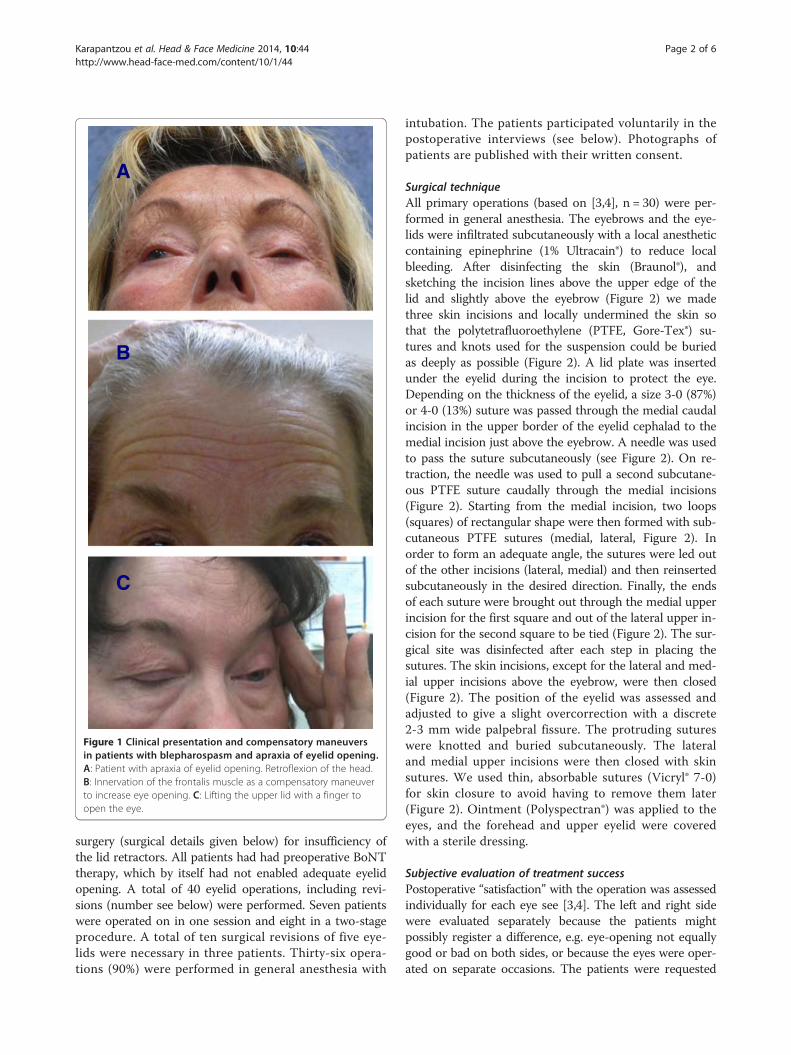
IntroductionThe treatment of essential blepharospasm with botulinumtoxin (BoNT) is a well-established method [1,2]. However,some patients suffer from a special form of blepharospasmthat renders them unable to open their eyes adequatelydespite local BoNT treatment. This phenomenon is due tothe inability to raise the eyelid (see Figure 1). This is re-ferred to in the literature as “apraxia of eyelid opening”,“levator inhibition blepharospasm” or “dystonic eyelidopening disorder” [3-6]. It leads to a compensatory con-strained posture such as retroflexion of the head to enablethe patient to see through the narrow palpebral fissure.The patients frequently exhibit photophobia, and performcertain compensatory movements to keep the palpebralfissure as wide as possible. Many patients increase vertical
* Correspondence: [email protected], University of Göttingen Medical Center, Göttingen, Germany4HNO-Klinik Universitätsmedizin Göttingen, Robert-Koch-Str. 40, 37075Göttingen, GermanyFull list of author information is available at the end of the article
© 2014 Karapantzou et al.; licensee BioMed CeCreative Commons Attribution License (http:/distribution, and reproduction in any mediumDomain Dedication waiver (http://creativecomarticle, unless otherwise stated.
traction by contracting the frontalis muscle to widen theopening of the palpebral fissure. Since BoNT treatmentalone will not provide any significant improvement of theeye opening, a number of more or less invasive surgicalprocedures have been introduced [3-5,7,8]. In our patientswith this condition we perform frontalis suspensionsurgery (“sling operation”) based on the technique ofRoggenkämper and Nüssgens [3,4]. In this report wedescribe this surgical procedure and present our re-sults of the operation as well as the assessments of thepatients with regard to the success of the treatmentand to their quality of life.
Patients and methodsGeneral informationThis retrospective analysis, which had the approval of ourinstitutional ethics review board (University of GöttingenMedical Center), encompasses 15 patients, nine womenand six men, who underwent bilateral frontalis suspension
ntral Ltd. This is an Open Access article distributed under the terms of the/creativecommons.org/licenses/by/4.0), which permits unrestricted use,, provided the original work is properly credited. The Creative Commons Publicmons.org/publicdomain/zero/1.0/) applies to the data made available in this
A
B
C
Figure 1 Clinical presentation and compensatory maneuversin patients with blepharospasm and apraxia of eyelid opening.A: Patient with apraxia of eyelid opening. Retroflexion of the head.B: Innervation of the frontalis muscle as a compensatory maneuverto increase eye opening. C: Lifting the upper lid with a finger toopen the eye.
Karapantzou et al. Head & Face Medicine 2014, 10:44 Page 2 of 6http://www.head-face-med.com/content/10/1/44
surgery (surgical details given below) for insufficiency ofthe lid retractors. All patients had had preoperative BoNTtherapy, which by itself had not enabled adequate eyelidopening. A total of 40 eyelid operations, including revi-sions (number see below) were performed. Seven patientswere operated on in one session and eight in a two-stageprocedure. A total of ten surgical revisions of five eye-lids were necessary in three patients. Thirty-six opera-tions (90%) were performed in general anesthesia with
intubation. The patients participated voluntarily in thepostoperative interviews (see below). Photographs ofpatients are published with their written consent.
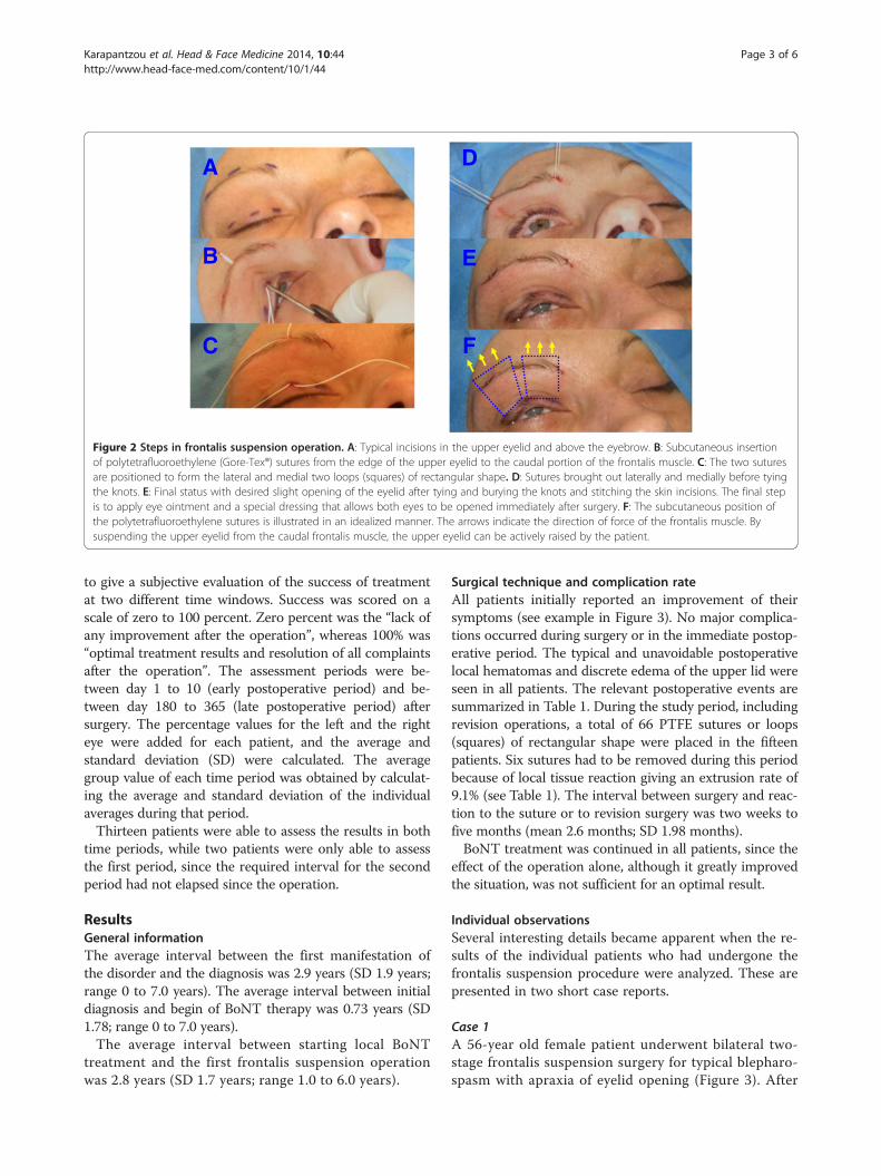
Surgical techniqueAll primary operations (based on [3,4], n = 30) were per-formed in general anesthesia. The eyebrows and the eye-lids were infiltrated subcutaneously with a local anestheticcontaining epinephrine (1% Ultracain®) to reduce localbleeding. After disinfecting the skin (Braunol®), andsketching the incision lines above the upper edge of thelid and slightly above the eyebrow (Figure 2) we madethree skin incisions and locally undermined the skin sothat the polytetrafluoroethylene (PTFE, Gore-Tex®) su-tures and knots used for the suspension could be buriedas deeply as possible (Figure 2). A lid plate was insertedunder the eyelid during the incision to protect the eye.Depending on the thickness of the eyelid, a size 3-0 (87%)or 4-0 (13%) suture was passed through the medial caudalincision in the upper border of the eyelid cephalad to themedial incision just above the eyebrow. A needle was usedto pass the suture subcutaneously (see Figure 2). On re-traction, the needle was used to pull a second subcutane-ous PTFE suture caudally through the medial incisions(Figure 2). Starting from the medial incision, two loops(squares) of rectangular shape were then formed with sub-cutaneous PTFE sutures (medial, lateral, Figure 2). Inorder to form an adequate angle, the sutures were led outof the other incisions (lateral, medial) and then reinsertedsubcutaneously in the desired direction. Finally, the endsof each suture were brought out through the medial upperincision for the first square and out of the lateral upper in-cision for the second square to be tied (Figure 2). The sur-gical site was disinfected after each step in placing thesutures. The skin incisions, except for the lateral and med-ial upper incisions above the eyebrow, were then closed(Figure 2). The position of the eyelid was assessed andadjusted to give a slight overcorrection with a discrete2-3 mm wide palpebral fissure. The protruding sutureswere knotted and buried subcutaneously. The lateraland medial upper incisions were then closed with skinsutures. We used thin, absorbable sutures (Vicryl® 7-0)for skin closure to avoid having to remove them later(Figure 2). Ointment (Polyspectran®) was applied to theeyes, and the forehead and upper eyelid were coveredwith a sterile dressing.
Subjective evaluation of treatment successPostoperative “satisfaction” with the operation was assessedindividually for each eye see [3,4]. The left and right sidewere evaluated separately because the patients mightpossibly register a difference, e.g. eye-opening not equallygood or bad on both sides, or because the eyes were oper-ated on separate occasions. The patients were requested
A
B
D
E
FC
Figure 2 Steps in frontalis suspension operation. A: Typical incisions in the upper eyelid and above the eyebrow. B: Subcutaneous insertionof polytetrafluoroethylene (Gore-Tex®) sutures from the edge of the upper eyelid to the caudal portion of the frontalis muscle. C: The two suturesare positioned to form the lateral and medial two loops (squares) of rectangular shape. D: Sutures brought out laterally and medially before tyingthe knots. E: Final status with desired slight opening of the eyelid after tying and burying the knots and stitching the skin incisions. The final stepis to apply eye ointment and a special dressing that allows both eyes to be opened immediately after surgery. F: The subcutaneous position ofthe polytetrafluoroethylene sutures is illustrated in an idealized manner. The arrows indicate the direction of force of the frontalis muscle. Bysuspending the upper eyelid from the caudal frontalis muscle, the upper eyelid can be actively raised by the patient.
Karapantzou et al. Head & Face Medicine 2014, 10:44 Page 3 of 6http://www.head-face-med.com/content/10/1/44
to give a subjective evaluation of the success of treatmentat two different time windows. Success was scored on ascale of zero to 100 percent. Zero percent was the “lack ofany improvement after the operation”, whereas 100% was“optimal treatment results and resolution of all complaintsafter the operation”. The assessment periods were be-tween day 1 to 10 (early postoperative period) and be-tween day 180 to 365 (late postoperative period) aftersurgery. The percentage values for the left and the righteye were added for each patient, and the average andstandard deviation (SD) were calculated. The averagegroup value of each time period was obtained by calculat-ing the average and standard deviation of the individualaverages during that period.Thirteen patients were able to assess the results in both
time periods, while two patients were only able to assessthe first period, since the required interval for the secondperiod had not elapsed since the operation.
ResultsGeneral informationThe average interval between the first manifestation ofthe disorder and the diagnosis was 2.9 years (SD 1.9 years;range 0 to 7.0 years). The average interval between initialdiagnosis and begin of BoNT therapy was 0.73 years (SD1.78; range 0 to 7.0 years).The average interval between starting local BoNT
treatment and the first frontalis suspension operationwas 2.8 years (SD 1.7 years; range 1.0 to 6.0 years).
Surgical technique and complication rateAll patients initially reported an improvement of theirsymptoms (see example in Figure 3). No major complica-tions occurred during surgery or in the immediate postop-erative period. The typical and unavoidable postoperativelocal hematomas and discrete edema of the upper lid wereseen in all patients. The relevant postoperative events aresummarized in Table 1. During the study period, includingrevision operations, a total of 66 PTFE sutures or loops(squares) of rectangular shape were placed in the fifteenpatients. Six sutures had to be removed during this periodbecause of local tissue reaction giving an extrusion rate of9.1% (see Table 1). The interval between surgery and reac-tion to the suture or to revision surgery was two weeks tofive months (mean 2.6 months; SD 1.98 months).BoNT treatment was continued in all patients, since the
effect of the operation alone, although it greatly improvedthe situation, was not sufficient for an optimal result.
Individual observationsSeveral interesting details became apparent when the re-sults of the individual patients who had undergone thefrontalis suspension procedure were analyzed. These arepresented in two short case reports.
Case 1A 56-year old female patient underwent bilateral two-stage frontalis suspension surgery for typical blepharo-spasm with apraxia of eyelid opening (Figure 3). After

Figure 3 Patient after bilateral frontalis suspension surgery. The eyes can be opened (A) and closed (B) without difficulty. The patient’sBoNT treatment was continued. Insert C shows the situation immediately after the unilateral operation of the left eye, identifiable by the smallhematoma. One can clearly see that the apraxia persists in the non-operated right eye and that eye opening is not possible in spite of innervationof the frontalis muscle. The position of the eyebrow is higher and the upper eyelid is completely closed. After the suspension surgery, the left,operated eye is already partly opened with only a low-intensity innervation. This is consistent with a lateralized control of the apraxia.
Karapantzou et al. Head & Face Medicine 2014, 10:44 Page 4 of 6http://www.head-face-med.com/content/10/1/44
the first operation we noticed that the levator inhibitioneffect appeared to differ between the two sides (Figure 3).When the patient attempted to raise her eyebrows we ob-served an increased innervation of the frontalis muscle onthe non-operated side but without a resulting adequate liftof the eyelid. A markedly slighter innervation of the fron-talis muscle of the treated side gave a much wider openingof the eyelid (Figure 3).
Case 2A 73-year old male patient with multifocal dystonia pre-sented with blepharospasm and apraxia of eyelid opening.Following successful frontalis suspension surgery the PTFEsutures were expulsed on both sides after two months(right side), respectively five months (left side). Despite re-peated revision operations with the aim of burying theknots the sutures eventually had to be removed. Despitethis the patient continued to be able to open his eyes.
Table 1 Type and incidence of postoperativecomplications
Postoperative complications Frequency
Suture extrusion 6 of 66 sutures (9.1%)
Suture granuloma 4 of 66 sutures (6.1%)
Lacrimation 2 of 40 operated eyelids (5.0%)
Infection 3 of 40 operated eyelids (7.5%)
The effect persisted for seven months (right eye), re-spectively 11 months (left eye).
Subjective rating of success of treatmentAll of the fifteen patients reported an initial postopera-tive improvement of eye opening.The average early postoperative (0 to 10 days) subjective
rating of therapeutic success was 74.6% (SD 26.4%; range20 to 100%). The average rating at the later period (180 to365 days) was 70.0% (SD 26.7%; range 0 to 100%).
DiscussionOne main approach in the treatment of essentialblepharospasm is to reduce muscle mass in the cranialportion of the orbicularis oculi muscle, often togetherwith parts of the procerus and the corrugator superciliimuscles [7]. These procedures can be combined withsuspension operations [8]. They are considerably moreinvasive than suspension operations alone, as performedin our own and other institutions [3-5].The polytetrafluoroethylene (PTFE, Gore-Tex®) sutures
gave satisfactory functional results. However, it is wellknown that PTFE sutures do not always integrate wellinto the tissue bed, and they can cause problems withtissue reactions and extrusion [9-11]. The extrusion rateof 9.1% and the 6.1% incidence of granulomas in our pa-tients are similar to those described by other authors [5].

Karapantzou et al. Head & Face Medicine 2014, 10:44 Page 5 of 6http://www.head-face-med.com/content/10/1/44
It should be mentioned that whenever a suture that wasintroduced using our method is extruded, a new one canbe inserted without any problems after a waiting periodof only a few weeks. In addition, if the traction effect ofthe suture decreases, it is possible to uncover the knotand “reposition” the upper eyelid.Fascia lata gives very good functional results compared
to other materials [12,13], but we prefer polytetrafluoro-ethylene because this is a minimally invasive option toelevate the upper lid in patients with blepharospam withapraxia of eye lid opening. The removal of fascia lata ismore invasive, an additional skin incision is needed inanother body region (thigh), and the results do not differmuch from the use of polytetrafluoroethylene in thisconnection [12,13].Our observations in individual patients were interest-
ing. In our surgically treated patients, we did not observethe known effect that a unilateral BoNT injection cancause bilateral improvement of blepharospasm. Theobserved phenomenon of a lateralized change in theability to raise the eyelid is an argument for a lateralized“organization” of the apraxia of eyelid opening. Anotherobservation was that adequate eye opening could persisteven after a suture had to be removed. This phenomenonhas not been described previously in the literature. In thecase described here, a new suture was implanted in a dif-ferent position after an appropriate waiting period. Theobserved phenomenon could be due to the developmentof filiform scar tissue that, for a limited time, could stillgive traction on the upper eyelid when the frontalismuscle contracted. This could also be an effect of theremaining sutures if not all had been removed. In thiscase, the remaining sutures maintain a connection be-tween the upper eyelid and the caudal part of the frontalismuscle.All patients had preoperative BoNT therapy, but this
alone was unable to allow sufficient elevation of the eye-lids. The patients rated the treatment results as an im-provement, which underscores the positive effect of theoperation. It was not possible to discontinue BoNTtreatment in any of the patients, since alleviation of theorbicularis oculi muscles spasms appears to be in vary-ing degrees an essential component for optimal results.It is relevant in this context that the BoNT injectionsduring postoperative treatment should not be placed inthe caudal portion of the frontalis muscle. This is veryimportant since the strength of the frontalis muscle andthe ability to raise the upper eyelid would be reduced ifthis recommendation were to be ignored. The study byGrivet et al. [14] is of interest in this context. The au-thors reported that the immediate postoperative im-provement of the “Functional Disability Scores” (FDS)was significantly greater in those patients with bleph-arospasm whose BoNT treatment had been discontinued
postoperatively compared to those whose treatment hadnot been interrupted. But the FDS of the patients withpostoperatively continued BoNT injections improvedlater to the level of the patients without postoperativeBoNT treatment. In their analysis, however, the authorsdid not stratify their patients by subtypes or by surgicalmethod. If such a total absence of spasms of the orbicu-laris oculi muscle is basically relevant in patients withapraxia of eyelid opening remains undecided. Our data,which show that continuing postoperative BoNT treat-ment was necessary in all patients, argue against thatassumption.The average subjective improvement ratings of our pa-
tients were between 75% and 70%, which is a relativelyhigh level see [3-5]. Roggenkämper and Nüssgens [3,4] ob-served a stable effect for their patient population as awhole, a result that is similar to the analysis of the follow-up period of our study. But this retrospective study doeshave some limitations. The results would have been morerobust had the improvement of eye opening described bythe patients been confirmed by objective measurements.A validated questionnaire and score for the patients’ self-assessment with focus on the degree of eye opening wouldalso have been useful. A further study with a greater num-ber of patients would be useful more detailed insights intothis rare and complex disease and determine the therapyoptions.In summary one can conclude that frontalis suspension
surgery using polytetrafluoroethylene (Gore-Tex®) suturesis a minimally invasive and effective option for the treat-ment of apraxia of eyelid opening in patients with essen-tial blepharospasm. The quality of daily life and generalpatient satisfaction is improved. The combination withbotulinum toxin injections seems to be of advantage foran optimal treatment outcome.
Competing interestsThe authors declare that they have no competing interests.
Authors’ contributionsCK treatment of patients, conceived the study, acquisition of data, analysisand interpretation of data. DD treatment of patients, participated in thedesign of the study, revising manuscript for important intellectual inputand content, statistical consultation. SR surgery of the patients, draftingmanuscript, revising manuscript for important intellectual input and content. RLconceived the study, surgery of the patients, drafting manuscript, acquisition ofdata, analysis and interpretation of data. All authors read and approved the finalmanuscript.
Author details1ENT-Department, University of Göttingen Medical Center, Göttingen, Germany.2Department of Neurology, University of Hannover Medical Center, Hannover,Germany. 3Department of Audiology and Phoniatrics, University of BerlinMedical Center, Berlin, Germany. 4HNO-Klinik Universitätsmedizin Göttingen,Robert-Koch-Str. 40, 37075 Göttingen, Germany.
Received: 9 August 2014 Accepted: 14 October 2014Published: 22 October 2014

Karapantzou et al. Head & Face Medicine 2014, 10:44 Page 6 of 6http://www.head-face-med.com/content/10/1/44
References1. Nüssgens Z, Roggenkämper P: Long-term treatment of blepharospasm
with botulinum toxin type A. Ger J Ophthalmol 1995, 4:363–367.2. Czyz CN, Burns JA, Petrie TP, Warkins JR, Cahill KV, Foster JA: Long-term
botulinum toxin treatment of benign essentaila blepharospasm,hemifacial spasm, and Meige syndrome. A J Ophthalmol 2013,156:173–177.
3. Roggenkämper P, Nüssgens Z: Frontalis suspension in the treatment ofessential blepharospasm unresponsive to botulinum toxin therapy. Firstresults. Ger J Ophthalmol 1993, 2:426–428.
4. Roggenkämper P, Nüssgens Z: Frontalis suspension in the treatment ofessential blepharospasm unresponsive to botulinum toxin therapy:long-term results. Graefe’s Arch Clin Exp Ophthalmol 1997, 235:486–489.
5. Wabbels B, Roggenkämper P: Long-term follow up of patients withfrontalis sling operation in the treatment of essentail blepharospasmunresponsive to botulinum toxin therapy. Graefe’s Arch Clin ExpOphthalmol 2007, 245:45–50.
6. Reichel G, Stenner A, Herrman W: Palpebrale Variante des Blepharospasmus- Abgrenzung zur Lidöffnungsapraxie und zur Inhibitionsstörung durchsynchrone EMG-Ableitungen. Akt Neurol 2009, 36:60–64.
7. Georgescu D, Vagefi MR, McMullan TFW, McCann JD, Anderson RL: Uppereyelid myectomy in blepharospasm with associated apraxia of lidopening. Am J Ophthalmol 2008, 145:541–547.
8. Patil B, Foss AJE: Upper lid orbicularis muscle strip and sequential browsuspension with autologous fascia lata is benefiacial for selectedpatients with essential blepharospasm. Eye 2009, 23:1549–1553.
9. Lemagne JM, Liu C: Complications of frontalis suspension usingpolytetrafluoroethylene (Gore-Tex). Orbit 1991, 10:29–31.
10. Takahashi Y, Leibovitch I, Kakzaki H: Frontalis suspension surgery in uppereyelid blepharoptosis. Open Ophthamology J 2010, 4:91–97.
11. Hayashi K, Katori N, Kasai K, Kamisasanuki T, Kokubo K, Ohno-Matsui K:Comparison of nylon monofilament suture and polytetrafluoroethylenesheet for frontalis suspension surgery in eyes with congenital ptosis.Am J ophthalmol 2013, 155:654–663.
12. Ben Simon GJ, Macedo AA, Schwarcz RM, Wang DY, McCann JD,Goldberg RA: Frontalis suspension for upper eyelid ptosis: evaluationof different surgical designs and suture materials. Am J Ophthalmol2005, 140:887–885.
13. Wasserman B, Springer DT, Helveston EM: Comparison of materials used infrontalis suspension surgery. Arch Ophthalm 2001, 118:687–691.
14. Grivet D, Robert PY, Thuret G, De Feligonde OP, Gain P, Maugery J, Adenis JP:Assessment of blepharospasm surgery using an improved disability scale:study of 138 patients. Ophthal Plast Reconstr Surg 2005, 21:230–234.
doi:10.1186/1746-160X-10-44Cite this article as: Karapantzou et al.: Frontalis suspension surgery totreat patients with essential blepharospasm and apraxia of eyelidopening-technique and results. Head & Face Medicine 2014 10:44.
Submit your next manuscript to BioMed Centraland take full advantage of:
• Convenient online submission
• Thorough peer review
• No space constraints or color figure charges
• Immediate publication on acceptance
• Inclusion in PubMed, CAS, Scopus and Google Scholar
• Research which is freely available for redistribution
Submit your manuscript at www.biomedcentral.com/submit