regulation of dna synthesis
TRANSCRIPT

Molecular Pharmacology course lecture No.2
regulation of DNA synthesis
(growth factors and growth factors’ inhibitors , licensing and geminin )
Dr. Omer Yahia Elhussein
B.sc. Pharmacy.
M.sc. Molecular Medicine

Control of nuclear DNA replication

1- Growth factors and inhibitors
• Within eukaryotes , DNA replication is controlled within the cell cycle.
• Most bacteria do not go through a well-defined cell cycle and instead continuously copy their DNA; during rapid growth this can result in multiple rounds of replication occurring concurrently.
• The G1/S checkpoint regulates whether eukaryotic cells enter the process of DNA replication and subsequent division.
• Cells which do not proceed through this checkpoint are quiescent in the "G0" stage and do not replicate their DNA.Regulation of Cell Division in Normal Cells
Peter J. Russell, iGenetics: Copyright © Pearson Education, Inc., publishing as Benjamin Cummings.

2- Cycline dependent kinase complex
• DNA replication is controlled within the context of the cell cycle.
• MPF (Maturation promoting factor) drives the cell into mitosis.
• Cyclin B1–Cdk1(The Mammalian MPF) activity during G2 prevents a cell from rereplicating DNA that has already been replicated earlier in the cell cycle.
Cell and Molecular Biology concepts and experiments 6 edition

3- Licensing: Positive control of Replication
• An ORC protein complex has been described as a “molecular landing pad” because of its role in binding the proteins required in subsequent steps.
• The Mcm ( licensing factor )proteins are loaded onto the replication origin at a late stage of mitosis, or soon thereafter.
• Studies indicate that the Mcm2–Mcm7 proteins are capable of associating into a ring-shaped complex that possesses helicase activity.
Cell and Molecular Biology concepts and experiments 6 edition

3- Geminin : Negative control ofreplication
• Geminin was identified as an inhibitor of DNA replication and substrate of the anaphase promoting complex (APC)
• Geminin is absent during G1 phase and accumulates through S, G2 phase and M phases of the cell cycle.
• At the start of the S-phase until late mitosis, geminin inhibits the replication factor Cdt1, preventing the assembly of the pre- replicative
complex.• Geminin therefore ensures that one and only
one round of replication occurs during each cell cycle.
sciencedirect.com

Molecular Pharmacology

Molecular PharmacologyCheck points defects as anticancer target:1. Spindle defects: (by using
anti-microtubule drugs)2. growth conditions: (by using
anti proliferative factors e.g. DNA damage, ionizing radiation and UV)
3. Replication faults: (by using anti-metabolites) .
4. Chromosome catenation: (by using topoisomerase inhibitors )

Antiploriflative Factors• Growth factors regulate behavior of
the cells including expression of genes required for growth and development.
• Mutation that constitutively activate proteins along this path way contributes to the development of many human tumors by deriving cellular proliferation.
• proteins communicate by adding phosphate groups to a neighboring protein, which act as an (on) or (off)

Types of oncogenesis in growth factor receptors

A. Increased growth factor production
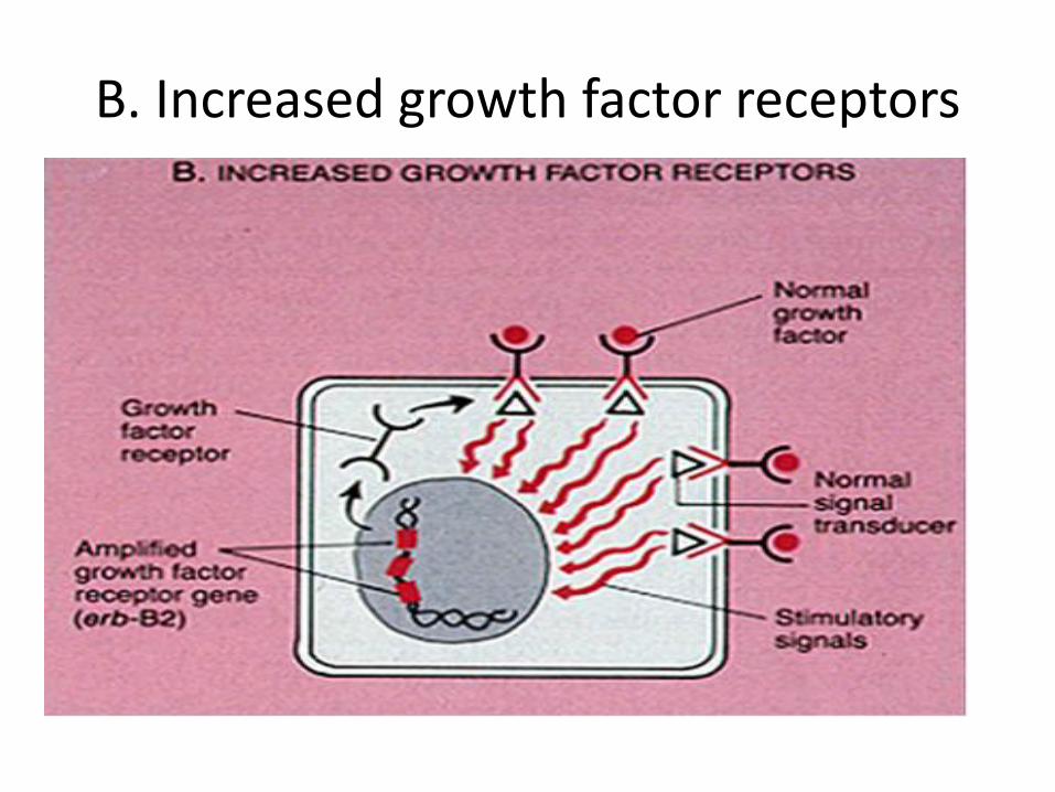
B. Increased growth factor receptors

C. Transducer Mutations

D. Mutant transcription factors

Molecular Pharmacology

Chronic myeloid leukemia
• It is a cancer of the white blood cells.
• It is a form of leukemia characterized by the increased and unregulated growth of predominantly myeloid cells in the bone marrow and the accumulation of these cells in the blood.
• CML is now largely treated with
targeted drugs called tyrosine kinase inhibitors (TKIs) which have led to dramatically improved long term survival rates since the introduction of the first such agent in 2001.

Gleevec
• Imatinib marketed by Novartis as Gleevec.
• It is a tyrosine-kinase inhibitor used in the treatment of multiple cancers, most notably Philadelphia chromosome-positive (Ph+) chronic myelogenous leukemia.
• Like all tyrosine-kinase inhibitors, imatinib works by preventing a tyrosine kinase enzyme, in this case BCR-Abl , from phosphorylating subsequent proteins and initiating the signaling cascade necessary for cancer development, thus preventing the growth of cancer cells and leading to their death by apoptosis.
Gleevec

Continue..
• Because the BCR-Abl tyrosine kinase enzyme exists only in cancer cells and not in healthy cells, imatinib works as a form of targeted therapy—only cancer cells are killed through the drug's action.
• In this regard, imatinib was one of the first cancer therapies to show the potential for such targeted action, and is often cited as a paradigm for research in cancer therapeutics.
• Its use is advised against in patients on strong CYP3A4 inhibitors such as clarithromycin, chloramphenicol and ketoconazole due to its reliance on CYP3A4 for metabolism.

Epidermal Growth Factors Receptors

Epidermal Growth Factors Receptors
• The identification of EGFR as an oncogene has led to the development of anticancer therapeutics directed against EGFR.
• Including
• Gefitinib , Erlotinib for lung cancer.
• Cetuximab for colon cancer.

Cetuximab
• It is an epidermal growth factor receptor (EGFR) inhibitor used for the treatment of metastatic colorectal cancer and head and neck cancer. Cetuximab is a chimeric (mouse/human) monoclonal antibody given by intravenous infusion
• The monoclonal antibodies block the extracellular ligand binding domain. With the binding site blocked, signal molecules can no longer attach there and activate the tyrosine kinase.

Gefitinib
• Gefitinib and Erlotinib directly target the EGFR.
• Patients have been divided into EGFR-positive and EGFR-negative , based upon whether a tissue test shows a mutation.
• EGFR-positive patients have shown a 60% response rate, which exceeds the response rate for conventional chemotherapy.

These EGFRI have an acne-like rash as a common side effect .
What is your evidence based suggestion
for the cause of this case ?

Natural EGFR inhibitors
Natural inhibitors include:
1. potato carboxypeptidase inhibitor (PCI). It is included in EGF family.
• Structural similarities with these factors can explain the antagonistic effect of PCI.
2. Grandinin is an ellagitannin found in oaks.
• It suppresses the phosphorylation of the epidermal growth factor receptor in human colon carcinoma cells.
Grandinin
potato

Vascular endothelial growth factor (VEGF)
• It is a signal protein produced by cells that stimulates vasculogenesis and angiogenesis.
• Serum concentration of VEGF is high in bronchial asthma and diabetes mellitus.

Types of VEGF and their functions
1. VEGF-A
• Angiogenesis
– ↑ Migration of endothelial cells
– ↑ mitosis of endothelial cells
– creation of blood vessel lumen
– creates fenestrations
• Chemotactic for macrophages and granulocytes
• Vasodilation (indirectly by NO release)

Continue …
2. VEGF-B : Embryonic angiogenesis (myocardial tissue,
specifically)
3. VEGF-C : Lymphangiogenesis
4. VEGF-D : Needed for the development of lymphatic
vasculature surrounding lung bronchioles

Breast cancer diagnosis


Alkylating antineoplastic agent

Do not mix between Thymineand Thiamine !
Nitrogenous bases :
• Pyrimidines :
– Cytosine (DNA, RNA)
– Uracil (RNA)
– Thymine (DNA)
• Purines
– Adenine (DNA, RNA)
– Guanine (DNA, RNA)

Where can sugar bind?

Do you know synthetic analogs for these pyrimidines ?

Preparing for nucleic acid synthesis
• DNA and RNA are nucleic acids, function in storage and transmission of genetic information.
• They are constructed of monomers called nucleotides.
• These nucleotides fuel the DNA replication, RNA transcription, mutation correction , damage and repair mechanisms.

Molecular Pharmacology

Alkylating antineoplastic agent
• They are originally derived from mustard gas used in the war.
• They have the ability to alkylate many molecules, including proteins , RNA and DNA.
• Alkylating agents work at any point in the cell cycle and thus are known as cell cycle-independent drugs.
US Army World War II Gas Identification Poster, ca. 1941–1945

Continue …
• The alkylating molecules may either bind twice to one strand of DNA (intrastrand crosslink), or , may bind once to both strands (interstrand crosslink).
• If the cell tries to replicate crosslinked DNA during cell division, or tries to repair it, the DNA strands can break.
• This leads to a form of programmed cell death called apoptosis.
Insights into the cross-linking mechanism of azinomycin B with DNA bases from hybrid QM/MM Computations

Continue …
• They impair cell function by forming covalent bonds with the amino, carboxyl, sulfhydryl, and phosphate groups in biologically important molecules.
• The most common toxicity of Alkilating agents;
Sterility
And
Secondary malignancy

Why Purines and Pyrimidines are important ?
• The sequence of amino acids in a polypeptide was specified by the sequence of nucleotides in the DNA of a gene.
• With the discovery of messenger RNA as an intermediate in the flow of information from DNA to protein, attention turned to the manner in which a sequence written in a ribonucleotide “alphabet” might code for a sequence in an “alphabet” consisting of amino acids.
Cell and Molecular Biology concepts and experiments 6 edition


Purines and Pyrimidines synthesis
• Nearly all organisms synthesize purines and pyrimidines "de novo“
• Many organisms also "salvage" purines and pyrimidines from diet and degradative pathways
• Ribose generates energy, but purine and pyrimidine rings do not
• Nucleotide synthesis pathways are good targets for anti-cancer/antibacterial strategies
http://www.d.umn.edu/~jfitzake/Lectures/DMED/Antineoplastics/DNASynthesisInhibitors/NucleotideBiochemistry.html

Conversion of Ribose-5-phosphate to PRPP
•The pentose sugar is always a ribose, which may be reduced todeoxyribose after nucleotide synthesis is complete.
•5-Phosphoribosyl-1-pyrophosphate (PRPP) is also involved insynthesis of pyrimidine nucleotides, NAD+, and histidine biosynthesis.
• Inhibited by AMP, GMP, and IMP
• Requires 4 ATP molecules

Purine de novo synthesis
• First step of purine synthesis is committed step
and rate limiting step.
•Intracellular concentrations of glutamine and PRPP control the reaction rate.
• Dependence on Glutamine in committed step (Glutamine PRPP amidotransferase ) lead to useAzaserine as anti tumor .
• Dependence on THF in two steps means that methotrexate and sulfonamides block purine synthesis
• Humans cannot synthesize folic acid , and must rely on external sources of this vitamin. Therefore, sulfa drugs do not interfere with human purine synthesis.
•The end product is Inosine- 5’- mono phosphate

Azaserine block purine synthesis
• De novo purine synthesis begins with the conversion of ribose-5-phosphate to 5-phosphoribosyl-1-pyrophosphate (PRPP), a reaction catalyzed by PRPP synthetase(PRPS).
• The first committed step in purine synthesis is the formation of 5-phosphoribosylamine via the enzyme Glutamine PRPP
amidotransferase• Azaserine – Glutamine analog-
inhibits Glutamine PRPP amidotransferase and act as Anti-tumor.

Sulfonamides block Microorganism purine synthesis
• Sulfonamides are structural analogs of para-aminobenzoic acid that competitively inhibit
bacterial synthesis of folic acid
• Because purine synthesis requires tetrahydrofolate asa coenzyme, the sulfa drugs slow down this pathway in bacteria.
• In methotrexate this hydrogen group is (methyl group)

Folic acid inhibitor
• Sulfonamides competitively inhibit ( Dihydropteroate synthetase ) in the synthesis of folic and, thereby, decrease the synthesis of nucleotides needed for the replication of DNA.
• Dihydrofolate reductase is competitively inhibited bymethotrexate, a folic acid analogue used to treatpsoriasis, rheumatoid arthritis and neoplastic diseases.

Drugs target folate synthesis
1. FOLATE BIOCHEMISTRY(e.g., METHOTREXATE) Tetrahydrofolate is synthesized by two mechanisms:
• Conversion of folate to dihydrofolate and dihydrofolate to tetrahydrofolate is catalyzed by dihydrofolate reductase (DHFR).• Methyltetrahydrofolate from liver stores is converted to tetrahydrofolate, a reaction that requires VITAMIN B12.
Two steps in the conversion of 5-phosphoribosylamine to IMP (purine synthesis) use tetrahydrofolate as a carbon donor.
Tetrahydrofolate is also involved in the generation of dTMP from dUMP (pyrimidine synthesis) – this reaction is catalyzed by thymidylate synthase
http://www.d.umn.edu/~jfitzake/Lectures/DMED/Antineoplastics/DNASynthesisInhibitors/NucleotideBiochemistry.html

Mycophenolate (cellcept)
• Mycophenolate is derived from the fungus Penicillum stoloniferum.
• Mycophenolate mofetil is metabolized in the liver to its active moiety mycophenolic acid. It inhibits inosine monophosphate dehydrogenase, the enzyme that controls the rate of synthesis of guanine monophosphate in the denovo pathway of purine synthesis used in the proliferation of B and T lymphocytes; downstream it interferes with leukocyte adhesion to endothelial cells through inhibition of E- selectin , P- selectin , and intercellular adhesion molecule -1.

Mycophenolate (cellcept)
• High levels shut down de novopurine synthesis is caused by AMP, GMP.
• If both AMP and GMP are present in adequate amounts, the de novo pathway of purine synthesis is turned off at the amidotransferase step.
Mycophenolic acid

Purine synthesisclinical importance
Gout:
During purines degradation ; Nucleotidasesand nucleosidases release ribose and phosphates and leave free bases .
Xanthine oxidase and guanine deaminase route everything to xanthine.
Xanthine oxidase converts xanthine to uricacid .
Allopurinol , which inhibits Xanthine oxidase, is a treatment of gout .
•Pentostatin inhibits adenosine deaminase-a critical pathway in purine metabolism.

Why do cancer patients takehigh doses of allopurinol?

crystal
deposition
hyperuricemia
protein binding
receptor
bindingcytokine
release
influx of PMN’s
crystals
engulfed
inflammation
Acute attacks are treated with colchicineand indomethecin for 3 weeks.
Long-term treatment with allopurinolreduces the amount of uric acid in circulation.
PMN is critical component
of crystal-induced
inflammation

Drugs induced goutkinetically !
Certain drugs can cause secondary gout due to actions on the kidney that affect uric acid reabsorption , secretion, and excretion.
• Diuretics, due to their actions on the kidney, can increase uric acid reabsorption and lead to hyperuricemia and resultant gout.
• High doses of aspirin ( >3 g/day) are uricosuric .
• Pyrazinamide, ethambutol, and niacin are other agents associated with gout because they suppress uric acid secretion.

Hydroxyurea asanti cancer
• The drug hydroxyurea destroys the free radical required for enzymic activity of ribonucleotide reductase, and thus inhibits the generation of substrates for DNA synthesis.
• For reductase to continue to produce deoxyribonucleotides, the disulfide bond created during the production of the 2'-deoxy carbon must be reduced.
• Hydroxyurea has been used in the treatment of cancers such as chronic myelogenous leukemia.

Drugs target conversion of ribonucleotides to deoxyribonucleotides
4. CONVERSION OF RIBONUCLEOTIDES TO DEOXYRIBONUCLEOTIDES (e.g., HYDROXYUREA)
This reaction is catalyzed by ribonucleotide reductase.
NOTE : Hydroxyurea, an antitumor drug, is therapeutically useful in SICKLE CELL ANEMIA because it increases circulating levels of Hb F, which decreases RBC sickling. This leads to decreased frequency of painful crises and reduces mortality.
http://www.d.umn.edu/~jfitzake/Lectures/DMED/Antineoplastics/DNASynthesisInhibitors/NucleotideBiochemistry.html

To BE CoNTINUED…….
15 MIN. BREAK

Purine synthesis salvagepath way
• Salvage pathways collect hypoxanthine and guanine and recombine them with PRPP to form nucleotides in the HGPRT reaction.
(hypoxanthine guanine phosphoribosyl transferase)
• Two enzymes are involved:
adenine phosphoribosyl transferase (APRT) and hypoxanthine-guanine phosphoribosyl transferase (HGPRT)
• Both enzymes use PRPP as the source of the ribose 5-phosphate group.
• The release of pyrophosphate and its subsequent hydrolysis by pyrophosphatase makes these reactions irreversible
• Absence of HGPRT is cause of Lesch-Nyhan syndrome.

Azathioprine
• Azathioprine, a mercaptopurine analog of adenine and hypoxanthine
• It is a prodrug that is converted first to 6 mercaptupurine (6-MP) which in turn can be converted to 6- mercaptopurine nucleotides leading to an inhibition of denovo purine synthesis.
• Three enzymes play major roles in the metabolism of azathioprineand mercaptopurine
1- xanthine oxidase (XO)2- thiopurine methyltransferase
(TPMT)3- purine pathway salvage enzyme
hypoxanthine–guanine phosphoribosyltransferase (HGPRT).

Azathioprine
• Azathioprine (AZA) is converted to 6-mercaptopurine (6-MP) in the liver via a glutathione-dependent process accelerated by glutathione-S-transferase.
• 6-MP undergoes further metabolism by
1. Xanthine oxidase (XO).
2. Thiopurine methyltransferase (TPMT).
3. Hypoxanthine guanine phosphoribosyltransferse (HPRT) within the red blood cells.
• In red blood cells it is subsequently converted by TPMT to 6-methyl-mercaptopurine ribonucleotides or by Inosine-5′- monophosphate dehydrogenase (IMPDH) to 6-thioguanine nucleotides.
• Deficiency of TPMT activity leads to accumulation of 6-thioguanine nucleotides, which may cause bone marrow toxicity.
http://en.wikipedia.org/wiki/File:AZA_metabolism.svg
IMPDH
RBC

Drugs target purine synthesis…Summary …
3. PURINE SYNTHESIS (e.g., 6-MP, 6-TG) De novo purine synthesis begins with the conversion of ribose-5-phosphate to 5-phosphoribosyl-1-pyrophosphate (PRPP), a reaction catalyzed by PRPP synthetase (PRPS). The first committed step in purine synthesis is the formation of 5-phosphoribosylamine via the enzyme glutamyl amidotransferase (GPAT).
IMP and GMP can also be created by via the “salvage pathway” whereby PRPP is combined with hypoxanthine or guanine bases (including 6-MP and 6-TG) by the actions of hypoxanthine-guanine phosphoribosyl transferase (HGPRT).
6-MP and 6-TG (and their naturally occurring analogues) inhibit guanylyl kinase, preventing the conversion of GMP to GDP and causing "pseudofeedback inhibition" of PRPS, GPAT, HGPRTand the 2 steps that lead to the formation of XMP and AMP from IMP.
One route for the degradation of purine nucleotides (and 6-MP and 6-TG) occurs via conversion of IMP to uric acid. Two steps in that process, conversion of hypoxanthine to xanthine and xanthine to uric acid, are catalyzed by the enzyme xanthine oxidase. This enzyme is inhibited by ALLOPURINOL.
http://www.d.umn.edu/~jfitzake/Lectures/DMED/Antineoplastics/DNASynthesisInhibitors/NucleotideBiochemistry.html

Pyrimidine synthesis
• Thymidylate SynthetaseThe enzyme use N5,N10-methylene tetrahydrofolate (CH3-THF) as “a methyl donor”.
• This form of folate derives its carbon from serine:
• Serine + THF ----> Glycine + CH3-THF
• 5-Fluorouracil is a pyrimidine analog which is converted metabolically to its toxic form, fluorodeoxyuridylate(F-d UMP).
• As cells metabolically activate the drug it acts as a "suicide" inhibitor of Thymidylate Synthetase.
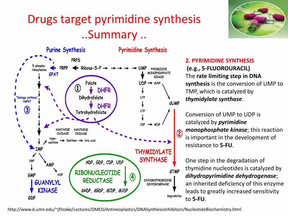
Drugs target pyrimidine synthesis..Summary ..
2. PYRIMIDINE SYNTHESIS(e.g., 5-FLUOROURACIL)The rate limiting step in DNA synthesis is the conversion of UMP to TMP, which is catalyzed by thymidylate synthase.
Conversion of UMP to UDP is catalyzed by pyrimidine monophosphate kinase; this reaction is important in the development of resistance to 5-FU.
One step in the degradation of thymidine nucleotides is catalyzed by dihydropyrimidine dehydrogenase; an inherited deficiency of this enzyme leads to greatly increased sensitivity to 5-FU.
http://www.d.umn.edu/~jfitzake/Lectures/DMED/Antineoplastics/DNASynthesisInhibitors/NucleotideBiochemistry.html


Discussion

Thank you
