protein isolation and determination of amino acid...
TRANSCRIPT

CHAPTER 3
Protein Isolation and Determination of Amino Acid Sequence
To understand the structure and function of a protein, one must know the number and kinds of amino acids present in the protein and their order (called sequence or primary structure). This information is necessary to understand the effects of mutations, mechanisms of enzyme-catalyzed reactions, and chemical synthesis of species-specific pep- tides that may eliminate undesirable hypersensitivity re- actions. Studies of amino acid sequences in proteins have aided in the understanding of the evolutionary develop- ment of living systems. Another application of amino acid sequence determination is in recombinant DNA techno- logy. A desired DNA coding for a given polypeptide can be constructed from knowledge of the precise sequence of that polypeptide (Chapter 23). Through the use of avail- able technologies of sequence determination and peptide synthesis, peptide fragments have been produced that are identical to part of large proteins present on the surface of a virus or other pathogen. In the appropriate host, such peptides elicit antibody production that is effective against the active pathogen and are potentially useful in the de- velopment of synthetic vaccines (Chapter 35). Viruses for which immunogenic peptides are being synthesized and clinically applied are hepatitis B, influenza, rabies, mouse leukemia, and hoof-and-mouth disease. Bovine in- sulin (M.W. 5,700) was the first protein to be completely sequenced (Sanger, 1955). Sanger was also the first to deduce the base sequence of a DNA molecule obtained
from phage 4~X 174 (a bacterial virus; see Chapter 23). Bovine insulin comprises two peptide chains of 21 and 30 amino acids each, linked by two interchain disulfide bonds.
In 1960, Hirs, Moore, Stein, and Anfinsen described the first primary structure of the enzyme ribonuclease (M.W. 13,700), which has a single peptide chain of 124 amino acid residues and four intrachain disulfide bonds. These investigators established many of the techniques still used in sequence analysis, such as the use of ion exchange resins for separation of peptides and amino acids.
3.1 Quantitative Determination of Proteins
UV absorption at 280 nm is an inaccurate method of protein determination because proteins have different amounts of tyrosine and tryptophan residues, and nucleic acids also absorb at 280 nm.
In the biuret test, the sample is treated with an alkaline copper sulfate reagent that produces a violet color and requires a peptide with at least two peptide bonds. The vi- olet color is produced through formation of a coordination complex (between peptide nitrogen atoms and cupric ion) that is analogous to the structure of the complex of biuret with cupric ion, as shown below:
35

36 CHAPTER 3 Protein Isolation and Determination of Amino Acid Sequence
NH2 I C = O I
2 NH + Cu 2+ I C--O I NH2
Biuret
O H H H H,O % \N / \WL- d / / ~ , , . . . _.. '% m \
,NH: +. ;NH C--N" "N--C
J / J % . I \ , , \ \ Un MI-I m O
Violet complex
l 1 1 H31~ I - A il B I C I COO -
Assume that cleavage of the above polypeptide with a specific cleaving agent yields four peptides, A, B, C, and D. These peptides are separated and sequenced but it is yet not known whether the correct order is ABCD or ACBD. A second hydrolytic procedure gives rise to three peptides: E, F, and G.
In the Folin-Ciocalteu reaction, the protein reacts with the phosphomolybdotungstic acid reagent to give a blue color through reaction with tyrosyl residues. The sensitivity of this method depends on the amino acid com- position of proteins in each sample.
Protein content, particularly in urine or cerebrospinal fluid, may also be estimated by methods based on precipi- tation using sulfosalicylic acid (an anionic protein pre- cipitant) or heat. The turbidity, which is a measure of protein concentration, can be quantitated by spectropho- tometric absorbance methods or light scattering analysis. Absorbance of a hydrophobic indicator dye that binds to protein and changes color is also used.
Specific proteins in biological fluids can be estimated by more discriminating methods, such as electrophoresis, specific binding techniques, or immunochemical methods.
3.2 Determinat ion of Primary Structure
The determination of the primary structure of a protein consists of the following steps:
1. Obtain a pure protein. 2. Determine the amino acid composition and molecular
weight of the pure protein. From amino acid composition and molecular weight data, calculate the number of residues of each amino acid present per protein molecule to the nearest whole number.
3. Reduce disulfide bonds to sulfhydryl groups. 4. Determine amino terminal (N-terminal) and carboxy
terminal (C-terminal) amino acids. (A unique residue for each terminus suggests that the native protein contains only one peptide chain.)
5. Fragment aliquots of the polypeptide by enzymatic or chemical hydrolysis and separate the peptide mixtures into individual fragments. (This process will yield overlapping sets of smaller peptides.)
6. Sequence each fragment and, by analyzing the overlapping parts, assemble the sequence of the original protein. The logic for arranging overlapping peptides is as follows:
- - - - E +---F -+ G- - -
+ .... i i I l _ A-B B-C C-D
break break break
COO -
By comparison of the amino acid sequence of fragments E and G, the terminal fragments A and D can be matched with their respective adjacent peptides, B and C. Alterna- tively, the order of the peptides B and C can be deduced from the overlapping sequence in E
Purification of a protein is indispensable for establish- ing its amino acid sequence. Mixtures of proteins will yield mixtures of peptides and ambiguous amino acid po- sitions; a unique amino acid sequence can be obtained only from a pure protein. Protein purification techniques exploit differences in size, shape, charge, solubility, and specific binding affinity of the proteins. The optimal combination of techniques is usually reached by trial and error.
3.3 Separat ion of Proteins
Proteins are separated on the basis of differences in their size, shape, charge, solubility, and binding affinity for other molecules. Most separations begin with proteins in solution. However, when proteins are an integral part of an organelle's structure, it is first necessary to extract them from membranous elements. Chemicals used to extract proteins from membranous particles include dissociating agents (e.g., urea and mercaptoethanol), chelating agents (e.g., ethylenediaminetetraacetic acid [EDTA]), and or- ganic detergents (e.g., sodium deoxycholate, sodium lau- ryl sulfate, and Triton X-100).
Since the total quantity (or activity) of a protein in a tissue sample is difficult to determine, the original (100%) quantity of protein at the start of a purification is usually based on measurements made on an aliquot of the initial homogenate. Each step in a purification process should remove extraneous protein and retain most of the protein of interest. A pure protein preparation is operationally de- fined as one that maintains a high activity per gram of pro- tein following several purification steps, i.e., the optimum

SECTION 3.3 Separation of Proteins 37
specific activity of the protein. Alternatively, a pure pro- tein cannot be further subdivided by the methods described below, e.g., chromatography or electrophoresis.
The initial purification steps usually separate proteins according to general classification, e.g., fibrous (insolu- ble) or globular (soluble). The fibrous and globular desig- nations are related to shape and solubility. Globular pro- teins are spherical or ellipsoidal and make up the majority of known proteins. Fibrous proteins contain one or more polypeptide chains. Their molecules are elongated and asymmetrical with lengths that may be many times their di- ameters. Lateral cross-linking between adjacent polypep- tides, by a variety of types of chemical bonding, confers mechanical strength and water insolubility to fibrous pro- teins; consequently, they are found in connective, elastic, and contractile tissues as well as in hair and skin. An initial aqueous extraction procedure tends to partition globular proteins into the soluble fraction and fibrous proteins into the "insoluble pellet" remaining after centrifugation.
Separation by Molecular Size
Protein can be separated on the basis of their size by d i a l y s i s , g e l f i l t r a t i o n , and m e m b r a n e f i l t r a t i o n . Small molecules (e.g., NaC1, amino acids, and sucrose) origi- nally present or added during the separation of organelles can be removed by dialysis through a semipermeable membrane. Dialysis membranes are prepared from cel- lophane or collodion and contain pores that permit pas- sage of solute molecules whose molecular weight is less than ~5000. Thus, proteins of high molecular weight are retained within a dialysis bag, whereas low-molecular- weight solutes diffuse through the pores into the fluid (dialysate) outside of the bag. Complete removal of low- molecular-weight solutes requires repeated changes of dialysate. Rapid removal of low-molecular-weight solutes and simultaneous concentration of the protein solution can be accomplished by applying pressure to the dialysis solu- tion (or vacuum to the dialysate). Such a process is known as u l t r a f i l t r a t i o n (Figure 3-1). The principles of membrane filtration are the same as those of dialysis except that syn- thetic membranes with specified pore sizes are used.
In gel filtration (or molecular sieving), a column is packed with hydrated, insoluble gel particles with known pore sizes; the protein solution is passed through the col- umn and the effluent solution is collected in fractions that will contain solutes of different sizes (Figure 3-2). The vol- ume of the column is essentially divided into two phases: the gel phase (within the pores) and the solvent phase (outside the gel particles). As a solution migrates in the column, the solute molecules that can penetrate the pores of the gel are distributed both within the gel and outside it. The particles that are larger than the pore size are excluded
Hydrodynamic force
[ Solvent
:::!~ ::!:!::ii:::::~i:::ii~. :iii'~. :i Small,
. . . . . . . . �9 ............... ~i:!~!.::~!;::~:@: Semi- . ::i~.?~-:..~)# :.~:L~"~ - - permeable :i::::i:: i i I :i:i:i:i il ::i!::i:::iiiiiii: membrane
6 J
Large, --impermeable
molecules
i ..:.
�9 . . . . . . ~ . . . . ."
6 !:i#ii ii!ii iii!iiiii',i iiii:i :;::!::;::i~i!!Oi::~iii!!O::iiiiiii::~:~:#l
..... ::::i:! ......... ! ~!~ ~ ::~:::~:i i ~ :~::~i:~ ........
F I G U R E 3-1 Separation and concentration of high-molecular-weight solutes from low-molecular-weight solutes by application of hydrodynamic force over the solution above a semipermeable membrane (ultrafiltration).
from the gel phase flow through the column more rapidly and appear first in the effluent. Commonly used gel parti- cles are inert cross-linked dextrans or agarose, which are commercially available with a wide range of exclusion limits. Molecular sieving is effective in the purification of macromolecules and if the column has been calibrated by elution of solutes of known molecular weight, it can also be used in the estimation of molecular weights of proteins or other solutes.
Separation by Chromatography
Chromatographic separations of proteins are based upon the differential partitioning of solute molecules due to their differences in affinity between a moving solvent phase and a fixed or supportive phase. In gas chromatography, the mobile phase is a gas, whereas in liquid chromatography it is a liquid. Gas chromatography is not useful in pro- tein purification because proteins cannot be converted to gases without decomposition. Liquid chromatography of proteins is performed on a variety of mechanically differ- ent stationary phases, e.g., paper, finely divided particles coated onto a glass or a plastic surface (thin-layer chro- matography), or beads packed in a column.
Many chemically different stationary phases are used in liquid chromatography of proteins. Ion exchange c h r o -
m a t o g r a p h y uses an ion exchange resin, and the proteins are eluted with buffer solutions differing in ionic strength

38 CHAPTER 3 Protein Isolation and Determination of Amino Acid Sequence
OOOo �9 _oJ Sam~,e I �9 �9 O�9 O gO0 O O'WOq~' zone DOg O e,_,_[ :iii!ii!iiii!~;ii!Qiii!iiiiii!!i~i~: I .iii:.:,:.,~::?.!',!',!iiiiiii,, ....... .,,iii!i',i':~!~:..[ i~ ~ ~i�9 !!iiii!~i ~ l i d ',
G:~!i ::. ii",i!iii::~i%!i'.':~:::. '.:: ....... Ge, ,~ i~.:!::,::Gi ;~.!,,ii!!ii:,..:,:.,::.~ pa,~de~
,
:~i!i;i:,ii~:~:~: !:iiiiiil;i!iiii',i;iiiil :~i!ii~:::Qi:i UIiI',Q
. . . . . . . . . . . . . . . . . . . . . . . . . . . . . . . . . . . . . . . . . . .
�9 i ii!i; ~; i::::;~iiii :ii !i!~i',~:: ~!iiiiii !iii',! !;i iii!iiiiiiiii;iiii!iiiiiiii !iii!ill ii!!iiiii;i!ii!ii iii!iiiiiiiii!i i
!ii !i!!i!ii! iiii!i! i!
,..,0 0
The pores of the gel particle exclude the larger solutes but admit smaller ones. Individual solute molecules are represented by the open circles.
ilJi!iiiiiiiiiiiiiiilQiiiiii!iiiii~il il
ii!i!i i!iiii!! i !ii!i]
F I G U R E 3-2 Gel filtration for separation of solutes by size. Solute molecules smaller than the diameter of the pores of the gel particles enter them and are retained for a longer time. Larger solute molecules cannot penetrate the pores of the gel particles and are eluted off the column first.
and pH. The resins are inert polymers to which ionizable groups have been attached; resins with negative charges are cation exchangers, and those with positive charges are anion exchangers. Two ion exchange resins frequently used in protein purification are diethylaminoethylcel- lulose (DEAE-cellulose, an anion exchanger) and car- boxymethylcellulose (CM-cellulose, a cation exchanger). A protein with a net positive charge at a given pH will com- bine with the negative groups of a cation exchange resin, and its flow will be retarded; a protein with a net nega- tive charge will migrate through the cation exchange resin unimpeded. Cationic proteins in a mixture going through the column will compete with one another for binding to the negatively charged groups of the resin. The relative migration rates of different molecules depend on three factors: their individual affinities for the charged sites on
the resin, the degree of ionization of the functional groups attached to the resin, and the chemical properties and con- centrations of competing low-molecular-weight ions, e.g., potassium and sodium. For example, NaC1 at the appropri- ate concentration can displace a cationic protein through competition between sodium ions (Na+) and the positively charged groups of the protein for the negatively charged sites on the resin (Figure 3-3). Changing the pH of the elut- ing buffer can also bring about desorption of proteins from the resin through neutralization of the ionizable R-groups of amino acid residues. Decreasing pH will elute proteins in the order of their decreasing is�9 point values during anion exchange chromatography.
Affinity chromatography takes advantage of specific affinities between protein molecules and analogues of bi- ological molecules that are covalently bound to the column

SECTION 3.3 Separa t i on of Pro te ins 39
Adsorp t ion ( ' :h ~ i . ~ . - . - " ~ . - ' . < ~ ~
, , , , . . . . . , . . . . . , . . . .
. . . . . . . . . . . . .
, ~ iN:i!ii!!i!ii~.:.:~!~..".'-:'!ii? k,LJ' ' , , ~lt, ~ . .,,4~!iiii! ......................
Ion e x c h a n g e (desorp t ion)
CI- x~,x\ \\x
GI- ,,e"
,,01-
Na + ~ ,~ . . . . . .
',,, ..'-'.!!!i'iii!
a + :i~'."!ii N ":i~!!i
(b)
Elut ion
CI - Na+
CI" Na
F I G U R E 3-3 Steps involved in the ion exchange chromatographic separation of proteins. (a) At an appropriate pH, a protein with positively charged groups is adsorbed to the resin by electrostatic bonding. (b) As the concentration of NaC1 is increased in the solvent flowing through the column, Na + competes with the positively charged protein for binding with the negatively charged groups or the resin, and desorption of the protein from the resin particles occurs. (c) The released protein is carried away with the flow of the solvent.
matrix (e.g., enzyme-substrate, hormone-receptor, or antigen-antibody interactions). The analogues on the col- umn are usually small molecules resembling enzyme sub- strates, hormones, or antigens. When a protein solution is applied to the column, only those proteins with a high affinity for the matrix are bound. Proteins that do not specifically bind pass rapidly through the column. The bound proteins can be eluted by altering the pH or ionic strength of the eluent or by adding excess quanti- ties of the ligand, e.g., hormone, antigen, or enzyme sub- strate or inhibitor. Affinity chromatography is useful in the purification of enzymes, hormones and their receptor sites, immunoglobulins (Chapter 35), and nucleic acids (Chapter 23).
Affinity Tag Chromatography
Affinity tag chromatography permits purification of re- combinant proteins from growth media or from cell lysates. New chromatography techniques take advantage of DNA cloning that produces recombinant fusion pro- teins and allows such proteins to be easily purified. Re- combinant proteins can be engineered to contain affinity tag sequences to create a fusion protein. The tag possesses unique affinity characteristics that serve as the basis for subsequent purification. Affinity chromatography is car- ried out using the immobilized ligand of the tag, which yields a highly purified fusion protein. A variety of affin- ity tag sequences are used such as hexa-histidine for metal chelate separation, enzyme tags that allow isolation using immobilized substrate, or epitope sequences for separa- tion by an immobilized monoclonal antibody. An enzyme cleavage site is usually included between the tag and pro- tein for removal of the tag from the fusion protein after purification. Once an effective purification strategy has been established for one fusion protein, it can be used for any protein that is engineered to include the same tag.
One example of this method is the use of glutathione S-transferase (GST) as the affinity tag. The fusion protein is engineered to contain a thrombin cleavage site between GST and the protein N terminus for subsequent removal of the GST (Figure 3-4). The sample is first run through a capture column consisting of the natural substrate for GST, glutathione bound to agarose. The column is then washed, this removing all unbound molecules and cellular debris. A large amount of free glutathione is then added to the col- umn. The free glutathione outcompetes the agarose-bound glutathione for GST causing elution of the fusion protein. The eluted protein is then run through a second column consisting of immobilized thrombin, which will remove the GST affinity tag by cutting at the thrombin cleavage site. An enzyme column is used instead of added enzyme because the bound enzyme maximizes the interaction be- tween substrate and enzyme. Furthermore, it eliminates the need for an enzyme removal step and enables the en- zyme column to be reused. The eluant from the thrombin column is passed through a gel filtration column that sep- arates the protein from the affinity tag based on molecular size. Finally, the eluant is fractionated and analyzed for purity.
Separation by Electrophoresis
Electrophoresis separates charged proteins on the basis of their different mobilities in an electric field. When a solu- tion of proteins is subjected to an electrical potential, the

40 CHAPTER 3 Protein Isolation and Determination of Amino Acid Sequence
I I 0 0 0 0 0 0
0 0 0 0 0 0 0 0 0 0 0 0 0
0 0 0 0 0 0 0 0 0 0 0 0 0
0 0 0 0 0 0 0 0 0 0 0 0 0
0 0 0 0 0 0 0 0 0 0 0 0 0
0 0 0 0 0 0 0 0 0 0 0 0 0
0 0 0 0 0 0 0 0 0 0 0 0 0
0 0 0 0 0 0 0 0 0 0 0 0 0
0 0 0 0 0 0 0 I I
Capture column (glutathione bound to agarose)
I I Po~176176176176176 o~176176176176176 Po~176176176176176 o~176176176176176 oOoOo~176 o~176176176176176 o~176176176176176 ~176176176176 o~176176176176176 ?00000000000%?
I I Thrombin column
(cleavage of GST and protein)
IQQQE
DDD~
D!oO~-C�9 ~QC D~
DDDD )QQQDC]_ DDDD o o o 8t .o: o: o_- 8t
) QQ~I D DD
(
Separation and 1 purification column
GST Protein
F I G U R E 3-4 A schematic diagram of the steps in the purification of a protein fused with glutathione S-transferase. Protein purification using this procedure utilizes three columns and is fully automatic.
charged protein molecules migrate toward either the anode or the cathode. Factors that influence the rate of migration are pH, composition of the medium through which migra- tion occurs, and size and shape of the protein molecule. A protein that does not migrate in an electric field at a given pH has no net charge at that pH. That pH is called the isoelectric point (pI) of the protein. The pI value is characteristic for each protein (Table 3-1). In a solution at a pH value above its pI, a protein will have a net nega- tive charge; below its pI, a protein will have a net positive charge. The electrophoresis technique known as isoelec- tricfocusing separates proteins on the basis of differences in their isoelectric points. Proteins whose pI values dif- fer by as little as 0.02 pH unit can be separated by this technique. In isoelectric focusing, a mixture of proteins is placed in a pH gradient; in the presence of an electric field, each protein migrates to a position corresponding to its pI and comes to rest in a narrow band at that pH. The pH gra- dient is established by placing in an electric field an aque- ous mixture of synthetic low-molecular-weight (300-600) ampholytes (oligomers of aliphatic amines, amino acids, and carboxylic acids). On application of an electric poten- tial, the ampholytes migrate and come to rest according to their respective isoelectric points. If proteins are mixed with the ampholytes, the proteins migrate to the positions of their respective isoelectric points within the ampholyte gradient and can thereby be separated and concentrated into narrow bands (Figure 3-5).
Electrophoretic techniques also yield estimates of the molecular weights of proteins and nucleic acids. The de- tergent sodium dodecyl sulfate (SDS) and proteins form
SDS-protein complexes that migrate in polyacrylamide gels according to their molecular weights. SDS dissociates multi subunit proteins into individual polypeptide chains. Each denatured chain has a uniform negative charge per unit mass of protein, since the total negative charge of the sulfonic acid groups of SDS, which are uniformly located along the surface of the protein, far exceeds the
T A B L E 3-1
Isoelectric Points of Some Proteins
Protein pl
Lysozyme 11.0 Cytochrome C 10.6 Pancreatic ribonuclease 9.6 Chymotrypsinogen 9.5 Myoglobin 7.0 Human growth hormone (somatotropin) 6.9 Hemoglobin 6.8 Human serum immunoglobulins 6.4-7.2 Carboxypeptidase 6.0 Catalase 5.6 Fibrinogen 5.5 fl-Lactoglobulin 5.2 Urease 5.1 Human serum albumin 4.8 Egg albumin 4.6 Thyroglobulin 4.6 Pepsin 1.0

SECTION 3.4 Capillary Electrophoresis 41
To high voltage
Anode
I
ii!iiii!!iiiiiiiiiil;i~i~;iiiiii~iii~%ili ' ~iiiiiiji!iiii~i!Qii!iii i!i!iiiiiiiiiii iiiiiiiiii!iiii , , i!ii Di.eren. ii!!~iiiiiiiO!~~ ~ ~ proteins
. . . . . . . . . . . . . . . . . . . . . . . . . . . . . . . . . . . . . . . . . . . . . . . . . . . . . . . . . . . . . . . . . . .
iiiiiii! iiiiiiiiiiii!!iiiiiiiiiiiili!iii !iiiiiii!iii!ii!iiiiiiiiiiiiiijii iiiiii~i!iiiii!ii!:ii!iiiii!i!!i!i Proteinsat .:i!iiiii:iiiil i!: !i:; i !~ ! il;iill isoelectric
iii!i!i iiiiii !iiiiiiill 0o,o,s
lli !iii : !ii i:ii i!i
iiiiiiiiii!!iiiiii!ii !i!iii!iiil .i!i!ii~iiii!i!i~i ] - A~luPt~~176
Izl ======================================================== _ . . . . . . . . . . . . . . . . . . . . . . . . . . . . . . . . . . . . . . . . . . . . . . . . .
: : : : : : : : : : : : : : : : : : : : : : : : : : : : : : : : : : : : : : : : : : : : : : : : : : : : : ========================================================
~ii! i l ! i! i i i~i~i~ii i! i~ii i i i ! i! i l i~i!~. : . : . : . : . : . : . : . : . , ::::::::::::::::::::::::::::::::::::::::::::::::::::: pH
- - 2 .,.iiiii!i!iiigiiiiiiiiiiiiiiiiiiiijiiiiiiiii~iiiiiii~i I ii i!l-,iiiii i i i!iiiiiiii : : : : : : : : : : : : : : : : : : : : : : : : : : : : : : : : : : : : : : : : : : : : : : : : : : :
/iiiiiiiliiii!i!iiii!ii!iiiii!iiiiiii!iiiiiiiiiiiiii; - s : : : : : : : : : : : : : : : : : : : : : : : : : : : : : : : : : : : : : : : . . . . . : :
.: ii - ~ . . . . . . . . . . . . . . . . . . . . . . . . . . . . . . . . . . . . . . . . . . . . . . . . . . . . . . .
= . : . : . : . . . . . . . . . . . . . . . . . . . . . . . . . . . . . . . . . . . . . . . . . . . . . . . : . : . : . : . : . : . : . : . : . : . : . : . : . : . : . : . : . : . : . : . : . : . : . : . : . : . : . :
~i~i~!i i : i !!! i~i:!~i~i~ii!! i! i i i i ! i! i : i : i : i : i : : : i : i : i - - 8
- . . . . . . , . . . . , . . . . . . . . . . , . . . . . , . . . . . . . . . + . . + . . . . . . : . : . : . : . : . : . : . : . : . : . : . : . : . : . : . : . : . : . : . : . : . : . : . : . : . : . : .
:!:iliiiiii!ili!i!i!!iiii!!iii~i!iii!i!i!i!iii!!i!ii!ii.. --11 . . . . . . . . . . . . . . . . . . . . . . . . . . . . . . . . . . . . . . . . . . . . . . . . . . . . . . .
. . . . . . . . . . . . . . . . . . . . . . . . . . . . . . . . . . . . . . . . . . . . . . . . . . . . . .
. . : . : . : . : . : . : . : . . . : . : . : . : . : . : . : . : . : . : . : . : . : . : . : . : . : . : . : . , . . . . . . . . . . . . . . . . . . , . . . . . . . . . . . . . . . . . . . . . . . . . . . . . . . . ._ .
. : : . : . : . . . . . . . . . . : . : . : . . . . . . . . . . . . . . . . . . . : . . . . . . . . . . ~ : : . : . : . : . : : . : . : . : . : . : . : . : . : . : . : . : . : . : . : . : . : . : . : . : . . . , , . . . . . . . . . . . . . . . . r :
Cathode (a) (b)
F I G U R E 3-5 Separation of proteins by isoelectric focusing. (a) Proteins with different isoelectric points are mixed with an appropriate ampholyte solution. (b) When an electric potential is applied, the ampholytes migrate to their pI values establishing a pH gradient, while the proteins migrate to the positions of their respective isoelectric points.
intrinsic charge of the amino acid residues. SDS-protein complexes, all of which are rod-shaped, migrate toward the anode rapidly if they are small, but slowly if they are large. The polyacrylamide medium retards migration according to the size of the rigid rods. The molecular weight of a protein is established by comparing its rate of migration with those of a number of proteins of known molecular weight.
3.4 Capillary Electrophoresis
Capillary electrophoresis is a technique that can be used to analyze and separate proteins. It has a high resolving power that exceeds other electrophoretic techniques and is capable of distinguishing between proteins that differ only slightly in amino acid composition or glycosylation.
Capillary electrophoresis is similar to high-performance liquid chromatography (HPLC) in high level of resolution, speed, on-line detection, and automation. However, it functions like slab gel electrophoresis in which proteins are separated based on mobility differences in an electric field.
In capillary electrophoresis, protein samples are first in- jected onto a fused silica microcolumn. The diameter of the fused silica microcolumns ranges between 50 and 100 #m. The free silanol groups of the fused silica become ionized at pH values greater than 2, causing the inside surface of the column to be negatively charged; the charge density is pH dependent. The micro-column sustains electric fields that are at least 50-fold more powerful than those used in slab gel electrophoresis. As a result of the high-intensity electric field, separation of proteins is more readily accom- plished and the increased sensitivity enables microcharac- terization of proteins. Due to the small size of the column, nanoliter quantities can be analyzed and separation occurs within minutes, whereas in slab gels it may take several hours. As the samples come off the column, proteins are measured by a specialized detector.
Separation by Solubility
Proteins may also be purified by selective precipitation, which is usually accomplished by changing the salt con- centration (ionic strength) of the solution. Many proteins that are readily soluble in aqueous solutions at low salt concentration exhibit decreased solubility with increased salt concentration. Protein precipitation by increasing salt concentration is known as the salting-out phenomenon. In this process, the salt ions compete with the protein molecules for interaction with solvent (water) molecules, such that the affinity between protein molecules increases as water molecules are "removed" from the surface func- tional groups of the proteins. Eventually, the decreased polymer-solvent association results in precipitation of the protein. Since different proteins have different surface functional groups, proteins can be differentially precipi- tated at different salt concentrations. The related salting-in phenomenon, which increases the solubility of some pro- teins by addition of dilute salt solution, is believed to be due to interactions between salt ions and the charged groups on protein molecules, which minimize protein-protein (precipitation) interaction and maximize protein-solvent (dissolution) interaction.
Ammonium sulfate, (NH4)2SO4, is the most commonly used compound for salting out of proteins because it is very soluble (706 g/L) and has four ionic charges per molecule. Precipitations are generally performed slowly with cold solutions to minimize protein denaturation due to the heat
42 CHAPTER 3 Protein Isolation and Determination of Amino Acid Sequence
released on mixing and to allow time for the formation of precipitates.
3.5 Amino Acid Composition
Determination of the amino acid sequence of a purified protein begins with determination of the amino acid com- position, i.e., the number of moles of each amino acid residue present per mole of protein, as follows.
The protein is completely hydrolyzed by acid (6 N HC1, 24 hours or longer at 110~ under vacuum or inert gas) to its constituent amino acids and the resultant hydrolysate is evaporated to dryness. The amino acid composition is de- termined on protein hydrolysates obtained after 24, 48, and 72 hours of acid treatment. The content of amino acids with bulky aliphatic side chains such as isoleucine, leucine, and valine, which undergo slow hydrolysis, is calculated from an extrapolation of the hydrolysate data to infinite time. The content of hydroxyl-containing amino acids, which are slowly destroyed during hydrolysis, is obtained by a corresponding extrapolation to zero time. Since cysteine, cystine, and methionine residues are somewhat unstable to hydrolysis, these residues are oxidized to cysteic acid and methionine sulfone, respectively, with performic acid before quantitative analysis. Cysteine, or half-cystine, is quantitated as a derivative such as carboxymethyl cys- teine after reduction and alkylation, a necessary pre- requisite to subsequent sequence analysis. Tryptophan
loss due to oxidation during hydrolysis can be greatly reduced by the inclusion of reducing agents--usually low- molecular-weight thiols--or oxygen-halide scavengers in the acid hydrolysis procedure. Tryptophan is substantially preserved if hydrolysis is performed with strong acids that do not contain halogen (e.g., methane sulfonic acid) or with strong alkali (e.g., NaOH). During acid hydrolysis, asparagine (Asn) and glutamine (Gin) are hydrolyzed to aspartic acid (Asp) and glutamic acid (Glu), respectively, and NH3. Thus, Asn and Gin do not appear in the elution profile, and the Asp and Glu quantities include Asn and Gin, respectively.
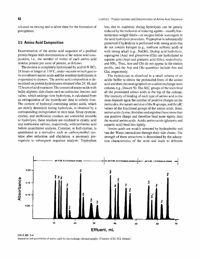
The hydrolysate is dissolved in a small volume of an acidic buffer to obtain the protonated form of the amino acid and then chromatographed on a cation exchange resin column, e.g., Dowex 50. The SO 3 groups of the resin bind all the protonated amino acids at the top of the column. The intensity of binding of each type of amino acid to the resin depends upon the number of positive charges on the molecules, the nature and size of the R-groups, and the pK' values of the functional groups of the amino acids. Basic amino acids (lysine, histidine and arginine) have more than one positive charge and therefore bind more tightly than the neutral amino acids. Acidic amino acids (glutamic and aspartic acid) bind less tightly.
Amino acids are weakly attracted by hydrophobic and van der Waals interactions through their side chains. The strength of these attractions is determined by the adsorp- tion characteristics of the resin and leads to different
to r-
. Q L _
o
. Q
L pH 3.28, 0.2 N Sodium citrate
0.20
0.15
O.
o.1o-4 | III II I
0.05
0 . g
I i
10 20 30 40 50
Effluent, mL F I G U R E 3-6 Separation and quantitation of amino acids by ion exchange chromatography. (Courtesy of Dr. N.S. Reimer.)
60 70

SECTION 3.6 Amino Acid Sequence Determination 43
elution times for each amino acid. The amino acids are differentially eluted by the use of a stepwise pH gradient varying between pH 3 and pH 5 (Figure 3-6). As each amino acid elutes, it is reacted with ninhydrin at 100~ to produce a deep blue or purple color (yellow for pro- line and hydroxyproline). The color intensity is measured spectrophotometrically to quantitate the amino acids.
The process of separation and quantitation of amino acids has been automated. In one automated method, a single cation exchange resin column separates all the amino acids in the protein hydrolysate. The analyzer is capable of detecting as little as 1-2 nmol of an amino acid and a complete analysis can be obtained in about 4 hours. In newer procedures, the complete analysis can be performed in about lhour and permit detection of as little as 1-2 nmol of an amino acid. Picomole amounts of amino acids can be determined when the separated amino acids are coupled to fluorescent reagents such as o-phthalaldehyde. Amino acid separation and quantitation can also be accomplished by reverse-phase high-pressure liquid chromatography of amino acid derivatives--a rapid and sensitive procedure.
3.6 A m i n o A c i d S e q u e n c e D e t e r m i n a t i o n
Determination of the amino acid sequence of a protein involves the following steps"
1. Identification of the N- and C-terminal amino acid residues,
2. Cleavage of any disulfide bonds present, 3. Limited cleavage of the peptide into overlapping
smaller fragments, 4. Purification of the fragments, and 5. Their stepwise cleavage into individual amino acid
residues.
Identification of the N-Terminal Residue
Determination of the N-terminal residue is carried out by labeling the free unprotonated a-amino groups. Three alternative labeling reagents are used: 2,4- dinitrofluorobenzene (DNFB; Sanger's reagent), dansyl chloride (1-dimethylaminonaphthalene-5-sulfonyl chlo- ride), and phenylisothiocyanate (PITC; Edman's reagent). DNFB and dansyl chloride react with free amino groups under basic conditions. The labeled peptide is hy- drolyzed with acid to yield the labeled N-terminal residue and other free amino acids (Figure 3-7). The 2,4- dinitrophenyl amino acid derivatives (DNP-amino acids) have a yellow color and are separable by chromatographic methods and identifiable by comparison with reference DNP-amino acids. DNFB reacts with the e-amino groups of lysyl residues to yield s-DNP-lysine after hydrolysis.
�9 N-Terminal lysine produces ol, s-di(DNP)-lysine, whereas an internal lysine produces a derivative with only one dini- trophenyl group (s-DNP-lysine).
O2N F~NO2 +
2,4- Dinitroflouro- benzene (DNFB)
H O H O H O H O i II I II I II I II D
H2NmCmC~ N ~ C ~ C ~ N m C ~ C ~ N J C ~ C m O I H I H I H I R1 ~ R3 R,
Tetrapeptide
HF ~ Base
H O
O2N--~\ / 7 - -N - -C- -C- - N \ \ / / H I H
..... "-...NO 2 R,
H O H O H O I II I II I II
- - C - - C - - N - - C - - C - - N - - C - - C - - O I H I H I R 2 R 3 R4
Acid hydrolysis Free amino acids
. , .
I H O H O H O I . +. I , i ii / / . - , - . - c - O2N C OH + H3N--CmCmOH + +
I I "-.NO 2 R, R2 R3
F I G U R E 3-7 Determination of N-terminal amino acid residues by use of 2,4-dinitrofluorobenzene (Sanger's reagent).
H O I II m
H3N--CmC--OH I R,

44 CHAPTER 3 Protein Isolation and Determination of Amino Acid Sequence
H3C ~ ~CH3 N
SO2 H O H O H O H O I I II I II I II I Ii
CI + H 2 N m C - - C m N - - C m C ~ N ~ C - - C - - N - - C - - C m O - Dansylchloride I H I H I H I
R, ~ R3 R, Tetrapeptide
HCI 4 Base
H3C ~ j C H ~ N
SO2 H O H O H O H O I I II I II I II I II
HN--C-- C-- N--C--C-- N--C--C-- N--C-- C-- 0- I H I H I H I R, R= P~ R,
H3C ~ j C H 3 N
SO2 H I I
Acid hydrolysis
Free amino acids
H H H I I I
HN--CmCOOH + +H~I--CmCOOH+ +H~I mCmCOOH + +H3N mCmCOOH I I I I P~ P~ F~ R,
Dansyl derivative of N-terminal amino acid (highly fluorescent)
FIGURE 3-8 Determination of N-terminal amino acid residues by use of dansyl chloride.
Treatment of a peptide with dansyl chloride fol- lowed by hydrolysis yields a dansyl derivative of the N-terminal amino acid and other unlabeled amino acids (Figure 3-8). The dansyl amino acid is separated and identified by chro- matographic methods. The dansyl procedure is about 100 times more sensitive than the DNFB method because the dansyl amino acids are highly fluorescent and therefore detectable in minute quantities.
In the Edman procedure, PITC reacts under ba- sic conditions with the free o~-amino group to form a phenylthiocarbamoyl peptide (Figure 3-9). Treatment with anhydrous acid yields the labeled terminal amino residue plus the remainder of the peptide. In this pro- cess, the terminal amino acid is cyclized to the cor- responding phenylthiohydantoin derivative (PTH-amino
acid), which can be identified by gas chromatography, reverse-phase high-pressure liquid chromatography, thin- layer chromatography, or as the free amino acid after hydrolysis.
A significant advantage of the Edman procedure is that on removal of the N-terminal residue, the remaining pep- tide is left intact and its N-terminal remaining peptide group is available for another cycle of the procedure. This procedure can thus be used in a stepwise manner to estab- lish the sequence of amino acids in a peptide starting from the N-terminal.
Identification of the C-Terminal Residue
The C-terminal residue is determined by the use of either a chemical reagent or the enzyme carboxypeptidase. The

SECTION 3.6 Amino Acid Sequence Determination 45
i II I N=C=s §
I I II I R, H O R 3
Phenylisothiocyanate Peptide (PITC)
t
II I II I N -- C --N ~C~,, C~N ~ CH ~c~N
I i II i R, H O R s
I Acid
I N ~ C ~ + H ~ N ~ C H ~ c ~ N ~ c ~ .
O---C ~ H NH
I H O R3
R1 Phenylthiohydantoin derivative of N-terminal amino acid (PTH-amino acid)
F I G U R E 3-9 Determination of the N-terminal residue by the Edman procedure. After removal of the N-terminal amino acid, the remainder of the peptide remains intact and a new N-terminal amino acid is available for removal by the next reaction cycle.
chemical reagent hydrazine forms aminoacyl hydrazides with every residue except the C terminus (Figure 3-10). The C terminus is thus readily identified by chromato- graphic procedures. The disadvantage of hydrazinolysis is that the entire sample is used to determine just one residue.
Carboxypeptidase is an exopeptidase that specifically hydrolyzes the C-terminal peptide bond and releases the C-terminal amino acid. Two problems are associ- ated with its use: the substrate specificity of the en- zyme and the continuous action of the enzyme. The continuous action may yield the second, third, and ad- ditional residues from some chains even before the ter- minal residues on every chain are quantitatively released. Thus, it may be difficult to determine which residue is the C terminus. However, monitoring the sequential re- lease of amino acids can often reveal the sequence of several residues at the C terminus. Concerning speci- ficity, carboxypeptidase A releases all C-terminal residues except Lys, Arg, and Pro; carboxypeptidase B cleaves C-terminal Arg and Lys residues; and carboxypeptidase C hydrolyzes C-terminal Pro residues. Thus, more than one method may be needed to establish the C-terminal amino acid.
Selective Hydrolysis Methods
Cleavage of disulfide bonds occurs before hydrolysis of the protein into peptides. Disulfide bonds may be cleaved oxidatively, or they may be reduced and alkylated. Treat- ment of the native protein with performic acid, a powerful oxidizing agent, breaks disulfide bonds and converts cys- tine residues to cysteic acid (Figure 3-11). Reduction of the disulfide linkage by thiols, such as/3-mercaptoethanol, yields reactive sulfhydryl groups. These groups may be stabilized by alkylation with iodoacetate or ethyleneimine to yield the carboxymethyl or aminoethyl derivative, respectively.
Hydrolysis of a protein into peptides can be ac- complished by group-specific chemical and enzymatic reagents (Table 3-2). N-Bromosuccinimide and cyanogen bromide hydrolyze proteins at tryptophan and methionine (Figure 3-12) residues, respectively. Trypsin hydrolyzes
H O H O H O H O I II I II I II I II
*H3N m C m C m N m C - - C - - N - - C ~ C m N ~ C ~ C ~ O- I H I H I H I R1 R2 R3 R~
NH, NH = (hydrazine)
H O H O H O H O ! II I il I II I II
+H3N~C~C~NHNH 2 + +H3N~C~C~NHNH 2 ++H3N--C~C~NHNH 2 ++H3N~C~C~O- I I I I R 1 R 2 R 3 R4
t ............. ~ C- terminal Aminoacyl hydrazides
F I G U R E 3-10 Determination of C-terminal amino acid residues by use of hydrazine.
amino acid

~6 CHAPTER 3 Protein Isolation and Determination of Amino Acid Sequence
TABLE 3-2 Hydrolysis of Polypeptides at Specific Sites by Selective Reagents
FIGURE 3-11 Cleavage of a disulfide bond (cystine residue) by performic acid.
the peptide linkage on the C-terminal side of lysine and arginine residues. Purification of the hydrolysis prod- ucts is often the most challenging aspect of sequence determination.
Anion and cation exchange chromatography, paper chromatography, and electrophoresis are useful, and reverse-phase high-pressure liquid chromatography is used increasingly because of its speed and sensitivity. The purified peptides are analyzed for amino acid composition and terminal residues. Small peptides may be sequenced directly, but large peptides must be further hydrolyzed. Proteases such as chymotrypsin, pepsin, and papain, which are much less specific than trypsin, hydrolyze the peptides formed on tryptic digestion. The amino acid sequences of the purified peptides may be determined by the sequential Edman procedure.
Another sequencing technique is indirect analysis fol- lowing nucleic acid sequencing of a DNA or RNA fragment corresponding to a specific protein. The uni- versal genetic code provides information for translating a nucleic acid sequence into an amino acid sequence. This method will not correctly identify amino acid se- quences from proteins that undergo posttranslational mod- ification or proteins derived from eukaryotic genes with in- tervening sequences that are not translated (Chapter 25). However, it is a rapid means of corroborating sequenc- ing data obtained by the classic slower methods described above.
Cleavage site
N-terminal . . . / N \ c / C - ~ " / C \ / ..... C-terminal amino acid N C amino acid
residue / \ I II residue H R1 H O
Amino acid Amino acid residue 1 residue 2
(carboxyl side) (amino side)
Cleavage Reagent Cleavage Site
Enzymes Trypsin Chymotrypsin Thermolysin
Chemicals Cyanogen bromide 2-Nitro-5-
thiocyanobenzoate N-B romosuccinimide
Hydroxylamine Mild acid hydrolysis
(70% HCOOH or 0.1 N HC1)
Lys or Arg at amino acid 1 Phe, Trp, or Tyr at amino acid 1 Leu, lie, or Val at amino acid 2
Met at aminio acid 1 Cys at amino acid 2
Tryptophan or tyrosine at amino acid 1
R l =Asn; R 2=Gly R 1 = Asp; R 2 = Pro
Peptide Sequence Confirmation
Once the sequence has been determined, the proper arrangement of individual peptides in the protein can be established by identifying the overlapping sequences be- tween peptides obtained by different cleavage procedures (Figure 3-13). The ultimate confirmation of sequence de- termination is protein synthesis. Chemical synthesis of peptides and proteins of known amino acid sequence can be accomplished by an elegant, automated, solid- phase procedure developed by Merrifield et al. Synthesis begins with the C-terminal amino acid, with each succes- sive residue added in a stepwise manner. The C-terminal amino acid is covalently bound to a solid phase by a reac- tion between the carboxyl group and a chloromethyl group linked to a phenyl group of the resin polystyrene. Since

SECTION 3.6 Amino Acid Sequence Determination 47
Peptide
NH Methionine residue
CH--CH2mCH2mS~CH3 I ~/5_
C ~ - - - O N ~ C - - B r Cyanogen bromide
HN"
CHR I
Peptide
Pel~tide
NH I
CH ~ C H 2 I I
o~ C ~ o J C H 2
Peptidyl homoserine lactone
FIGURE 3-12
Peptide I NH I A .
CH- - CH=---CH=mS--CH3 I ~ I _
C - - O N ~ C Br
I CHR I
Peptide
~ CH3~SCN Methylthiocyanate
Peptide I NH
H2 0 I " CH ~ C H 2
§ I I ~ H3 Br- C ~ jCH=
II, o CHR HN Br- I I
Peptide CHR Amino acyl peptide P|ptidee
Cleavage of a peptide chain at methionine residue by cyanogen bromide. The methionine residue is modified to a carboxy-terminal homoserine lactone residue.
the amino group of an amino acid is reactive, it must be protected by a blocking group (tertiary butyloxycarbonyl; t-BOC), so that it does not react with the chloromethyl groups. The t-BOC is later removed by acid, enabling the amino group to participate in peptide bond formation. The degradation products of t-BOC, isobutylene and carbon dioxide, are removed as gases. The carboxyl group of a second amino acid, added as a t-BOC derivative, reacts
Peptides obtained from trypsin cleavage: Ala-Gly-Glu-Lys
Gly-Ala-Met-Arg Ile-VaI-Phe
Peptides obtained from cyanogen cleavage: Ala-Gly-Glu-Lys-Gly-Ala-homoserine lactone*
Arg-Ile-VaI-Phe
*This residue is derived from methionyl residue.
The complete sequence deduced from the above overlapping peptides- Ala-Gly-Glu-Lys-Gly-Ala-Met-Arg-Ile-VaI-Phe
FIGURE 3-13 Deduction of peptide sequence from analysis of overlapping sequences of component peptides.
with the amino group of the anchored amino acid in the presence of the condensing agent, dicyclohexylcarbodi- imide (DCC). DCC, which removes H20 from the two functional groups forming a peptide bond, is converted to dicyclohexylurea. In the next cycle, the t-BOC of a second amino acid of the solid-phase dipeptide is sim- ilarly removed, and a third t-BOC-amino acid is added along with DCC. The stepwise process of peptide syn- thesis is continued until the desired peptide is formed (Figure 3-14). The finished peptide can be easily cleaved from the polystyrene resin without affecting the peptide linkages.
Advantages of the solid-phase method are amenability to automation, the almost 100% yield of product for each reaction, the ease of removal of excess reagents and waste products by washing and filtration of resin particles, the lack of need for purification of inter- mediates, and speed. Peptides or proteins that have been synthesized by the solid-phase method include ribonuclease, bradykinin, oxytocin, vasopressin, somato- statin, insulin, and the /3-chain of hemoglobin. The se- quence analyses for these substances were confirmed by demonstrating that the synthetic products, constructed on the basis of sequence data, had the same biological ac- tivities as those of the corresponding natural substances.

48 CHAPTER 3 Protein Isolation and Determination of Amino Acid Sequence
_ _ . . ~ . ~ t-BOC- Solid '~ ; ~ /~ protected amino group
support ~/~--- C H 2- Cl r l o, ,, c.., - - - - N - - C - - O - - C - - C H 3
I I i R, CH 3
CHE--O-- -- --N--C--O--C--CH3 I
" -~ R, CH 3
§ c o ,
CH,
CHE--O--C--C--N--H I R,
I II I H 0 H
Dicyclohexyl urea
O H H II I I
H O-- -C- -C- -N- - t -BOC I R2
Incoming blocked amino acid
O H H H H II I I I I
CH 2---- O - - C - - C - - N - - C - - C - - N - - t - B O C I II I R, O R2 l H ,C- -C- - -CH, + CO 2
I CH~
~ O H H H H \ // \ \ II I I I I ~ " '~--- CH 2--- O--C~C-- N--C-- C-- N-- H
/ \ ........ / i II i _ ~ / " " R, O R,
F I G U R E 3-14 Steps in the chemical synthesis of a polypeptide by the solid-phase technique.
Coupling of the C-terminal residue to the solid support
Removal of the protective group by acidification
Covalent joining of two amino acids by using the condensing agent
Removal of the protective group
Dipeptidal group anchored to the resin and ready for another cycle of amino acid addition
Fmoc Solid.Phase Peptide Synthesis Solid-phase polypeptide synthesis that uses the t-BOC
group to protect the N~-amino group is a major proce- dure used by protein chemists. However, another solid- phase procedure is frequently used for the chemical synthesis of peptides and proteins of interest; this is the 9-fluorenylmethoxycarbonyl (Fmoc) procedure (Figure 3-15). The Fmoc method for solid-phase peptide synthesis uses the Fmoc group for protecting the N~-amino group. In contrast to the t-BOG group method which requires acid for removal of the N~-amino protective group, the Fmoc group can be removed by a mild base. A piperidine solution in N,N-dimethylformamide or N-methylpyrrolidone is
generally used for the removal of the Fmoc group; an- hydrous trifluoroacetic acid is used to remove the t-BOC group. This Fmoc procedure is technically simpler and chemically less complex than the t-BOC procedure. Be- cause the Fmoc protecting group can be removed by base, the linkage of the peptide to the resin support does not have to be stable under acidic conditions as in the t-BOC procedure.
The Fmoc procedure offers more flexible reaction con- ditions and more reagent options. Because of the milder conditions of peptide synthesis, the Fmoc procedure is widely used for the synthesis of modified polypep- tides that are phosphorylated, sulfated, or glycosylated.

Supplemental Readings and References 49
FIGURE 3-15 Schematic representation of 9-fluorenylmethoxycarbonyl (Fmoc) solidphase polypeptide synthesis. (1) Amino-acid containing Fmoc as N ~-amino protecting group is attached to the insoluble polystyrene resin. (2). The Fmoc protecting group is removed by piperidine in a N,N-dimethylformamide (DMF) solution. (3). N~-amino group of amino acid attacks dicyclohexylcardodiimide (DCC)-activated carboxyl group of amino acid 2 to form a peptide bond. (4). Deprotection of N ~-amino group of the newly integrated amino acid by piperidine in the DMF solution the synthesized polypeptide is ultimately released from likage to the resin by treatment with hydrogen fluoride (HF) or trifluoroacetic acid (TFA). Steps (2) and (3) are repeated until the specific polypeptide chain is synthesized.
Since the Fmoc procedure uses a base-for protection of the N-amino group, acid-labile compounds are used to protect the side chains. Side chain protecting com- pounds generally use a t-butyl moiety such as t-butyl ethers for Ser, Thr, and Tyr; t-butyl esters for Asp and Glu; and the t-BOC group for His and Lys, respectively. Also, the trityl group is used to protect Cys, Asn, and Gln; the 4-methoxy 2,3,6,-trimethylbenzenesulphonyl or the 2,2,5,7,8,-pentamethylChroman-6- sulfonyl
groups have been used to protect the Arg guanidino group.
Supplemental Readings and References
R Alewood, D. Alewood, L. Miranda, et al.: Rapid in situ neutralization protocols for t-Boc and Fmoc solid-phase chemistries. Methods in Enzy- mology 289, 14 (1997).
J. A. Borgia and G. B. Fields: Chemical synthesis of proteins. Trends in Biotechnology 18, 243 (2000).

50 CHAPTER 3 Protein Isolation and Determination of Amino Acid Sequence
K. A. Denton and S. A. Tate: Capillary electrophoresis of recombinant proteins. Journal of Chromatography B 697, 111 (1997).
C. Jones, A. Patel, S. Griffin, J. Martin, et al.: Current trends in molecular recognition and bioseparation. Journal of Chromatography A 707, 3 (1995).
B. L. Karger, Y-H. Chu, and E Foret: Capillary electrophoresis of proteins and nucleic acids. Annual Review of Biophysical Biomolecular Structure 24, 579 (1995).
M. Lynch, R Lynch, N. Gordon, D. Whitney, et al.: Strategies for rapid purification of fusion proteins: Protein targets for drug discovery. American Biotech. Lab. 16, 8 (1998).
K. H. Mayo: Recent advances in the design and construction of synthetic peptides: for the love of basics or just for the technology of it. Trends in Biotechnology 18, 212 (2000).
D. A. Wellings and E. Atherton: Standard Fmoc protocols. Methods in Enzymology 289, 44 (1997).