production of temporary bone scaffold reinforced...
TRANSCRIPT

PRODUCTION OF TEMPORARY BONE SCAFFOLD REINFORCED WITH
OPEFB-CMC FROM OIL PALM EMPTY FRUIT BUNCH
ELIZA BINTI HJ. M. YUSUP
A thesis submitted in fulfillment of the requirement for award the degree of
Doctor of Philosophy
Faculty of Mechanical and Manufacturing Engineering
Universiti Tun Hussein Onn Malaysia
DECEMBER 2015

iii
For my parents, husband and children:
Hj. M. Yusup B. Hj. Ridhwan,
Hjh. Basimah Bte. Hj. Pahing,
Noor Nasriq Bin Selamat,
Nur Balqis Yazmin Binti Yazmi,
Muhammad Adib Fahmi Bin Yazmi,
Muhamad Alif Iskandar Bin Noor Nasriq,
Nur Awatif Afiah Binti Noor Nasriq

iv
ACKNOWLEDGEMENT
Praise be to Allah, Alhamdulillah.
Firstly, I would like to express my sincere appreciation to my supervisor, Assoc.
Prof. Dr. Shahruddin Bin Hj. Mahzan @ Mohd. Zin and co-supervisors, Prof. Dr.
Wan Rosli Wan Daud and Dr. Baharuddin Bin Mohammad for the guidance, support
continual encouragement, discussion, contribution to ideas and advice given
through-out the duration for this research. I gratefully acknowledged the financial
support from Highest Education of Ministry, Malaysia and funding from RAGS
research grant. Not to forget to Prof. Dr. Julian R. Jones from Imperial College
London for his guidance and support during attachment programme.
I would like to dedicate this thesis to my parents, Hj. M. Yusup B. Hj. Ridhwan and
Hjh. Basimah Bte. Hj. Pahing and parents-in-law, Selamat B. Abd. Ghani and
Hasiah Bt. Md. Alip. To my husband, Noor Nasriq B. Selamat and all my beautiful
children; Aqish, Adib, Alif and Awatif for understanding my situation during this
hard and difficult time. Not to forget all my siblings for help me in many ways.
I also would like to thank all my colleagues at UTHM and USM for advice and
support. To all technicians involve in this research including in UTHM and also
USM.
Finally, to everyone that was helped during my PhD research. May ALLAH blessed
all of us.
v
ABSTRACT
Bone fracture is a common injury because of its nature position that is mostly closest to
skin and exposed to excessive compression and depression. Current treatment for bone
fracture employs the scaffolding approaches which are specifically positioned for a
certain period of time. These allow the defective bones to undergo proper healing
processes. However, these scaffolds have two issues that need to be addressed; the
material’s compatibility and degradability. Previously, there was poor interaction
between the Chitosan (CS) and Hydroxyapatite (HA)/nano HA (nHA) phases causing
the composite to have poor physico-chemical properties. This research used
Carboxymethylcellulose (CMC) as the reinforcement material for CS / HA or nHA
composite scaffold. The main objective is to produce CMC from Oil Palm Empty Fruit
Bunch (OPEFB) for temporary biodegradable bone scaffold from a combination of
CMC, CS and HA/nHA. Series of experiments were done including extracting CMC
from the OPEFB, fabricating composite scaffold by a co-solution method followed by
freeze-drying approach to produce a porous bone implant. The final procedure was to
analyse the CMC and scaffold produced by various analyses and tests including FTIR,
SEM, EDX, TGA and compressive-modulus for its mechanical characteristics. The
findings indicated that the strength has increased within 32 – 50 kPa with CMC content
compared to chitosan scaffold alone which was only recorded at 0.042 – 0.7 kPa. With
the additional of Calcium Phosphate the results only recorded from 0.024 kPa until 2
kPa. The composite scaffold was also successfully constructed with lots of pores,
allowing the scaffold to demonstrate preferential proliferation and extracellular matrices
and generate mineralised bones. The investigation was extended to in-vitro test
involving Simulated Body Fluid (SBF) solution to evaluate the biodegradation rate and
vi
the growing of apatite layer during immersion. The implant had exhibited
biodegradation feature parallel to new bone formation. The ability in attracting Calcium
(Ca) and Phosphate (P) elements for apatite layer development on its surface was also
proven with the calculated value of Ca/P ratio that has identical value with the theory, at
1.67.

vii
ABSTRAK
Patah tulang adalah kecederaan biasa kerana kebiasaannya, ianya terletak paling dekat
dengan kulit menyebabkan pendedahan yang melampau pada tekanan yang tidak
disengajakan. Rawatan terkini untuk patah tulang menggunakan pendekatan perancah
yang berada pada kedudukan yang khusus untuk tempoh masa yang tertentu. Ini
membolehkan tulang yang rosak untuk menjalani proses penyembuhan semula. Walau
bagaimanapun, perancah ini mempunyai dua isu yang perlu ditangani; keserasian bahan
dan degradasi. Sebelum ini, wujud interkasi yang lemah di dalam fasa antara Chitosan
(CS) dan Hydroxyapatite (HA) / nano HA (nHA) menyebabkan komposit mempunyai
ciri-ciri fiziko-kimia yang lemah. Kajian ini menggunakan carboxymethylcellulose
(CMC) sebagai pengukuh untuk CS / HA atau nHA perancah komposit. Objektif utama
adalah untuk menghasilkan CMC dari Minyak Sawit Tandan Buah Kosong (OPEFB)
untuk perancah tulang sementara yang boleh terbiodegradasi sendiri daripada gabungan
CMC, CS dan HA / nHA. Beberapa siri eksperimen telah dilakukan termasuk
mengekstrak CMC dari OPEFB, merekabentuk perancah komposit dengan kaedah co-
solution diikuti oleh pendekatan beku-pengeringan untuk menghasilkan implan tulang
yang berliang. Prosedur akhir adalah untuk menganalisis CMC dan perancah komposit
yang dihasilkan melalui pelbagai analisis dan ujian termasuk FTIR, SEM, EDX, TGA
dan mampatan-modulus untuk ciri-ciri mekanikal. Dapatan kajian menunjukkan bahawa
kekuatan ini telah meningkat di antara 32-50 kPa bersama kandungan CMC berbanding
perancah chitosan sahaja hanya direkodkan pada 0,042-,7 kPa. Dengan tambahan
Kalsium fosfat keputusan hanya direkodkan daripada 0,024 kPa sehingga 2 kPa.
Perancah komposit ini juga telah berjaya dibina dengan banyak liang, membolehkan sel-
sel tulang untuk memulakan percambahan dan matriks extracellular
viii
dan menjana semula tulang yang baru. Siasatan itu telah dilanjutkan kepada ujian in-
vitro yang melibatkan larutan Simulated Body Fluid (SBF), kaedah untuk menilai kadar
biodegradasi dan pertumbuhan lapisan apatite semasa rendaman. Implan tersebut telah
menunjukkan cirri-ciri biodegradasi selari dengan pembentukan tulang baru. Keupayaan
dalam menarik Kalsium (Ca) dan fosfat (P) elemen untuk pembangunan lapisan apatite
di permukaannya juga dibuktikan dengan mengira nisbah Ca/P yang mempunyai nilai
yang sama dengan teori, pada 1.67.

ix
TABLE OF CONTENT
DECLARATION ii
DEDICATION iii
ACKNOWLEDGEMENT iv
ABSTRACT v
ABSTRAK vii
TABLE OF CONTENT ix
LIST OF TABLES xiii
LIST OF FIGURES xiv
LIST OF SYMBOLS AND ABBREVIATIONS xviii
CHAPTER 1 INTRODUCTION 1
1.1 Background 1
1.2 Problem Statement 4
1.3 Objective 6
1.4 Scope of Research 7
1.5 Contribution to Knowledge 8
1.6 Organization of thesis 9
CHAPTER 2 LITERATURE REVIEW 10
2.1 Introduction 10
2.2 Bone and Bone Tissue 11
x
2.2.1 Compact and spongy bone 13
2.2.1.1 Constitution 14
2.2.1.2 Histogenesis and bone growth 15
2.3 Bone Fracture 15
2.4 Bone Formation 17
2.5 Bone Graft 18
2.5.1 Allograft 18
2.5.2 Autograft 19
2.5.3 Synthetic bone graft 21
2.6 Development of Scaffolds 23
2.6.1 Characteristics 23
2.6.2 Biomaterials: Synthetic and natural 24
2.6.2.1 Metal-based biomaterials 24
2.6.2.2 Ceramic-based biomaterials 28
2.6.2.3 Polymer-based biomaterials 29
2.6.3 Scaffold fabrication method 36
2.6.3.1 Ideal combination of bioceramics and
natural polymer: Strong composite
41
2.7 Characterization of Composite Scaffold 43
2.7.1 Mechanical test 43
2.7.2 Biological factors of porous scaffold 44
CHAPTER 3 METHODOLOGY 49
3.1 Introduction 49
3.2 Overview of the Research Work 50
3.3 Preparation of Dissolving Pulp 54
3.3.1 Pre-hydrolysis process 54
3.3.2 Soda pulping 55
3.3.3 Preparation of Oxygen-Ozone-Peroxide (OZP)
pulp
56
3.4 Carboxymethylation of OZP pulp 59

xi
3.4.1 Conventional method 60
3.4.2 Alternative method 62
3.5 Preparation of a Temporary Bone Scaffold Implant 65
3.6 OPEFB-CMC and Scaffold Characterization 66
3.6.1 Mechanical testing 67
3.6.2 Microstructural properties 69
3.6.2.1 Fourier Transform Infrared
Spectroscopy (FTIR) analysis
69
3.6.2.2 Scanning Electron Microscope (SEM)
analysis
70
3.6.2.3 Energy Dispersion (EDX) Analysis 70
3.6.2.4 Porosity measurement 70
3.6.3 Physical properties 71
3.6.3.1 Thermogravimetric analysis (TGA) 71
3.6.3.2 In-vitro test: Preparation of
simulated body fluid (SBF) liquid
71
3.6.3.3 In-vitro test: Apatite-forming ability,
weight loss and swelling ability test
73
CHAPTER 4 ANALYSIS FOR OPEFB-CMC 76
4.1 Feedstock for Synthesis of Carboxymethyl Cellulose 76
4.2 Cellulose Dissolution 79
4.3 Synthesis of Carboxynthyl Cellulose
(Carboxymethylation)
81
4.3.1 Evidence of carboxymethylation 81
4.3.1.1 Fourier transform infra-red (FTIR)
analysis
82
4.3.1.2 Thermogravimetric analysis (TGA) and
derivative thermogravimetry (DTG)
84
4.3.2 Alternative method of carboxymethylation 87
4.3.2.1 Fourier transform infra-red (FTIR) 87

xii
analysis
4.3.2.2 Thermogravimetric analysis (TGA) and
derivative thermogravimetry (DTG)
88
4.3.3 Analysis of Degree of Substitution (DS) 90
CHAPTER 5 CHARACTERIZATION FOR BONE SCAFFOLD 92
5.1 Physical Characterization of Composite Scaffolds 92
5.1.1 Mechanical testing: Compression test 93
5.1.2 Compositional analysis of composite scaffolds 96
5.1.3 Thermal analysis of composite scaffold (TGA) 101
5.2 Pore Morphology of Composite Scaffolds 107
5.2.1 Pore size of the scaffold by SEM 109
5.2.2 Porosity content and swelling ability 112
5.3 Mechanical Strength and Porosity Content 114
5.4 In-vitro simulation test for composite scaffolds 116
5.4.1 Biodegradability: Weight loss 117
5.4.2 Apatite layer 119
CHAPTER 6 CONCLUSIONS AND RECOMMENDATIONS 127
6.1 Conclusion 127
6.2 Future Recommendations 130
REFERENCES 131
VITAE 156
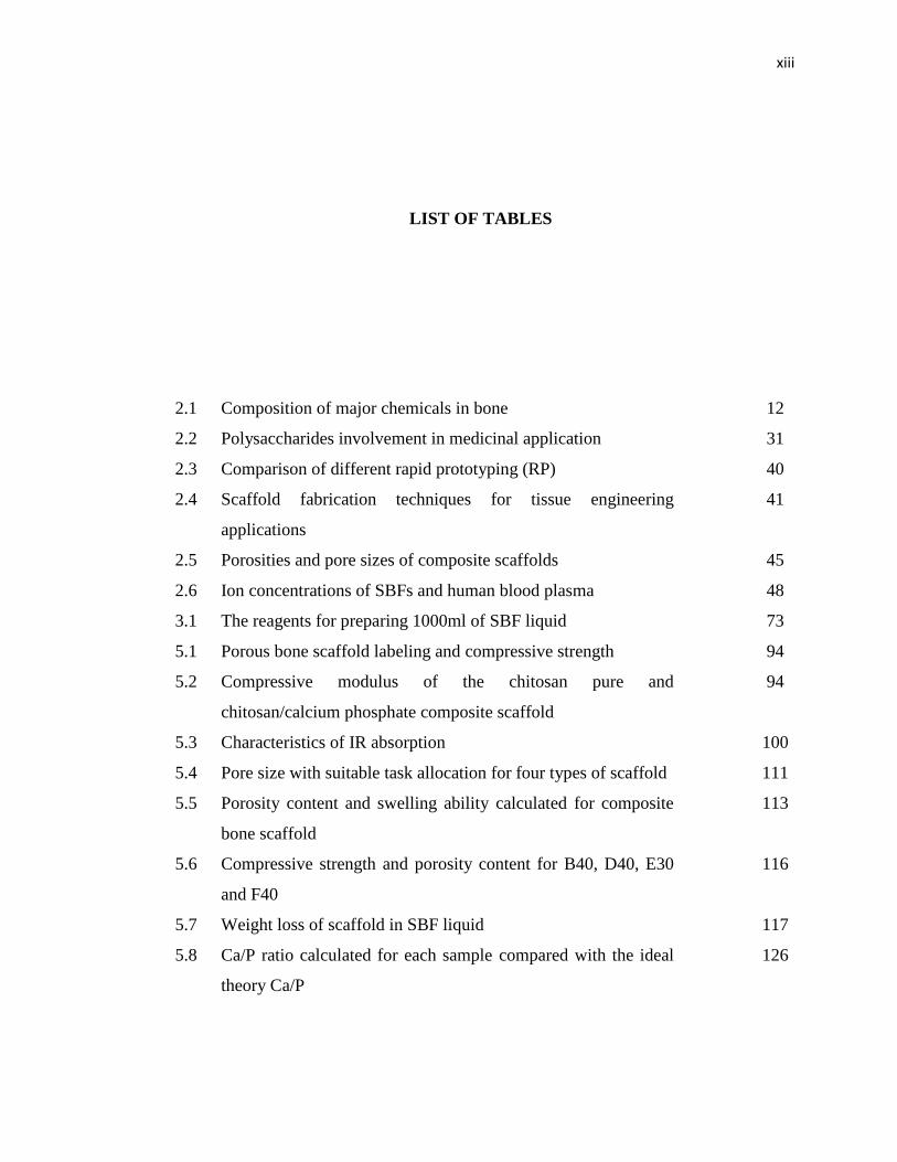
xiii
LIST OF TABLES
2.1 Composition of major chemicals in bone 12
2.2 Polysaccharides involvement in medicinal application 31
2.3 Comparison of different rapid prototyping (RP) 40
2.4 Scaffold fabrication techniques for tissue engineering
applications
41
2.5 Porosities and pore sizes of composite scaffolds 45
2.6 Ion concentrations of SBFs and human blood plasma 48
3.1 The reagents for preparing 1000ml of SBF liquid 73
5.1 Porous bone scaffold labeling and compressive strength 94
5.2 Compressive modulus of the chitosan pure and
chitosan/calcium phosphate composite scaffold
94
5.3 Characteristics of IR absorption 100
5.4 Pore size with suitable task allocation for four types of scaffold 111
5.5 Porosity content and swelling ability calculated for composite
bone scaffold
113
5.6 Compressive strength and porosity content for B40, D40, E30
and F40
116
5.7 Weight loss of scaffold in SBF liquid 117
5.8 Ca/P ratio calculated for each sample compared with the ideal
theory Ca/P
126

xiv
LIST OF FIGURES
1.1 The chemical structure of (a) CMC, and (b) Chitosan 5
2.1 (a) Macroscopic features of bone structure, (b) Composition of
the bone in volume percent
12
2.2 Cross-section of bone 13
2.3 Stages in the healing of a bone fracture 17
2.4 Autograph procedure for tracheal 20
2.5 Porous bone scaffold 22
2.6 Schematic diagram showing the different functions of a tissue
engineering scaffold depending on its porosity and pore
structure
22
2.7 Major maxillofacial defect after tumor resection and radiation
therapy
25
2.8 A typical compressive stress-strain curve of the 45S5 Bioglass-
based foams sintered at 1000°C for 1h
28
2.9 Chemical structure of chitosan 32
2.10 Cellulose structure with anhydroglucose units visible at C2, C3
and C6
33
2.11 Sodium hydroxide aqueous dissolved in distilled water for
cellulose activation and producing alkali cellulose
35
2.12 Monochloroacetic acid as etherifying agent 35

xv
2.13 Schematic representation for four commonly used chitosan
scaffold fabrication methods
38
2.14 SEM images for Trabecular bone and scaffold fabricated from
PDLLA by salt leaching technique
46
2.15 SEM images show pores for scaffold and apatite layer was
formed on the surface of the scaffold after coating with
bioactive materials
47
3.1 Part 1 – Preparation of dissolving pulp from Oil Palm Empty
Fruit Bunch (OPEFB-CMC)
51
3.2 Part 2 – Simplified diagram for CMC synthesis and analyses 52
3.3 Part 3 – Simplified diagram for scaffold fabrication, tests and
analyses
53
3.4 Illustration layout for oxygen bleaching equipment 57
3.5 Compressed oxygen tank, ozone generator and hermetic vessel:
equipments diagram for ozone bleaching
58
3.6 DP – OZ pulp 59
3.7 Diagram experimental set-up at the early stage of
carboxymethylation in water bath
61
3.8 Instrumental set-up for carboxymethylation of cellulose in the
last stage of the process
61
3.9 Experimental set-up for improvement method of preparing
CMC
64
3.10 Porous bone scaffold fabrication process 66
3.11 The standard dimension for test specimen 68
3.12 The testing machine for compression test (ASTM D695-96) 68
3.13 An example of a specimen in the SBF 74
4.1 FTIR spectra of OPEFB – cellulose pulp (OZP pulp) 77
4.2 X – ray diffraction patterns of OPEFB – cellulose pulp (OZP
pulp)
78
4.3 Cellulose solution in mixture solutions of (A) Urea/NaOH and
(B) TBAF/DMSO
79

xvi
4.4 FTIR spectrum of the OPEFB – cellulose in TBAF/DMSO
mixture solvent
80
4.5 Non – derivatizing solvent of TBAF/DMSO reacts in disrupting
the hydroxyl groups of cellulose
81
4.6 FTIR spectra of OPEFB – CMC and commercial CMC 83
4.7 TGA and DTG of OPEFB – cellulose pulp, conventional
OPEFB – CMC and commercial CMC
86
4.8 FTIR spectra of alternative OPEFB – CMC 88
4.9 TGA and DTG thermograms of alternative OPEFB – CMC 89
5.1 Compressive strength versus CMC concentration of scaffold for
(a) Sample B, (b) Sample D, (c) Sample E and (d) Sample F
95
5.2 FTIR wavelength for B40 composite scaffold 96
5.3 FTIR wavelength for D40 composite scaffold 97
5.4 FTIR wavelength for E30 composite scaffold 98
5.5 FTIR wavelength for F40 composite scaffold 99
5.6 Composite scaffold of (a) B40, (b) D40, (c) E30 and (d) F40 100
5.7 TGA curves and its derivatives analysis for chitosan 102
5.8 TGA curves and its derivatives analysis for Hydroxyapatite
(HA)
103
5.9 TGA curves and its derivatives analysis for nano HA 104
5.10 TGA analysis and its derivatives for composite scaffold of B40.
The identical pattern also shows for D40 and E30
105
5.11 TGA analysis and its derivatives for composite scaffold of F40 106
5.12 Example of porous scaffold fabricated in this research 108
5.13 SEM images for B40, D40, E30 and F40 110
5.14 Porous scaffold composite with an open pore structure (a) B40
composite scaffold and (b) chitosan/gelatin composite scaffold
112
5.15 Compressive strength versus porosity content for all specimens 115
5.16 Degradation rate for B40, D40, E30 and F40 composite scaffold 118
5.17 Composite bone scaffold of B40 before and after incubation in
Simulated Body Fluid (SBF)
121
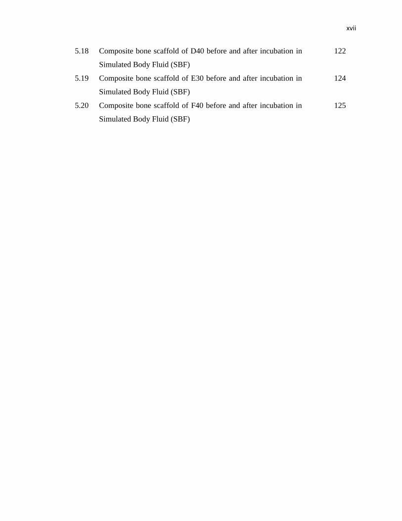
xvii
5.18 Composite bone scaffold of D40 before and after incubation in
Simulated Body Fluid (SBF)
122
5.19 Composite bone scaffold of E30 before and after incubation in
Simulated Body Fluid (SBF)
124
5.20 Composite bone scaffold of F40 before and after incubation in
Simulated Body Fluid (SBF)
125

xviii
LIST OF SYMBOLS AND ABBREVIATIONS
Abs Absorbance of the peak sample at a particular wavelength
AGU Anhydroglucose units
ad air dried
ASTM American Society of Testing and Materials
B Scaffold fabricated from CMC commercial, CS and HA
B40 Scaffold with 40% CMC conventional content, CS and HA
CA Cellulose Acetate
CaCl2 Calcium Chloride
CED Cupriethylenediamine
CH2COOH Carboxymethyl groups
CH4O Methanol
C2H4O2 Acetic acid
C2H6O Ethanol
ClCH2CO2H Monochloroacetic acid
CMC Carboxymethyl cellulose
CP Cellulose Phosphate
CS Chitosan
D Scaffold fabricated from CMC conventional, CS and nHA
D40 Scaffold with 40% CMC conventional content, CS and nHA
DMSO Dimethyl Sulfoxide
DP Dissolving pulp
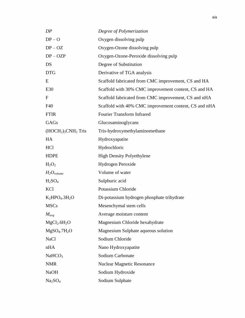
xix
DP Degree of Polymerization
DP – O Oxygen dissolving pulp
DP – OZ Oxygen-Ozone dissolving pulp
DP – OZP Oxygen-Ozone-Peroxide dissolving pulp
DS Degree of Substitution
DTG Derivative of TGA analysis
E Scaffold fabricated from CMC improvement, CS and HA
E30 Scaffold with 30% CMC improvement content, CS and HA
F Scaffold fabricated from CMC improvement, CS and nHA
F40 Scaffold with 40% CMC improvement content, CS and nHA
FTIR Fourier Transform Infrared
GAGs Glucosaminoglycans
(HOCH2)3CNH2 Tris Tris-hydroxymethylaminomethane
HA Hydroxyapatite
HCl Hydrochloric
HDPE High Density Polyethylene
H2O2 Hydrogen Peroxide
H2Ovolume Volume of water
H2SO4 Sulphuric acid
KCl Potassium Chloride
K2HPO4.3H2O Di-potassium hydrogen phosphate trihydrate
MSCs Mesenchymal stem cells
Mavg Average moisture content
MgCl2.6H2O Magnesium Chloride hexahydrate
MgSO4.7H2O Magnesium Sulphate aqueous solution
NaCl Sodium Chloride
nHA Nano Hydroxyapatite
NaHCO3 Sodium Carbonate
NMR Nuclear Magnetic Resonance
NaOH Sodium Hydroxide
Na2SO4 Sodium Sulphate

xx
od oven dried
OPEFB Oil Palm Empty Fruit Bunch
OZP Oxygen-Ozone-Peroxide
PP Polypropylene
SBF Simulated Body Fluid
SEM Scanning Electron Microscope
TAPPI Technical Association of the Pulp and Paper Industry
TBAF Tetrabutylammonium Flouride
TCF Totally Chlorine-Free
TE Tissue Engineering
TGA Thermogravimetric Analysis
V Volume
VNaOH Volume of aqueous NaOH
Voverall Volume of overall liquid
wa Initial weight of scaffold (dry weight)
wb Weight after dried in an oven
wd Dry weight of scaffold before immersed in ethanol
Wo Weight after immersed in SBF
Ww Dry weight of scaffold
ww Weight of scaffold after immersed in ethanol
XRD X-Ray diffraction
Greek letters
ρethanol Density of ethanol
CHAPTER 1
INTRODUCTION
1.1 Background
Bone is notably created to support and protect various organs in a body. It produces
red and white blood cells and also stores minerals for living things; humans and
animals. Mechanical functions of bones are for protection where bones protect
internal organs. For instance, the skull is protecting the brain and the ribs are
protecting the heart and lungs. In addition, bones also provide a structural body
frame to keep the body supported.
Dynamically, as referred to the web from The Cleveland Clinic Foundation
(2013), bones trigger movement for the body, where it provides a leverage system for
skeletal muscles, tendons, ligaments, and joints function together to generate and
transfer forces. So, individual body parts or the whole body can be manipulated in
three dimensional spaces. It obviously shows that bones are an eventful structure for
all living things for survival to execute daily and routine activities.
The characteristics of bones are very interesting and unique. It bends when it
receives sudden, unpredictable forces up to its own limitation (Riggs & Melton,
1995). However, bones are prone to impact from unwanted forces. If the forces
exerted against a bone exceeded its limit, bones could not withstand the forces and
starts to break. This phenomenon occurs as bones are only covered with very thin

2
skin and less fat surrounding them, hence provide them with little absorption during
higher impact events. Despite easily crack problems, bones are able to regenerate and
redeveloped (Yamamuro, 1995). The newly generated bones provide the same functions
and strength as normal bones. Bone regeneration is a continuous process and happens
for an entire life. Unfortunately, the regeneration process decreases slowly with the
addition of age.
Bone healing is a complex process. The time required for ossification or process
of bone healing are dependent and can be affected by many factors including types of
bone fracture and dependent on the patient’s age and their nutritional status (Alvarez &
Nakajima, 2009) . Since bone healing is a natural process, the period of time taken to
cure is of concern. Therefore, several proactive curings are taken to assist the process of
bone healing.
Autograft and allograft techniques are frequently used in order to overcome the
bone fracture problem. Autograft is a technique of replacing the fractured bone with the
healthy bone from the same person. The advantages of autograft are it provides bone
cells and growth factors that are essential for healing and bone regeneration, no risk of
disease transfer and no risk of rejection (Silber et. al., 2003; Myeroff & Archdeacon,
2011; Oppenheim, Segal & Spitzer, 2002). Despite the advantages of autograft, the
patients are required to have double surgical operations from two different sites in the
same body host. This caused double pain to the patients as well as increasing the
traumatic experiences of the patients (Valliant & Jones, 2011).
As for options, allograft technique is introduced. This technique involves the
bone transplant from different host or a bone bank. Allograft provides safer alternative
to patients who are at higher risk of complications under anesthesia. The surgeon would
not take a long time to harvest and prepare the autograft, complete the reconstruction
faster thus avoid having longer period of surgery (Mahony & Jones, 2008).
Synthetic bone graft substitution brings new phenomena in orthopaedic and
tissue engineering after more findings were discovered as an effort in curing the bone
defect. Moore, Graves & Bain (2001) quoted that a variety of synthetic bone graft
substitutes have been developed during the past 30 years with the aim to minimize the
risk of postoperative infection and fractures as well as the potential risk of disease

3
transmission as it is from synthetic origin. Moreover, synthetic bone grafts also
contribute in osteoinductive and osteostimulative (osteointegration) (Moore et. al., 2001)
which is an essential attribute for bone regeneration stage, offering biodegradable
properties, an ample supply for bone substitute and available in a wide range of size and
shape. Unfortunately, most synthetic bone grafts do not provide sufficient mechanical
strength like ceramics and they are not osteogenic.
Another type of bone treatment is by metallic implants. In this process, metal
plates were used rather than the actual bones. Normally, metal plates used were stainless
steel and titanium and Cobalt based alloys (Schmutz, Quach-Vu & Gerber, 2008). They
show a high corrosion resistance due to their stable passive layer. However, they also
have some benefits; superior in mechanical properties such as hardness and stiffness
compared to other materials such as polymer and visible during x-ray (Schmutz et. al.,
2008). Metallic implants were used in many treatments and were fairly successful, but
problems related to stress shielding during post-healing and fatigue and loosening of the
implant limit its function. Moreover, second surgery is usually required in order to
remove the metallic implant after healing, and it increases the risk of the operation and
the expense to the patient (Middleton & Tipton, 2000).
The above treatments have mentioned several benefits and drawbacks of the
treatments. It has been a desire for biodegradable implants to be developed that will
eventually biodegrade itself. Upon degradation process, ion releases are able to
encourage surrounding cells to form new bone formation more rapid at a preferred rate.
According to Pilliar et. al. (2001), the controllable rate of new bone formation is
necessary in order for the defect site to eventually be replaced by a newly formed natural
bone and strong enough to fulfil required load-bearing. The new bone can at least be
functional during the early stage of the post - implantation period, before significant
bone ingrowth and the replacement has occurred.
Most metallic materials are not biodegradable, which bring polymeric materials
more benefits than the metal implants because it eliminates the need for a second
operation and can prevent some problems associated with stress shielding. Sundararajan,
Ma & Howard (1999), Pilliar et. al. (2001), Langer & Vacanti (1993), Hubbel (1995),
Hellman (1997) and Niklason & Langer (1997) have stated that the tissue engineering

4
approach to repair and regenerate is founded upon the use of polymer scaffolding which
serve to support, reinforce and in some cases organize the regenerating tissue. So, the
reconstruction of new bone is more effective and well organized.
There is a need for the development of new biodegradable materials to be used in
orthopaedics and as scaffolding for hard tissue enginnering (Mano et. al., 1999).
Polymers are often used as matrix in bone scaffold composite. For example,
lignocellulosic fibers obtained from renewable resources where it is composed from
carbohydrate polymers is one of the example of natural polymer. An example of
carbohydrate polymer is cellulose. It is the abundant renewable resource that has become
of more and more interest as reinforcement in composites. This is because they are
biodegradable and harmless for the ecological system. Furthermore, they have promising
mechanical properties and are less expensive than conventional synthetic polymers
(Zimmermann, Pohler & Geiger, 2004).
1.2 Problem Statement
Bones are important organs to ensure smooth movement for daily activities but it is
prone to get fractured since it is surrounded by thin skin and less fat. That makes it
easily exposed to get harmed. Bone implant is the second most replaced organ in the
body after blood where approximately 2.2 million bone graft procedures are performed
worldwide each year (Giannoudis, Dinopoulos & Tsiridis, 2005). Moreover, the
estimated cost of these procedures approaches $2.5 billion per year (Desai, 2007). While
bone transplantation and tissue reconstruction are highly successful therapies for a
variety of bone diseases and fracture problems, a shortage of donor bone tissue limits
their application (Jones & Hench, 2001).
Due to the serious circumstances, the vital alternative is to create an implant
fabricated from synthetic and also natural sources. Extracellular matrices (ECMs) of
hard tissue are composed of organic (collagen type I and small amount of GAGs-
glycosaminoglycans) and inorganic phases (mainly nano hydroxyapatite crystals – nHA)
(Zhao et. al., 2002).

5
Nano scale HA is known to own excellent biocompatibility based on its close
chemical and crystal resemblance to bone material (Hench, 1998; Suchanek &
Yoshimura, 1998; Gomez-Vega et. al., 2000). While that, chitosan (CS) can accelerate
the bone formation because of the similarity to GAGs in structure (Seol et. al., 2004; Di,
Sittinger & Risbud, 2005; Madihally & Matthew, 1999; Yamane et. al., 2005; Loke et.
al., 2000).
However, there is a poor interaction between CS and HA/nHA phases causing
the composite to have poor physico-chemical properties. Due to the fact that normally,
for interface improvement between HA/nHA and CS, the second organic polymer acts as
reinforced phase in HA/nHA-based composite is essential (Jiang, Li & Xiong, 2009b).
Carboxymethyl cellulose (CMC) possesses very similar structure to CS structure which
creates strong ionic cross-linking action between CMC and CS (Xiao et. al., 2006; Qiu
& Li, 2005). This evidence has been supported by Latif, Anwar & Noor (2007) as shown
in Fig. 1.1.
Figure 1.1: The chemical structure of (a) CMC, and (b) Chitosan (Latif et. al., 2007)
Briefly, CMC, also known as cellulose gum, is a cellulose derivative with
carboxymethyl groups (-CH2-COOH). The functional group is bound to some of the
hydroxyl groups (-OH) of the glucose monomers that make up the cellulose backbone.
(a)
(b)

6
The availability of CMC sources is undoubted. In this research, it was extracted
from Oil Palm Empty Fruit Bunch (OPEFB). Empty Fruit Bunch (EFB) from palm oil
waste is a potential raw material. This is because palm oil has made an impressive and
sustained growth in the global market over the past four decades, and it is projected that
in 2016-2020, the average annual production of palm oil in Malaysia will reach 15.4
million tonnes (Teoh, 2000; Abdullah & Sulaiman, 2013).
Sulaiman et. al. (2010) indicated that large amount of oil palm residues that can
be re-utilised were dumped. This resulted in millions of ringgit energy value wasted
each year with approximate loss of about 6,379 million ringgit (Sulaiman et. al., 2010).
Due to the environmental concerns over properly disposing the waste, OPEFB could be
converted into useful material in biomedical engineering.
Therefore, a novel approach of the composite with the additional of CMC as a
natural polymer in order to reinforce CS and HA was created to address the limitations
of the previous sample. For the scaffold to integrate with surrounding tissue, it should
imitate the structure and morphology of the natural bone tissue (Stevens et. al., 2007).
Thus, there is strong ionic cross-linking action between CMC and chitosan and it is able
to produce better composite for bone scaffold. The strong ionic cross-linking between
CMC and chitosan is possible to occur because chitosan is a cationic polymer whereas
CMC is an anionic polymer where by their combination, a strong ionic bond is created
to produce stronger composite.
1.3 Objective
The aim of this research is to produce CMC from OPEFB as biomaterial for temporary
bone scaffold reinforced with chitosan and HA/nHA. In order to achieve the aim, several
objectives have been highlighted as follow:
(1) To evaluate and analyse the performance of the OPEFB-CMC as the
reinforcement material to strengthen chitosan and HA/nHA, as a porous
composite scaffold,
7
(2) To investigate the strength of composite by compression test and physical
characteristics,
(3) To evaluate the degradation time, apatite layer formation, porosity measurement
and swelling ability through in-vitro test simulation.
1.4 Scope of Research
The scope of this research includes:
(1) To produce CMC that was synthesized from oil palm waste, the empty fruit
bunch. It was chosen because it dissolved easily in water because in order to
utilize cellulose widely in any application, cellulose must be converted to soluble
derivatives. The fabrication process is also at lower cost, easy and safe to
produce. Analyses involved are FTIR and XRD.
(2) To produce porous scaffold fabricated from natural polymer and HA/nHA with
the attendance of chitosan for better physico-chemical properties.
(3) To investigate the mechanical properties and focus only on compression test in
evaluating the effectiveness of CMC as a potential material in bone scaffold. The
analysis involved is TGA analysis.
(4) To analyse the morphology of the scaffold including its porosity content either at
the surface or inside the scaffold. FTIR, SEM and EDX analyses will be
implemented to examine this.
(5) To simulate the biodegradation rate of bone scaffold and the growing of apatite
layer by immersion of samples in Simulated Body Fluid (SBF) liquid for in-vitro
test.
8
1.5 Contribution to Knowledge
CMC in this research was produced through two different methods from OPEFB. The
first method was followed by conventional and commercial product processes. It used
aqueous Sodium Hydroxide (NaOH) for cellulose activation. Improvement in fabricating
process of CMC using solid NaOH approach for cellulose activation with the
polysaccharide was priorly dissolved in Tetrabutylammonium Flouride
(TBAF)/Dimethyl Sulfoxide (DMSO). So, there is no requirement to use aqueous NaOH
to obtain CMC with higher Degree of Substitution (DS).
Conventionally, CMC wide application was in fields of membrane separation,
coating, film and textile (Fauzi, Wan Daud & Mohamad Ibrahim, 2014) while CMC is
famous in the application of food, pharmaceuticals, toothpaste, detergents, oil drilling
mud, paper coating (Ambjornsson, Schenzel & Germgard, 2013; Stigsson, Kloow &
Germgard, 2001) and others. This research attempts to use the material in orthopaedic
applications for temporary bone scaffold. The manufacturing of the product itself was
very environmentally friendly since no chlorine was involved during bleaching process,
easy and safe to produce in the laboratory.
The scaffold fabrication by previous co-solution method added all the materials
together in distilled water before uniformly agitated on hotplate. Since OPEFB-CMC
state is not identical to commercial products, a new method has been developed. In order
to dissolve and uniformly disperse the OPEFB-CMC in distilled water, there is a
requirement to sonicate it before mixed with other materials. This resulted in more
strength composite.
9
1.6 Organization of Thesis
This thesis contains six chapters. Chapter 1 is the introduction explaining briefly about
the background, problem statement, objective, scope and contribution of the research.
Chapter 2 explains the literature review of current and latest studies. This
includes explanation about bone structure, problems involving bones, the materials
commonly used for bone tissue engineering, fabricating of bone implant and some
review about the analyses and tests for bone scaffold.
Chapter 3 describes the methodology carried out during the experimental work. It
comprises of generally two main parts which are materials used and procedures in
fabricating bone scaffolds.
Chapter 4, provide the results and discussions of characterization of CMC from
oil palm waste, the empty fruit bunch.
Chapter 5 is the analysis results for porous bone scaffold fabricated from the
combination of organic materials which are chitosan and CMC and inorganic material,
hydroxyapatite and its nano size.
Chapter 6 is the final chapter of the thesis consisting of the conclusion and future
recommendations. It summarizes the overall findings from the experiments done. The
conclusions reflected the achievements of the listed objectives obtained throughout the
study. Finally, future recommendations for the research are listed for improvement of
future study in the same field of study.

10
CHAPTER 2
LITERATURE REVIEW
This chapter discusses some important features related to previous works done by other
researchers. This includes the literature reviews on tissue engineering, bone fractures,
materials used for bone replacements as well as the procedures of bone generations. This
chapter gives some information for readers on the research conducted herein.
2.1 Introduction
The conjunction of a combination of cells, engineering and materials methods, together
with suitable biochemical and physico-chemical features to improve or replace
biological functions are known as Tissue Engineering (TE) technology. It has gained so
much attention since it used the combination of biology, engineering and material
science in providing suitable biochemical and physiochemical factors to achieve better
improvement while replacing biological functions. TE involves attempts to mimic
specific biochemical and physical functions combining cells within artificially-created
support systems (Carrico et. al., 2008).
11
Engineering factors are commonly associated with the repair or replacement of
tissue portions or the whole tissue itself, for example, bone, cartilage, blood vessel, etc.
It includes the mechanical and structural properties to operate properly. The concept is
that tissues and organs can be “engineered” to be used for transplantation, which could
provide revolutionary and stimulating for self healing.
All inclusive explanation involving TE applied to bone based on natural sources
is the main focus of this research. This begins with a brief explanation on bone
constitution and characteristics, followed by more detailed clarification on material
selection, production of scaffolds and important features necessary to produce good
scaffold to be used as temporary bone implant. The scaffold will be evaluated through
several analyses and tests to clarify its effectiveness.
2.2 Bone and Bone Tissue
Bones are the rigid organs that form parts of the endoskeleton of vertebrates that
function to move, support and protect such vital organs. It is an excellent and inimitable
structural composite (Mobini et. al., 2012) composed of two major phases; collagen
fibers, serves as an organic matrix with biological apatite precipitated along the collagen
fibrils as reinforcing constituent (Park & Larks, 2007; Murugan & Ramakrishna, 2005;
Mobini et. al., 2012; Ramakrishna et. al., 2001; Olah et. al., 2006) as shown in Fig. 2.1,
designed by nature. The high elastic modulus hydroxyapatite mineral comprises
approximately 70% of the dry bone mass and contributes significantly to the bone
stiffness (Olah et. al., 2006).
Since bones are original composite, it has also come in a variety of shapes and
have a complex internal and external structure. They possess special attributes such as
lightweight, yet strong and hard. Moreover they have to fulfil many other functions
including mineral storage, acid-base balance, detoxification and sound transduction
(Carrico et. al., 2008).
12
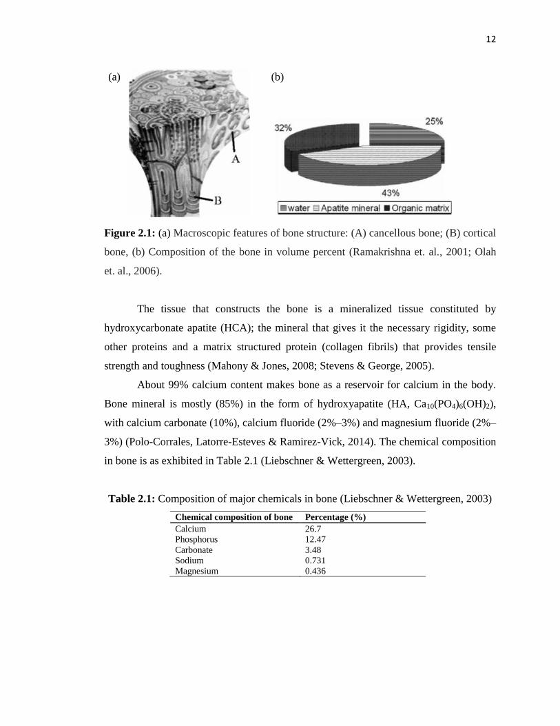
(a) (b)
Figure 2.1: (a) Macroscopic features of bone structure: (A) cancellous bone; (B) cortical
bone, (b) Composition of the bone in volume percent (Ramakrishna et. al., 2001; Olah
et. al., 2006).
The tissue that constructs the bone is a mineralized tissue constituted by
hydroxycarbonate apatite (HCA); the mineral that gives it the necessary rigidity, some
other proteins and a matrix structured protein (collagen fibrils) that provides tensile
strength and toughness (Mahony & Jones, 2008; Stevens & George, 2005).
About 99% calcium content makes bone as a reservoir for calcium in the body.
Bone mineral is mostly (85%) in the form of hydroxyapatite (HA, Ca10(PO4)6(OH)2),
with calcium carbonate (10%), calcium fluoride (2%–3%) and magnesium fluoride (2%–
3%) (Polo-Corrales, Latorre-Esteves & Ramirez-Vick, 2014). The chemical composition
in bone is as exhibited in Table 2.1 (Liebschner & Wettergreen, 2003).
Table 2.1: Composition of major chemicals in bone (Liebschner & Wettergreen, 2003)
Chemical composition of bone Percentage (%)
Calcium 26.7
Phosphorus 12.47
Carbonate 3.48
Sodium 0.731
Magnesium 0.436

13
2.2.1 Compact and spongy bone
The cross section of bones shown in Fig. 2.2 presents the transparent area of bone
(Stevens, 2008). Compact bone is tightly packed, situated at the outer layer which nearly
approaches the solid state of bone. It contributes 80% of the total bone mass of an adult
skeleton and contains 5% – 30% of pores. This tissue, also known as a dense bone due to
its minimal gaps and spaces, gives bones their smooth, white and solid appearance.
Figure 2.2: Cross-section of bone (Stevens, 2008)

14
The remaining 20% bone mass is from spongy bones since it contains a lot of
pores (sponge look-alike), estimated between 30% – 90%, with nano size where the
regeneration of new bone formation begins here. In trabeculae, it corresponds to the
areas with the lacuna (void or space) and contains osteocyte, the mature bone cells
besides having nerves and veins for blood circularization and providing for cell seeding
too (Clarke, 2008; Erikson, Axelrod & Melsen, 1994).
2.2.1.1 Constitution
The matrix builds up the major constituent of bone surrounding the cells. The inorganic
matter in the matrix represents about 50% of its dry weight. Crystalline mineral salts and
calcium are almost constituted of hydroxyapatite. The organic part of matrix is Type I
collagen. In terms of mimicking natural tissue, an ideal polymer for tissue scaffolds
would be type I collagen as it has excellent mechanical properties and comprises over
90% of the organic component of bone (Iler, 1979; Pereira et. al., 2005; Valliant &
Jones, 2011).
The main difference that distinguishes the matrix of a bone from other cell is its
hardness. The matrix is initially laid down as non-mineralized osteoid (manufactured by
osteoblasts). Mineralization involves the secretion of alkaline phosphatase by osteoblasts
vesicles. As referred to web of Boundless, Born (2015), regarding to the cells, there are
three different types: osteocytes (internal bone cells), osteoblasts (bone creation) and
osteoclasts (bone resorption).
Osteocytes, which synthesize the organic components of the matrix (type I
collagen, proteoglycans and glycoproteins), are exclusively located at the surfaces of
bone tissue. Some of them, when surrounded by newly formed matrix become
osteocytes and create an empty space, named lacuna as referred to Fig. 2.2. These
osteocytes, found each one in a different lacuna, have a kind of extensions – a network
of thin canaliculi – able to pass molecules from cell to cell. Osteoclasts, which are
multilnucleated giant cells involved in the resorption and remodeling of bone tissue, are
derived from the fusion of bone marrow – derived cells, which secret specific enzymes

15
that promote the digestion of collagen and dissolution of calcium salt crystals (Carneiro,
2005).
2.2.1.2 Histogenesis and bone growth
Bone can be formed in two different ways, there are by direct mineralization of matrix
secreted by osteoblasts (intramembraneous ossification) or by deposition of bone matrix
on a pre-existing cartilage matrix (endochondral ossification). In both processes, the
bone tissue that appears first is primary, or woven. It is a temporary tissue and is soon
replaced by the definitive lamellar secondary tissue, in a process that allows maintaining
the bone shape while it grows. The rate of bone remodelling (bone turnover) is very
active in young children, where it can be 200 times faster than that in adults. Bone
remodelling in adults is a dynamic physiologic process that occurs simultaneously in
multiple locations of the skeleton, not related to bone growth (Carneiro, 2005).
The relevance of bone TE lies on replacing the organism in a function it cannot
naturally perform. The body’s bone regenerative capacity is insufficient to heal severely
injured bone portions.
2.3 Bone Fracture
In medical, bone failure always refers to bone fracture, a condition of which the
existence of small cracks or break in the continuous bones. In general, the common
cause for bone fracture can be summarized as follows (Krucik, 2012):
1) Traumatized incidents such as a sports injury, vehicle accidents and falls.
2) Acquired diseases of bone such as osteoporosis.
3) Anomalies formation of bone in a congenital disease such as osteogenesis
imperfection and brittle bone disease.

16
Traumatized incidents contributed to more than half problems in the whole
world. It is commonly caused by activities that place stresses on bones and joints like
harsh and overwhelming sports and tremendous vehicle accidents. Disease such as
osteoporosis is also main contributor for bone fracture problem too.
Jacob et. al. (2008) clarify that osteoporosis is a disease characterized by low
bone mass and deterioration of bone structure that increases the risk of fracture. It effects
directly from the unhealthy lifestyle such as lack of daily nutrition taken needed by
bones to keep healthy.
Jacob et. al. (2008) also mentioned a rate of 26 in 100 women and 4 in 100 men
had been told by their doctors they had osteoporosis where the rate show an increment
than those found a decade earlier due to the increasing of bone mass testing and
extensive educational and awareness effort. Nevertheless, in 2004, only 16 percent of
persons admitted to the hospital were diagnosed with osteoporosis proved that
osteoporosis is significantly under-diagnosed (Jacob et. al., 2008).
Moreover, a brittle bone disease also known as Osteogenesis Imperfecta presents
at birth and occur commonly among babies who have a family history of the disease.
The worst case, it could cause hearing loss, respiratory or heart failure, spinal cord and
brain stem problems and permanent deformities (Kivi & Solan, 2012).
Furthermore, the nutritional status is usually directly related to the age of the
patient. The absorption of nutrient for elderly patient is decreased because the bone is
going through aging, which resulted in low collagen in the bone that finally causing low
bone formation. Bonjour, Schurch & Rizzoli (1996) stated that deficiency in both
micronutrients for example calcium and vitamin D and macronutrients for example
protein can accelerate age-dependent bone lose, increase the propensity to fall by
impairing movement coordination and affect protective mechanisms that reduce the
impact of falling.

17
2.4 Bone Formation
Clearly, bone regeneration naturally occur is continuously for entire life. Fig. 2.3 shows
general bone formation which clearly shows four stages in bone fracture healing process.
The stage (1) is the hematoma formation which describes the formation of blood clots
from blood leakages. During this stage, bone cells without nutrition died. Stage (2) is the
fibrocartilage formation. During this stage, fibrocartilage callus forms a splint. Tissues
with new capillaries are granulated whereas dead tissue is disposed. The fibrocartilage
callous forms connective tissue fibres, cartilage and some bony matrix. Stage (3) is
called the bony callus formation. Here, the osteoblasts and osteoclasts migrate into the
tissues and divide, replace the cartilage with bony callus. Finally, in the stage (4),
remodelling of bony matrix according to the stresses placed on bone takes place.
Permanent patch finally is produced resulted in bone healing completely.
Figure 2.3: Stages in the healing of a bone fracture. (1) Hematoma formation, (2)
Fibrocartilage callus formation, (3) Bony callus formation and finally (4) Bone
remodelling (Kivi & Solan, 2012)
Due to the advancement in medical treatments, several attempts have been made
to encourage bone formation during healing processes. This involves with bone graft,
bone synthetic graft with the assistant of foreign things such as metallic implants, and
the most recent by using biodegradable and bioactive materials such as polymers and
ceramics (Hench, 1991).
18
2.5 Bone Graft
Bone graft is one of the famous grafts after blood and skin implant (Valliant & Jones,
2011). This is another valuable and successful alternative when facing bone failure, such
as fracture and break. Bone graft is a surgical procedure needed when someone suffered
bone fracture related problems. It has been confronted with an unlimited amount of
challenges since the first successful graft had been performed in the 1870’s (Hing,
Wilson & Buckland, 2007). There are two prominent techniques in bone grafting
including allograft and autograft. Apart from these two techniques, synthetic bone graft
is also of interest when dealing with bone fractures (Bauer, 2007).
2.5.1 Allograft
Allograft possesses osteoinductive properties where few mature osteoblasts survive the
transplantation (Cypher & Grossman, 1996) but adequate numbers of precursor cells do
(Triffit, 1996). It is from these precursor cells that the osteogenic potential is derived.
Though it only could be recognized when the graft is utilized in either a morsellized or
dimineralized form (Moore et. al., 2001). This is because allograft is a surgical
operation which takes bone from another person, commonly from elderly, or also from a
bone bank as the main supplier. However, taking bone from an elderly is quite a risky as
the mechanical properties are rather low, as a result of decreased protein and collagen
contained.
Betz (2002) also has outlined several difficulties arises from having this
treatment. Since the defect bone was substituted with bone retrieved from the bone
bank, there are several issues related to minor immunogenic rejection and disease
transmissions, which unfortunately still unresolved. These bones have higher risk
contain less nutrition because in order to lower the risk of disease transmission and also
rejection, all bones are irradiated to kill all cells, leaving only the bone matrices
(Valliant & Jones, 2011).
19
Although the risk is low, the probability the bones to get infected (Moore et. al.,
2001) are still there. The irradiation process also damages the collagen structure, which
causes a severe reduction in terms of the fractures (about 64%) with rate up to 19
percent (Moore et. al., 2001) and fatigue loading (about 87%) (Costain & Crawford,
2009). However, the failure percentage depends on the dosage during the irradiation
process. The bones are also commonly harvested from elderly that cause bone density to
be low and cause poor mechanical properties (Seebach et. al., 2010).
Bone bank is more risky with complication such as non-union problem (Moore
et. al., 2001). Coalition or union of allograft is difficult to assess and inconsistency has
been found between clinical, radiological and histological union. Costly, time
consuming bone banking as well as the possible danger of viral and bacterial transmitted
diseases are disadvantages that were also concluded by Schieker et. al. (2006).
2.5.2 Autograft
In avoiding problems created by allograft, autograft is seen as a better curing for its
availability and avoidance of morbidity by autogenous graft. Autograft is largely useful
for large bone defects, which require structural support, or when insufficient autogenous
graft volume is available. The operation provides all three elements for generating and
maintaining bone tissue, namely osteogenic progenitor cells, osteoinductive growth
factors, and osteoconductive matrices (Schieker et. al., 2006). However, Schieker and
his co-workers also mentioned that this method is still restricted due to limited quantity
and an additional secondary operative procedure.
Autograft is limited in terms of more operative time, limited availability and
significant morbidity related to blood loss, wound complications, local sensory loss and
most importantly, chronic pain as the technique involves double surgery on the same
host– on the defected and donor sites. Pain persists more than three months on the donor
site, causing trauma to the patients and it seems proportional to the extent of dissection
required to obtain the graft. The second site of surgery, which is at the donor site usually
requires more time to heal as it needs further treatment under maximum supervision.
20
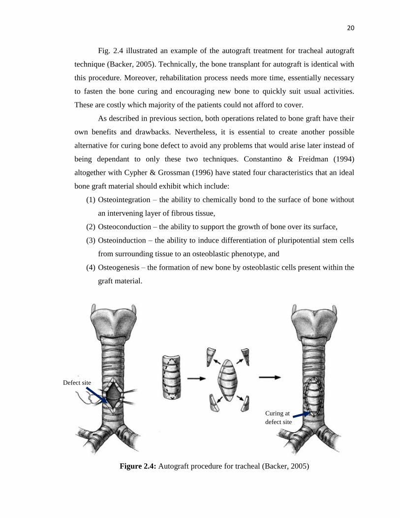
Fig. 2.4 illustrated an example of the autograft treatment for tracheal autograft
technique (Backer, 2005). Technically, the bone transplant for autograft is identical with
this procedure. Moreover, rehabilitation process needs more time, essentially necessary
to fasten the bone curing and encouraging new bone to quickly suit usual activities.
These are costly which majority of the patients could not afford to cover.
As described in previous section, both operations related to bone graft have their
own benefits and drawbacks. Nevertheless, it is essential to create another possible
alternative for curing bone defect to avoid any problems that would arise later instead of
being dependant to only these two techniques. Constantino & Freidman (1994)
altogether with Cypher & Grossman (1996) have stated four characteristics that an ideal
bone graft material should exhibit which include:
(1) Osteointegration – the ability to chemically bond to the surface of bone without
an intervening layer of fibrous tissue,
(2) Osteoconduction – the ability to support the growth of bone over its surface,
(3) Osteoinduction – the ability to induce differentiation of pluripotential stem cells
from surrounding tissue to an osteoblastic phenotype, and
(4) Osteogenesis – the formation of new bone by osteoblastic cells present within the
graft material.
Figure 2.4: Autograft procedure for tracheal (Backer, 2005)
Defect site
Curing at
defect site
21
2.5.3 Synthetic Bone Graft
The awareness of bone shortage by applying either allograft or autograft technique in
curing bone defect is making researches around the world to figure out several
alternatives. The most effective one is by creating a material (other than human sources),
that could imitate the ability of natural bone. Synthetic is an unnatural substantial made
artificially from materials like ceramic and glass materials. There is also natural or
synthetic polymer and the combination of both polymer and ceramic producing stronger
composites and lighter which are similar to natural bones (Zimmermann et. al., 2011).
Bauer (2007) said that the material involved with the fabrication of synthetic
bone graft should have biological properties like osteointegration which has the ability
to chemically bond with the bone surface. In order to implement the task perfectly,
osteoinduction which promotes bone formation along its surface when it is placed into
the bone would take part followed by differentiation of stem cells from surrounding
tissues into osteoblast cells. Osteoblasts are unique cells designed to support the
formation of new bones. This is known as osteogenesis.
Synthetic bone substituting graft appearances are diversified such as in the form
of bone cement filling, membrane and among them, bone scaffold is the most preferred
because Chen, Roether & Boccaccini (2008) highlighted that bone scaffold is highly
porous, three-dimensional (3D) which exhibit tailored porosity, pore size and
interconnectivity for vascularization. Fig. 2.5 exhibited µCT image of a silica/γ-PGA
hybrid foam scaffold from previous works (Valliant & Jones, 2011).
Moreover, porous bone scaffold could priorly seeded with cell culture during in
vitro activity. The implementation is related to the application of a tissue-engineered
implant surface to permanently stabilize implants by coating the prosthesis with cells or
tissue before implantation (Burg, Porter & Kellam, 2000). Reconstructive orthopaedic
surgeries took credit with this benefit because it always have high possibility incidences
of failure secondary to large bone defect (Burg et. al., 2000; Dekker et. al., 1998).

22
Figure 2.5: Porous bone scaffold (Valliant & Jones, 2011)
However, porosity and pore structure are the key parameters which are
determining the properties and the applicability of scaffolds for tissue engineering. In
Fig. 2.6 shows a summary of different functions related to the pore structure that must be
tailored to the particular tissue under consideration (Boccaccini & Maquet, 2003).
Figure 2.6: Schematic diagram showing the different functions of a tissue engineering
scaffold depending on its porosity and pore structure (Boccaccini & Maquet, 2003)
Sufficient pore volume: tissue expansionSpatial pore orientation and continuity:
reconstruction of tissue framework
Macropores (100µm): cell infiltration, invasion of blood vessels, building of
tissue layers.
Micropores (10-50µm): cell adhesion, diffusion of oxygen and nutrients, waste
clearance
Mechanical stability
Physical properties of porous scaffolds

23
It is a tricky and challenging task to fulfil all the essential requirements in
producing good porous scaffold. All important aspects should take into consideration
that would related from the materials chosen, fabricating process, the design and not to
forget the characteristics in developing a scaffold that will be explained in detail in the
next section.
2.6 Development of Scaffolds
The development in fabricating scaffolds indicate positive improvement since it was first
invented decades ago (Gösta, 2000). Several important attributes are taken into
consideration such as the characteristics of the bone scaffold, the materials chosen,
scaffold architecture and its fabrication with the main aim is to produce a tough even
having high porosity content.
2.6.1 Characteristics
Vascularization is essentially necessary because large scaffolds need blood vessels for
successful bone regeneration. Blood vessels can be grown in vitro prior to implantation
so they will connect the host vessels after implantation. To achieve that, stem cells and
endothelial cells were seeded inside a gel and scaffolds were soaked in it for about one
day for veins to be traced.
An ideal bone scaffold should at least exhibit some of these general attributes
(Jones, Lee & Hench, 2006; Jones, Gentlemen & Polak, 2007; Jones et. al., 2009):
1) Act as a template for bone growth in 3D and has an interconnected pore structure
to allow 3D bone ingrowth.
2) Resorbs at the same rate as the bone is repaired, producing degradation products
that are non-toxic and that can be excreted easily by the body.
3) Biocompatible (non-toxic) and promotes cell adhesion, stimulating new bone
growth (osteogenesis).
24
4) Bonds to the host bone without the formation of scar tissue, creating a stable
interface.
5) Exhibit mechanical properties matching those of the host bone after in vitro
tissue culture.
6) Made from a processing technique that can produce irregular shapes to match
that of the bone defect.
7) Has the potential to produce commercially and sterilized to the required
international standards for clinical use.
2.6.2 Biomaterials: Synthetic and Natural
Synthetic is defined as something that chemically or sometimes naturally made
exclusively to imitate the natural product. The terminology of biomaterials are product
that interacts with biological systems. Synthetic biomaterials could be simplified as
something that synthesized chemically to mimic the natural product so the products
produced are able to interact with biological system. Materials that synthetically
synthesized in order to fabricate bone scaffold commonly from metals, ceramics, glass,
chemically synthesized polymers, natural polymers and combinations of these materials
to form composites (Karageorgiou & Kaplan, 2005).
2.6.2.1 Metal-based biomaterials
In attempting to repair the skeletal systems, surgeons must endeavor to replicate the
static and dynamic responses of the bone. Bone exhibits a higher flexural strength and
flexural modulus than polymeric materials but is weaker and more deformable than
metals (Dunn & Casper, 1985). Therefore, there is vital circumstances in searching the
most suitable material for temporarily substitute defect bone.

131
REFERENCES
Abdullah, N. and Sulaiman, F. (2013). The oil palm wastes in Malaysia (Chapter 3).
In. Biomass now – sustainable growth and use, INTECH. Unpublished.
Alvarez, K. and Nakajima, H. (2009). Review: Metallic scaffolds for bone
regeneration, Materials, pp. 790 – 832.
Ambjornsson, H. A., Schenzel, K. and Germgard, U. (2013). Carboxymethyl
cellulose produced at different mercerization conditions and characterized by
NIR FT Raman Spectroscopy in combination with multivariate analytical
methods, BioResources, 8 (2), pp. 1918 – 1932.
American Society for Testing and Materials. Standard test method for compressive
properties of rigid plastics. United States, D695. 1996.
Archana, D., Upadhyay, L., Tewari, R. P., Dutta, J., Huang, Y. B. and Dutta, P. K.
(2013). Chitosan-pectin-alginate as a novel scaffold for tissue engineering
applications, Indian Journal of Biotechnology, 12, pp. 475 – 482.
Ashby, M. F. (1984). The Mechanical Properties of Cellular Solids, Metallurgical
Transactions A, 14 (9), pp. 1755 – 1769.
Backer, C. L. Compression of the trachea by vascular rings. In: Shields, T. W.,
LoCicero, J., Ponn, R. and Rusch, V. W. General Thoracic Surgery (6th
Eds.). Lippincott Williams & Wilkins. 2005.
Bagambisa, B., Joos, U. and Schilli, W. (1993). Mechanisms and structure of the
bond between bone and hydroxylapatite ceramics, Journal of Biomedical
Materials Research, 27, pp. 1047 – 1055.
132
Bauer, T. W. (2007). Bone graft substitutes, Skeletal Radiol, 36, pp. 1105 – 1107.
Betz, R. R. (2002). Limitations of autograft and allograft: new synthetic solutions.
Orthopedics. Retrieved at September 1st, 2014 from http://europepmc.org.
Biswal, D. R. and Singh, R. P. (2004). Characterisation of Carboxymethyl cellulose
and polyacrylamide graft copolymers, Carbohydrated Polymers, 57, pp. 379
– 387.
Boccaccini, A. R., Chatzistavrou, X., Mohamad Yunos, D. and Calitano, V.
Biodegradable polymer-bioceramic composite scaffolds for bone tissue
engineering. Proceedings of 17th
International Conference on Composite
Structures ICCM17. 27th
July – 31st July 2009. Edinburgh, Scotland: IOM
Communications Ltd. 2013. pp. 1 – 9.
Boccaccini, A. R. and Maquet, V. (2003). Bioresorable and bioactive
polymer/bioglass composites with tailored pore structure for tissue
engineering applications, Composites Science and Technology, 63, pp. 2417 –
2429.
Bonjour, J-P., Schurch, M-A. and Rizzoli, R. (1996). Nutritional aspects of hip
fractures, Bone, 18 (3), pp. 139 – 144.
Borden, M., Attawia, M. and Laurencin, C. T. (2002). The sintered microsphere
matrix for bone tissue engineering: in vitro osteoconductivity studies, Journal
of Biomedical Material Res., 61, pp. 421 – 429.
Born, K. L. (2015). Cell types in bone. Retrieved on 17th November, 2015. From
https://www.boundless.com.
Branda, S. S., Chu, F., Kearns, D. B., Losick, R. and Kolter, R. (2006). A major
protein component of the Bacillus subtilis biofilm matrix, Molecular
Microbiology, 59 (4), pp. 1229 – 1238.
Brånemark, R., Brånemark, P-I., Rydevik, B and Myers, R. R. (2001).
Osseointegration in skeletal reconstruction and rehabilitation, 38(2).
133
Brodke, D. S., Gollogly, S., Alexander, M. R., Nguyen, B. K., Dailey, A. T. and
Bachus, A. K. (2001). Dynamic cervical plates: biomechanical evaluation of
load sharing and stiffness, Spine, 26 (12), pp. 1324 – 1329.
Burg, K. J. L., Porter, S, and Kellam, J. F. (2000). Biomaterial developments in bone
tissue engineering, Biomaterials, 21, pp. 2347 – 2359.
Cai, X., Tong, H., Shen, X., Chen, W., Yan, J. And Hu, J. (2009). Preparation and
characterization of homogeneous chitosan-polylactic acid/hydroxyapatite
nanocomposite for bone tissue engineering and evaluation of its mechanical
properties, Acta Biomaterialia, 5 (7), pp. 2693 – 2703.
Carneiro, J. E. (2005). Basic histology, 11th
ed., McGraw – Hill.
Carrico, A. C., Farracho, M., Nunes, C., Ruela, A. M. and Semedo, J., (2008). Bone
tissue engineering: Production of scaffolds. In: Introduction to engineering
biomaterials. Unpublished.
Chandy, T. and Sharma, C. (1990). Chitosan as a biolmaterial, Journal of
Biomaterial Artificial Cell Artificial. Organs, 18(1), pp. 1 – 24.
Chen, Q. Z., Thompson, I. D. and Boccaccini, A. R. (2006). 45S5 Bioglasss-derived
glass–ceramic scaffolds for bone tissue engineering, Biomaterials, 27, pp.
2414 – 2425.
Chen, Q., Roether, J. A. and Boccaccini, A. R. (2008). Tissue Engineering Scaffolds
from Bioactive Glass and Composite Materials. In. Ashammakhi, N., Reis, R.
and Chiellini, F. (Eds.). Topics in Tissue Engineering.
Chipara, M., Lozano, K., Hernandez, A. and Chipara, M. (2008). TGA analysis of
polypropylene-carbon nanofibers composites, Polymer Degradation and
Stability, 93 (4), pp. 871 – 876.
Cho, S. B., Nakanishi, K., Kokubu, T., Soga, N., Ohtsuki, C., Nakamura, T., Kitsugi,
T. and Yamamuro, T. (1995). Dependence of apatite formation on silica gel
on its structure: effect of heat treatment, Journal of the American Ceramic
Society, 78, pp. 1769 – 1774.

134
Chu, C.C., Pratt, L., Zhang, L., Hsu, A. and Chu, A. (1993). A Comparison of a New
Polypropylene Suture with Prolene, Journal of Applied Biomaterials, 4, pp.
169 – 181.
Clarke, B. (2008). Normal bone anatomy and physiology, Clinical Journal of The
American Society of Nephrology, pp. 131 – 139.
Constantino, P. D. and Freidman, C. D. (1994). Synthetic bone graft substitutes,
Otolaryngol. Clin. North Am., 27, pp. 1037 – 1073.
Cooke, F. W. (1992). Ceramics in orthopaedic surgery, Clinical orthopaedic, 276,
pp. 135 – 146.
Costain, D. J. and Crawford, R. W. (2009). Fresh-frozen vs. Irradiated allograft bone
in orthopaedic reconstructive surgery, Injury, 40 (12), pp. 1260 – 1264.
Cruz, S. E. S. B., Beelen, P. M. G., Silva, D. S., Pereira, W. E., Beelen, R. and
Beltrao, F. S. (2007). Characterization of condensed tannin of the species
manicoba (Manihot pseudoglaziovii), flor-de-seda (Calotropis procera),
feijao-bravo (Capparis flexuosa) and jureminha (Desmanthus virgatus), Arq.
Bras. Med. Vet. Zootec., 59 (4), pp. 1038 – 1044.
Cypher, T. J. and Grossman, J. P. (1996). Biological principles of bone graft healing,
Journal of Foot and Ankle Surgery, 35, pp. 413 – 417.
Czaja, W. K., Young, D. J., Kawecki, M. and Brown, R. M. (2007). The future
prospects of microbial cellulose in biomedical applications,
Biomacromolecules, 8, pp. 1 – 12.
Dekker, P. J., Ryan, M. T., Brix, J., Muller, H., Honlinger, A. and Pfanner, N.
(1998). Preprotein translocase of the outer mitochondrial membrane:
Molecular dissection and assembly of the general import pore complex,
Molecule, Cell & Biology, 18 (11), pp. 6515 – 6524.
Den Boer Frank, C., Wippermann, B. W. and Blokhuis, T. J. (2003). Healing of
segmental bone defects with granular porous hydroxyapatite augmented with
recombinant human osteogenic protein-1 or autologous bone marrow,
Journal Orthop. Res., 21(3), pp. 521-528.

135
Desai, B. M. (2007). Osteobiologics, Am. Journal of Orthopaedics, 36, pp. 8 – 11.
Di, M. A., Sittinger, M. and Risbud, M. V. (2005). Chitosan: a versatile biopolymer
for orthopaedic tissue-engineering, Biomaterials, 26, pp. 5983 – 5990.
Dunn, R. L. and Casper, R. A. Methods of producing biodegradable prothesis and
products thereform. U. S. Patent EP 0146398 A2. 1985.
Dwek, R. A. (1996). Glycobiology: Toward Understanding the Function of Sugars,
Chem. Rev., 96 (2), pp. 683 – 720.
Entcheva, E., Bien, H., Yin, L., Chung, C. Y., Farrel, M. and Kostov, Y. (2004).
Functional cardiac cell constructs on cellulose-based scaffolding,
Biomaterials, pp. 5753 – 5762.
Eriksen, E. F., Axelrod, D. W. and Melsen, F. (1994). Bone Histomorphometry.
Raven Press. pp. 1 – 12.
Fan, H., Hui, J., Duan, Z., Fan, D., Mi, Y., Deng, J., and Li, H. (2014). Novel
scaffolds fabricated using Oleuropein for bone tissue engineering, BioMed
Research International, pp. 1 – 11.
Fauzi, M. D., Wan Daud, W. R. and Mohamad Ibrahim, M. N. (2014). Synthesis and
characterization of Cellulose Acetate from TCF Oil Palm Empty Fruit Bunch
Pulp, BioResources, 9 (3), pp. 4710 – 4721.
Flautre, B., Descamps, M., Delecourt, C., Blary, M. C. and Hardouin, P. (2001).
Porous HA ceramic for bone replacement: role of the pores and
interconnections – experimental study in the rabbit, Journal of Material
Science and Material Medicine, 12, pp. 679 – 682.
Fricain, J. C., Granja, P. L., Barbosa, M. A., de Jeso, B., Barthe, N., and Baquey, C.
(2002). Cellulose phosphates as biomaterials. In vivo biocompatibility
studies, Biomaterials, 23, pp. 971 – 980.
Fuentes, S., Retuert, J. and Gonzalez, G. (2008). Preparation and properties of
chitosan hybrid films from microwave irradiated solutions, Journal of
Chileon Chemical Society, 53, pp. 1511 – 1514.
136
Fukuya, M. N., Senoo, K., Kotera, M., Yoshimoto, M. and Sakata, O. (2014).
Enhanced oxygen barrier of poly (ethylene oxide) films crystalline-oriented
by adding cellulose single nanofibers, Polymer, 55 (25), pp. 5843 – 5846.
Giannoudis, P. V., Dinopoulos, H. and Tsiridis, E. (2005). Bone substitutes: An
update, Injury, 36, pp. 20 – 27.
Gibson, L. and Ashby, M. (1988). Cellular materials: structure and prom., Pergamon
Press.
Goa, K. L. and Benfield, P. (1994). Hyaluronic-acid – a review of its pharmacology
and use as a surgical aid in ophthalmology, and its therapeutic potential in
joint disease and wound-healing, Drugs, 47 (3), pp. 536 – 566.
Gomez-Vega, J. M., Saiz, E., Tomsia, A. P., Marshall, G. W. and Marshall, S. J.
(2000). Bioactive glass coatings with hydroxyapatite and Bioglass particles
on Ti-based implants. 1. Processing, Biomaterials, 21, pp. 105 – 111.
Gösta, G. (2000). Measurement of Bone Metal Contact (BMC) in Retrieved
Maxillofacial Osseointegrated Implants, Acta Otolaryngol, 543, pp. 114 –
117.
Granja, P.L. and Barbosa, M. A. (2001). Cellulose phosphates as biomaterials.
Mineralization of chemically modified regenerated cellulose hydrogels,
Journal of Material Science, 36, pp. 2163 – 2172.
Granja, P. L., Pousegu, L., Petraud, M., De Jeso, B., Baquey, C and Barbosa, M.
(2001). Cellulose phosphates as biomaterials. I. Synthesis and
characterization of highly phosphorylated cellulose gels, Journal of Applied
Science, 82, pp. 3341 – 3353.
Harris, L. D., Kim, B. S. and Mooney, D. J. (1998). Open pore biodegradable
matrices formed with gas foaming, Journal of Biomedical Materials Res., 42
(3), pp. 396 – 402.
Heinze, Th. (1998). New ionic polymers by cellulose functionalization.
Macromolecular Chemistry and Physics, 199, pp. 2341 – 2364.
137
Heinze, T., Liebert, P. and Klufers, F. (1999). Carboxymetylation of cellulose in
unconventional media, Cellulose, 6, pp. 153 – 165.
Heinze, Th., Dicke, R., Koschella, A., Kull, A. and Koch, W. (2000). Effective
preparation of cellulose derivatives in a new simple cellulose solvent,
Macromolecular Chemistry and Physics, 201, pp. 627 – 631.
Heinze, Th., Erler, U., Nehls, I. and Klemm, D. (1994). Determination of the
substituent pattern of heterogeneously synthesized carboxymethyl cellulose
by using high-performance liquid chromatography. Angewandte
Makromoleculare Chemie, 215, pp. 93 – 106.
Hench, L. L. (1998). Bioactive materials: the potential for tissue regeneration,
Journal of Biomedical Materials Research, 41 (4), pp. 511–518.
Hench, L. L., Wheeler, D. L. and Greenspan, D. C. (1998). Molecular control of
bioactivity in sol–gel glasses, Journal of Sol–Gel Sci Tech, 13 (1-3), pp. 245–
250.
Hench, L. L. (1991). Bioceramics: From concept to clinic, Journal of American
Ceramic Society, 74 (7), pp. 1487 – 1510.
Hench, L. L. (2013). Chronology of Bioactive Glass Development and Clinical
Applications, New Journal of Glass and Ceramics, 3, pp. 67 – 73.
Hellman, K. B. (1997). Bioartificial organs as outcomes of tissue engineering,
scientific and regulatory issues, Ann NY Acad Sci, 831, pp. 1-9.
Hing, K. A., Wilson, L. F. and Buckland, T. (2007). Comparative performance of
three ceramic bone graft substitutes, The Spine Journal, 7, pp. 475 – 490.
Hou, D., Suzuki, K., Wolfgang, W.J., Clay, C., Forte, M., Kidokoro, Y. (2003).
Presynaptic impairment of synaptic transmission in Drosophila embryos
lacking Gs(alpha), J. Neurosci. 23(13), pp. 5897 – 5905.
Huang, Y-C. and Mooney, D. J. (2006). Gas foaming to fabricate polymer scaffolds
in tissue engineering. In. Ma, P. X. and Elisseeff, J. Scaffolding in tissue
engineering. London: Taylor & Francis Group. pp. 155 – 167.

138
Hubbell, J. A. (1995). Biomaterials in tissue engineering, Bio/Technol, 13, pp. 565-
575. Industrial and Engineering Chemistry, 41, pp. 2828–2831.
Hulberts, S. F., Young, F. A., Matthews, R. S., Klawitter, J. J., Talbert, C. D. and
Stelling, F. H. (1970). Potential of ceramic materials as permanently
implantable skeletal prosthesis, Journal of Biomedical Material Res., 4, pp.
433.
Hutmacher, D. W. (2000). Scaffold in bone tissue engineering and cartilage,
Biomaterials, 21, pp. 2529 – 2543.
Iqbal, M., Saeed, A. and Zafar, S. I. (2009). FTIR spectrophotometry, kinetics and
adsorption isotherms modelling, ion exchange, and EDX analysis for
understanding the mechanism of Cd2+
and Pb2+
removal by mango peel
waste, Journal of Hazardous Materials, 164 (1), pp. 161 – 171.
Iler, R. K. (1979). The chemistry of silica: solubility, polymerization, colloid and
surface properties and biochemistry of silica, John Wiley and Sons, Inc., New
York, pp. 185–189, pp. 209–213, pp. 366 – 367.
Ishaug, S. L., Crane, G. M., Miller, M. J., Yasko, A. W., Yaszemski, M. J. and
Mikos, A. G. (1997). Bone formation by three-dimensional stromal osteoblast
culture in biodegradable polymer scaffolds, Journal of Biomedical Materials
Res., 36, pp. 17 – 28.
Ishaug-Riley, S. L., Crane, G. M., Curlek, A., Miller, M. J., Yasko, A. W.,
Yaszemski, M. J. and Mikos, A. G. (1997). Ectopic bone formation by
marrow stromal osteoblast transplantation using poly (DL – lactic – co –
glycolic acid) foams implanted into the rat mesentery, Journal of Biomedical
Material Res., 36, pp. 1 – 8.
Isogai, A., Usuda, M., Kato, T., Uryu, T. and Atalla, R. H. (1989). Solid-state
CP/MAS carbon-13 NMR, Macromolecules, 22 (7), pp. 3168 – 3172.
Itala, A. L., Ylanen, H. O., Ekholm, C., Karlsson, K. H. and Aro, H. T. (2001). Pore
diameter of more than 100 micron is not requisite for bone ingrowth in
rabbits, Journal of Biomed. Mater. Res., 58 (6), pp. 679 – 683.
139
Jacob, J. J., Anderson, G. B. J., Bell, J-E., Weinstein, S. L., Dormans, J. P., Gnatz, S.
M., Lane, N., Puzas, J. E., Claire, E. W. S. & Yelin, E. H. (2008). The burden
of musculoskeletal diseases in the United State. Bone and Joint Decade.
Jiang, L., Li, Y. and Xiong, C. (2009a). A novel composite membrane of chitosan –
carboxymethyl cellulose polyelectrolyte complex membrane filled with nano-
hydroxyapatite. I. Preparation and properties, Journal of Material Science:
Material Medicine, 20, pp. 1645 – 1652.
Jiang, L., Li, Y. and Xiong, C. (2009b). Preparation and biological properties of a
novel composite scaffold of nano-hydroxyapatite/chitosan/carboxymethyl
cellulose for bone tissue engineering, Journal of Biomedical Science, 16 (65),
pp. 1 – 10.
Jiang, L., Li, Y., Zhang, L. and Wang, X. (2009c). Preparation and characterization
of a novel composite containing carboxymethyl cellulose used for bone
repair, Materials Science and Engineering C, 29, pp. 193 – 198.
Jones, J. R., Gentlemen, E. and Polak, J. (2007). Bioactive Glass Scaffolds for Bone
Regeneration, Elements, 3, pp. 393 – 399.
Jones, J. R. and Hench, L. L. (2001). Biomedical materials for the new millenium: A
perspective on the future. Journal of Material Science and Technology, 17,
pp. 891 – 900.
Jones, J. R., Lee, P. D. and Hench, L. L. (2006). Hierarchical porous materials for
tissue engineering, Philosophical Transactions of the Royal Society A-
Mathematical Physical and Engineering Science, 364, pp. 263 – 281.
Jones, J. R., Lin, S., Yue, S., Lee, P. D., Hanna, J. V., Smith, M. E. and Newport, R.
J. (2009). Bioactive glass scaffolds for bone regeneration and their
hierarchical characterization, Proc. IMechE, 224, Part H: J. Engineering in
Medicine, pp. 1373 – 1387.
Karageorgiou, V. and Kaplan, D. (2005). Review: Porosity of 3D biomaterial
scaffold and osteogenesis, Biomaterials, 26, pp. 5474 – 5491.
140
Katthagen, B. D. and Mittelmeier, H. (1984). Experimental animal investigation of
bone regeneration with collagen-apatite, Arch Orthop Trauma Surg., 103(5),
pp. 291 – 302.
Kavya, K. C., Jayakumar, R., Nair, S. and Chennazi, K. P. (2013). Fabrication and
characterization of chitosan/gelatin/nSiO2 composite scaffold for bone tissue
engineering, International Journal of Biological Macromolecules, 59, pp. 255
– 263.
Kawakami, T., Antoh, M. and Hasegawa, H. (1992). Experimental study on
osteoconductive properties of a chitosan-bonded hydroxyapatite
selfhardening paste, Journal of Biomaterials, 13(11), pp. 759 – 763.
Kennady, M. C., Tucker, M. R., Lester, G. E. and Buckley, M. J. (1989).
Histomorphometric evaluation of stress shielding in mandibular continuity
defects treated with rigid fixation plates and bone grafts, Int. J. Oral
Maxillofac. Surg., 18 (3), pp. 170 – 174.
Kennedy, J. F. and Knil, C. J. (2003). Biomaterials utilised in medical textiles: An
overview. In. Anand, S. C., Kennedy, J. F., Miraftab M. and Rajendran, S.
(Eds). Medical textiles and biomaterials for healthcare. England: Woodhead
Publishing Limited. pp. 3 – 22.
Khan, Y., El-Amin, S. F. and Laurencin, C. T.. In vitro and in vivo evaluation of a
novel polymer-ceramic composite scaffold for bone tissue engineering,
Proceedings of the 28th IEEE, EMBS Annual International Conference. New
York City, USA: 2006. pp. 529 – 530.
Kim, H.-W., Knowles, J. C. and Kim, H.-E. (2004). Hydroxyapatite/poly (e-
caprolactone) composite coatings on hydroxyapatite porous bone scaffold for
drug delivery, Biomaterials, 25, pp. 1279 – 1287.
Kivi, R. and Solan, M. (2012). Brittle Bone Disease (Osteogenesis Imperfecta).
Extracted at September 25, 2014, from Healthline website.
141
Koempel, J. A., Patt, B. S. and O'Grady, K. (1998). The effect of recombinant human
bone morphogenetic protein-2 on the integration of porous hydroxyapatite
implants with bone, Journal Biomed Mater Res, 41(3), pp. 359 – 363.
Kokubu, T. (1991). Bioactive glass ceramics: properties and applications,
Biomaterials, 12, pp. 155 – 163.
Kokubu, T. and Takadama, H. (2006). How useful is SBF in predicting in vivo
bioactivity, Biomaterials, 27, pp. 2907 – 2915.
Kong, L., Gao, Y., Cao, W., Gong, Y., Zhao, N. and Zhang, X. (2005). Preparation
and characterization of nano-hydroxyapatite/chitosan composite scaffolds,
Wiley Periodicals, Inc.
Kong, L., Gao, Y., Lu, G., Gong, Y., Zhao, N. and Zhang, X. (2006). A study on the
bioactivity of chitosan/nano-hydroxyapatite composite scaffolds for bone
tissue engineering, European Polymer Journal, 42, pp. 3171 – 3179.
Konishi, S., Nakamura, H. and Seki, M. (2002). Hydroxyapatite granule graft
combined with recombinant human bone morphogenic protein-2 for solid
lumbar fusion, Journal of Spinal Disorders & Techniques, 15(3), pp. 237 –
244.
Krucik, G. (2012). What cause fractures?. Retrieved from www.healthline.com, on
July 6th
2015.
Kuberanm B. and Lindhardt, R. (2000). Carbohydrate based vaccines, Journal of
Curr. Organism Chemistry, 4, pp. 653 – 677.
Langer, R. and Vacanti, J. P. (1993). Tissue engineering, Science, 260, pp. 920 –
926.
Latif, A., Anwar, T. and Noor, S. (2007). Two – step synthesis and characterization
of carboxymethylcellulose from rayon grade wood pulp and cotton linter,
Journal of Chemical Society Pak., 29 (2), pp. 143 – 150.
Lavernia, C. and Schoenung, J.M. (1991). Calcium Phosphate Ceramics as Bone
Substitutes, Ceramic Bulletin, 70, pp. 95 – 100.

142
Leake, D. (1987). Clinical applications of biomaterials in maxillofacial, plastic and
reconstructive surgery. In. Pizzoferrato, A. (Ed). Biomaterials and Clinical
Applications. Amsterdam: Elsevier Science Publishing Company Inc. pp.
777 – 783.
LeGeros, R. Z. (2002). Properties of osteoconductive biomaterials: calcium
phosphates, Clinical Orthopaedic Relation Res., 395, 81 – 98.
Leh, C. P., Wan Daud, W. R., Zainuddin, Z. & Tanaka, R. (2008). Optimization of
oxygen delignification in production of totally chlorine free cellulose pulps
from oil palm empty fruit bunch fiber, Industrial Crops and Products, 28,
260 – 267.
Lei, B., Shin, K.-A., Moon, Y.-W., Noh, D.-Y., Koh, Y.-H., Jin, Y. and Kim, H.-E.
(2012). Synthesis and bioactivity of sol-gel derived porous, bioactive glass
microspheres using chitosan as novel biomolecular template, Journal of The
American Ceramic Society, 95 (1), pp. 30 – 33.
Leon Y Le’, C. A. (1998). New perspectives in mercury porosimetry, Advances in
colloid and interface Science, 76 – 77, pp. 341 – 372.
Levengood, S. K. L. & Zhang, M. (2014). Chitosan-based scaffolds for bone tissue
engineering, Journal of Materials Chemistry B, 2, 3161 – 3184.
Liebert, T. and Heinze, Th. (1998). Induced phase separation: a new synthesis
concept in cellulose chemistry. In. Heinze, Th. And Glasser, W. G. (Eds.),
Cellulose derivatives: modification, characterization and nanostructures.
ACS Symposium Series, 688, pp. 61 – 72.
Liebschner, M. A. K. and Wettergreen, M. A. (2003). Optimization of bone scaffold
engineering for load bearing. In. Ashammakhi, N and Ferretti, P. Topics in
Tissue Engineering, pp. 1.
Lien, S. M., Ko, L. Y. and Huang, T. J. (2009). Effect of pore size on ECM secretion
and cell growth in gelatin scaffold for articular cartilage tissue engineering.
Acta Biomaterial 5, 670.
143
Lindhardt, R. J. and Toida, T. (1997). Carbohydrates in Drug Design. In. Witczak, Z.
J., Nieforth, K. A. (Eds). New York: Marcel Dekker.
Li, S. H., deGroot, K. and Layrolle, P. (2002). Bioceramic scaffold with controlled
porous structure for bone tissue engineering, Key Engineering Material, 218
– 220, pp. 25 – 30.
Li, Z., Ramaya, H. R., Hauchb, K. D., Xiaoc, D. and Zhang, M. (2005). Chitosan–
alginate hybrid scaffolds for bone tissue engineering, Biomaterials, 26, pp.
3919 – 3928.
Loke, W. K., Lau, S. K., Yong, L. L., Khor, E. and Sum, C. K. (2000). Wound
dressing with sustained anti-microbial capability, Journal of Biomedical
Material Resources, 53(1), pp. 8 – 17.
Lombardo, L., Reeves, R., Ribeiro, A. and Leach, J. B. (2007). Crosslinked
carboxymethyl cellulose hydrogels: Versatile platforms for studying cellular
behavior in 3D biomaterials, Society for Biomaterials, pp. 1.
Lu, S., Liu, M. and Ni, B. (2010). An injectable oxidized carboxymethyl cellulose /
N – succinyl – chitosan hydrogel system for protein delivery, Chemical
Engineering Journal, 160, pp. 779 – 787.
Madihally, S. V. and Matthew, H. W. T. (1999). Porous chitosan scaffolds for tissue
engineering, Biomaterials, 20, pp. 1133 – 1142.
Mahony, O. and Jones, J. R. (2008). Porous bioactive nanostructured scaffolds for
bone regeneration: a sol-gel solution, Nanomedicine, 3(2), pp. 233 – 245.
Mahony, O., Tsigkou, O.,Ionescu, C., Minelli, C., Ling, L., Hanly, R., Smith, M. E.,
Stevens, M. M. and Jones, J. R. (2010). Silica-Gelatin Hybrids with
Tailorable Degradation and Mechanical Properties for Tissue Regeneration,
Adv. Funct. Mater., 20, pp. 3835 – 3845.
Ma, P. X. and Zhang, R. Y. (2001). Microtubular architecture of biodegradable
polymer scaffolds, Journal of Biomedical Material Res., 56, pp. 469 – 477.

144
Mano, J. F., Vaz, C. M., Mendes, S. C., Reis, R. L. and Cunha, A .M. (1999).
Dynamic mechanical properties of hydroxyapatite reinforced and porous
starch-based degradable biomaterials, Journal of Materials Science:
Materials in Medicine, 10, pp. 857 – 862.
Martson, M., Viljanto, J., Hurme, T., Laippala, P. and Saukko, P. (1999). Is cellulose
sponge degradable or stable as implantation material? An in vivo
subcutaneous study in the rat, Biomaterials, 20 (21), pp. 1989 – 1995.
Matassi, F., Botti, A., Sirleo, L., Carulli, C. and Innocenti, M. (2013). Porous metal
for orthopaedics implants, Clinical Cases Mineral and Bone Metabolism, 10
(2), pp. 111 – 115.
Middleton, J. C. and Tipton, A. J. (2000). Synthetic biodegradable polymers as
orthopedic devices, Biomaterials, 21, pp. 2335 – 2346.
Mikos, A. G., Thorsen, A. J., Czerwonka, L. A., Bao, Y., Langer, R., Winslow, D. N.
and Vacanti, J. P. (1994). Preparation and characterization of Poly (L – lactic
acid) foams, Polymer, 35, pp. 1068 – 1077.
Mobini, S., Solati-Hashjin, M., Peirovi, H. and Samadikuchaksaraei A. (2012).
Synthesis and characterization of fiber reinforced polymer scaffolds based on
natural fibers and polymer for bone tissue engineering application, Iranian
Journal of Biotechnology, 10 (3), pp. 184 – 190.
Moore, W. R., Graves, S. E. and Bain, G. I. (2001). Synthetic bone graft substitutes,
ANZ J. Surg., 71, pp. 354 – 361.
Mueller, W. M. A. and Rogers, L. N. (1953). Summative Cupriethylenediamine
Fractionation of Cellulose, Industrial and Engineering Industry, 45 (11), pp.
2522 - 2526
Murugan, R. and Ramakrishna, S. (2005). Development of nanocomposites for bone
grafting. Composite Science and Technology, 65, pp. 2385 – 2406.
Muzzarelli, R., Baldassarre, V. and Conti, F. (1988). Biological activity of chitosan:
ultrastructural study, Journal of Biomaterials, 9(3), pp. 247 – 252.

145
Myeroff, C. and Archdeacon, M. (2011). Autogeneous bone graft: Donor sites and
techniques. Journal of Bone Joint Surgery Am., 93, pp. 2227 – 2236.
Nalla, R. K., Kinney, J. H. and Ritchie, R. (2003). Mechanistic fracture criteria for
failure of human cortical bone, Nature Materials, 2, pp. 164 – 169.
Nichols, H. L., Zhang, N., Zhang, J., Shi, D., Bhaduri, S. and Wen, X. (2007).
Coating nanothickness degradable films on nanocrystalline hydroxyapatite
particles to improve the bonding strength between nanohydroxyapatite and
degradable polymer matrix, Journal of Biomed Mater Res, 82A(2), pp. 373–
382.
Niklason, L. E. and Langer, R. S. (1997). Advances in tissue engineering of blood
vessels and other tissuesm, Transplant Immunol, 5, pp. 303 – 306.
Niyas, A. M. I., Sankar, S., Mohammed, K. P., Hayath, B. S. K. and Sastry, T. P.
(2015). Evaluation of biomaterial containing renegerated cellulose and
chitosan incorporated with silver nanoparticles, International Journal of
Biology and Macromolecules, 72, pp. 680 – 686.
Ogino, M., Ohuchi, F. and Hench, L. L. (1980). Compositional dependence of the
formation of calcium phosphate films on bioglass, Journal of Biomedical
Material Res., 14, pp. 55 – 64.
Ogushi, Y., Sakai, S. and Kawakami, K. (2007). Synthesis of enzymatically-gellable
Carboxymethylcellulose for biomedical applications, Journal of Bioscience
and Bioengineering, 104 (1), pp. 30 - 33.
Olah, L., Filipczak, K., Jaegermann, Z., Czigany, T., Borbas, L., Sosnowski, S.,
Ulanski, P. and Rosiak, J. M. (2006). Synthesis, structural and mechanical
properties of porous polymeric scaffolds for bone tissue regeneration based
on neat poly(e-caprolactone) and its composites with calcium carbonate,
Polym. Adv. Technol., 17, pp. 889 – 897.
Oldani, C. and Dominguez, A. (2012). Titanium as a biomaterial for implants. In.
Fokter, S. Recent advances in arthroplasty, InTech. Unpublished.
146
Oliveira, A. A. R., Gomide, V. S., Fatima Leite, M., Mansur, H. S. and Magalhaes
Pereira, M. (2009). Effect of Polyvinyl Alcohol content and after synthesis
neutralization on structure, mechanical properties and cytotoxicity of sol-gel
derived hybrid foams, Materials Research, 12 (2), pp. 239 – 244.
Oppenheim, J., Segal, D. And Spitzer, D. (2002). Persistant iliac crestdonor site pain:
Independant outcome assessment, Neurosurgery, 51, pp. 854 – 855.
Osaka, A., Miura, Y., Takeuchi, K., Asada, M. and Takahashi, K. (1991). Calcium
hydroxyapatite prepared from calcium hydroxide and orthophosphoric acid,
Journal of Mater Sci Mater Med, 2(1), pp. 51 – 55.
Oyane, A., Kim, H. M., Furuya, T., Kokubu, T., Miyazaki, T. and Nakamura, T.
(2003). Preparation and assessment of revised simulated body fluids. Journal
of Biomedical Material Res., 65A, pp. 188 – 195.
Parfitt, A. M. (1975). Effect of cellulose phosphate on calcium and magnesium
homeostasis: Studies in normal subjects and patients with latent
hypoparathyroidism, Clinical Science and Molecular Medicine, 49, pp. 83 –
90.
Park, H., Choi, B., Nguyen, J., Fan, J., Shafi, S., Klokkevold, P. and Lee, M. (2013).
Anionic carbohydrate-containing chitosan scaffolds for bone regeneration,
Carbohydrate Polymers, 97, pp. 587 – 596.
Park, J. and Larks, R.S. (2007). Composite as biomaterials. In. (3rd
ed). Biomaterials
an introduction, New York: Springer. pp. 207 – 223.
Pushpamalar, V., Langford, S. J., Ahmad, M. and Lim, Y. Y. (2004).
Characterisation of carboxymethyl cellulose and polyacrylamide graft
copolymer, Carbohydrate Polymer, 57, pp. 379 – 387.
Pereira, M., Jones J. R., Orefice, J. and Hench, L. (2005). Preparation of bioactive
glass-polyvinyl alcohol hybrid foams by the sol-gel method, Journal of
Material Science: Materials in Medicine, 16, pp. 1045 – 1050.

147
Pilliar, R. M., Filiaggi, M. J., Wells, J. D., Grynpas, M. D. and Kandel, R. A. (2001).
Porous calcium polyphosphate scaffolds for bone substitute applications – in-
vitro characterization, Biomaterials, 22, pp. 963 – 972.
Polo-Corrales, L., Latorre-Esteves, M. and Ramirez-Vick, J. E. (2014). Scaffold
design for bone regeneration, Journal of Nanoscience and Nanotechnology,
14 (1), pp. 15 – 56.
Pramanik, S., Mohd. Hanif, A. S., Pingguan-Murphy, B. and Abu Osman, N. (2013).
Change of heat treated bovine bone: A comparative study, Materials, 6, pp.
65 – 75.
Qiu, X.L. and Li, G.M. (2005). Preparation of low molecular weight
heparinchitosan-sodium carboxymethyl cellulose microcapsules and its drug-
release performances, Chinese Journal of Pharmaceuticals, 36, pp. 690 –
693.
Ramakrishna, S., Mayer, J., Wintermantel, E. and Leong, K. W. (2001). Biomedical
applications of polymer-composite materials: a review, Composite Science
and Technology, 61, pp. 1189–1224.
Ramos, L. A., Frollini, E. and Heinze, Th. (2005). Carboxymethylation of cellulose
in the new solvent dimethylsulfoxide /tetrabutylammonium fluoride,
Carbohydrate Polymers, 60, pp. 259 – 267.
Raucci, M.G., Guarino, V. and Ambrosio, L. Polymeric composites, prepared by sol-
gel method, with spatial gradients of hydroxyapatite bioactive signals, ICCM
17 Congress, 27 – 31 July 2009, Edinburgh, Scotland: 2009.
Reeves, R., Ribeiro, A., Lombardo, L., Boyer, R. and Leach, J. B. (2010). Synthesis
and characterization of carboxymethyl cellulose – methacrylate hydrogel
cells scaffolds, Polymers, 2, pp. 252 – 264.
Riggs, B. L. and Melton, L. J. (1995). The worldwide problem of Osteoporosis:
Insights afforded by epidemiology, Bone, 17 (5), pp. 505 – 511.

148
Ruzene, D. S., Gonçalves, A. R., Teixeira, J. A. and Pessoa De Amorim, M. T.
(2007). Carboxymethylcellulose obtained by ethanol/water organosolv
process under acid conditions, Applied Biochemistry and Biotechnology,
136–140, pp. 573 – 582.
Saake, B., Horner, S., Kruse, Th., Puls, J., Liebert, T. and Heinze, Th. (2000).
Detailed investigation on the molecular structure of carboxymethyl cellulose
with unusual substitution pattern by means of an enzyme-supported analysis,
Macromolecular Chemistry and Physics, 201, pp. 1996 – 2002.
Sabir, M. I., Xu, X. and Li L. (2009). A review on biodegradable polymeric materials
for bone tissue engineering applications, Journal of Material Science, 44, pp.
5713 – 5724.
Sanghamitra, S., James, D. M. and Dimitris, S. A. (2013). Review of Cellulose Non-
Derivatizing Solvent Interactions with Emphasis on Activity in Inorganic
Molten Salt Hydrates. Journal of Sustainable Chemistry and Engineering, 1,
pp. 858-870.
Schieker, M., Seitz, H., Drosse, I., Seitz, S. and Mutschle, W. (2006). Biomaterials as
scaffold for bone tissue engineering, European Journal of Trauma, 32, pp.
114 – 124.
Schmutz, P., Quach-Vu, N-C. and Gerber, I. (2008). Metallic medical implants:
Electrochemical characterization of corrosion processes, The Electrochemical
Society Interface, pp. 35 – 40.
Schwartz, I., Robinson, B. P., Szachowicz, E. H. and Brekke, J. (1995). Calvarial
bone repair with porous D, L – polylactide, Otolaryngol, Head Neck Surgery,
112, pp. 707 – 713.
Seebach, C., Schultheiss, J., Wilhelm, K., Frank, J. and Henrich, D. (2010). Injury,
41, pp. 731 –738.

149
Seidi, A. and Ramalingam, M. (2012). Protocols for biomaterial scaffold fabrication.
In. Ramalingam, M., Haidar, Z., Ramakrishna, S., Kobayashi, H. and Haikel,
Y. Integrated biomaterials in tissue engineering, Massachusetts: Scrivener
Publishing LLC and John Wiley & Sons Inc. pp. 1 – 23.
Sendemir, A. and Altintag, S. Production of hydroxyapatite reinforced polymer
composites for biomedical applications, IEEE, 2nd International Biomedical
Engineering Days, 1998, pp. 114 – 117.
Seol, Y. J., Lee, J. Y., Park, Y. J., Lee, Y. M., Young, K., Rhyu, I. C., Lee, S. J.,
Han, I. S. B. and Chung, C. P. (2004). Chitosan sponges as tissue engineering
scaffolds for bone formation, Biotechnology Lett., 26, pp. 1037 – 1041.
Shahini, A., Yazdimmaghani, M., Walker, K. J., Eastman, M. A., Hatami-Marbini,
H., Smith, B. J., Ricci, J. L., Madihally, S. V., Vashee, D. and Tayebi, L.
(2014). 3-dimensional conductive nanocomposite scaffold for bone tissue
engineering, International Journal of Nanomedicine, 9, pp. 167 – 181.
Shikinami, Y., Hata, K. and Okuno, M. (1996). Ultrahigh-strength resorbable
Implants made from bioactive ceramic particles/poly-lactide composites,
Bioceramics, 9, pp. 391.
Shogren, R. L., Peterson, S. C., Evans, K. O. and Kenar, J. A. (2011). Preparation
and characterization of cellulose gels from corn cobs, Carbohydrate
Polymers, 86 (3), pp. 1351 – 1357.
Silber, J., Anderson, D., Daffner, S., Brislin, B., Leland, J., Hilibrand, A., Vaccaro,
A. and Albert, T. (2003). Donor site morbidity after anterior iliac crest bone
harvest for single-level anterior cervical discectomy and fusion, Spine, 28, pp.
134 – 139.
Silverstein, R. M., Bassler, G. C. and Morrill, T. C. (1981). Spectrometric
identification of organic compounds, John Wiley and Sons.
Stevens, B., Yang, Y., Mohandas, A., Stucker, B. and Nguyen, K. T. (2007). A
Review of Materials, Fabrication Methods, and Strategies Used to Enhance
Bone Regeneration in Engineered Bone Tissues, Journal of Biomedical
Materials Research Part B: Applied Biomaterials, pp. 573 – 582.
150
Stevens, M. M. (2008). Biomaterials for bone tissue engineering, Materials Today,
11 (5), pp. 18 – 25.
Stevens, M. M. and George, J. H. (2005). Exploring and engineering the cell surface
interface, Science, 310, pp. 1135 – 1138.
Stigsson, V., Kloow, G. and Germgard, U. (2001). Historical overview of CMC
production on anindustrial scale, Paper Asia, 17(10), pp. 16 – 21.
Streicher, R.M. (1991). Materials for Artificial Joints, SULZERmedica Technical
Review, pp. 13 – 17.
Suchanek, W. and Yoshimura, M. (1998). Processing and properties of
hydroxyapatite-based biomaterials for use as hard tissue replacement
implants, Journal of Material Res., 13, pp. 94 – 117.
Sulaiman, F., Abdullah, N., Gerhauser, H. and Shariff, A. (2010). A perspective of
oil palm and its wastes, Journal of Physical Science, 21 (1), pp. 67 – 77.
Sundararajan, V., Ma, D. and Howard, W. T. (1999). Porous chitosan scaffolds for
tissue engineering, Biomaterials, 20, pp. 1133 – 1142.
Sun, J. S., Liu, H. C., Chang, W. H. S., Li, J., Lin, F. H. and Tai H. C. (1998).
Influence of hydroxyapatite particle size on bone cell activities: an in vitro
study, Journal of Biomedical Material Res., 39, pp. 390 – 397.
Technical Association of the Pulp and Paper Industry (TAPPI) Standard,
Determination of equilibrium moisture in pulp, paper and paperboard for
chemical analysis, T 550 om-08, 2012.
Teixeira, S., Rodriguez, M. A., Pena, P., De Aza, A. H., Ferraz, M. P. and Monteiro,
F. J. (2008). Physical characterization of hydroxyapatite porous scaffolds for
tissue engineering, Materials Science and Engineering C, pp. 1 – 5.
Teoh, C. H. (2000). A bridged report produced for the WWF Forest information
system database under project MY 0057 – Policy Assessment of Malaysia
Conservation Issues, Kinabatangan.

151
Thanaphat, P., Thunyakitpisal, P. and Tachaboonyakit, W. (2008). Chitosan/Calcium
Phosphate composites scaffolds prepared by membrane diffusion process,
Journal of Metals, Materials and Minerals, 18 (2), pp. 67 – 71.
The cleveland clinic foundation (2013). Diseases and Conditions. Retrieved on
November 17, 2014, from http://my.clevelandclinic.org
Thomas Jr., W. C. (1978). Use of phosphates in patients with calcareous renal
calculi, Kidney International, 13, pp. 390 – 396.
Tjellstrom, A. (1989). Osseointegrated systems and their application in the head and
neck, Advance Otolaryngol Head Neck Surgery, 3, pp. 39 – 70.
Tjellstrom, A., Granstrom, G. and Bergstrom, K. (1996). Osseointegrated implants
for craniofacial prostheses. In: Weber, R. S., Miller, M. J. and Goepfert, H.,
(eds). Basal and squamous cell skin cancers of the head and neck. Baltimore:
Philadelphia: London: Paris: Williams and Wilkins. pp. 313 – 330.
Tjellström, A., Jansson, K. and Brånemark, P-I., (1992). Craniofacial defects. In:
Worthington, P. and Brånemark, P-I. Advanced Osseointegration Surgery:
Applications in the Maxillofacial Region. Chicago: Quintessence, pp. 293 –
312.
Triffitt, J. T. (1996). The stem cell of the osteoblast. In: Bilizekian, J., Raisz, L. and
Rodou, G. Principles of Bone Biology. San Diego, CA: Academic. pp. 39 –
50.
Tuzlakoglu, K. and Reis, R. L. (2008). Chitosan-based scaffolds in orthopaedic
applications. In. Neves, N. M., Mano, J. F., Gomes, M. E., Marques, A. P.
and Azevedo, H. S. Natural-based polymers for biomedical applications,
Cambridge England: Woodhead Publishing. pp. 357 – 373.
Unthoff, H. K., Biosvert, D. and Finnegan, M. (1994). Cortical porosis under plates,
Reaction to unloading or to necrosis?, Journal of Bone and Joint Surgery
Am., 76 (10), pp. 1507 – 1512.

152
Valliant, E. M. and Jones, J. R. (2011). Softening bioactive glass for bone
regeneration: sol–gel hybrid materials, Soft Matter, 7, pp. 5083.
VandeVord, P. J., Matthew, H. W., DeSilva, S. P., Mayton, L., Wu, B. and Wooley,
P. H. (2002). Evaluation of the biocompatibility of a chitosan scaffold in
mice, Journal of Biomedical Materials Research, 59 (3), pp. 585 – 590.
Varki, A. (1993). Biological roles of Oligosaccharides: all of the theories are correct,
Glycobiology, 3 (2), pp. 97 – 130.
Varum, K. M., Myhr, M. M., Hjerde, R. J. N. and Smidsrod, O. (1997). In vitro
degradation rates of partially N – acetylated chitosans in human serum,
Carbohydrate Research, 299 (1 – 2), pp. 99 – 101.
Vogt, S., Larcher, Y., Beer, B., Wilke, I. and Schnabelrauch, M. (2002). Fabrication
of highly porous scaffold materials based on functionalized oligolactides and
preliminary results on their use in bone tissue engineering, Cells and
Materials, 4, pp. 30 – 38.
Wake, M. C., Patrick Jr., C. W. and Mikos, A. G., (1994). Pore morphology effects
on the fibrovascular tissue growth in porous polymer substrates, Cell
Transplant 3, pp. 339.
Wan Daud, W. R., Leh, C. P., Zainuddin, Z., and Tanaka, R. (2003). Optimisation of
soda pulping variables for preparation of dissolving pulps from oil palm fiber,
Holzforschung, 57, pp. 106 – 113.
Wan Daud, W. R., Roslan, R. and Ghazali, A. (2011). Synthesis and characterization
of cellulose phosphate from oil palm empty fruit bunches microcrystalline
cellulose, Carbohydrate Polymers, 84, pp. 262 – 267.
Wang, J. and Somasundaran, P. (2005). Adsorption and conformation of
carboxymethyl cellulose at solid-liquid interfaces using spectroscopic, AFM
and allied techniques, Journal of Colloid and Interface Science, 291, pp. 75 –
83.

153
Wang, X., Hu, J., Liang, Y. and Zeng, J. (2012). TCF bleaching character of soda-
anthraquinone pulp from oil palm frond, BioResources, 7 (1), pp. 275 – 282.
Wang, X., Ma, J. and Wang, Y. (2002). Bone repair in radii and tibias of rabbits with
phosphorylated chitosan reinforced calcium phosphate cements, Journal of
Biomaterials, 23, pp. 4167 – 4176.
Wattanutchariya, W. and Changkowchai, W. (2014). Characterization of porous
scaffold from chitosan-gelatin/hydroxyapatite for bone grafting. Proceedings
of the International Multiconference of Engineers and Computer Scientists,
Hong Kong, 2.
Whang, K., Healy, K. E. and Elenz, D. R. (1999). A novel method to fabricate
bioabsorbable scaffolds with novel microarchitecture. Tissue Engineering, 5
(1), pp. 35 – 51.
Whang, K., Thomas, G. H. and Healy, K. E. (1995). A novel method to fabricate
bioabsorable polymer scaffolds, Polymer, 36, pp. 837 – 842.
Wise, L. E., Murphy, M., & D’Addieco, A. A. (1946). Chlorite holocellulose, its
fractionation and bearing on summative wood analysis and on studies on the
hemicelluloses, Paper Trade, 122, pp. 35 – 43.
Wu, L., Zhang, H., Zhang, J. and Ding, J. (2005). Fabrication of 3D porous scaffolds
of complicated shape for tissue engineering. I. Compression moulding based
on flexible-rigid combined mold, Tissue Engineering, 11 (7 – 8), pp. 1105 –
1114.
Xiao, H.J., Hou, C.L., Guan, S.B. and Liu, Y.P. (2006). Preparation and evaluation
of chitosan-carboxymethylcellulose membrane for prevention of
postoperative intestinal adhesion: an experimental study, Academic Journal
Sec. Mil. Med. Univ., 27, pp. 755 – 759.
Xie, E., Hu, Y., Chen, X., Bai, J., Ren, L. and Zhang, Z. A Novel Nanocomposite
and its Application in Repairing Bone Defects. Proceedings of the 3rd IEEE
International Conference on Nano/Micro Engineered and Molecular Systems,
Sanya, China, 2008. pp. 943 – 946.
154
Xing, Q., Zhao, F., Chen, S., McNamara, J., DeCoster M. A. and Lvov, Y. M.
(2010). Porous biocompatible three-dimensional scaffolds of cellulose
microfiber / gelatin composites for cell culture, Acta Biomater., 6, pp. 2132 –
2139.
Yamamuro, T. Development and clinical application of artificial bone and bioactive
bine cement in Japan. Biomedical Engineering Conference. 7 – 9 April 1995.
Shreveport, Los Angeles: IEEE. 1995.
Yamane, S., Iwasaki, N., Majima, T., Funakoshi, T., Masuko, T., Harada, K.,
Minami, A., Monde, K. and Nishimura, S. I. (2005). Feasibility of chitosan-
based hyaluronic acid hybrid biomaterial for a novel scaffold in cartilage
tissue engineering, Biomaterials, 26, pp. 611 – 619.
Yuan, H., Chen, N., Lü, X., Zheng, B., Cui, W. and Song, X. (2005). Natural
hydroxyapatite/chitosan composite for bone substitute materials. Proceedings
of the 2005 IEEE Engineering in Medicine and Biology 27th Annual
Conference, Shanghai, China, pp. 4888 – 4891.
Yusup, E. M., Mahzan, S., Jafferi, N. and Chung, W. B. (2015). The effectiveness of
TBAF / DMSO in dissolving oil palm empty fruit bunch – cellulose
phosphate, Journal of Medical and Bioengineering, 4 (2), pp. 165 – 169.
Zakharov, N. A., Ezhova, Zh. A., Koval, E. M. and Kanlinnikov, V. T. (2005).
Hydroxyapatite – carboxymethyl cellulose nanocomposite biomaterials,
Inorganic materials, 41 (5), pp. 592 – 599.
Zhang, L., Ruan, D. and Gao, S. (2002). Dissolution and regeneration of cellulose in
NaOH/Thiourea aqueous solution, Journal of polymer science, Part B:
Polymer Physics, 40, pp. 1521 – 1529.
Zhang, L., Ruan, D. and Zhou, J. (2001). Structure and Properties of Regenerated
Cellulose Films Prepared from Cotton Linters in NaOH/Urea Aqueous
Solution, Ind. Eng. Chem. Res., 40, pp. 5923-5928.

155
Zhang, Y. and Zhang, M. (2004). Cell growth and function on calcium phosphate
reinforced chitosan scaffolds, Journal of Materials Science, Materials in
Medicine, 15 (3), pp. 255 – 260.
Zhao, F., Yin, Y. J., William, W. L., Leong, J. C., Zhang, W. Y., Zhang, J. Y.,
Zhang, M. F. and Yao, K. D. (2002). Preparation and histological evaluation
of biomimetic three-dimensional hydroxyapatite/chitosan-gelatin network
composite scaffolds, Biomaterials, 23, pp. 3227 – 3234.
Zhbankov, R. G. (1966). Infrared spectra of cellulose and iits derivatives. New York:
Consultants Bereau, Plenum Publishing Corporation.
Zhou, J., Chang, C., Zhang, R. and Zhang, L. (2007). Hydrogels Prepared from
Unsubstituted Cellulose in NaOH/Urea Aqueous Solution, Macromol. Biosci,
7, pp. 804 – 809.
Zheng, B. Z. (2004). Study of bone-rehabilitation composite from natural source,
Biomedical and Engineering Department of Southeast University: Thesis
Phd.
Zimmermann, K. A, LeBlanc, J. M., Sheets, K. T., Fox, R. W. and Gatenholm, P.
(2011). Biomimetic design of a bacterial cellulose/hydroyapatite
nanocomposite for bone healing applications, Materials Science and
Engineering C, 31, pp. 43 – 49.
Zimmermann, T., Pohler, E. and Geiger, T. (2004). Cellulose fibrils for polymer
reinforcement, Advanced Engineering Materials, 6 (9), pp. 754 – 761.