practical strategies to reduce nosocomial transmission to
TRANSCRIPT
REVIEW Open Access
Practical strategies to reduce nosocomialtransmission to healthcare professionalsproviding respiratory care to patients withCOVID-19Ramandeep Kaur1, Tyler T. Weiss1, Andrew Perez1, James B. Fink1, Rongchang Chen2, Fengming Luo3,Zongan Liang3, Sara Mirza1 and Jie Li1*
Abstract
Coronavirus disease (COVID-19) is an emerging viral infection that is rapidly spreading across the globe. SARS-CoV-2belongs to the same coronavirus class that caused respiratory illnesses such as severe acute respiratory syndrome(SARS) and Middle East respiratory syndrome (MERS). During the SARS and MERS outbreaks, many frontlinehealthcare workers were infected when performing high-risk aerosol-generating medical procedures as well aswhen providing basic patient care. Similarly, COVID-19 disease has been reported to infect healthcare workers at arate of ~ 3% of cases treated in the USA. In this review, we conducted an extensive literature search to developpractical strategies that can be implemented when providing respiratory treatments to COVID-19 patients, with theaim to help prevent nosocomial transmission to the frontline workers.
Keywords: Nosocomial infection, Respiratory care, Aerosol-generating procedures
IntroductionCoronavirus disease (COVID-19) cases were first re-ported to the World Health Organization on December31, 2019 [1]. Since then, this illness has spread exponen-tially in over 200 countries. As of June 9, 2020, therewere 7,039,918 confirmed cases of the COVID-19 dis-ease globally [2]. Even though the exact mode ofCOVID-19 transmission has been debatable, the route ofCOVID-19 transmission is reported to be from person-to-person contact and exposure to respiratory droplets(> 5–10 μm) [3], whereas airborne transmission (< 5 μm)
during aerosol-generating procedures remains under in-vestigation [4, 5]. Based on the initial data reported [6–12], around 5–30% of COVID-19 patients develop signsof severe respiratory distress requiring intensive careunit (ICU) admission to receive advanced respiratorysupport in terms of oxygen therapy, non-invasive and in-vasive ventilatory support with prone positioning(Table 1).Standard droplet and contact precautions (gowns,
gloves, mask) are known to reduce the risk of contract-ing severe acute respiratory syndrome (SARS) [13] butnot under all circumstances, especially when performinghigh-risk procedures such as intubation [14]. A recentsystematic meta-analysis showed that a physical distanceof 1 m or more and wearing a mask is optimum to re-duce person-to-person virus transmission and to keephealthcare workers (HCWs) from contracting the SARS-CoV-2 infection [15]. During the SARS outbreak, many
© The Author(s). 2020 Open Access This article is licensed under a Creative Commons Attribution 4.0 International License,which permits use, sharing, adaptation, distribution and reproduction in any medium or format, as long as you giveappropriate credit to the original author(s) and the source, provide a link to the Creative Commons licence, and indicate ifchanges were made. The images or other third party material in this article are included in the article's Creative Commonslicence, unless indicated otherwise in a credit line to the material. If material is not included in the article's Creative Commonslicence and your intended use is not permitted by statutory regulation or exceeds the permitted use, you will need to obtainpermission directly from the copyright holder. To view a copy of this licence, visit http://creativecommons.org/licenses/by/4.0/.The Creative Commons Public Domain Dedication waiver (http://creativecommons.org/publicdomain/zero/1.0/) applies to thedata made available in this article, unless otherwise stated in a credit line to the data.
* Correspondence: [email protected] abstract publication/presentation: JL presented partial content in thespecial COVID-19 webinar invited by the International Society for Aerosols inMedicine on March 19, 2020.1Division of Respiratory Care, Department of Cardiopulmonary Sciences, RushUniversity Medical Center, 1620 W Harrison St, Tower LL1202, Chicago, IL60612, USAFull list of author information is available at the end of the article
Kaur et al. Critical Care (2020) 24:571 https://doi.org/10.1186/s13054-020-03231-8

Table
1Use
ofrespiratory
interven
tions
inCOVID-19patient
popu
latio
n
Stud
yHuang
etal.[5]
(n=41)
Wuet
al.[6]
(n=
201)
Wanget
al.[7]
(n=138)
Guanet
al.[8]
(n=1099)
Yang
etal.[9]
(n=52)
Arentzet
al.[10]
(n=21)
Grasselietal.[11]
(n=1591)
Richardson
etal.[12]
(n=5700)
Stud
ydesign
Prospe
ctivecoho
rt,
sing
lecenter
Retrospe
ctive
coho
rt,single
center
Retrospe
ctive,
sing
le-cen
tercase
series
Retrospe
ctive
data
from
552
hospitalsin
China
Retrospe
ctivecoho
rt,
sing
lecenter
Retrospe
ctivecoho
rt,
sing
lecenter
Retrospe
ctivecase
series
Retrospe
ctivecase
series
Stud
ypop
ulation
Con
firmed
COVID-
19casesfro
mDec
16,2019,to
Jan2,
2020
Con
firmed
COVID-
19casesfro
mDec
25,2019,to
Jan26,
2020
Con
firmed
COVID-19cases
from
Jan1to
Jan
28,2020
Con
firmed
COVID-19cases
throug
hJanu
ary
29,2020
Criticallyillbconfirm
edCOVID-19casesfro
mlate
Dec
2019
toJan26,
2020
Con
firmed
COVID-19
casesadmitted
toICU
from
Feb20
toMarch
5,2020
Criticallyilllabo
ratory-
confirm
edCOVID-19cases
from
Feb20
toMarch
18,
2020
Con
firmed
COVID-19
casesadmitted
to12
hospitalsin
New
York
City
from
March
1to
April4,2020
Age
49(IQ
R,41–58)
51(IQ
R,43–60)
56(IQ
R,42–68)
47(IQ
R35–58)
59.7(±
13.3)
70(IQ
R,43–92)
63(IQ
R,56–70)
63(IQ
R,52–75)
ICU
admission
13(32%
)53
(26.4%
)36
(26%
)55
(5%)
52(100%)
21(100%)
1591
(100%)
373/2634
(14.2%
)
Acu
terespiratory
distress
synd
rome
12(29%
)84
(41.8%
)27
(19.6%
)37
(3.4%)
35(67%
)20
(95%
)NR
NR
Oxygen
therap
y27
(66%
)98
(49%
)106(77%
)454(41.3%
)NR
NR
13/1300(1%)
1584/5693(27.8%
)
High-flo
wna
sal
cann
ula
10(24%
)aNR
NR
NR
33(63.5%
)1(4.8%)
NR
NR
Non
-invasive
ventilation
10(24%
)a61
(30%
)15
(10.9%
)56
(5.1%)
29(56%
)4(19%
)137/1300
(11%
)NR
Invasive
mecha
nical
ventilation
2(5%)
5(2.5%)
17(12.3%
)25
(2.3%)
22(42%
)15
(71%
)1150/1300(88%
)320/2634
(12.2%
)
Pron
eposition
ventilation
NR
NR
NR
NR
6(11.5%
)8(38%
)240/875(27%
)NR
ECMO
2(5%)
1(0.5%)
4(2.9%)
5(0.5%)
6(11.5%
)NR
5/498(1%)
NR
Abb
reviations:ICU
intensivecare
unit,
IMVinvasive
mecha
nicalv
entilation,
ECMOextracorpo
real
mem
bran
eoxyg
enation,
NRno
trepo
rted
a Rep
ortedas
NIV
orHFN
Cuse;
bde
fined
asthosead
mitted
toICUrequ
iring
mecha
nicalv
entilationor
hadFiO2≥0.6
Kaur et al. Critical Care (2020) 24:571 Page 2 of 13

frontline HCWs were infected via nosocomial transmis-sion due to failure to implement adequate infection con-trol precautions, especially when performing aerosol-generating medical procedures (AGMPs) [16–18], suchas bronchoscopy, intubation, suctioning, invasive andnon-invasive ventilation (NIV), bag mask ventilation,and nebulization [19–21]. In a prospective study,Macintyre et al. [22] reported that clinicians who per-formed AGMPs were at greater risk of acquiring theinfection as compared to those who were not involvedin such procedures [adjusted relative risk (RR) 2.90,95% confidence interval (CI) 1.42–5.87]. ConsideringSARS-CoV-2 belongs to the same family as SARS,frontline clinicians delivering AGMPs to COVID-19patients are likely at a similar high risk of transmis-sion and infection. According to the Centers for Dis-ease Control and Prevention (CDC), 95,860 (incidenceof 3%) HCWs have been reported to be infected withCOVID-19 in the USA, with at least 515 deaths as ofJuly 10, 2020 [23]. Until further high-quality evidence,including well-conducted randomized controlled trials,is available to demonstrate the definite role ofAGMPs in spreading nosocomial infection, it is bestto use the data available from past outbreaks to im-plement additional safeguards.In this review, we performed a comprehensive litera-
ture search to present practical strategies (Table 2) to re-duce the risk of nosocomial transmission whendelivering AGMPs to patients with COVID-19. Thesesuggestions are to be utilized in addition to the CDCrecommendations available for proper personal protect-ive equipment (PPE) for HCWs.
Literature search strategyA literature search was performed via PubMed andScopus databases using the following keywords:(“coronavirus” OR “COVID-19” OR “Severe AcuteRespiratory Syndrome” OR “SARS” OR “Middle EastRespiratory Syndrome” OR “MERS” OR “H1N1”)AND (“aerosol generating procedures” OR “nosoco-mial infection”). Publication types included system-atic review, meta-analysis, randomized clinical trials,and observation studies. The study population in-volved HCWs providing respiratory care includingAGMPs to patients infected with SARS, MERS, influ-enza A virus subtype H1N1 (H1N1), or COVID-19.In vitro studies investigating the role of exhaled airdispersion when providing AGMPs were also in-cluded. Published letters, book chapters, conferenceabstracts, and editorials were excluded. The litera-ture search was limited to articles published untilMay 2020. The detailed selection process conductedis shown in Fig. 1.
Literature findings and suggestionsA common clinical finding with COVID-19 is cough [7].Coughing, speaking, laughing, and breathing have beenassociated with generation of bio-aerosols capable ofcarrying the virus [46, 47]. The bio-aerosols can rangefrom 0.1 to 100 μm, and particles smaller than 1 μmhave been reported to disperse to greater distances andremain airborne for several hours [5, 48, 49]. Large parti-cles tend to settle directly on surfaces surrounding thepatients, with reports of surface swabs testing positiveacross the patient’s room [5, 50]. A recent experimentalstudy indicated that the COVID-19 virus can remain vi-able and infectious in aerosol for hours and on surfacefor days [51] and virus-laden aerosol deposition plays arole in surface contamination [4]. Some medical proce-dures that cause/irritate patients to cough or sneeze,such as bronchoscopy and nasal-pharyngeal suctioning,lead to the generation of bio-aerosols from patients. Incontrast, other medical procedures do not “generate”bio-aerosols but increase the dispersion of bio-aerosolsgenerated by infectious patients, such as NIV and high-flow nasal cannula (HFNC) oxygen therapy [5, 19].Based on CDC guidelines, HCWs performing AGMPsshould wear N95 or high-level respirators along with eyeprotection, gloves, and a gown. Furthermore, the num-ber of personnel entering patient’s room during AGMPsshould be limited and procedures should be ideally per-formed in an airborne infection isolation room [52].
Oxygen therapySupplemental oxygen therapy is essential for patientswith hypoxemic respiratory failure. While supplementaloxygen has not been shown to generate bio-aerosols,they may have a role in dispersing them. In an in vitrostudy using a human simulator with smoke (< 1 μmaerosol of solid particles) exhaled through airway, Huiet al. examined the exhaled air dispersion during oxygendelivery via nasal cannula. The results showed thatexhaled air dispersion increased as oxygen flow wasincreased from 1 to 5 L/min and substantial exposureoccurred within 1 m from the bed in a negative pressureventilation room [24]. A substantial increase in lateralexhaled air dispersion is reported as the oxygen flowsincreased [25–27]. The same group of researchers usinga similar model reported that both nonrebreather andair-entrainment masks increased exhaled air dispersion[25–27]. Exhaled air dispersion distance was further withthe air-entrainment mask than simple and nonrebreathermasks [53].Placing a simple surgical mask on patient’s face has
been reported to reduce the exhaled dispersion distance[28, 29] and the influenza A virus load [30] during acough. Surgical masks and N95 masks are similarly ef-fective at preventing influence virus exposure [30].
Kaur et al. Critical Care (2020) 24:571 Page 3 of 13
Placing either mask on a patient with confirmed COVID-19can help reduce the dispersion of bio-aerosols [15]. Basedon these findings, when a standard nasal cannula is used todeliver low-flow oxygen therapy, a surgical mask should beplaced over the patient’s face. The air-entrainment maskshould be avoided for patients with COVID-19, if possible.If higher delivered FIO2 is needed, a closed non-breathermask with a filter could be considered [54].Oxygen delivery via HFNC has become widely used in
patients with acute hypoxemic respiratory failure due toits benefits of meeting or exceeding patient inspiratoryflow demand, reducing oxygen dilution, and washing out
pharyngeal dead space [55]. HFNC has been shown toreduce the need for endotracheal intubation when com-pared to conventional oxygen delivery devices [56]. Tworetrospective studies examining the effects of HFNC inpatients with acute hypoxemic respiratory failure sec-ondary to COVID-19 showed that HFNC was able tomaintain adequate oxygenation and reduced the needfor NIV and mechanical ventilation [32, 57]. Exhaledsmoke dispersion, from a manikin during HFNC treat-ment, was shown to significantly increase with increasedflow rate [58]. Interestingly, the dispersion distance fromthe HFNC at 60 L/min was shorter than an air-
Table 2 Recommendations for providing respiratory care to COVID-19 patients
Respiratory intervention Evidence resource Recommendation
1 Oxygen therapy 5 in vitro [24–28]3 in vivo [29–31]
• Use nasal cannula and place a surgical/procedure mask on the patient's face• Avoid Venturi mask• Avoid nonrebreather mask unless it is filtered
2 High-flow nasal cannula 1 in vitro [32]2 in vivo [15, 31]
• Proper nasal cannula fitting• Place a surgical/procedure mask over HFNC on the patient's face (Fig. 2)
3 Nebulization 2 in vitro [33, 34]2 in vivo [22, 35]
• Use metered dosed inhaler with spacer when possible• Avoid using small volume nebulizer unless it is filtered (Fig. 3a, b)• Use nebulizer in line with HFNC or via ventilator
4 Lung expansion and airwayclearance therapy*
3 in vivo [22, 35, 36] • If using IPPB, place a filter between circuit and mask or mouthpiece, or onexpiratory port
• If possible, avoid cough inducing therapies such as intermittent percussiveventilation and cough assist
• During high-frequency chest wall oscillation therapy, place a surgical/procedure mask on the patient's face
5 Non-invasive ventilation* 2 in vitro [37, 38]2 in vivo [39, 40]
• Use tight fit oral mask without leaks, consider helmet or total face maskif available
• Avoid using nasal mask• When using non-heated-wire single-limb circuit, place a filter betweenthe non-vented mask and the expiratory port (Fig. 4a)
• If humidification is required, heated wire single-limb circuit with filterplaced at the expiratory port for non-invasive ventilator (Fig. 4b) or heatedwire dual-limb circuits with critical care ventilator can be utilized
6 Intubation and Invasiveventilation*
1 in vitro [41]4 in vivo [22, 39, 42, 43]
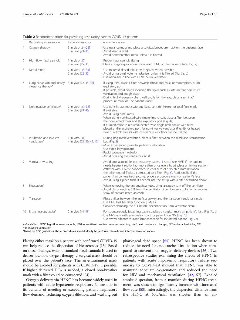
• During bag mask ventilation, place a filter between the mask and resuscitationbag (Fig. 5)
• Most experienced provider performs intubation• Use video-laryngoscope• Rapid sequence intubation• Avoid breaking the ventilator circuit
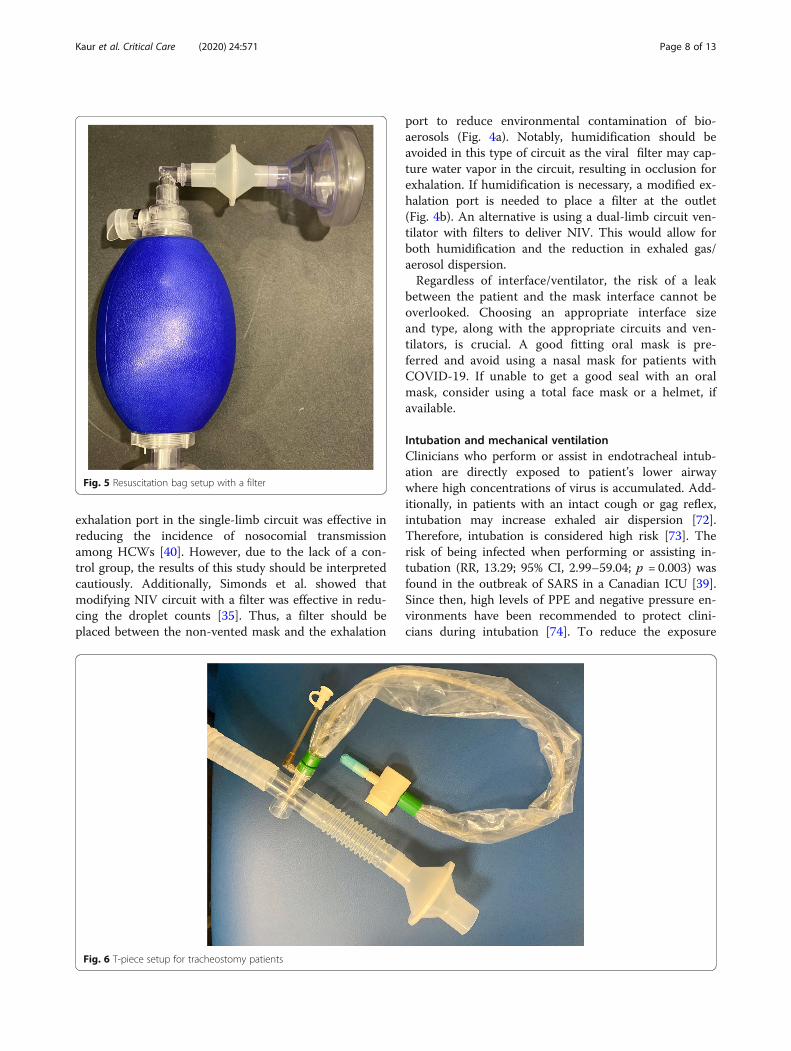
7 Ventilator weaning • Avoid cool aerosol for tracheostomy patient, instead use HME. If the patientneeds frequent suctioning (more than once every hour), place an in-line suctioncatheter with T-piece connected to cool aerosol or heated humidification,the other end of T-piece connected to a filter (Fig. 6). Additionally, if thepatient has cuffless tracheotomy, place a procedure mask on patient’s face
• Avoid using T-piece trials. If needed, use the setup with a filter described above
8 Extubation* • When removing the endotracheal tube, simultaneously turn off the ventilator• Avoid disconnecting ETT from the ventilator circuit before extubation to reducespray of contaminated aerosols
9 Transport • Place a filter between the artificial airway and the transport ventilator circuit• Use HME that has filter function (HME-F)• Consider clamping the ETT before disconnection from ventilator circuit
10 Bronchoscopy assist* 2 in vivo [44, 45] • For spontaneously breathing patients, place a surgical mask on patient's face (Fig. 7a, b)• Use NIV mask with examination port for patients on NIV (Fig. 7d)• Use swivel adapter to insert bronchoscope for intubated patient (Fig. 7c)
Abbreviations: HFNC high-flow nasal cannula, IPPB intermittent positive pressure breathing, HME heat moisture exchanger, ETT endotracheal tube, NIVnon-invasive ventilation*Based on CDC guidelines, these procedures should ideally be performed in airborne infection isolation rooms
Kaur et al. Critical Care (2020) 24:571 Page 4 of 13

entrainment or nonrebreather mask [53]. A randomizedcontrolled, crossover non-inferiority study trial reportedno difference in gram-negative bacterial and total bacter-ial counts between HFNC at 60 L/min and simple oxy-gen mask at 8 L/min when air sample collection plateswere placed at 0.4 or 1.5 m away from the patient [31].It is important to note that a substantial increase of ex-haled smoke dispersion was reported when the nasalcannula connection with patient nares was loose [58,59]. Because of these findings, it is suggested that a sur-gical or procedure mask be worn by patients receivingHFNC (Fig. 2). Regular checks on the proper positionand connection of the nasal cannula interface under themask are also necessary.
NebulizationAerosol therapy has been identified as a high-risk pro-cedure for nosocomial transmission, due to its active
generation of aerosol, which may carry viruses into theenvironment [19, 60]. Hui and colleagues found themaximum exhaled air dispersion distance was ≥ 0.45 mwhen a small volume jet nebulizer (SVN) was connectedto a mask at a gas flow of 6 L/min [33]. This distancewas even further than NIV at maximum settings (IPAP18 cmH2O, EPAP 4 cmH2O), using the same studymethod [33]. Two clinical observational studies alsofound droplet counts significantly increased immediatelyafter SVN started to generate aerosol, particularly, theaerosol/droplet count within small and medium sizerange 1–5 μm, when compared to the baseline level orother procedures including oxygen therapy and NIV [35]or bronchoscopy examination [44]. Nevertheless, theaerosol/droplets generated by a nebulizer may not con-tain a virus; however, if the nebulizer is contaminated,the aerosol can carry viruses to the surrounding environ-ment. McGrath and colleagues [34] found that mass
Fig. 1 Flow diagram of the literature search
Kaur et al. Critical Care (2020) 24:571 Page 5 of 13

concentrations of aerosols/droplets were significantly re-duced after placing a filter at the end of the mouthpiecefor nebulizers. Therefore, if aerosol therapy is indicatedfor COVID-19 patients, SVN should be avoided unlessfiltered, and inhalers including metered dose inhaler(MDI) and dry power inhalers (DPIs) are preferred forspontaneous breathing patients who can tolerate theiruse without generating additional cough [61].With MDI, a spacer with one-way valve is suggested
to reduce the need for coordination and to increase lung
deposition [62]. If patients are unable to use MDIs orDPIs, or the required medication is only available in theform of a solution, such as antibiotics, antivirals, muco-kinetics, or prostanoids, nebulizers via mouthpiece witha filter placed distal to the reservoir tubing (Fig. 3a andb) should be utilized. For patients who cannot tolerate amouthpiece or require medication administered over aprolonged period of time, such as continuous broncho-dilator for asthmatic patients [63] or inhaled epoproste-nol for patients with pulmonary hypertension orhypoxemia [64, 65], in-line placement of a nebulizerwith HFNC setup is recommended. This setup has twoadvantages: (1) more comfortable and better toleratedwhen compared to a mask or mouthpiece [63] and (2) asurgical mask to reduce the aerosol dispersion distanceor aerosol mass concentration can be placed on the pa-tient [53, 56]. When HFNC is utilized to deliver aerosoltreatment, gas flow needs to be set relatively low if pos-sible (10–20 L/min for adults and 0.25 L/kg/min for chil-dren), to improve the aerosol delivery efficiency [66, 67]and reduce the dispersion. Vibrating mesh nebulizers orvalved T-pieces for jet SVNs can reduce the need tobreak the ventilator circuit when nebulization is pro-vided during invasive ventilation.
Lung expansion and airway clearance therapyLittle evidence is available regarding lung expansiontherapy and nosocomial infection. Lung expansion ther-apy is designed to treat and prevent pulmonary atelec-tasis. Intermittent positive pressure breathing (IPPB)utilizes short-term positive pressure ventilation via maskor mouthpiece to promote lung expansion. Due to therisk of causing a cough response that might dispersebio-aerosols [21], IPPB should be used judiciously andwith filters placed between the breathing circuit and themask or mouthpiece.
Fig. 2 Wearing a surgical mask over high-flow high humiditynasal cannula
Fig. 3 a SVN setup with filter and one-way valve. b SVN setup with a filter
Kaur et al. Critical Care (2020) 24:571 Page 6 of 13

The number of particles emitted by cough from an in-fected patient was greater than that from a recoveredpatient (P < 0.001) [36]. Bronchial hygiene therapies suchas intermittent percussive ventilation and vibratory posi-tive expiratory pressure irritate the airway causing thepatient to cough forcefully, potentially emitting virus-laden aerosols. Placing a filter between these devices andpatient’s mouth is suggested. When intermittent percus-sive ventilation is utilized, nebulization via its integratednebulizer should be avoided as the filter placed betweenthe device outlet and patient will capture aerosols. Add-itionally, high-frequency chest wall oscillation can beused for secretion clearance. In addition to HCWs wear-ing proper PPE, a surgical or procedure mask worn bypatients receiving the therapy may be helpful. Overall, inpatients with confirmed COVID-19, avoid the indiscrim-inate use of bronchial hygiene therapies that may not beclinically indicated [61].
Non-invasive ventilationNIV has been utilized in 10–50% of COVID-19 patientsin published clinical reports [6–12]. Even though NIVdelivered by helmet was effective in terms of reducingintubation rate and 90-day mortality rate among ARDSpatients [68], its role in patients with severe ARDS re-mains controversial [69]. During the MERS outbreak,NIV was commonly used to treat acute hypoxic respira-tory failure, but it had a high failure rate and was not as-sociated with improved patient outcomes [70].
NIV produces a jet of exhaled gas through the exhal-ation port or leak from the connection of patient’s inter-face and ventilator, increasing dispersion distance ofpatient-generated bio-aerosol, and therefore it iscounted as an AGMP [20, 39]. Consequently, NIVshould be used with caution for COVID-19 patients andadditional modifications to minimize or reduce exhaledgas/aerosol dispersion are required.In an experimental study, Hui et al. reported signifi-
cant exhaled air dispersion within a 0.5-m radius of thehuman simulator receiving NIV and higher pressure set-tings increased the spread of exhaled air. However, theexhaled air dispersion is limited if the mask fit is appro-priate [37]. When comparing helmet to total face mask,Hui et al. [38] in another study demonstrated that NIVapplication via a double-limb circuit ventilator withfilters and a helmet with good seal was effective in redu-cing exhaled air dispersion. In contrast, NIV applied viaa total mask through a single-limb circuit ventilatorcaused increased exhaled air dispersion [38]. Non-ventedmasks were shown to have less air dispersion as com-pared to vented mask [71].Thus, when vented masks are used, additional precau-
tions for protection of the HCW may be appropriate.HCWs that are in close proximity to patients receivingNIV need to wear high respiratory personal protectionincluding N95 or powered air-purifying respirator(PAPR). Secondly, the NIV circuits can be modified toplace a filter. During the SARS outbreak, Cheung et al.demonstrated that a filter placed before the fixed
Fig. 4 a Non-heated single limb ventilator circuit. b Heated single limb ventilator circuit
Kaur et al. Critical Care (2020) 24:571 Page 7 of 13
exhalation port in the single-limb circuit was effective inreducing the incidence of nosocomial transmissionamong HCWs [40]. However, due to the lack of a con-trol group, the results of this study should be interpretedcautiously. Additionally, Simonds et al. showed thatmodifying NIV circuit with a filter was effective in redu-cing the droplet counts [35]. Thus, a filter should beplaced between the non-vented mask and the exhalation
port to reduce environmental contamination of bio-aerosols (Fig. 4a). Notably, humidification should beavoided in this type of circuit as the viral filter may cap-ture water vapor in the circuit, resulting in occlusion forexhalation. If humidification is necessary, a modified ex-halation port is needed to place a filter at the outlet(Fig. 4b). An alternative is using a dual-limb circuit ven-tilator with filters to deliver NIV. This would allow forboth humidification and the reduction in exhaled gas/aerosol dispersion.Regardless of interface/ventilator, the risk of a leak
between the patient and the mask interface cannot beoverlooked. Choosing an appropriate interface sizeand type, along with the appropriate circuits and ven-tilators, is crucial. A good fitting oral mask is pre-ferred and avoid using a nasal mask for patients withCOVID-19. If unable to get a good seal with an oralmask, consider using a total face mask or a helmet, ifavailable.
Intubation and mechanical ventilationClinicians who perform or assist in endotracheal intub-ation are directly exposed to patient’s lower airwaywhere high concentrations of virus is accumulated. Add-itionally, in patients with an intact cough or gag reflex,intubation may increase exhaled air dispersion [72].Therefore, intubation is considered high risk [73]. Therisk of being infected when performing or assisting in-tubation (RR, 13.29; 95% CI, 2.99–59.04; p = 0.003) wasfound in the outbreak of SARS in a Canadian ICU [39].Since then, high levels of PPE and negative pressure en-vironments have been recommended to protect clini-cians during intubation [74]. To reduce the exposure
Fig. 6 T-piece setup for tracheostomy patients
Fig. 5 Resuscitation bag setup with a filter
Kaur et al. Critical Care (2020) 24:571 Page 8 of 13

time to the SARS-CoV-2, the most experienced providershould perform intubation to avoid multiple attempts.Video-laryngoscope has shown to be useful when intub-ating patients with COVID-19 to increase the distancebetween the provider and the patient airway [74]. For adifficult airway, bronchoscopy is preferred to assist in-tubation, if a skilled provider is present [75]. Rapid se-quence intubation is also recommended, in order tominimize cough during the procedure [42, 76]. Aerosolboxes [77] as well as protective shields made of glass[78] have been described as practical barriers to limit ex-posure to patient’s exhaled droplets during intubation.While potentially useful, a documented reduction in dis-ease transmission has not been reported and concerns
regarding adequate airway view and appropriate ergo-nomics during intubation have been raised [79].Pre-oxygenation prior to intubation plays a crucial role
in avoiding complications during intubation. Multiplerandomized controlled trials have shown that theutilization of HFNC for pre-oxygenation can help reducethe incidence of hypoxemia during intubation [55, 80].The cost-effectiveness and the high risk of transmissionfrom high gas flows should be taken into consider-ation before using it for pre-oxygenation prior to in-tubation. The traditional method of using manualventilation via resuscitator and mask for patients priorto intubation also has some risks. Exhaled gas disper-sion distance has been shown to be 16–27 cm during
Fig. 7 a Bronchoscope insertion via the nose. b Bronchoscope insertion via the mouth. c Bronchoscope insertion via the endotracheal tube.d Bronchoscope insertion via the NIV mask
Kaur et al. Critical Care (2020) 24:571 Page 9 of 13
manual ventilation, which is similar to the distancebetween clinicians and the patient’s airway [41]. Pla-cing a filter between resuscitator and mask (Fig. 5)has been found to significantly reduce the exhaled gasdispersion distance [41, 43, 81].For patients requiring mechanical ventilation via
artificial airways, a ventilator with a dual-limb, heatedwire circuit in conjunction with filters placed at theventilator exhalation outlet is crucial [61, 73]. Inaddition, breaking the ventilator circuit connectionshould be limited and circuit changes should only bedone when visibly soiled [82].Approximately 8–13% mechanically ventilated patients
receive tracheostomy to facilitate the long-term need forventilatory support [83]. For patients with COVID-19,open tracheostomy is recommended over percutaneousdilational tracheostomy to reduce the risk of aerosoltransmission [84]. Recently, Pichi et al. described stand-ard steps to promote a safe and effective method whenperforming open tracheostomy in patients with COVID-19 [85]. Bertroche et al. [86] created a negative pressurecover to limit the exposure to the aerosols, but thesemethods need further investigation on the efficacy in re-ducing nosocomial infections.When transporting a mechanically ventilated COVID-
19 patient, it is suggested that a filter HME be placedbetween the artificial airway and the transport ventilatorcircuit [53]. Before pausing the ICU ventilator, considerclamping the endotracheal tube (ETT) to preventderecruitment and minimize the spread of bio-aerosolswhen transitioning patients from the ICU ventilator tothe transport ventilator [5, 87, 88]. When returning tothe ICU, clamp the ETT and leave the filter connectedto it to prevent accidental exposure. When ready fortransition to the ICU ventilator, disconnect the bacteriafilter and place the patient on the ventilator beforeunclamping ETT.
Weaning and extubationThe most common methods to perform a spontaneousbreathing trial are T-piece trial and pressure supportventilation (PSV). Subirà and colleagues [89] reportedthat successful extubation occurred in 82.3% of patientsin the PSV group compared to 74.0% in the T-piecegroup (difference, 8.2%; 95% CI, 3.4–13.0%; P = 0.001).With these findings, in conjunction with the need toavoid opening patient’s airway to the environment, PSVis preferred for COVID-19 patients. When a T-piece isneeded, HCWs should take safety precautions tominimize the exposure to a patient’s airway, such asusing the in-line suction catheter’s T-piece with one endconnecting humidified oxygen while the other end isconnected to a filter (Fig. 6). This setup can also be ap-plied for tracheostomy patients who are weaned from
mechanical ventilation, particularly for patients whoneed frequent suctioning (more than once an hour), asthis device keeps airway sealed and the filter protectsHCWs during suctioning. However, the filter can beclogged as it captures water vapor; hence periodicallychecking and replacing the filter are necessary. A filterHME can also be used to provide passive humidity [90]while humidified oxygen via a tracheostomy maskshould be avoided. Additionally, if the patient has a cuff-less tracheostomy tube in place, a procedure mask onpatient’s face may reduce bio-aerosol dispersion.The process of extubation induces a cough reflex
which may spread aerosols; therefore, it is imperativeto use proper precautions when removing an ETT[91]. During extubation, it is important to maintainthe connection of the ventilator circuit and suctioncatheter to the ETT, in order to avoid aerosol disper-sion from the ventilator circuit. The extubation pro-cedure should be performed by two HCWs. In anin vitro study, a clear plastic drape was shown to sig-nificantly reduce aerosol dispersion during the extuba-tion process; however, the feasibility of this practicerequires further investigation [92].
Bronchoscopy assistBronchoscopy examination is considered an AGMP andmay be related to an increased risk for transmission ofinfectious airborne particles [19]. Thompson et al. [45]found that bronchoscopy was associated with increasedprobability of aerosol generation and increased viral cop-ies among different AGMPs for H1N1-positive patients.O’Neil et al [44] found an increase in particle concentra-tion when a nebulized medication administration wasperformed before and after bronchoscopy, while bron-choscopy examination itself did not increase concentra-tion compared to baseline.According to the American Association for Bronchol-
ogy and Interventional Pulmonology guidelines, bron-choscopy procedures are relatively contraindicated forpatients with suspected or confirmed COVID-19 infec-tions when less invasive diagnostic procedures are in-conclusive [93]. Urgent bronchoscopy procedures shouldonly be considered if intervention is deemed as lifesavingin patients with (1) massive hemoptysis, (2) benign ormalignant severe airway obstruction, (3) suspicion ofsecondary infectious etiology, or (4) malignant conditionthat results in endobronchial obstruction. In the event aCOVID-19 patient requires bronchoscopic intervention,it is recommended that the patient be placed in negativepressure isolation room and personnel should don ap-propriate droplet precaution PPE, including a poweredair-purifying respirator or N95 mask [93].Some additional precautions might also be considered
to protect HCWs from exposure during bronchoscopy
Kaur et al. Critical Care (2020) 24:571 Page 10 of 13
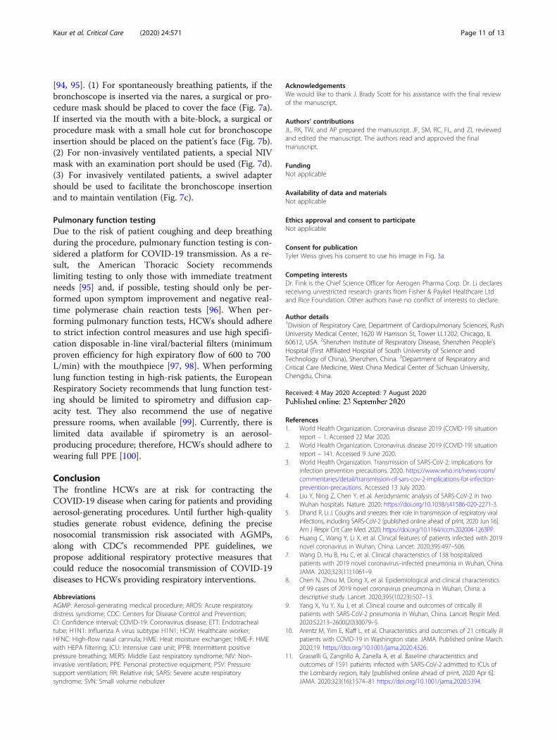
[94, 95]. (1) For spontaneously breathing patients, if thebronchoscope is inserted via the nares, a surgical or pro-cedure mask should be placed to cover the face (Fig. 7a).If inserted via the mouth with a bite-block, a surgical orprocedure mask with a small hole cut for bronchoscopeinsertion should be placed on the patient’s face (Fig. 7b).(2) For non-invasively ventilated patients, a special NIVmask with an examination port should be used (Fig. 7d).(3) For invasively ventilated patients, a swivel adaptershould be used to facilitate the bronchoscope insertionand to maintain ventilation (Fig. 7c).
Pulmonary function testingDue to the risk of patient coughing and deep breathingduring the procedure, pulmonary function testing is con-sidered a platform for COVID-19 transmission. As a re-sult, the American Thoracic Society recommendslimiting testing to only those with immediate treatmentneeds [95] and, if possible, testing should only be per-formed upon symptom improvement and negative real-time polymerase chain reaction tests [96]. When per-forming pulmonary function tests, HCWs should adhereto strict infection control measures and use high specifi-cation disposable in-line viral/bacterial filters (minimumproven efficiency for high expiratory flow of 600 to 700L/min) with the mouthpiece [97, 98]. When performinglung function testing in high-risk patients, the EuropeanRespiratory Society recommends that lung function test-ing should be limited to spirometry and diffusion cap-acity test. They also recommend the use of negativepressure rooms, when available [99]. Currently, there islimited data available if spirometry is an aerosol-producing procedure; therefore, HCWs should adhere towearing full PPE [100].
ConclusionThe frontline HCWs are at risk for contracting theCOVID-19 disease when caring for patients and providingaerosol-generating procedures. Until further high-qualitystudies generate robust evidence, defining the precisenosocomial transmission risk associated with AGMPs,along with CDC’s recommended PPE guidelines, wepropose additional respiratory protective measures thatcould reduce the nosocomial transmission of COVID-19diseases to HCWs providing respiratory interventions.
AbbreviationsAGMP: Aerosol-generating medical procedure; ARDS: Acute respiratorydistress syndrome; CDC: Centers for Disease Control and Prevention;CI: Confidence interval; COVID-19: Coronavirus disease; ETT: Endotrachealtube; H1N1: Influenza A virus subtype H1N1; HCW: Healthcare worker;HFNC: High-flow nasal cannula; HME: Heat moisture exchanger; HME-F: HMEwith HEPA filtering; ICU: Intensive care unit; IPPB: Intermittent positivepressure breathing; MERS: Middle East respiratory syndrome; NIV: Non-invasive ventilation; PPE: Personal protective equipment; PSV: Pressuresupport ventilation; RR: Relative risk; SARS: Severe acute respiratorysyndrome; SVN: Small volume nebulizer
AcknowledgementsWe would like to thank J. Brady Scott for his assistance with the final reviewof the manuscript.
Authors’ contributionsJL, RK, TW, and AP prepared the manuscript. JF, SM, RC, FL, and ZL reviewedand edited the manuscript. The authors read and approved the finalmanuscript.
FundingNot applicable
Availability of data and materialsNot applicable
Ethics approval and consent to participateNot applicable
Consent for publicationTyler Weiss gives his consent to use his image in Fig. 3a.
Competing interestsDr. Fink is the Chief Science Officer for Aerogen Pharma Corp. Dr. Li declaresreceiving unrestricted research grants from Fisher & Paykel Healthcare Ltdand Rice Foundation. Other authors have no conflict of interests to declare.
Author details1Division of Respiratory Care, Department of Cardiopulmonary Sciences, RushUniversity Medical Center, 1620 W Harrison St, Tower LL1202, Chicago, IL60612, USA. 2Shenzhen Institute of Respiratory Disease, Shenzhen People’sHospital (First Affiliated Hospital of South University of Science andTechnology of China), Shenzhen, China. 3Department of Respiratory andCritical Care Medicine, West China Medical Center of Sichuan University,Chengdu, China.
Received: 4 May 2020 Accepted: 7 August 2020
References1. World Health Organization. Coronavirus disease 2019 (COVID-19) situation
report – 1. Accessed 22 Mar 2020.2. World Health Organization. Coronavirus disease 2019 (COVID-19) situation
report – 141. Accessed 9 June 2020.3. World Health Organization. Transmission of SARS-CoV-2: implications for
infection prevention precautions. 2020. https://www.who.int/news-room/commentaries/detail/transmission-of-sars-cov-2-implications-for-infection-prevention-precautions. Accessed 13 July 2020.
4. Liu Y, Ning Z, Chen Y, et al. Aerodynamic analysis of SARS-CoV-2 in twoWuhan hospitals. Nature. 2020; https://doi.org/10.1038/s41586-020-2271-3.
5. Dhand R, Li J. Coughs and sneezes: their role in transmission of respiratory viralinfections, including SARS-CoV-2 [published online ahead of print, 2020 Jun 16].Am J Respir Crit Care Med. 2020; https://doi.org/10.1164/rccm.202004-1263PP.
6. Huang C, Wang Y, Li X, et al. Clinical features of patients infected with 2019novel coronavirus in Wuhan, China. Lancet. 2020;395:497–506.
7. Wang D, Hu B, Hu C, et al. Clinical characteristics of 138 hospitalizedpatients with 2019 novel coronavirus–infected pneumonia in Wuhan, China.JAMA. 2020;323(11):1061–9.
8. Chen N, Zhou M, Dong X, et al. Epidemiological and clinical characteristicsof 99 cases of 2019 novel coronavirus pneumonia in Wuhan, China: adescriptive study. Lancet. 2020;395(10223):507–13.
9. Yang X, Yu Y, Xu J, et al. Clinical course and outcomes of critically illpatients with SARS-CoV-2 pneumonia in Wuhan, China. Lancet Respir Med.2020:S2213–2600(20)30079–5.
10. Arentz M, Yim E, Klaff L, et al. Characteristics and outcomes of 21 critically illpatients with COVID-19 in Washington state. JAMA. Published online March.2020;19. https://doi.org/10.1001/jama.2020.4326.
11. Grasselli G, Zangrillo A, Zanella A, et al. Baseline characteristics andoutcomes of 1591 patients infected with SARS-CoV-2 admitted to ICUs ofthe Lombardy region, Italy [published online ahead of print, 2020 Apr 6].JAMA. 2020;323(16):1574–81 https://doi.org/10.1001/jama.2020.5394.
Kaur et al. Critical Care (2020) 24:571 Page 11 of 13

12. Richardson S, Hirsch JS, Narasimhan M, et al. Presenting characteristics,comorbidities, and outcomes among 5700 patients hospitalized withCOVID-19 in the New York City area. JAMA. 2020;323(20):2052–9.
13. Seto WH, Tsang D, Yung RW, et al. Effectiveness of precautions againstdroplets and contact in prevention of nosocomial transmission of severeacute respiratory syndrome (SARS). Lancet. 2003;361(9368):1519–20.
14. Scales DC, Green K, Chan AK, et al. Illness in intensive care staff after briefexposure to severe acute respiratory syndrome. Emerg Infect Dis. 2003;9(10):1205–10 https://doi.org/10.3201/eid0910.030525.
15. Chu DK, Akl EA, Duda S, et al. Physical distancing, face masks, and eyeprotection to prevent person-to-person transmission of SARS-CoV-2 andCOVID-19: a systematic review and meta-analysis. Lancet 2020; publishedonline June 1. https://doi.org/10.1016/S0140-6736(20)31142-9.
16. Gamage B, Moore D, Copes R, Yassi A, Bryce E, BC InterdisciplinaryRespiratory Protection Study Group. Protecting health care workers fromSARS and other respiratory pathogens: a review of the infection controlliterature. Am J Infect Control. 2005;33(2):114–21.
17. Suwantarat N, Apisarnthanarak A. Risks to healthcare workers with emergingdiseases: lessons from MERS-CoV, Ebola, SARS, and avian flu. Curr OpinInfect Dis. 2015;28(4):349–61.
18. Raboud J, Shigayeva A, McGeer A, et al. Risk factors for SARS transmissionfrom patients requiring intubation: a multicentre investigation in Toronto,Canada. PLoS One. 2010;5(5):e10717.
19. Judson SD, Munster VJ. Nosocomial transmission of emerging viruses viaaerosol-generating medical procedures. Viruses. 2019;11(10):940.
20. Tran K, Cimon K, Severn M, Pessoa-Silva CL, Conly J. Aerosol generatingprocedures and risk of transmission of acute respiratory infections tohealthcare workers: a systematic review. PLoS One. 2012;7(4):e35797.
21. Hui DS, Chan MT, Chow B. Aerosol dispersion during various respiratorytherapies: a risk assessment model of nosocomial infection to health careworkers. Hong Kong Med J. 2014;20(Suppl 4):9–13.
22. Macintyre CR, Seale H, Yang P, et al. Quantifying the risk of respiratoryinfection in healthcare workers performing high-risk procedures. EpidemiolInfect. 2014;142(9):1802–8.
23. Center for Disease Control and Prevention. Coronavirus disease 2019. Cases,data and surveillance. https://www.cdc.gov/coronavirus/2019-ncov/cases-updates/cases-in-us.html. Accessed 10 July 2020.
24. Hui DS, Chow BK, Chu L, et al. Exhaled air dispersion and removal isinfluenced by isolation room size and ventilation settings during oxygendelivery via nasal cannula. Respirology. 2011;16(6):1005–13.
25. Ip M, Tang JW, Hui DS, et al. Airflow and droplet spreading around oxygenmasks: a simulation model for infection control research. Am J InfectControl. 2007;35(10):684–9.
26. Hui DS, Ip M, Tang JW, et al. Airflows around oxygen masks: a potentialsource of infection? Chest. 2006;130(3):822–6.
27. Hui DS, Hall SD, Chan MT, et al. Exhaled air dispersion during oxygendelivery via a simple oxygen mask. Chest. 2007;132(2):540–6.
28. Hui DS, Chow BK, Chu L, et al. Exhaled air dispersion during coughing withand without wearing a surgical or N95 mask. PLoS One. 2012;7(12):e50845.
29. Milton DK, Fabian MP, Cowling BJ, Grantham ML, McDevitt JJ. Influenzavirus aerosols in human exhaled breath: particle size, culturability, and effectof surgical masks. PLoS Pathog. 2013;9(3):e1003205.
30. Johnson DF, Druce JD, Birch C, Grayson ML. A quantitative assessment ofthe efficacy of surgical and N95 masks to filter influenza virus in patientswith acute influenza infection. Clin Infect Dis. 2009;49(2):275–7.
31. Leung CCH, Joynt GM, Gomersall CD, et al. Comparison of high-flow nasalcannula versus oxygen face mask for environmental bacterial contaminationin critically ill pneumonia patients: a randomized controlled crossover trial. JHosp Infect. 2019;101(1):84–7.
32. Wang K, Zhao W, Li J, Shu W, Duan J. The experience of high-flow nasalcannula in hospitalized patients with 2019 novel coronavirus-infectedpneumonia in two hospitals of Chongqing, China. Ann Intensive Care. 2020;10(1):37 Published 2020 Mar 30. https://doi.org/10.1186/s13613-020-00653-z.
33. Hui DS, Chow BK, Chu LCY, et al. Exhaled air and aerosolized dropletdispersion during application of a jet nebulizer. Chest. 2009;135(3):648–54.
34. McGrath JA, O'Sullivan A, Bennett G, et al. Investigation of the quantity ofexhaled aerosols released into the environment during nebulisation.Pharmaceutics. 2019;11(2):75 Published 2019.
35. Simonds AK, Hanak A, Chatwin M, et al. Evaluation of droplet dispersionduring non-invasive ventilation, oxygen therapy, nebuliser treatment andchest physiotherapy in clinical practice: implications for management of
pandemic influenza and other airborne infections. Health Technol Assess.2010;14(46):131–72.
36. Lee J, Yoo D, Ryu S, et al. Quantity, size distribution, and characteristics ofcough-generated aerosol produced by patients with an upper respiratorytract infection. Aerosol Air Qual Res. 2019;19(4):840–53.
37. Hui DS, Hall SD, Chan MT, et al. Noninvasive positive-pressure ventilation: anexperimental model to assess air and particle dispersion. Chest. 2006;130(3):730–40.
38. Hui DS, Chow BK, Ng SS, et al. Exhaled air dispersion distances duringnoninvasive ventilation via different Respironics face masks. Chest. 2009;136(4):998–1005.
39. Fowler RA, Guest CB, Lapinsky SE, et al. Transmission of severe acuterespiratory syndrome during intubation and mechanical ventilation. Am JRespir Crit Care Med. 2004;169(11):1198–202.
40. Cheung TM, Yam LY, So LK, et al. Effectiveness of noninvasive positivepressure ventilation in the treatment of acute respiratory failure in severeacute respiratory syndrome. Chest. 2004;126(3):845–50.
41. Chan MTV, Chow BK, Lo T, et al. Exhaled air dispersion during bag-maskventilation and sputum suctioning - implications for infection control. SciRep. 2018;8(1):198.
42. Cai SJ, Wu LL, Chen DF. et al, Analysis of bronchoscope-guided trachealintubation in 12 cases with COVID-19 under the personal protectiveequipment with positive pressure protective hood. Chin J Tuberc Respir Dis.2020;43 Epub ahead of print. https://doi.org/10.3760/cma.j.cn112147-20200222-00153.
43. Christian MD, Loutfy M, McDonald LC, et al. Possible SARS coronavirustransmission during cardiopulmonary resuscitation. Emerg Infect Dis. 2004;10(2):287–93 https://doi.org/10.3201/eid1002.030700.
44. O'Neil CA, Li J, Leavey A, et al. Characterization of aerosols generated duringpatient care activities. Clin Infect Dis. 2017;65(8):1335–41.
45. Thompson K, Pappachan JV, Bennett AM, Mittal H, Macken S, Dove BK, et al.Influenza aerosols in UK hospitals during the H1N1 (2009) pandemic – the riskof aerosol generation during medical procedures. PLoS One. 2013;8(2):e56278.
46. Weber TP, Stilianakis NI. Inactivation of influenza A viruses in the environmentand modes of transmission: a critical review. J Inf Secur. 2008;57(5):361–73.
47. Lindsley WG, Blachere FM, Beezhold DH, et al. Viable influenza A virus inairborne particles expelled during coughs versus exhalations. InfluenzaOther Respir Viruses. 2016;10(5):404–13.
48. Stilianakis NI, Drossinos Y. Dynamics of infectious disease transmission byinhalable respiratory droplets. J R Soc Interface. 2010;7(50):1355–66.
49. Jensen PA, Lambert LA, Iademarco MF, Ridzon R, CDC. Guidelines forpreventing the transmission of Mycobacterium tuberculosis in health-caresettings, 2005. MMWR Recomm Rep. 2005;54(RR-17):1–141.
50. Bean B, Moore BM, Sterner B, Peterson LR, Gerding DN, Balfour HH Jr. Survivalof influenza viruses on environmental surfaces. J Infect Dis. 1982;146(1):47–51.
51. van Doremalen N, Bushmaker T, Morris DH, Holbrook MG, Gamble A,Williamson BN, et al. Aerosol and surface stability of SARS-CoV-2 ascompared with SARS-CoV-1. N Engl J Med. Published online 2020 Mar 17.https://doi.org/10.1056/NEJMc2004973.
52. Center for Disease Control and Prevention. https://www.cdc.gov/coronavirus/2019-ncov/hcp/infection-control-recommendations.html#take_precautions.
53. Li J, Fink JB, Ehrmann S. High-flow nasal cannula for COVID-19 patients: lowrisk of bio-aerosol dispersion. Eur Respir J. 2020;55(5):2000892.
54. Somogyi R, Vesely AE, Azami T, et al. Dispersal of respiratory droplets withopen vs closed oxygen delivery masks: implications for the transmission ofsevere acute respiratory syndrome. Chest. 2004;125(3):1155–7.
55. Li J, Jing GQ, Scott JB. Year in review 2019: high-flow nasal cannula (HFNC)oxygen therapy for adult patients. Respir Care. 2020;65(4):545–57.
56. Rochwerg B, Granton D, Wang DX, et al. High flow nasal cannula comparedwith conventional oxygen therapy for acute hypoxemic respiratory failure: asystematic review and meta-analysis. Intensive Care Med. 2019;45(5):563–72.
57. Patel M, Gangemi A, Marron R, et al. Use of high flow nasal therapy to treatmoderate to severe hypoxemic respiratory failure in COVID-19. medRxiv2020.05.22.20109355. https://doi.org/10.1101/2020.05.22.20109355.
58. Hui DS, Chow BK, Lo T, et al. Exhaled air dispersion during high flow nasalcannula therapy versus CPAP via different masks. Eur Respir J. 2019;53:1802339.
59. Leonard S, Atwood CW Jr, Walsh BK, et al. Preliminary findings on control ofdispersion of aerosols and droplets during high-velocity nasal insufflationtherapy using a simple surgical mask: implications for the high-flow nasalcannula [published online ahead of print, 2020 Apr 2]. Chest. 2020; https://doi.org/10.1016/j.chest.2020.03.043.
Kaur et al. Critical Care (2020) 24:571 Page 12 of 13

60. Sandrock C, Stollenwerk N. Acute febrile respiratory illness in the ICU:reducing disease transmission. Chest. 2008;133(5):1221–31.
61. Zhonghua Jie He He Hu Xi Za Zhi. Expert Consensus on PreventingNosocomial Transmission During Respiratory Care for Critically Ill PatientsInfected by 2019 Novel coronavirus pneumonia. Chin J Tuberc Respir Dis. 2020;17 Epub ahead of print. https://doi.org/10.3760/cma.j.issn.1001-0939.2020.0020.
62. Keeley D. Everyone with asthma should have a metered dose inhaler and aspacer. BMJ. 2018;360:k648.
63. Baudin F, Buisson A, Vanel B, Massenavette B, Pouyau R, Javouhey E. Nasalhigh flow in management of children with status asthmaticus: aretrospective observational study. Ann Intensive Care. 2017;7(1):55.
64. Li J, Harnois LJ, Markos B, et al. Epoprostenol delivered via high flow nasalcannula for ICU subjects with severe hypoxemia comorbid with pulmonaryhypertension or right heart dysfunction. Pharmaceutics. 2019;11(6):281.
65. Li J, Gurnani PK, Roberts KM, Fink JB, Vines D. The clinical impact of flowtitration on epoprostenol delivery via high flow nasal cannula for ICUpatients with pulmonary hypertension or right ventricular dysfunction: aretrospective cohort comparison study. J Clin Med. 2020;9(2):464.
66. Li J, Gong L, Fink JB. The ratio of nasal cannula gas flow to patientinspiratory flow on trans-nasal pulmonary aerosol delivery for adults: an invitro study. Pharmaceutics. 2019;11(5):225.
67. Li J, Gong L, Ari A, Fink JB. Decrease the flow setting to improve trans-nasalpulmonary aerosol delivery via “high-flow nasal cannula” to infants andtoddlers. Pediatr Pulmonol. 2019;54(6):914–21.
68. Patel BK, Wolfe KS, Pohlman AS, Hall JB, Kress JP. Effect of noninvasiveventilation delivered by helmet vs face mask on the rate of endotrachealintubation in patients with acute respiratory distress syndrome: arandomized clinical trial. JAMA. 2016;315(22):2435–41.
69. Bellani G, Laffey JG, Pham T, et al. Noninvasive ventilation of patients withacute respiratory distress syndrome. Insights from the LUNG SAFE study. AmJ Respir Crit Care Med. 2017;195(1):67–77.
70. Alraddadi BM, Qushmaq I, Al-Hameed FM, et al. Noninvasive ventilation incritically ill patients with the Middle East respiratory syndrome. InfluenzaOther Respir Viruses. 2019;13(4):382–90.
71. Hui DS, Chow BK, Lo T, et al. Exhaled air dispersion during noninvasiveventilation via helmets and a total facemask. Chest. 2015;147(5):1336–43.
72. Cheung JC, Ho LT, Cheng JV, Cham EYK, Lam KN. Staff safety duringemergency airway management for COVID-19 in Hong Kong [publishedonline ahead of print, 2020 Feb 24]. Lancet Respir Med. 2020;S2213–2600(20):30084–9 https://doi.org/10.1016/S2213-2600(20)30084-9.
73. Wax RS, Christian MD. Practical recommendations for critical care andanesthesiology teams caring for novel coronavirus (2019-nCoV) patients.Can J Anaesth. 2020; https://doi.org/10.1007/s12630-020-01591-x.
74. Yao W, Wang T, Jiang B, et al. Emergency tracheal intubation in 202patients with COVID-19 in Wuhan, China: lessons learnt and internationalexpert recommendations [published online ahead of print, 2020 Apr 10]. BrJ Anaesth. 2020; S0007–0912(20)30203–8.
75. Yam LY, Chen RC, Zhong NSSARS. ventilatory and intensive care.Respirology. 2003;8(Suppl):S31–5.
76. Zuo MZ, Huang YG, Ma WH. et al, Expert recommendations for trachealintubation in critically ill patients with noval coronavirus disease 2019[published online ahead of print, 2020 Feb 27]. Chin Med Sci J. 2020;https://doi.org/10.24920/003724.
77. Canelli R, Connor CW, Gonzalez M, Nozari A, Ortega R. Barrier enclosureduring endotracheal intubation. N Engl J Med. 2020;382(20):1957–8.
78. Chen C, Shen N, Li X, Zhang Q, Hei Z. New device and technique to protectintubation operators against COVID-19 [published online ahead of print,2020 May 8]. Intensive Care Med. 2020:1–3 https://doi.org/10.1007/s00134-020-06072-9.
79. Coronavirus disease 2019 (COVID-19): anesthetic concerns, including airwaymanagement and infection control. UpToDate. 2020. https://www.uptodate.com/contents/coronavirus-disease-2019-covid-19-airway-management-anesthesia-machine-ventilation-and-anesthetic-care. opens in new tab.
80. Jhou HJ, Chen PH, Lin C, Yang LY, Lee CH, Peng CK. High-flow nasalcannula therapy as apneic oxygenation during endotracheal intubation incritically ill patients in the intensive care unit: a systematic review and meta-analysis. Sci Rep. 2020;10(1):3541.
81. Chan MT, Chow BK, Chu L, Hui DS. Mask ventilation and dispersion ofexhaled air. Am J Respir Crit Care Med. 2013;187(7):e12–4.
82. Han J, Liu Y. Effect of ventilator circuit changes on ventilator-associatedpneumonia: a systematic review and meta-analysis. Respir Care. 2010;55(4):467–74.
83. Mehta AB, Syeda SN, Bajpayee L, Cooke CR, Walkey AJ, Wiener RS. Trends intracheostomy for mechanically ventilated patients in the United States,1993-2012. Am J Respir Crit Care Med. 2015;192(4):446–54.
84. Chao TN, Braslow BM, Martin ND, et al. Tracheotomy in ventilated patientswith COVID-19 [published online ahead of print, 2020 May 5]. Ann Surg.2020; https://doi.org/10.1097/SLA.0000000000003956.
85. Pichi B, Mazzola F, Bonsembiante A, et al. CORONA-steps for tracheotomy inCOVID-19 patients: a staff-safe method for airway management. Oral Oncol2020;105:104682. https://doi.org/10.1016/j.oraloncology.2020.104682.
86. Bertroche JT, Pipkorn P, Zolkind P, Buchman CA, Zevallos JP. Negative-pressure aerosol cover for COVID-19 tracheostomy [published online aheadof print, 2020 Apr 28]. JAMA Otolaryngol Head Neck Surg. 2020:e201081https://doi.org/10.1001/jamaoto.2020.1081.
87. Turbil E, Terzi N, Schwebel C, Cour M, Argaud L, Guérin C. Does endo-tracheal tube clamping prevent air leaks and maintain positive end-expiratory pressure during the switching of a ventilator in a patient in anintensive care unit? A bench study. PLoS One. 2020;15(3):e0230147.
88. Li YC, Lin HL, Liao FC, et al. Potential risk for bacterial contamination inconventional reused ventilator systems and disposable closed ventilator-suction systems. PLoS One. 2018;13(3):e0194246.
89. Subirà C, Hernández G, Vázquez A, et al. Effect of pressure support vs T-piece ventilation strategies during spontaneous breathing trials onsuccessful extubation among patients receiving mechanical ventilation: arandomized clinical trial [published correction appears in JAMA. 2019 Aug20;322(7):696]. JAMA. 2019;321(22):2175–82.
90. De Seta D, Carta F, Puxeddu R. Management of tracheostomy duringCOVID-19 outbreak: heat and moisture exchanger filter and closedsuctioning system [published online ahead of print, 2020]. Oral Oncol. 2020:104777 https://doi.org/10.1016/j.oraloncology.2020.104777.
91. Jee D, Park SY. Lidocaine sprayed down the endotracheal tube attenuatesthe airway-circulatory reflexes by local anesthesia during emergence andextubation. Anesth Analg. 2003;96(1) https://doi.org/10.1097/00000539-200301000-00058.
92. Matava CT, Yu J, Denning S. Clear plastic drapes may be effective at limitingaerosolization and droplet spray during extubation: implications for COVID-19. Can J Anaesth. 2020;67(7):902–4.
93. Wahidi MM, Lamb C, Murgu S, et al. American Association for Bronchologyand Interventional Pulmonology (AABIP) statement on the use ofbronchoscopy and respiratory specimen collection in patients withsuspected or confirmed COVID-19 infection. March 19th Updates. Accessedon 20 Mar 2020 at https://aabronchology.org/2020/03/12/2020-aabip-statement-on-bronchoscopy-covid-19-infection/.
94. Respiratory Care Committee of Chinese Thoracic Society. Expert consensuson preventing nosocomial transmission during respiratory care for criticallyill patients infected by 2019 novel coronavirus pneumonia. Zhonghua JieHe He Hu Xi Za Zhi. 2020;43(4):288–96.
95. Ferioli M, Cisternino C, Leo V, Pisani L, Palange P, Nava S. Protectinghealthcare workers from SARS-CoV-2 infection: practical indications. EurRespir Rev. 2020;29(155):200068.
96. ATS, Pulmonary function laboratories: advice regarding COVID-19. 2020.https://www.thoracic.org/professionals/clinical-resources/disease-related-resources/pulmonary-function-laboratories.php. Accessed 29 Mar 2020.
97. Bignamini E, Cazzato S, Cutrera R, et al. Italian pediatric respiratory societyrecommendations on pediatric pulmonary function testing during COVID-19 pandemic. Ital J Pediatr. 2020;46:68.
98. Zhi ZJHHHXZ, et al. Task force of Pulmonary Function Testing and ClinicalRespiratory Physiology, Chinese Association of Chest Physicians; PulmonaryFunction Testing Group, Respiratory Therapeutics Group. Chin Thorac Soc.2020;43(4):302–7.
99. Recommendation from ERS Group 9.1 (Respiratory function technologists/Scientists) Lung function testing during COVID-19 pandemic and beyond.https://ers.app.box.com/s/zs1uu88wy51monr0ewd990itoz4tsn2h. Accessed10 July 2020.
100. Hull JH, Lloyd JK, Cooper BG. Lung function testing in the COVID-19endemic. Lancet Respir Med. 2020;8(7):666–7.
Publisher’s NoteSpringer Nature remains neutral with regard to jurisdictional claims inpublished maps and institutional affiliations.
Kaur et al. Critical Care (2020) 24:571 Page 13 of 13