practical manual molecular biology bio302 hand outs... · practical manual molecular biology bio302...
TRANSCRIPT
Practical Manual
Molecular Biology BIO302
Department of Molecular Biology
Virtual University of Pakistan, Lahore,
Pakistan
Practical Manual
MOLECULAR BIOLOGY BIO302
2
CONTENTS
Introduction
Laboratory Safety
Protocol 1: Reagent Preparation; Preparation of Stock and Working Solutions.
Protocol 2: Isolation of Nucleic Acids
Protocol 3: Quantification of Nucleic Acid
Protocol 4: Polymerase Chain Reaction
Protocol 5: Agarose Gel Electrophoresis
Protocol 6: Restriction Digestion of DNA and Preparation of Restriction Maps
Protocol 7: Detection of Mutations by Restriction Fragment Length Polymorphism
Protocol 8: Preparation of Competent Cells
Protocol 9: Transformation of Bacteria with Plasmid DNA
Protocol10: Comparing Different Plasmids Using Molecular Weight Markers
Protocol11: Real-Time PCR Amplification
Appendix A: Useful resources
References
Practical Manual
MOLECULAR BIOLOGY BIO302
3
INTRODUCTION
Molecular biology techniques are applicable not only to modern medical practice but also to
the identification of genetically modified organisms, forensics, and quality assessment of
laboratory animals, pharmacogenomics, and other fields.
This procedure manual encompasses most widely used molecular biology techniques namely
DNA extraction, end-point and Real-Time PCR.
SAFETY CONSIDERATIONS
- Use personal protective equipment such as disposable gloves, lab coats, disposable
masks, etc.
- Handle all sharps with care and dispose of sharps in the sharps disposal containers.
- Handle hazardous chemicals and samples carefully. Blood and other body fluid must
be considered potentially hazardous. Biological waste should be disposed of in the
designated trash bags that could be incinerated later on.
- Decontaminate the work benches regularly and especially before and after work. For
decontamination, wipe the surfaces with 10% bleach followed by water.
MINIMUM STANDARDS AND CONTROLS
During DNA extraction, PCR and other experiments appropriate standards and
controls should be used to assure the quality of the results.
- Positive control to check the efficiency of the reagents, procedure and equipment.
- Negative Control to check the contamination
Validation of Critical Reagents and Procedures
All technical procedures and critical reagents should be tested and validated before
performing the actual case work or research work experiments.
Calibration of Instruments
All instruments should be calibrated according to required schedule and before performing
the validation studies, case work and research experiments.
Practical Manual
MOLECULAR BIOLOGY BIO302
4
Protocol 1:
PREPARATION OF REAGENTS
The following general instructions are applicable in the preparation of all reagents.
Use graduated cylinders or pipettes closest to the volume being measured for
preparing liquid reagents.
Store all reagents in sterile containers unless otherwise noted. Label all reagents with name
of reagent, date prepared, initials of scientist that prepared reagent, lot number, and
expiration date. Record each preparation in the lab’s reagent logbook.
1M Tris-HCl [Tris(Hydroxymethyl)aminomethan] pH8
Tris base 121.1g
H2O to 800ml
Adjust to desired pH with concentrated HCl.
Mix and add H2O to 1 Liter.
Store at room temperature
0.5 M EDTA (Ehylenediamine Tetraacetic Acid) pH 8.0
Na2EDTA.2H2O 186.1g
H2O to 700ml
Adjust pH to 8.0 with 10M NaOH (almost 50ml)
Mix and add H2O to 1 Liter.
Store at room temperature
10M NaOH
NaOH 400 g
H2O to 1 Liter
Store at room temperature
10 mg/ml Ethidium Bromide Ethidium Bromide 0.2 g H2O to 20ml Mix well and store at 4
oC in dark.
TE (Tris 10 mM-EDTA 2mM) pH 8.0 (Lysis Buffer)
1M Tris-HCl ph 8.0 10 ml 0.5 M EDTA pH 8.0 4 ml
H2O to 1 Liter
Store at room temperature
Practical Manual
MOLECULAR BIOLOGY BIO302
5
Low TE (Tris 10 mM-EDTA 0.2 mM) pH 8.0 for DNA storage
1M Tris-HCl pH 8.0 10 ml
0.5 M EDTA pH 8.0 0.4 ml
H2O to 1 Lite
Store at room temperature
Proteinase K (20mg/ml)
Proteinase K 100 mg lyophilized powder
Ultra-pure H2O to 5 ml
Aliquot and store at approximately -20C.
CAUTION: Powder and solutions of Proteinase K can be irritating to
mucous membranes.
SDS 10% w/v
Sodium dodecyl sulfate 100g H2O
to 700ml Heat to approximately 65oC to
dissolve.
Bring to a final volume of 1.0 L with ultra pure water.
Store at room temperature.
CAUTION: SDS can be irritating to mucous membranes. Wear safety
glasses, mask and gloves when handling.
TEN buffer (10mM Tris, 2mM EDTA, 400 mM NaCl)
1 M Tris-HCl ph 8.0 10 ml
5M NaCl 80 ml
0.5M EDTA 4 ml
H2O to 1 Liter
Store at room temperature.
50x TAE (Tris-Acetate-EDTA) Electrophoresis Stock buffer
Tris base 242g
Glaciall acetic acid 57.1 ml
0.5 M EDTA pH 8.0 100ml
H2O to 1 Liter
Store at room temperature
Practical Manual
MOLECULAR BIOLOGY BIO302
6
1x TAE (Tris 40mM, Acetate 20mM, EDTA 2mM) Electrophoresis working
buffer
50x TAE 10 ml
H2O to 500 ml
The pH of diluted buffer is 8.3.
Store at room temperature.
10x TBE (Tris 90mM-Borate 90mM-EDTA 2mM) Electrophoresis buffer
Tris base 108g
Boric Acid 55g
0.5M EDTA pH 8.0 40 ml H2O to 1 Liter
Store at room temperature
2x Gel Loading Dye
2% Bromophenol blue 0.25 ml
2% Xylene cyanol 0.25 ml
Glycol 7ml
H2O 10ml
Store at room temperature
5M Sodium Chloride
Sodium Chloride 292.2 g
H2O to 1 Liter
Store at room temperature.
6M Sodium Chloride
Sodium Chloride 351g
H2O to 1 Liter
Store at room temperature.
Practical Manual
MOLECULAR BIOLOGY BIO302
7
Protocol 2:
DNA Extraction from Whole Blood Principle
The extraction of DNA involves three main steps that are cell lysis, protein separation, and
DNA purification. Cell lysis is usually performed by incubation of cell in buffer containing
detergent and protease. Cellular proteins are salted out or phase separated using organic
solvents. Finally DNA is isolated and purified either by alcohol precipitation or adsorption
with silica and elution.
Reagents required
TE buffer (10mM Tris, 2mM EDTA, pH 8.0)
TEN buffer (10 mM Tris, 2mM EDTA, 400mM NaCl)
10% SDS
Proteinase-K solution 20mg/ml
6M NaCl
Phenol-Choloroform-Isoamylalcohol (PCI) (25:24:1)
Absolute Ethanol or Isopropanol
75% Ethanol
Low TE buffer (10mM Tris, 0.2mM EDTA)
Consumables required
Filter barrier tips 200 µl
Filter barrier tips 1000 µl
Wide bore tips 1000 µl
Falcon tubes 15 ml
Microcentrifuge tubes 1.5 ml
Equipment required
Centrifuge for 15 ml falcon tubes
Microcentrifuge for 1.5 ml tubes
Adjustable micropipettes 1 ml and 200 µl
Procedure
1. Add 1 ml chilled TE buffer to 200 µl blood. Mix by inverting the tube several times.
2. Spin at 4000 rpm for 15 min at room temperature.
3. Discard the supernatant and add 900 µl chilled TE buffer. Re-suspend the pellet by
vigorous shaking by hand.
4. Spin at 4000 rpm for 15 min at room temperature.

Practical Manual
MOLECULAR BIOLOGY BIO302
8
5. Discard the supernatant and add 800 µl TE buffer. Re-suspend the pellet by vigorous
shaking by hand.
6. Spin at 4000 rpm for 15 min at room temperature.
7. Discard the supernatant and add 200 µl TEN/A1 buffer, 20 µl SDS (10% solution) and
10 µl Proteinase-K solution. Re-suspend the pellet by shaking and vortex mixing.
8. Incubate the mixture at 56°C overnight.
9. Next day, place the tubes on ice and add 50 µl 6M NaCl. Shake the tube vigorously and
place on ice again for 15 min.
10. Spin at 4000 rpm for 15 min to pellet down the salts and proteins.
11. Transfer the supernatant in a fresh properly labeled 1.5-ml centrifuge tube.
12. Add equal volume of chilled isopropanol and invert the tubes gently till DNA is
visible.
13. Spin at 8000 rpm for 1 min at room temperature. Discard supernatant.
14. Add 200 ml absolute ethanol and vortex for 15 sec.
15. Spin at 8000 rpm for 1 min at room temperature.
16. Add 200 ml 75% ethanol and vortex for 15 sec.
17. Spin at 8000 rpm for 1 min at room temperature.
18. Discard the supernatant and add 100 µl low TE buffer or sterile distilled water to
dissolve the DNA pellet. Incubate at 72°C for 30 min.
19. Store DNA at -20°C.
Alternate steps for protein precipitation
First 8 steps are same as above.
9. Add equal volume of Phenol-Choloroform-Isoamylalcohol (PCI) solution. Mix the
contents by inverting gently. Leave at room temperature for 5 min.
10. Centrifuge at 13000 rpm for 10 min to form three layers.
11. Carefully take upper aqueous layer containing DNA with 1ml pipette and transfer to a
fresh properly labeled 1.5 ml centrifuge tube.
Follow step 12 onwards as given in the inorganic protocol.

Practical Manual
MOLECULAR BIOLOGY BIO302
9
NOTE: For more purification, organic and inorganic protein precipitation can be
combined i.e., Precipitation by 6M NaCl followed by the phenol-chloroform-isoamyl
alcohol purification.
Practical Manual
MOLECULAR BIOLOGY BIO302
10
Protocol 3:
DNA quantification by spectrophotometry using NanoDrop Principle:
Nucleic Acids (nucleotides, RNA, ssDNA, and dsDNA) all absorb light at 260 nm
wavelength; therefore spectrometry at 260nm is useful to quantify DNA or RNA in solutions
according to Beer-Lamberts law.
Readings should be taken at wavelengths of 260 nm and 280 nm. The reading at 260 nm
allows calculation of the concentrate on of nucleic acid in the sample.
1 O.D. at 260 nm for double-stranded DNA = 50 ng/µl of dsDNA
1 O.D. at 260 nm for single-stranded DNA = 20-33 ng/ul of ssDNA
1 O.D. at 260 nm for RNA molecules = 40 ng/µl of RNA
The reading at 280 nm gives the amount of protein in the sample.
Pure preparations of DNA and RNA have OD260/OD280 values of 1.8 to 2.0, respectively.
If there is contamination with protein or phenol, this ratio will be significantly less than the
values given above, and accurate quantitation of the amount of nucleic acid will not be
possible.
So typically, dilute sample 1 µl in 100 µl so the dilution factor is 100. Put whole 100 µl in
spectrophotometer cuvette. The DNA concentration read will then be:
OD260 X 50 ng/ul x dilution factor
For example, if have OD260 = 1.6. Then the concentration is:
1.6 x 50 ng/ul x 100 = 8000 ng/ul or 8 ug/ul.
Equipment Required
NanoDrop 2000
Vortex Mixer
Pipettes covering 1-1000µL range
Cuvets and sample tubes
Procedure Nucleic acid samples can be easily checked for concentration and quality using the NanoDrop 2000/2000c spectrophotometer. To measure nucleic acid samples select the
Nucleic Acid application from the home screen.
Nucleic Acid Calculations For nucleic acid quantification, the Beer-Lambert equation is modified to use a factor with units of ng-cm/microliter.
Practical Manual
MOLECULAR BIOLOGY BIO302
11
The modified equation used for nucleic acid calculations is the following:
c = (A * ε)/b
c = the nucleic acid concentration in ng/microliter
A = the absorbance in AU
ε = the wavelength-dependent extinction coefficient in ng-cm/microliter
b= the pathlength in cm
The generally accepted extinction coefficients for nucleic acids are:
• Double-stranded DNA: 50 ng-cm/μL
• Single-stranded DNA: 33 ng-cm/μL
• RNA: 40 ng-cm/μL
When the pedestal mode is selected, the NanoDrop 2000/2000c spectrophotometer uses short
path lengths between 1.0 mm to 0.05 mm to enable measurement of concentrated samples
without dilution. Note: Absorbance data shown in reports is archived as displayed on the software screen. The Nucleic Acid application absorbance values are normalized to a 1.0 cm (10.0 mm) path for all pedestal and cuvette measurements.
Measurement Concentration Ranges The NanoDrop 2000/2000c will accurately measure purified dsDNA samples <15,000 ng/μL without dilution. The software automatically utilizes the optimal path length to measure the
absorbance of each sample. Refer to ―Measurement Ranges‖ for additional information.
The small sample volume option is available when samples have 10 mm equivalent
absorbance values of 3.0 or higher (>150 ng/μL dsDNA.)
Unique Screen Features The right pane displays features specific to the Nucleic Acid application. Task bars in the left pane not described below are described in ―Software Overview.‖
The spectral display shows data for the current sample normalized to a 10 mm path for all
measurements including measurements made with any cuvette path length.
The following features are to the right of the spectral display:
• Sample ID - field into which a sample ID is entered. The appropriate sample ID should be
entered prior to each measurement.
• Type - a drop down list from which the user may select the (color-keyed) type of nucleic
acid being measured. Options include DNA-50 for dsDNA, RNA-40 for RNA, and ssDNA-
33 for single-stranded DNA.
Additional options include Oligo DNA and Oligo RNA which utilize the appropriate
extinction coefficient based upon user-defined base sequences. The Custom option allows the
user to enter an extinction coefficient between 15 and 150.
Practical Manual
MOLECULAR BIOLOGY BIO302
12
• Conc - concentration based on absorbance at 260 nm and the default or user defined
extinction coefficient. Concentration units may be selected from the adjacent drop-down box.
Refer to ―Nucleic Acid Calculations‖ for more details.
• A260 - displays absorbance at 260 nm normalized to a 10 mm pathlength.
• A280 - displays absorbance at 280 nm normalized to a 10 mm pathlength.
• 260/280 - ratio of absorbance at 260 nm and 280 nm. The ratio of absorbance at 260 and
280 nm is used to assess the purity of DNA and RNA. A ratio of ~1.8 is generally accepted as
―pure‖ for DNA; a ratio of ~2.0 is generally accepted as ―pure‖ for RNA. If the ratio is
appreciably lower in either case, it may indicate the presence of protein, phenol or other
contaminants that absorb strongly at or near 280 nm. See ―260/280 Ratio‖ in ―Diagnostics
and Troubleshooting‖ for more details on factors that can affect this ratio.
• 260/230 - ratio of absorbance at 260 nm and 230 nm. This is a secondary measure of nucleic
acid purity. The 260/230 values for a ―pure‖ nucleic acid are often higher than the respective
260/280 values and are commonly in the range of 1.8-2.2. If the ratio is appreciably lower,
this may indicate the presence of co-purified contaminants.
• Baseline correction - if selected, the default wavelength for the bichromatic normalization
is 340 nm. The user can manually enter a different wavelength for the bichromatic
normalization of the absorbance data. In either case, the baseline is automatically set to the
absorbance value of the sample at the selected wavelength. All wavelength data will be
referenced off this value.
Note: If a baseline correction is not selected, the spectra may be offset from the baseline and
the calculated concentration will change accordingly.
Making Nucleic Acid Measurements 1. Select the Nucleic Acid application from the main menu. If the wavelength verification window appears, ensure the arm is down and click OK.
2. Select the type of sample to be measured from the Type drop-down list. The default setting
is DNA-50.
3. Choose the concentration units from the drop-down list adjacent to the color coded
concentration box. The default units are ng/μL.
4. A default wavelength of 340 nm is automatically used for a bichromatic normalization.
Select an alternative reference wavelength or choose not to have the spectrum normalized by
de-selecting the baseline correction box.
- Select the file drop-down option Use current settings as default as a convenient way to
limit set-up time for each new workbook.
5. Select Add to report to automatically include all measurements in the current report. The
default setting is for all samples to be added to reports. The Add to report checkbox must be
selected prior to a measurement to save the sample data to a workbook. 6. Select Overlay spectra to display multiple spectra at a time.
Practical Manual
MOLECULAR BIOLOGY BIO302
13
7. Establish a blank using the appropriate buffer. The blank solution generally is the buffer
that the molecule of interest is suspended or dissolved in. This solution should be the same
pH and of a similar ionic strength as the sample solution.
Pedestal Option: Pipette 1-2 μL of the appropriate blanking solution onto the bottom
pedestal, lower the arm and click the Blank button.
Cuvette Option (Model 2000c only): Insert the cuvette noting the direction of the light path
indicated by the etched arrow. The optical beam (2 mm) is directed 8.5 mm above the bottom
of the cuvette. Refer to the cuvette manufacturer for volume recommendations.
Note: The arm must be down for all measurements, including those made with cuvettes. It is
recommended that cuvettes be removed from the instrument prior to making a pedestal
measurement to ensure that the pedestal arm can move to the proper starting position.
8. Enter a Sample ID in the appropriate field, load the first sample as described for the blank
above and click Measure.
Note: A fresh aliquot of sample should be used for each measurement.
After the measurement: - Simply wipe the upper and lower pedestals using a dry laboratory wipe and the instrument is ready to measure the next sample.
- When using the cuvette option, remove the cuvette, rinse thoroughly and dry between
samples.
Practical Manual
MOLECULAR BIOLOGY BIO302
14
Protocol 4:
Polymerase Chain Reaction (PCR) amplification Principle:
Polymerase Chain Reaction is an in-vitro method for exponential amplification of a target
portion of template DNA, which involves incorporation of nucleotides by DNA polymerase
during thermal cycling
Reagents Required:
PCR master mix including Taq polymerase, dNTPs, MgCl2 and buffer. or
Taq DNA Polymerase, dNTPs, MgCl2 and PCR buffer separately.
PCR primers (Forward and Reverse)
PCR grade water
Negative and Positive Controls
Equipment Required:
Thermal Cycler with analysis software
Vortex Mixture
Microcentrifuge
Pipettes
PCR safety cabinet
Consumables:
PCR tubes/strips/plates according to equipment compatibility and requirement
Filtered pipette tips
1.5 ml centrifuge tubes
Procedure:
1. Label the PCR tubes for samples and controls. In case of quantification experiments, tube
will also be labeled for standards.
2. Thaw the PCR reagents and prepare PCR reaction mix. A generalized recipe of PCR is
given in the following table. The amount of ingredients may vary according to the desired
protocol and manufacturer’s instructions. Calculate the volume of total reaction mix
required for the whole batch including samples, controls and standards.
Practical Manual
MOLECULAR BIOLOGY BIO302
15
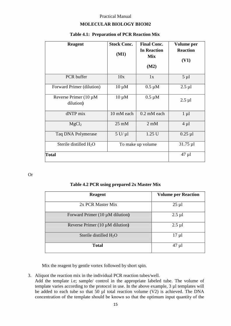
Table 4.1: Preparation of PCR Reaction Mix
Reagent Stock Conc.
(M1)
Final Conc.
In Reaction
Mix
(M2)
Volume per
Reaction
(V1)
PCR buffer 10x 1x 5 µl
Forward Primer (dilution) 10 µM 0.5 µM 2.5 µl
Reverse Primer (10 µM
dilution)
10 µM 0.5 µM
2.5 µl
dNTP mix 10 mM each 0.2 mM each 1 µl
MgCl2 25 mM 2 mM 4 µl
Taq DNA Polymerase 5 U/ µl 1.25 U 0.25 µl
Sterile distilled H2O To make up volume 31.75 µl
Total 47 µl
Or
Table 4.2 PCR using prepared 2x Master Mix
Reagent Volume per Reaction
2x PCR Master Mix 25 µl
Forward Primer (10 µM dilution) 2.5 µl
Reverse Primer (10 µM dilution) 2.5 µl
Sterile distilled H2O 17 µl
Total 47 µl
Mix the reagent by gentle vortex followed by short spin.
3. Aliquot the reaction mix in the individual PCR reaction tubes/well.
Add the template i.e; sample/ control in the appropriate labeled tube. The volume of
template varies according to the protocol in use. In the above example, 3 µl templates will
be added to each tube so that 50 µl total reaction volume (V2) is achieved. The DNA
concentration of the template should be known so that the optimum input quantity of the
Practical Manual
MOLECULAR BIOLOGY BIO302
16
template DNA can be used for PCR reaction. Optimal amounts of template DNA in the
50 μl reaction volume are 0.01-1 ng for both plasmid and phage DNA, and 0.1-1 μg for
genomic DNA. Higher amounts of template increases the risk of generation of non-
specific PCR products. Lower amounts of template reduces the accuracy of the
amplification.
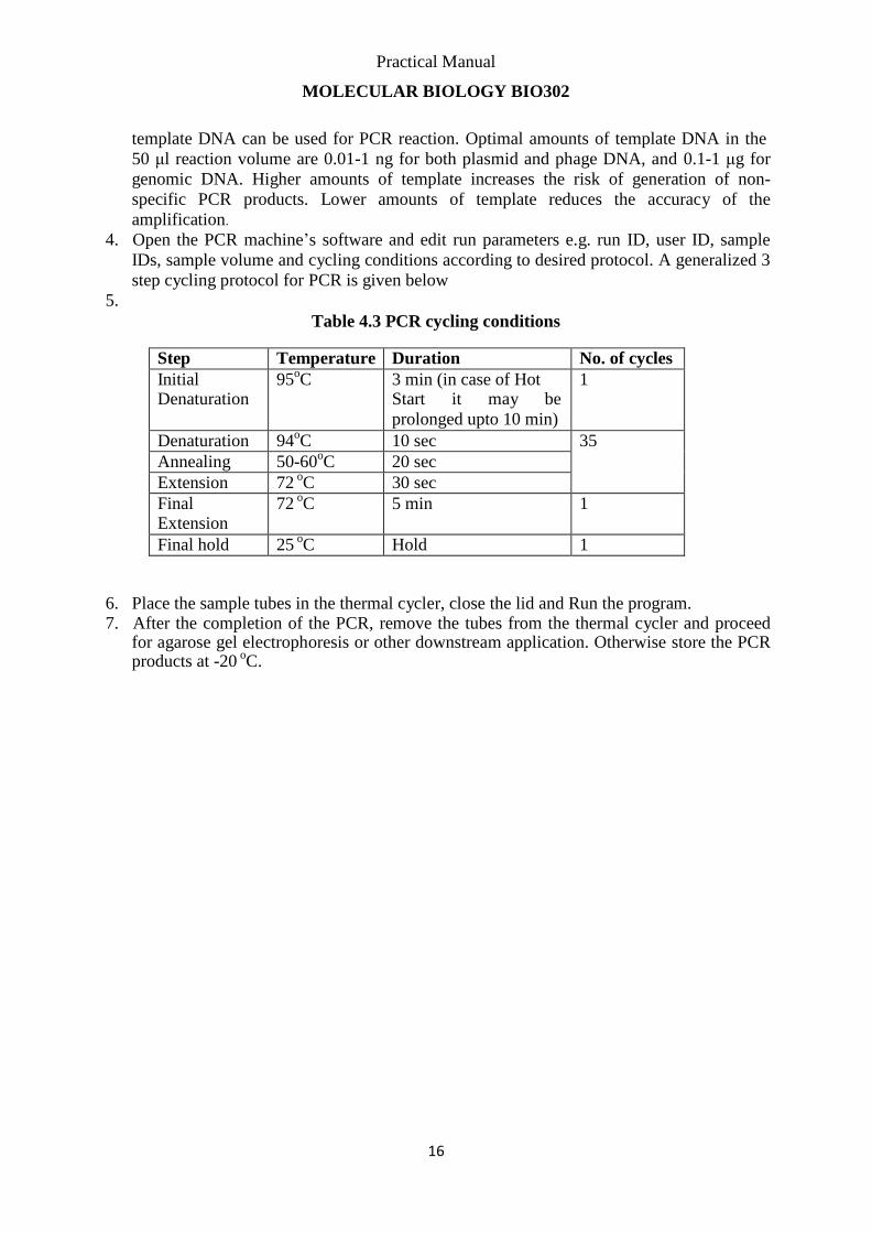
4. Open the PCR machine’s software and edit run parameters e.g. run ID, user ID, sample
IDs, sample volume and cycling conditions according to desired protocol. A generalized 3
step cycling protocol for PCR is given below
5.
Table 4.3 PCR cycling conditions
Step Temperature Duration No. of cycles
Initial Denaturation
95oC 3 min (in case of Hot
Start it may be
prolonged upto 10 min)
1
Denaturation 94oC 10 sec 35
Annealing 50-60oC 20 sec
Extension 72 oC 30 sec
Final Extension
72 oC 5 min 1
Final hold 25 oC Hold 1
6. Place the sample tubes in the thermal cycler, close the lid and Run the program. 7. After the completion of the PCR, remove the tubes from the thermal cycler and proceed
for agarose gel electrophoresis or other downstream application. Otherwise store the PCR products at -20
oC.
Practical Manual
MOLECULAR BIOLOGY BIO302
17
Protocol 5:
Agarose Gel Electrophoresis Principle Nucleic acid molecules are size separated by the aid of an electric field where negatively charged molecules migrate toward anode (positive) pole. The migration flow is determined
solely by the molecular weight where small weight molecules migrate faster than larger ones
(Sambrook & Russel 2001). In addition to size separation, nucleic acid fractionation using
agarose gel electrophoresis can be an initial step for further purification of a band of interest.
Extension of the technique includes excising the desired ―band‖ from a stained gel viewed
with a UV transilluminator (Sharp et al.,1973)
Equipment Required
An electrophoresis chamber and power supply
Gel casting trays, which are available in a variety of sizes and composed of UV-
transparent plastic. The open ends of the trays are closed with tape while the gel is
being cast, then removed prior to electrophoresis.
Sample combs, around which molten agarose is poured to form sample wells in the gel.
Transilluminator (an ultraviolet lightbox), which is used to visualize ethidium
bromide-stained DNA in gels. NOTE: always wear protective eyewear when
observing DNA on a transilluminator to prevent damage to the eyes from UV light.
Pipettes ---- covering 1 to 100 ul range
Reagent Required
Electrophoresis buffer, usually Tris-acetate-EDTA (TAE) or Tris-borate-EDTA (TBE).
DNA sizing standard/ladder
Loading buffer, which contains something dense (e.g. glycerol) to allow the sample to
"fall" into the sample wells, and one or two tracking dyes, which migrate in the gel
and allow visual monitoring or how far the electrophoresis has proceeded.
Ethidium bromide, a fluorescent dye used for staining nucleic acids. NOTE: Ethidium
bromide is a known mutagen and should be handled as a hazardous chemical – wear
gloves while handling.
Procedure: To pour a gel, agarose powder is mixed with electrophoresis buffer to the desired concentration, then heated in a microwave oven until completely melted. Most commonly,
ethidium bromide is added to the gel (final concentration 0.5 ug/ml) at this point to facilitate
visualization of DNA after electrophoresis. After cooling the solution to about 60C, it is
poured into a casting tray containing a sample comb and allowed to solidify at room
temperature.
After the gel has solidified, the comb is removed, using care not to rip the bottom of the
wells. The gel, still in its plastic tray, is inserted horizontally into the electrophoresis chamber
Practical Manual
MOLECULAR BIOLOGY BIO302
18
and just covered with buffer. Samples containing DNA mixed with loading buffer are then
pipetted into the sample wells, the lid and power leads are placed on the apparatus, and a
current is applied. You can confirm that current is flowing by observing bubbles coming off
the electrodes. DNA will migrate towards the positive electrode, which is usually colored red.
The distance DNA has migrated in the gel can be judged by visually monitoring migration of
the tracking dyes. Bromophenol blue and xylene cyanol dyes migrate through agarose gels at
roughly the same rate as double-stranded DNA fragments of 300 and 4000 bp, respectively.
When adequate migration has occurred, DNA fragments are visualized by staining with
ethidium bromide. This fluorescent dye intercalates between bases of DNA and RNA. It is
often incorporated into the gel so that staining occurs during electrophoresis, but the gel can
also be stained after electrophoresis by soaking in a dilute solution of ethidium bromide. To
visualize DNA or RNA, the gel is placed on an ultraviolet trans-illuminator. Be aware that
DNA will diffuse within the gel over time, and examination or photography should take place
shortly after cessation of electrophoresis.
Migration of DNA Fragments in Agarose Fragments of linear DNA migrate through agarose gels with a mobility that is inversely proportional to the log10 of their molecular weight. In other words, if you plot the distance
from the well that DNA fragments have migrated against the log10 of either
their molecular weights or number of base pairs, a roughly straight line will appear.
Circular forms of DNA migrate in agarose distinctly differently from linear DNAs of the
same mass. Typically, uncut plasmids will appear to migrate more rapidly than the same
plasmid when linearized. Additionally, most preparations of uncut plasmid contain at least
two topologically-different forms of DNA, corresponding to supercoiled forms and nicked
circles. The image to the right shows an ethidium-stained gel with uncut plasmid in the left
lane and the same plasmid linearized at a single site in the right lane.
Several additional factors have important effects on the mobility of DNA fragments in
agarose gels, and can be used to your advantage in optimizing separation of DNA fragments.
Chief among these factors are:
Agarose Concentration: By using gels with different concentrations of agarose, one can
resolve different sizes of DNA fragments. Higher concentrations of agarose facilitate
separation of small DNAs, while low agarose concentrations allow resolution of larger
DNAs.
Voltage: As the voltage applied to a gel is increased, larger fragments migrate proportionally
faster than small fragments. For that reason, the best resolution of fragments larger than about
2 kb is attained by applying no more than 5 volts per cm to the gel (the cm value is the
distance between the two electrodes, not the length of the gel).
Electrophoresis Buffer: Several different buffers have been recommended for electrophoresis
of DNA. The most commonly used for duplex DNA are TAE (Tris-acetate- EDTA) and TBE
(Tris-borate-EDTA). DNA fragments will migrate at somewhat different rates in these two
buffers due to differences in ionic strength. Buffers not only establish a pH, but provide ions
to support conductivity. If you mistakenly use water instead of buffer, there will be
essentially no migration of DNA in the gel! Conversely, if you use concentrated buffer (e.g. a
10X stock solution), enough heat may be generated in the gel to melt it.
Effects of Ethidium Bromide: Ethidium bromide is a fluorescent dye that intercalates between
bases of nucleic acids and allows very convenient detection of DNA fragments in gels, as
shown by all the images on this page. As described above, it can be incorporated into agarose

Practical Manual
MOLECULAR BIOLOGY BIO302
19
gels, or added to samples of DNA before loading to enable visualization of the
fragments within the gel. As might be expected, binding of ethidium bromide to
DNA alters its mass and rigidity, and therefore its mobility.
Practical Manual
MOLECULAR BIOLOGY BIO302
20
Protocol 6:
Preparation of chemically competent cells Principle:
A single colony of DH5α cells will be inoculated into 5 mL LB broth and grow overnight
at 37˚C with shaking at 120 rpm.
The overnight grown culture will be diluted to 100 times and incubated at 37˚C until the
absorbance at 660 nm reached 0.4 (almost 2 hours).
The cells will be incubated on ice for 5 min then culture will be transfer to a precooled
centrifuged tube under sterilized condition.
Cells will be spun down using a precooled rotar at 5500 rpm for 5 min at 4˚C.
The supernatant will discarded and cells will gently resuspend in 20 mL of ice cold 50
mM CaCl2 (Appendix IV) and will be incubated on ice for 40 min.
Then the cells will centrifuge again at 5500 rpm for 5 min at 4˚C.
The supernatant will be discarded and cells will again resuspend in 2 mL ice cold CaCl2
and store on ice at 4ºC until needed (Sambrook and Russell 2001).
Preparation of LB broth (1% Trypton, 0.5% NaCl, 0.5 % Yeast extract)
Trypton (1 g), NaCl (0.5 g) and yeast extract (0.5 g) will be dissolved in distilled water and
make the volume upto 100 mL (Appendix V). The media will be transferred to test tubes (5
mL), 100 mL conical flask (10 mL) and 250 mL conical flask (50 mL). The flasks and tubes
were sealed with cotton plugs and aluminium foil and then will be autoclaved.
Preparation of IPTG, X-Gal, Ampicillin plates
The autoclaved and solidified agar was melted completely in microwave oven. When the agar
became bearable to hand then 133 µL of IPTG (0.1 M), 130 µL of X-Gal (20 mg/mL) and 100
µL of ampicillin (100mg/mL) will be added in 100 ml of LB Agar. The medium will be poured
into the plates and allow to solidify under sterilized condition.
Practical Manual
MOLECULAR BIOLOGY BIO302
21
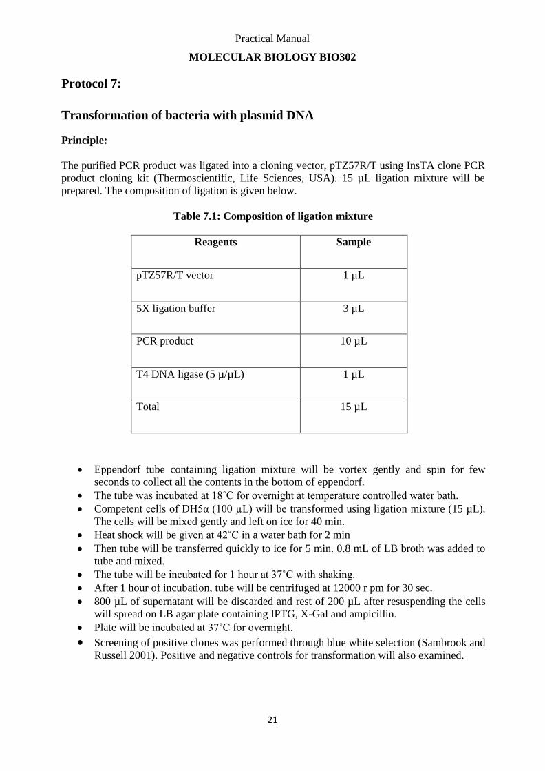
Protocol 7:
Transformation of bacteria with plasmid DNA Principle:
The purified PCR product was ligated into a cloning vector, pTZ57R/T using InsTA clone PCR
product cloning kit (Thermoscientific, Life Sciences, USA). 15 µL ligation mixture will be
prepared. The composition of ligation is given below.
Table 7.1: Composition of ligation mixture
Reagents Sample
pTZ57R/T vector 1 µL
5X ligation buffer 3 µL
PCR product 10 µL
T4 DNA ligase (5 µ/µL) 1 µL
Total 15 µL
Eppendorf tube containing ligation mixture will be vortex gently and spin for few
seconds to collect all the contents in the bottom of eppendorf.
The tube was incubated at 18˚C for overnight at temperature controlled water bath.
Competent cells of DH5α (100 µL) will be transformed using ligation mixture (15 µL).
The cells will be mixed gently and left on ice for 40 min.
Heat shock will be given at 42˚C in a water bath for 2 min
Then tube will be transferred quickly to ice for 5 min. 0.8 mL of LB broth was added to
tube and mixed.
The tube will be incubated for 1 hour at 37˚C with shaking.
After 1 hour of incubation, tube will be centrifuged at 12000 r pm for 30 sec.
800 µL of supernatant will be discarded and rest of 200 µL after resuspending the cells
will spread on LB agar plate containing IPTG, X-Gal and ampicillin.
Plate will be incubated at 37˚C for overnight.
Screening of positive clones was performed through blue white selection (Sambrook and
Russell 2001). Positive and negative controls for transformation will also examined.
Practical Manual
MOLECULAR BIOLOGY BIO302
22
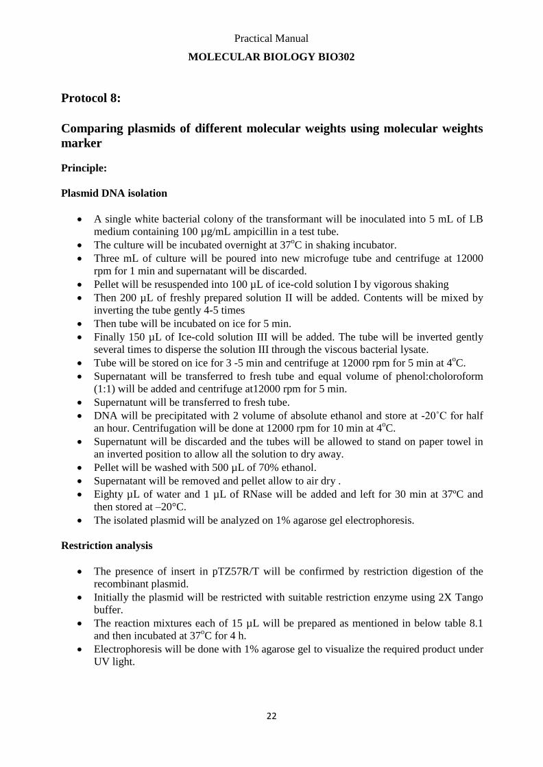
Protocol 8:
Comparing plasmids of different molecular weights using molecular weights
marker Principle:
Plasmid DNA isolation
A single white bacterial colony of the transformant will be inoculated into 5 mL of LB
medium containing 100 µg/mL ampicillin in a test tube.
The culture will be incubated overnight at 37oC in shaking incubator.
Three mL of culture will be poured into new microfuge tube and centrifuge at 12000
rpm for 1 min and supernatant will be discarded.
Pellet will be resuspended into 100 µL of ice-cold solution I by vigorous shaking
Then 200 µL of freshly prepared solution II will be added. Contents will be mixed by
inverting the tube gently 4-5 times
Then tube will be incubated on ice for 5 min.
Finally 150 µL of Ice-cold solution III will be added. The tube will be inverted gently
several times to disperse the solution III through the viscous bacterial lysate.
Tube will be stored on ice for 3 -5 min and centrifuge at 12000 rpm for 5 min at 4oC.
Supernatant will be transferred to fresh tube and equal volume of phenol:choloroform
(1:1) will be added and centrifuge at12000 rpm for 5 min.
Supernatunt will be transferred to fresh tube.
DNA will be precipitated with 2 volume of absolute ethanol and store at -20˚C for half
an hour. Centrifugation will be done at 12000 rpm for 10 min at 4oC.
Supernatunt will be discarded and the tubes will be allowed to stand on paper towel in
an inverted position to allow all the solution to dry away.
Pellet will be washed with 500 µL of 70% ethanol.
Supernatant will be removed and pellet allow to air dry .
Eighty µL of water and 1 µL of RNase will be added and left for 30 min at 37ºC and
then stored at –20°C.
The isolated plasmid will be analyzed on 1% agarose gel electrophoresis.
Restriction analysis
The presence of insert in pTZ57R/T will be confirmed by restriction digestion of the
recombinant plasmid.
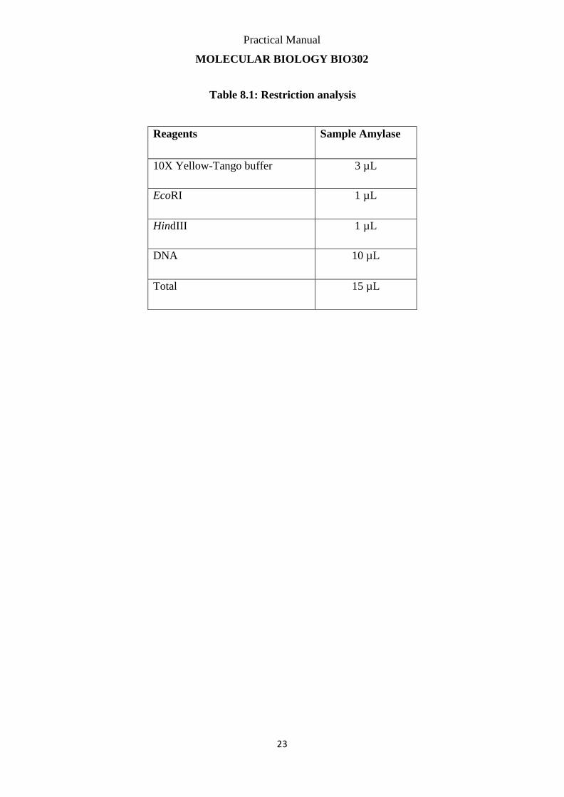
Initially the plasmid will be restricted with suitable restriction enzyme using 2X Tango
buffer.
The reaction mixtures each of 15 µL will be prepared as mentioned in below table 8.1
and then incubated at 37oC for 4 h.
Electrophoresis will be done with 1% agarose gel to visualize the required product under
UV light.
Practical Manual
MOLECULAR BIOLOGY BIO302
23
Table 8.1: Restriction analysis
Reagents Sample Amylase
10X Yellow-Tango buffer 3 µL
EcoRI 1 µL
HindIII 1 µL
DNA 10 µL
Total 15 µL

Practical Manual
MOLECULAR BIOLOGY BIO302
24
Protocol 9:
Real-Time Quantitative PCR Amplification Principle Real-time Polymerase Chain Reaction (PCR) is the ability to monitor the progress of the PCR
as it occurs (i.e., in real time). Data is therefore collected throughout the PCR process, rather
than at the end of the PCR. The higher the starting copy number of the nucleic acid target, the
sooner a significant increase in fluorescence is observed. In contrast, an endpoint assay (also
called a ―plate read assay‖) measures the amount of accumulated PCR product at the end of
the PCR cycle. Main applications of Real-Time PCR include Qualitative analysis or
plus/minus scoring, Absolute Quantification, Relative Quantification and Genotyping.
The TaqMan probe principle relies on the 5´–3´ exonuclease activity of Taq polymerase to
cleave a dual-labeled probe during hybridization to the complementary target sequence and
fluorophore-based detection. As in other quantitative PCR methods, the resulting
fluorescence signal permits quantitative measurements of the accumulation of the product
during the exponential stages of the PCR; however, the TaqMan probe significantly increases
the specificity of the detection. TaqMan probes were named after the videogame PacMan
(Taq Polymerase + PacMan = TaqMan) as its mechanism is similar.
Reagents Required:
PCR master mix including Taq polymerase, dNTP, MgCl2 and buffer (in case
of one-step reverse-transcriptase PCR, the master mix also contains the Reverse Transcriptase enzyme for initial step of cDNA synthesis from RNA template)
PCR primers
Labeled Probe or DNA binding dyes
PCR grade Water
DNA/RNA standards (for quantification assays)
Negative and Positive Controls
Equipment Required:
Real-Time Thermal Cycler with analysis software
Vortex Mixture
Microcentrifuge
Pipettes
PCR safety cabinet
Consumables:
Optically clear PCR tubes/strips/plates according to equipment compatibility
Filtered pipette tips
1.5 ml centrifuge tubes
Practical Manual
MOLECULAR BIOLOGY BIO302
25
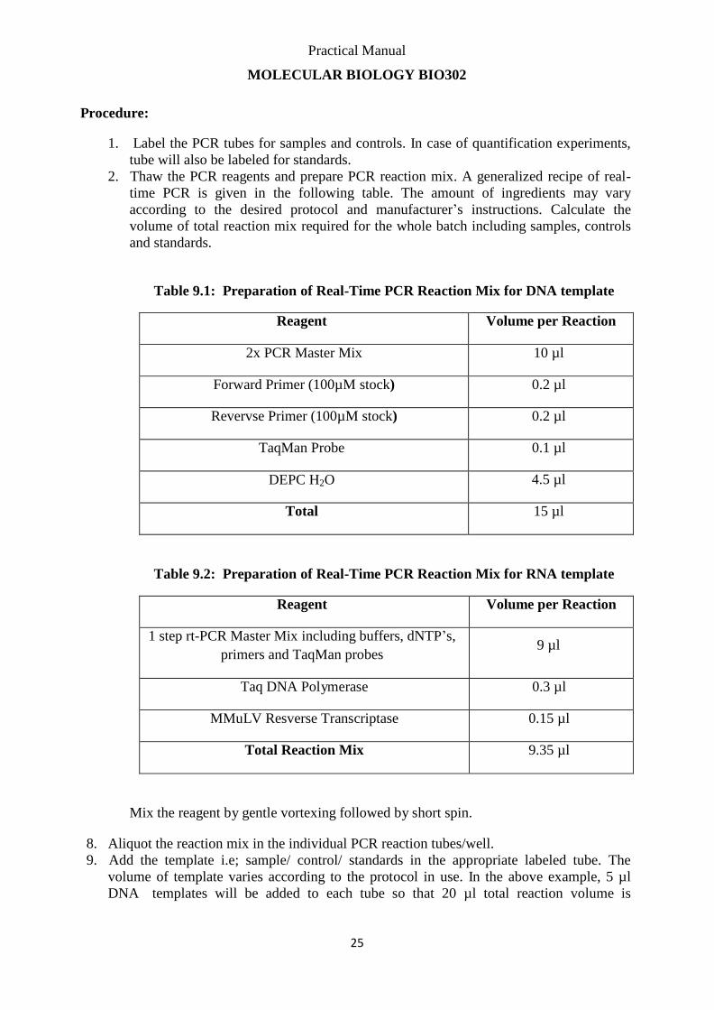
Procedure:
1. Label the PCR tubes for samples and controls. In case of quantification experiments,
tube will also be labeled for standards.
2. Thaw the PCR reagents and prepare PCR reaction mix. A generalized recipe of real-
time PCR is given in the following table. The amount of ingredients may vary
according to the desired protocol and manufacturer’s instructions. Calculate the
volume of total reaction mix required for the whole batch including samples, controls
and standards.
Table 9.1: Preparation of Real-Time PCR Reaction Mix for DNA template
Reagent Volume per Reaction
2x PCR Master Mix 10 µl
Forward Primer (100µM stock) 0.2 µl
Revervse Primer (100µM stock) 0.2 µl
TaqMan Probe 0.1 µl
DEPC H2O 4.5 µl
Total 15 µl
Table 9.2: Preparation of Real-Time PCR Reaction Mix for RNA template
Reagent Volume per Reaction
1 step rt-PCR Master Mix including buffers, dNTP’s,
primers and TaqMan probes
9 µl
Taq DNA Polymerase 0.3 µl
MMuLV Resverse Transcriptase 0.15 µl
Total Reaction Mix 9.35 µl
Mix the reagent by gentle vortexing followed by short spin. 8. Aliquot the reaction mix in the individual PCR reaction tubes/well.
9. Add the template i.e; sample/ control/ standards in the appropriate labeled tube. The
volume of template varies according to the protocol in use. In the above example, 5 µl
DNA templates will be added to each tube so that 20 µl total reaction volume is
Practical Manual
MOLECULAR BIOLOGY BIO302
26
achieved. Whereas, in case of RNA, 6 µl template sample will be added to achieve 15 µl
total reaction volume. 10. Open the Real-PCR machine’s software and edit run parameters e.g. sample IDs, plate
map, sample volume, detection wavelengths and cycling conditions according to desired protocol. A generalized cycling protocol for TaqMan assay is given below
11. Table 9.3: Reaction conditions for DNA templates
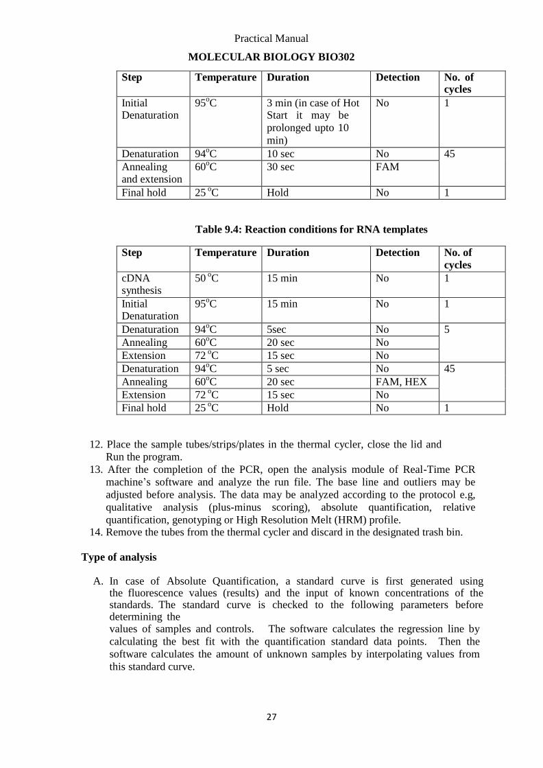
Practical Manual
MOLECULAR BIOLOGY BIO302
27
Step Temperature Duration Detection No. of
cycles
Initial Denaturation
95oC 3 min (in case of Hot
Start it may be
prolonged upto 10
min)
No 1
Denaturation 94oC 10 sec No 45
Annealing and extension
60oC 30 sec FAM
Final hold 25 oC Hold No 1
Table 9.4: Reaction conditions for RNA templates
Step Temperature Duration Detection No. of
cycles
cDNA synthesis
50 oC 15 min No 1
Initial Denaturation
95oC 15 min No 1
Denaturation 94oC 5sec No 5
Annealing 60oC 20 sec No
Extension 72 oC 15 sec No
Denaturation 94oC 5 sec No 45
Annealing 60oC 20 sec FAM, HEX
Extension 72 oC 15 sec No
Final hold 25 oC Hold No 1
12. Place the sample tubes/strips/plates in the thermal cycler, close the lid and
Run the program.
13. After the completion of the PCR, open the analysis module of Real-Time PCR
machine’s software and analyze the run file. The base line and outliers may be
adjusted before analysis. The data may be analyzed according to the protocol e.g,
qualitative analysis (plus-minus scoring), absolute quantification, relative
quantification, genotyping or High Resolution Melt (HRM) profile. 14. Remove the tubes from the thermal cycler and discard in the designated trash bin.
Type of analysis
A. In case of Absolute Quantification, a standard curve is first generated using the fluorescence values (results) and the input of known concentrations of the standards. The standard curve is checked to the following parameters before determining the values of samples and controls. The software calculates the regression line by
calculating the best fit with the quantification standard data points. Then the
software calculates the amount of unknown samples by interpolating values from
this standard curve.
Practical Manual
MOLECULAR BIOLOGY BIO302
28
a. Slope—indicates amplification efficiency. The range for slope as
recommended is -2.9 to -3.3.
b. R
2—is the correlation coefficient, and indicates the statistical
significance of the standard curve. An R2
value of
approximately 0.99 is desirable.
c. Intercept— is the point at which the standard curve touches the Y-
axis. It indicates the expected Ct value for a sample with a specific
quantity according to the method.
B. In Relative Quantification, a comparative analysis is performed between the
fluorescent signals of different templates in the sample labeled with separate dyes. A
common use of relative quantification is in the gene expression analysis after reverse
transcription and amplification of mRNA targets.
C. Plus-minus scoring analysis involves the qualitative assessment of presence of the
template on the basis of the rise of amplification curve beyond base line. The samples
are considered Negative if the amplification graph fails to rise during the PCR.
D. Genotyping analysis involves the detection of a particular DNA sequence
polymorphism /mutation by the specific labeled probe. Different genotypes have to be
labeled by different dyes for genotyping analysis to work.
E. High Resolution Melt Analysis makes use of DNA melting behavior according to
number and sequence of nucleotides in the PCR product. HRM analysis involves the
DNA binding dye chemistry and it is performed after the completion of PCR. Any
variation in target DNA will result a shift in the HRM temperature peak and therefore
genotype may be identified.
Practical Manual
MOLECULAR BIOLOGY BIO302
29
Appendix A: Useful Recourses
EQUIPMENT AND REAGENT SUPPLIERS
World Wide Scientific
Office#1-2,1st Floor, Syed Plaza, 30-Ferozepur Road
Lahore
Mobile: 03009476496
Phone: 042-37552355, Fax: 042-37553255
Email: [email protected]
GE Healthcare Life Sciences http://www.gelifesciences.com
Scientific Supplies, Saleemi Chambers
15 Edward (Mouj Darya) Road , P.O.Box 2179
LAHORE Pakistan Phone: + 92 42 3732 4449(Punjab), Fax: + 92 42 3732 4722(Punjab), Email:
Bio-Rad Laboratories http://www.bio-rad.com/
Life Science (Research, Education, Process Separations, Food Science) Bio-Rad SNC Office No. 1002/1003, Golden Tower, Buhaira Chorniche
Sharjah, United Arab Emirates
Phone: +971 6 574 8328
Fax: +971 6 574 9273
For general information, quotation requests, please contact:
E-mail: [email protected]
Life Technologies (ABI) https://www.lifetechnologies.com/pk/en/home.html
Analytical Measuring Systems (Private) Limited
AMS House 14-C Main Sehar Commercial Avenue, Lane.04, DHA-VII, Karachi-75500,
Email: [email protected] , Phone: +92-21-35345581, Fax: +92-21-35345582
Practical Manual
MOLECULAR BIOLOGY BIO302
30
Promega Corporation http://worldwide.promega.com/
Molecular Products Co. (Agent and Distributor)
Office No. 208, 2nd
Floor, Nafees Arcade, Plot No. SC-14 University Road, Karachi-74800 , Pakistan Tel: +92 21 34922501, 34922502, 321 8752522, Fax: +92 21 34922501 E-mail Address: [email protected], [email protected]
Qiagen https://www.qiagen.com/pk/
Briogene Pvt Ltd, Office No 303, 3rd Floor,
Progressive Centre, Plot No 30-A, Block-6,
P.E.C.H.S., Karachi 75400, Pakistan
Tel: +9221 34559046-7, Fax: +9221 34316380
Website: www.briogene.com
Sigma Aldrich http://www.sigmaaldrich.com
M.S. Traders, Lahore, Pakistan, Phone: 92 42 636 0663, Fax: 92 42 636 0292
Email: [email protected]
Si-Scientific
Ms. Shumyla Usman, Lahore, Pakistan, Phone: 92 42 578 2163, Cell: 92 301 842 8369,
Email: [email protected]
Analytical Measuring System Pvt. Ltd. Karachi, Pakistan, Phone: 92 21 35345581, Fax: 92 21
5345582
Email: [email protected]
Beckman Coulter https://www.beckmancoulter.com
Scientific Supplies
57A, Block 2
P.E.C.H.S.
P.O. Box 8956,
75400 Karachi
Pakistan
phone: +92-21-3455 5617
alt phone: +92-21-3455 4236
fax: +92-21-3455 7446
email: [email protected]
Practical Manual
MOLECULAR BIOLOGY BIO302
31
Biometra http://www.biometra.de/index.php/contact.html
Scientific Supplies (Pvt) Ltd. 57-A, Block 2, P.E.C.H.S., Karachi-75400, Pakistan
Phone: +92 21 455 5617, +92 21 455 4236, Fax: +92 21 455 7446
scientific-supplies.com.pk
Merck Millipore http://www.merckmillipore.com
Merck (Private) Limited D-7, Shaheed e Millat Road, Karachi
Pakistan
Tel.: +9221 111 523 523
National Ware House (Lahore) Address : Plot No. 75 -M Quaid-e-Azam Industrial Estate Township, Kot Lakhpat Lahore.
Phone : (92) 42 - 111-523-523
Fax : (92) 42 - 35150830
Practical Manual
MOLECULAR BIOLOGY BIO302
32
DNA Sequencing and Genotyping Services
DNA Sequencing and Synthesis Facility at CAMB
CAMB DNA core facilities, Centre for Applied Molecular Biology, 87-West Canal Bank Road, Thokar Niaz baig, Ministry of Science and Technology,
Lahore, Pakistan-53700 Phone Office: 042-5293141-6 Ext. 116, Fax: 042-5293149
E mail: [email protected]
Bioinformatics Resources NCBI BLAST Tool for sequence alignment
http://blast.ncbi.nlm.nih.gov/Blast.cgi
Ensembl
http://www.ensembl.org/index.html
Primer 3 tool for primer designing
http://biotools.umassmed.edu/bioapps/primer3_www.cgi
Mega6 Sequence Alignment Tool
http://www.megasoftware.net/mega.php
Practical Manual
MOLECULAR BIOLOGY BIO302
33
References Molecular Cloning: A Laboratory Manual by Joseph Sambrook, David William Russell,
CSHL Press, 2001 - Science - 2344 pages
Manufacturers’ Manuals for Spectrophotometer, Thermal Cycler, Real-Time PCR System
and Genetic Analyzer.