posteriortibialfractures - the podiatry · pdf filefigure 3a mortise view of weber b fracture...
TRANSCRIPT

The opinions of the author are his personal beliefs and should not beconstrued as reflecting official US Army Medical Department policy.
Fractures of the posterior malleolus as isolated injuriesare most uncommon. They are usually associated withdisruption to the other malleolli commonly producing thetrimalleolar fracture. The posterior malleolus fracture mostcommonly occurs with the supination-external rotation andpronation-external rotation fracture patterns. Athough notcommon, they can occur with pronation-abduction fracturesand are not usually large. Posterior malleolus fractures arenot observed with the supination-adduction injuries (1).
The ankle joint involves three bones, the tibia, fibula,and talus. The distal tibia is quadrilateral in configurationending in the articular surface forming the superior aspect ofthe ankle joint. The articular surface is inclined at about 15degrees. The posterolateral aspect of the distal tibia isreferred to as the posterior malleolus. The medial extensionis the medial malleolus and there is a concaved recess on thelateral aspect for accommodation of the fibula. The anterioraspect of the tibia extends further lateral as the tubercle ofChaput than the posterior aspect. The posterior aspect ofthe tibia extends more inferiorly than the anterior. Thisposterior portion of the tibia has been referred to as theposterior malleolus or Volkmann’s tubercle. Medially, thedeltoid ligament attaches to the medial malleolus. Theinferior anterior tibiofibular (anterior syndesmotic ligament)ligament attaches to the anterolateral tibia (tubercle ofChaput) extending to the anterior aspect of the lateralmalleolus. The posterior inferior tibiofibular ligamentattaches to the posterior malleolus and the posterior aspectof the lateral malleolus. The interosseous ligament issituated in the syndesmotic space between the distal tibiaand fibula (2-4).
The biggest question is the size threshold at whichthe posterior malleolus fracture needs to undergo openreduction with internal fixation (ORIF). The traditionalanswer has been when it is 25-30% of the tibial articularsurface as seen on the lateral radiograph (5-7). It isrecognized that ankle fractures resulting in posteriordislocationmay produce larger posterior malleolus segments.One problem with large posterior malleolus fractures is thedifficulty of maintaining a closed reduction. The foot mayneed to be placed into plantarflexion to hold the reduction(5). While the posterior malleolus contributes to stabilizing
the talus against posterior subluxation, it also can contributeto syndesmostic stability (8).
It is recognized that the exact size of the posteriormalleolus fracture may be hard to determine on plain lateralradiographs. An externally rotated lateral view may help.However, a CT scan will always be useful in determining theactual configuration (9). If the posterior malleolus fracture islarge enough to fix, it usually exits along the posteromedialaspect of the tibia or the medial malleolus. This fracture linecan sometimes be appreciated on the AP or mortise views.While it has been recognized that trimalleolar fractures doworse than bimalleolar, the role of the posterior malleolusis hard to assess (5, 10). More recently, there has been somediscussion that posterior malleolus fractures less than 25%should undergo ORIF as they may increase the riskof arthritis (11).
Once the determination has been made to performORIF, the next decision is how to approach the fracture. Thetwo options are the posteromedial versus the posterolateralincision. The posteromedial approach is usually performedwith the patient supine (Figure 1). It follows the posterioraspect of the tibia and curves along the inferior aspect of themedial malleolus essentially following the course of theposterior tibial tendon.
The sheath for the posterior tibial tendon is opened andthe tendon is retracted posteriorly. The periosteum along theposterior aspect of the tibia is reflected exposing the fracture.The fracture can now be distracted and any osteochondralfragments in the ankle joint excised. If the medial malleolusis fractured, it is likewise exposed. A fractured medialmalleolus can be distracted inferiorly, which will actuallyallow visualization of the articular portion of the posteriormalleolus fracture. If the deltoid ligament is ruptured, thenaccess to the ankle joint can be obtained through theanteromedial aspect of the joint. However, it may be difficultto visualize the posterior tibial articular surface. Sometimes,an arthroscope can be placed through the widened medialjoint space, which can allow visualization of the posteriormalleolus. It is usually done before exposing the fibula butthis is a surgeon’s choice.
The fibula undergoes ORIF through a standard lateralapproach. The AO theory of the vassel principle would haveyou believe that anatomic reduction of the fibula will
POSTERIOR TIBIAL FRACTURES
George S. Gumann, DPM
C H A P T E R 30

automatically reduce the posterior malleolus fracture becauseboth are attached to the posterior syndesmotic ligament.While this sometimes will occur, it is more common that theposterior malleolus fracture needs to be directly manipulated.It will need to be pulled inferiorly and/or rotated to achievean anatomic reduction. The quality of the reduction isassessed by the cortical margin along the posterior aspect ofthe tibia. Comminution of this cortex could be problematicfor assessing reduction, but this rarely occurs.
The posterolateral approach is performed with thepatient prone (Figure 2). By placing a foam block under the
leg, it can actually be rotated to produce a lateral position.The approach is placed midway between the peronealtendons and the Achilles tendon. The sural nerve will needto be protected. The dissection to the posterior malleolus isquite deep. The flexor hallucis tendon will need to beretracted medially. The periosteum is dissected along themargin of the fracture for exposure. The fibular fracture isexposed, debrided, anatomically reduced and internallyfixated. The fibular plate can be applied posteriorly for ananti-glide function or laterally for neutralization. If the plateis to be applied laterally, it can sometimes be difficult to place
CHAPTER 30152
Figure 1A Mortise view of a supination-externalrotation stage IV fracture sustained in a parachutejump.
Figure 1B. Lateral view.
Figure 1C. CT scan demonstrating a large displacedposterior malleolus fracture.
Figure 1D. CT scan showing displacement.
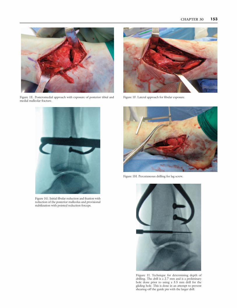
CHAPTER 30 153
Figure 1G. Initial fibular reduction and fixation withreduction of the posterior malleolus and provisionalstabilization with pointed reduction forceps.
Fiigure 1I. Technique for determining depth ofdrilling. The drill is a 2.7 mm and is a preliminaryhole done prior to using a 3.5 mm drill for thegliding hole. This is done in an attempt to preventshearing-off the guide pin with the larger drill.
Figure 1E. Posteromedial approach with exposure of posterior tibial andmedial malleolar fracture.
Figure 1F. Lateral approach for fibular exposure.
Figure 1H. Percutaneous drilling for lag screw.
CHAPTER 30154
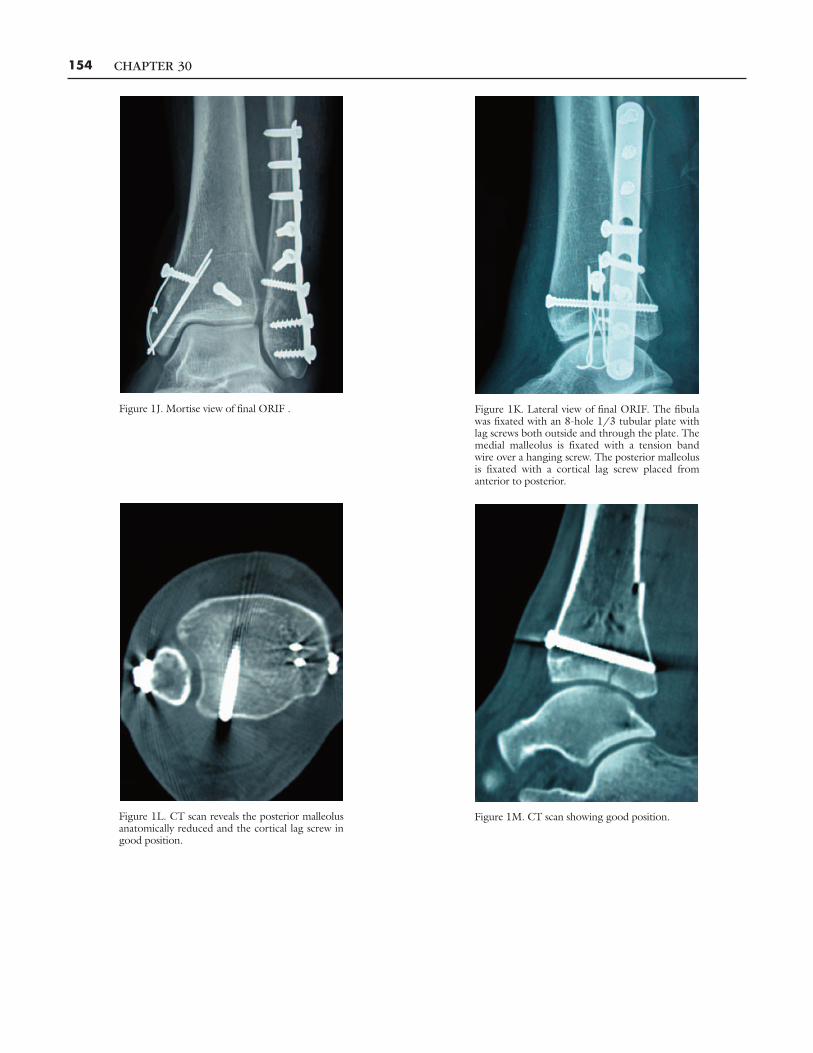
Figure 1J. Mortise view of final ORIF . Figure 1K. Lateral view of final ORIF. The fibulawas fixated with an 8-hole 1/3 tubular plate withlag screws both outside and through the plate. Themedial malleolus is fixated with a tension bandwire over a hanging screw. The posterior malleolusis fixated with a cortical lag screw placed fromanterior to posterior.
Figure 1L. CT scan reveals the posterior malleolusanatomically reduced and the cortical lag screw ingood position.
Figure 1M. CT scan showing good position.

a lag screw from anterior to posterior. The posteriormalleolus fracture is then reduced using the cortical marginsunder direct visualization. It should be noted thatosteochondral fragments cannot be removed from this ap-proach. Also, a medial malleolus fracture is hard to reduceand fixate from this position.
The second decision is how to fixate the posteriormalleolus. With the posteromedial approach, the posteriormalleolus fracture is fixated by placing the screw or screwsfrom anterior to posterior (Figure 1). This is commonlydone percutaneously. Care needs to be maintained not toiatrogenically injure any of the neurovascular or tendonstructures along the anterior aspect of the distal tibia.Sometimes, the screws can be placed through either theposteromedial incision if retracted far enough laterally orthrough the lateral incision used to fixate the fibular fractureif it can be retracted enough medially. The screws arenormally 3.5 mm cortical and can be either cannulated ornoncannulated. The use of cannulated instrumention alongwith fluoroscopic visualization aids in the preparation forscrew placement.
A cannulated screw is easy to place and easy to retrieveif necessary. It is possible to employ cannulatedinstrumentation but then place a noncannulated screw.Sometimes, a washer may be necessary to preventsubsidence of the screw past the anterior tibial cortex. Thiscan occur with osteoporotic bone or over zealoustightening of the screw. The question is always how far toover-drill for a lag effect but not accidently go through theposterior cortex. Basically, one needs to exercise tactilecontrol and use fluoroscopic imaging in the lateralprojection. This can allow visualization of the depth of the3.5 mm drill bit. Be careful not to shear-off the guide pin.However, one normally reduces the fibula first and manysurgeons will apply a lateral neutralization plate. Thisusually obscures the reduction of the posteriormalleolus; it may also interfere with calculation for over-drilling the lag screw. To avoid this issue, sometimes thefibula can be partially fixated with lag screws to allowlateral fluoroscopic viewing or a posterior anti-glide platecan be applied. The use of partially threaded cancellousscrews placed from anterior to posterior should not beemployed because of the difficulty of identifying if thethreads pass on the far side of the fracture.
Through a posterolateral approach, the posteriormalleolus can be fixated with lag screws placed from
posterior to anterior or with a plate (Figure 2). The screwscan be either 3.5 mm cortical placed with a lag effect or 4.0mm partially threaded cancellous screws. Depending on thesize of the fracture, one or two screws can be employed. Asupplemental technique is the placement of an anti-glidescrew over a washer or a one-hole plate at the apex ofthe fracture. This is placed to help prevent proximaldisplacement of the fracture. A plate can also be used forfixation with lag screws going through the distal holesproviding more robust stability in an anti-glide function.This can be standard 1/3 tubular plate or a specificallydesigned posterior tibial plate.
At our institution, a variation is seen in which thefracture involves the entire posterior aspect of the tibia (12)(Figure 3). These injuries are thought of as “mini-pilonfractures.” This fracture pattern occurs generally as the resultof parachuting. Therefore, there is an axial loading as well asrotational forces influencing the mechanism of injury. Thefracture may be a single large piece or may have an additionalsagittal fracture line dividing it into two pieces. Therewill be one fragment posterolaterally and the other post-eromedially. The posteromedial segment is most commonlythe larger especially when considering the superior extensioninto the tibial metaphysis. However, they can be relativelythe same size or sometimes the posterolateral fragment canbe larger. They are usually interdigited in the mid-fractureline, the periosteum at this level intact, and effectivelyoperate as one piece. On occasion, they may act as twoindividual fracture fragments. There are usually osteochondralfragments within the ankle joint. There may be also be atransverse fracture of the medial malleolus. This fracturepattern is most commonly approached posteromedially forthe tibia and laterally for the fibula. This is necessary toexpose the ankle joint for removal of the osteochondralfragments. It allows for excellent visualization to reduce theposteromedial tibial fracture component and place internalfixation. The screws are oriented slightly from post-eromedial to anterolateral. In addition, a cortical lag screw isusually placed from anterior to posterior to capture the morelateral portion of the fracture.
Postoperatively, the patient is immobilized in either afracture brace or a short-leg cast depending on the quality ofthe bone and stability of the fixation. An initial period ofnonweightbearing is necessary usually for 4-8 weeks. Whento begin range of motion exercises and physical therapy is atthe discretion of the attending surgeon.
CHAPTER 30 155

CHAPTER 30156
Figure 2D. Posterolateral approach to the ankle with the fibula fixated witha posterior plate.
Figure 2A Mortise view of a supination-externalrotation fracture sustained in a parachute jump.
Figure 2B. Lateral view.
Figure 2C. CT scan demonstrating the posteriormalleolus and degree of displacement.

CHAPTER 30 157
Figure 2E. Posterior malleolus fixated with aposterior plate.
Figure 2F. Mortise view of ORIF.
Figure 2G. Lateral view of ORIF. Figure 3A Mortise view of Weber B fracturedislocation of the ankle.
CHAPTER 30158
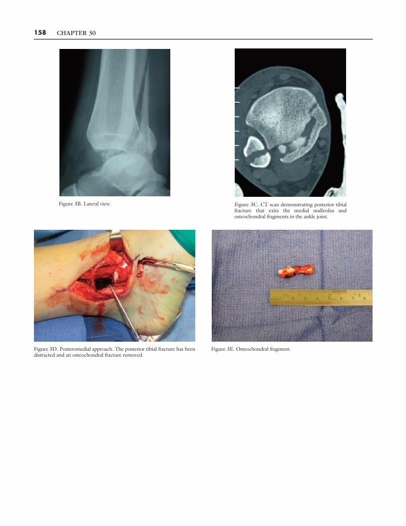
Figure 3B. Lateral view. Figure 3C. CT scan demonstrating posterior tibialfracture that exits the medial malleolus andosteochondral fragments in the ankle joint.
Figure 3D. Posteromedial approach. The posterior tibial fracture has beendistracted and an osteochondral fracture removed.
Figure 3E. Osteochondral fragment.

REFERENCES1. Lauge-Hansen N. Fractures of the ankle: analytic, historic survey as thebasis of new experimental, roentgenologic, and clinical investigations.Arch Surg 1948;56:259-317.
2. Davidovitch RI, Egol KA. Ankle fractures. In Rockwood CA andGreenDP. Fractures in Adults, Lippincott Williams & Wilkins Philadelphia;2010.
3. Hamilton W. Traumatic disorders of the of the ankle. New York:Springer-Verlag. 1984.
4. Haraguchi N, Haruyama H, Toga H, et al. Pathoanatomy of posteriormalleolus fractures of the ankle. J Bone Joint Surg Am 2006;88:1085-92.
5. Mast JW, Teipner WA. A reproducible approach to the internal fixationof adult ankle fractures: rationale, technique, and early results. OrthopClin North Am 1980;11:661.
6. Michelson JD. Current concepts review fractures of the ankle. J BoneJoint Surg Am 1990;77:142-52.
7. Macko VW, Matthews LS, Zwirkoski P, et al. The joint-contact area ofthe ankle. The contribution of the posterior malleolus. J Bone JointSurg Am 1991;73:347-51.
8. Gardner MJ, Brodsky A, Briggs SM, et al. Fixation of the posteriormalleolar fractures provides greater syndesmotic stability. Clin OrthopRel Res 2006;447:165-71.
9. Magid D,Michelson JD, Ney DR, et al. Ankle fractures: comparison ofplain films and two- and three-dimensional CT scans. Am J Roentgenol1990;154:1017-23.
10. Bauer M, Bergstrom B, Hemborg A et al. Malleolar fractures:nonoperative versus operative treatment. A controlled study. ClinOrthop Rel Res 1985;17:27.
11. Jaskulka RA, Ittner G, Schedl R. Fractures of the posterior tibialmargin: their role in the prognosis of malleolar fractures, J Trauma1989;29:1565-70.
12. Lei W, Shi Z, Zhang C, et al. Trimalleolar fracture with involvement ofthe entire posterior plafond. Foot Ankle Int 2011;32:774-81.
CHAPTER 30 159
Figure 3F. Posteromedial plate applied to the distal tibia with a lag screw indistal hole and medial malleolus fixated with tension band wire.
Figure 3G. Mortise view of ORIF. The long fibularfracture was fixated with three cortical lag screws.The posterior tibial fracture was fixated with aposteromedial plate and a cortical lag screw appliedfrom anterior to posterior. The medial malleolus wasfixated with a tension band wire.
Figure 3H. Lateral view of ORIF.