positions in human serum albumin which involve the indole
TRANSCRIPT

THE JOURNAL OF BIOLOGICAL CHEMISTRY Vol. 250, No. 17, Issue of September 10, pp. 6711-6719, 1975
Printed in U.S.A.
Positions in Human Serum Albumin which Involve
the Indole Binding Site
SEQUENCE OF 107-RESIDUE FRAGMENT*
(Received for publication, December 19, 1974)
KANWAL K. GAMBHIR,$ RAPIER H. MCMENAMY,§ AND FRANK WATSON
From the Department of Biochemistry, The Medical and Dental Schools, State University of New York at Buffalo, Bu$alo, New York id214
SUMMARY
The first 107 residues of Fragment C of human serum albumin have been sequenced and two positions at which affinity labels block the indole site determined. Histidine 23 is the position of blockage by bromoacetyl-r.-tryptophan and lysine 67 is the position of blockage by .5-dimethylamino- naphthalene- 1 -sulfonyl chloride and probably pyridoxal-S’- phosphate. The presence of an indole ligand at the binding site markedly reduces incorporation of the label into the above lysyl residue, and in the case of 5-dimethylamino- naphthalene-1-sulfonyl chloride, increases incorporation into three other positions, lysine residues 13, 39, and 84. It is concluded that binding of the indole ligand on the site brings about conformational changes in the albumin struc- ture exposing new reactive positions for $dimethylamino- naphthalene-1-sulfonyl chloride. There is a large accumula- tion of basic and hydrophobic residues and no glycine, serine, threonine, valine, aspartate, or cysteine residues in the sequence 10 to 43. Lysine 71 has been identified by amino acid analyses and sequence studies as the position acetylated by acetylsalicylic acid (HAWKINS, D. R., PINCKARD, N., CRAWFORD, C. P., AND FARR, R. S. J. Clin. Invest. (1969) 48, 536), establishing the structural relationships of two major ligand binding sites on albumin. The lone tryptophan is at position 86. Evidence indicates that within residues 1 to 86 of Fragment C and within residues of the A- Phe fragment (M, = ~10,000)~ the latter known to be ad- jacent to Fragment C in the whole albumin structure, exists the major binding sites of all ligands for human serum al- bumin.
* This investigation was supported by National Science Foun- dation Research Grant GB-7224 and by a grant from the James H. Cummings Foundation. An initial report was presented at the 57th Annual Meeting of the Federation of American Societies for Experimental Biology, April, 1973. Part of the data are taken from a thesis submitted by K. K. Gambhir for his Ph.D. degree.
$ Present address, Department of Obstetrics, Gynecology and Medicine, Howard University, Washington, D. C. 20001.
Q From whom reprints should be requested.
It was found earlier that reacting human serum albumin with affinity labels bromoacetyl-L-tryptophan, dansyli chloride, or pyridoxal S/-phosphate, led to a blockage of the binding of acetyl- n-tryptophan (1). It was further found that the greater part of each label was incorporated into Fragment C (-11, = -18,000). Thus, at conditions of 1:l molar ratio of labeling agent to al- bumin 65 to 95% of the label was incorporated into Fragment C, accounting for essentially an equivalent stoichiometric blocking of the binding site when bromoacetyl-L-tryptophan was used as a reagent, and 35 to 50% of the equivalent stoichiometric block- ing when dansyl chloride or pyridoxal 5’-phosphate were used as the labeling reagents. If labeling was conducted in the presence of indolepropionate, a ligand strongly bound by albumin, the in- dole site was largely protected. Hromoacetyl-L-tryptophan re- acted with imidazole groups, dansyl chloride reacted with e-amino lysyl(s) and tyrosyl-OH group(s), and pyridoxal 5’.phosphate re- acted with e-amino lysyl group(s).
The indole binding site on albumin is unusual in several as- pects. First, the above described labeling agents which block the indole site all have diverse structural features. The site, thus, is apparently readily adaptable to different types of ligands. A fur- ther example of this adaptability is shown with thyroxine, a com- pound much different from the indole compounds in shape and structural groups, yet, as shown by Tritsch (2), binding competi- tively with L-tryptophan. Another unusual feature of the site is that fatty acids at low concentrations inhibit the indole site in the alkaline pH region, but have little effect on indole binding in the neutral and acid pH regions (3). Fatty acid inhibition at low concentration is not directly competitive with indole binding. In addition to providing a broad accommodation to different types of ligand, there is also evidence that occupation of the site by an indole compound provides considerable protection to the albumin. Thus, the reaction of trypsin with albumin is much retarded when albumin is associated with L-tryptophan (4). Also, acetyl-n-tryp- tophan has, for many years, been known to stabilize albumin against thermal denaturation. Finally, it has recently been postu-
1 The abbreviations used are: dansyl, the prefix for 5-dimethyl- aminonaphthalene-1-sulfonyl group; TPCK, n-I-tosylamido-2- phenylethyl chloromethyl ketone; acetyl-n-tryptophan-HSA, al- bumin reacted with bromoacetyl-r-tryptophan; dansyl-HSA, albumin reacted with dansyl chloride; pyridoxal-P-HSA, albumin reacted with pyridoxal 5’.phosphate and reduced with sodium borohydride; PTH, phenylthiohydantoin.
6711
by guest on March 25, 2018
http://ww
w.jbc.org/
Dow
nloaded from

6712
lated that the indole binding site plays a role in neural regulation
(5). For these reasons, it is of much interest to study further the
structure and other features of the indole binding site.
In the current study, a major part of Fragment C has been se- quenced, and the positions of the labeled residues determined.
Information is provided on several regions of the peptide chain which are involved in the indole binding site. The natures of the
side groups of the residues near the binding region, which likely
supply the forces for binding, are considered.
EXPERIMEIGTAL PROCEDURE
The source of human serum albumin, affinity labeling condi- tions, fragmentation and isolation of Fragment C were as de- scribed previously (1). Affinity labels bromoacetyl-L-tryptophan, dansyl chloride, and pyridoxal 5’-phosphate, were reacted with protein at 1:l molar ratios in the presence and absence of indole- propionate. Binding studies were conducted to evaluate the ex- tent to which the affinity labels blocked the site, and the albumin preparations were then cleaved to isolate Fragment C.
ZZetEuction-Fragment C was reduced as previously described except iodoacetate or 4-vinylpyridine (6) were used to react with the freed --SH groups, providing S-carboxymethylated and S-B- (4-pyridyl)ethylated C fragments, respectively. The peptide was separated from the reagents by passing the solutions through a Sephadex G-25 (coarse) column equilibrated with 0.01% triethyl- amine and concentrating the separated peptide zone by a Diaflo apparatus using a UM-2 Ultrafilter (Amicon Corp.).
Performic Acid Osidation-At -5”, Fragment C (75 to 100 mg) was dissolved in a solution containing 4.5 ml of twice recrystal- lized formic acid and 0.5 ml of methanol. A performic acid solution, prepared by allowing to stand 1 hour at room temperature a mix- ture of 5 ml of formic acid and 0.5 ml of 309$ hydrogen peroxide (Mallinckrodt reagent grade) and 0.01 ml of liquid phenol (Mal- linckrodt), cooled to -5”, was added, and the reaction carried out for 00 min. The reaction solution was then placed on a 3.5 X GO cm column of Sephadex G-25 (coarse) equilibrated at 0” with 1% propionic acid. Just before the protein zone was eluted from the column, the column was placed at room temperature and moni- tored at 280 nm to detect the protein fraction. After collection, the fraction was concentrated in a Diaflo apparatlls.
Maleylation-l~ed~lced or oxidized Fragment C was dissolved in 5 ml of water, its container placed in an ice bath and, with the pH maintained at 8.5 to 0 by addition of 2 N NaOH, 2 parts of maleic anhydride (w/w) were added in small aliqllots over a 15. min period. After standing for an additional 30 min, the reaction solution was diluted three times to 60 ml with water, and concen- trated each time to approximately 5 ml in a Diaflo apparatlls.
‘I’rypsin Reaction-To assure more complete destruction of chymotryptic activity, trypsin (TPCK-treated, Worthington) was reacted shortly before-&e with a further aliquot of TPCK. To 2.5 mg of the trvnsin in 0.25 ml of 0.2 N NaHCO, were added 20 ~1 of a-27? TPCK solution (w/w) in acetone. After-3 hours, the pH was reduced to a value of 3.0 with 0.1 N HCI. The enzyme solu- tion was used within 16 hours.
To a 3-ml solution of reduced or oxidized, maleylated Fragment C (75 to 100 mg), after flushing with N, and pH adjustment to 9.0, was added 0.1 ml of TPCK-treated trypsin solution. The pH was maintained at 9.0 by addition of 0.1 N NaOH for 5 min. A second aliquot of trypsin solution was added, and the pH again maintained at 9.0 by addition of NaOH solution (the volume of NaOII solution added after the second addition of enzyme solu- tion was usually very small). After 5 min, with good mixing, 2.5 ~1 of diisopropylphosphorofluoridate were added and allowed to react for 20 min. The solution was placed on a Sephadex G-100 column (5 X 210 cm), and eluted with a solution containing 0.1 N
NH,HCO, and 0.01 N NH*OH. With nonmaleylated peptides, the trypsin reactions were car-
ried out under essentially the same conditions, except the reaction times were extended 2 to 5 hours.
Chymo&/psin Xeaclion-Reactions with maleylated peptides were carried ollt at pH 8.0 by two additions of chymotrypsin (Worthington) at two 20-min intervals (l:lOO, w/w). At the end of the reaction, 50 ~1 of phenylmethaneslllfonylfluoride solution (2 mg freshly added to 1 ml of methanol) were added. After 20
min, the digested peptide was placed on Sephadex G-100, and eluted with asolution of 0.1 N NHdHC03 and 0.01 N NHdOH.
Demaleylation-After the enzyme digestions and separation of the fragments, demaleylation was conducted at pH 2.5 to 3.0 for 40 hours at 45”.
cu-Protease Reaction-At a 1:50 ratio (w/w), pH 8.0, 0.01 M
CaC12, ol-protease (Pierce Chemical Co.) was reacted with the peptides for 2 hours. The pH of the solution was then reduced to 3.0.
Separation of Peptides-Fractionation of peptides was con- ducted on a column (0.9 X 15 cm) of Dowex 50-X8 (Spinco resin AA15) using volatile buffers following the general methods de- scribed by Hill and Delaney (7) and by paper chromatography and electrophoresis. In the separation of dansyl peptides, the strategy was first to find a system in which the dansyl peptide could be separated from other peptides by one-dimensional move- ment on paper. After the separation and before the paper was completely dry (dansyl zones elute very poorly if allowed to dry on the chromatograms), the zone was cut out and eluted by cen- trifugation. The apparatus for the latter consisted of a 12-ml centrifuge tube with a serated conical disc fitted approximately 3 cm from the bottom of the tube. The paper with the dansyl zone was placed on the disc, wetted with a small amount of 507, pyri- dine, allowed to stand several minutes, briefly centrifuged, and the extraction step repeated. After several extractions with the pyri- dine solution, two or more extractions were then carried out with 107, acetic acid.
Reaction with N-Bromosuccinimide-Fragment C (20 mg) was dissolved in 1 ml of H20, the container placed in an ice bath, and 30 mg of succinic anhydride added in small aliquots over a IO-min Deriod with the nH maintained at 8 to 9 bv addition of N NaOH. kfter 1 hour, the’ pH was reduced to 4.5, and the solution allowed to stand 2 hours. Twice recrystallized urea (1 g) was added to the solution, the pH readjusted to 4.5, and 120 ~1 of N-bromosuccin- imide solution (0.2 g/20 ml of HzO) added. (This amount of N- bromosuccinimide was 2 times the amount necessary to obtain a minimum reading at 280 nm.) After 5 min, the pH of the solution was raised to 9.0, and the solution placed on a column (2 X 30 cm) of Yephadex G-25 (coarse) and eluted with 0.005 s NHdOH. The protein solution obtained from the column was freeze-dried and oxidized with performic acid.
Amino Acid Anulllses-Analyses of the normal and modified amino acids were conducted on a Beckman 120 B amino acid ana- lyzer using a two column methodology as described before (1). Hydrolysis was conducted at llO”, 22 hours in G N HCI or 4 N methanesulfonic acid containing 0.2yo 3.(2.aminoethyl)indole (Pierce Chemical Co.). In each instance, aliquots of the hydroly- sate solutions were diluted 1:30 with water, and the solutions placed directly on the resin columns.
Sequencing--Large peptides were analyzed on a Beckman Se- quencer (model 89OC), modified with the optimal accelerated se- quencing kit and Nz flushing system. The Quadrol program recom- mended by the manufacturer was used. All solvents and reagents were from Pierce Chemical Co. For peptides less than 50 amino acid residues, 4-sulfophenylisothiocyanate was generally used to assist in keeping the peptide in the sequenator cup (8). Coupling with this reagent was carried out directly in the cup. After the peptide was dried, It1 and 1~3 were placed in the manual positions, Quadrol buffer was added to the lo-mm mark (low speed), the CUP
was stopped, opened, and 25 mg of 4.sulfophenylisothiocyanate was placed in the center of the cup. The cup was started, and when up tb speed, the cover was replaced. With the effluent to waste, N? was flushed through the cell for 1 min. The cup was then set at high speed and R2 w’ts manually added to the 24-mm mark. The reaction was then started at Step 12 on the program and allowed to proceed to the end of the drying step after the ethyl acetate wash (Step 36 on the program). The program was then restarted from Step 1 with RI and 1~3 in their automatic positions.
The methods of Gray (9) and Sauer et al. (10) were used for manual sequencing.
PTH Analysis-2-Anilino&thiazolinone derivatives of the cleaved amino acids were converted to the PTH derivatives and extracted with ethyl acetate by the procedure described in the Sequencer Operator Manual. Aliquots of the ethyl acetate SOIU-
tion were applied to 20 X 20 cm silica gel plates (Eastman NO. 6060) along with three PTH standards consisting of amino acids leucine, proline, tyrosine, aspartate, asparagine; isoleucine,
by guest on March 25, 2018
http://ww
w.jbc.org/
Dow
nloaded from

phenylalanine, lysine, glutamate, serine; and valine, alanine, lysine, threonine, glutamine. The standards, applied at 2.5 nmol, were alternated with unknowns in applications across the plate. Repetitive aliquots (-35 ~1) of unknowns were applied at the same spot until the fluorescent intensity reached approxi- mately that of the standards. The amount applied was recorded. Eight unknowns were usually run on each gel plate. Ascending development was carried out in Solvent V (heptane 58 ml, pro- pionic acid 17 ml, and ethylene chloride 25 ml) of Jeppsson and Sjoquist (11). The dried chromatograms were first viewed under ultraviolet light to identify as many zones as possible, then sprayed with a 1-butanol solution containing 0.5 g/100 ml of nin- hydrin followed by heating of the plates in an oven at 110” for 10 min. This latter treatment assisted further identification of some of the zones by color differences (12). Overlaps were easily followed by this method. Sequencing experiments were termi- nated when the background became too high to recognize the un- known zones. When leucine, isoleucine, and sometimes valine and proline were indicated, their identification was confirmed by gas chromatography using the method of Pisano et al. (13) as indicated in the Sequencer Operator Manual. PTH derivatives of arginine, histidine, and cysteic acid, and sometimes lysine (when 4.sulfo- phenylisothiocyanate was used), were usually identified by amino acid analysis after hydrolysis with 58$& HI in sealed, evacuated tubes at 140” for 24 hours. In some instances, arginine was identi-
,~‘-‘-,-~,~,-l~~o~,-,-,-,-,-l-,-,-,-,. -I-, 1600 2800 3200 3600
mL FIG. 1. Elution profile of tryptic digest of carboxymethylated
Fragment C (100 mg). Column Sephadex G-100 (5 X 210 cm), eluting buffer 0.1 N NHrHCO,/O.Ol N NHaOH.
6713
fied by phenanthrenequinone (14) and histidine by p-anisidine (15) as described by Hermodson et al. (G).
RESULTS
Peptide Separations-The elution profile of trypsin-digested, maleylated carboxymethylated Fragment C is shown in Fig. 1. Zones a, b,, and c were found to contain single peptides with NHz-terminal residues, Phe, Tyr, and Cys. Amino acid analyses are given in Table I. Zone b, appeared to be a mixture of two fragments; however, fractions taken from the leading and trailing shoulders had identical NHz-terminal groups, and upon hydroly- sis, had identical amino acid contents. The reason for the evident splitting of this zone was not clear. The relative amounts in the split zones also varied; in some instances, the leading peak was much reduced. Zone d was freeze-dried, demaleylated, and chro- matographed on the peptide resin (Fig. 2). Amino acid analyses of Zones dl, dB, and da are reported in Table I. The other zones in this elution contained multiple peptides at low concentrations and were not further considered.
The elution profile of trypsin-digested, maleylated S-fl(4- pyridyl)ethylated Fragment C is given in Fig. 3. When compared with Fig. I, a new zone appeared between Zones c and d, Zone e. This zone was further purified by concentration and passing the second time over Sephadex, G-100, or by demaleylating and chromatographing on the peptide resin (Fig. 4). Comparison of the amino acid composition of the peptides in Zone b, and e from the S-fl(4-pyridyl)ethylated Fragment C digest with the peptide from b, of the S-carboxymethylated Fragment C digest indicated that the latter was equal to the sum of the former two (Table II). Splitting the peptide b, at its internal arginine apparently is in- hibited by S-carboxymethylation at cysteinyl residues. Splitting readily occurred with S-@(4-pyridyl)ethylated and performic acid- oxidized Fragment C preparations.
The fragments isolated accounted for all of the eight arginines in Fragment C. Peptide a, which contained a homoserine residue, and was, thus, the COOH-terminal peptide, contained an internal
TABLE I Amino acid composition (nanomoles) of tryptic arginine-cleaved peptides from Fragment C
LYS His Arg Cys””
ASP Thr Ser GlU Pro GUY Ala Val Ile Leu Tyr Phe HSer Tre Total Residues nmo le s Recovered
31 6 11 2
6 1 30 6 38 7 12 2 18 3 48 10
8 2 5 1
27 5 15 3 12 2 35 6
5 1 10 2
5 1
b a
Best integer
60 6
22 2 30 3 38 3 13 1 27 3 53 6
1c 1
19 2 37 8
55 6 9 1
16 2
60 46
4.6 3.6
* Ref. 16. ** As S-carboxymethyl-L-cysteine.
a
Best integer
c
Best integer
83 2 34 1 42 1 43 1 73 2 68 2
128 3
93 2
44 1 LO6 2
69 2 100 2
21
1.3
*1 Best
integer
26 1 117 1
20 1 27 1
30 1
4
2.2
d2 Best
integer
120 1
$20 4 106 1
118 1
LO6 1
9
97 2 96 2
156 3
2.6 I 3.9
d3 Best
integer
48 1 46 1 92 2
51 1 104 2
115 2
16 156
Total Fragment C residues residues*
16 4 8
10 13
5 7
21 5 3
22 4 3
17 6
10 1 1
16 4 8
11 13
5 8
23 5 3
21 5 3
18 5 9
1
159
by guest on March 25, 2018
http://ww
w.jbc.org/
Dow
nloaded from
6714
60
Or
80 100 120 140 160 480 200 220 240
Fractions
',-'-,-~~'-,-'~~oo'-'-'-'-'-,-'-'-'-'-'-, 16Cil 2800 3200 3Mm
me FIG. 3. Elution profile of tryptic digest of maleylated, S-p(4-
pyridyl)ethylated Fragment C (100 mg). Column Sephadex G-100 (5 X 210 cm). Eluting buffer 0.1 M NHdHCO,,O/.Ol N NHcOH.
4 1’ I-I-+-l-l 120 130 140 150
Fraction
FIG. 4. Chromatography of Zone e on Dowex 50-X8, column, 0.9 X 15 cm. Gradient was 250 ml of 12.5% acetic acid v/v, 250 ml of 0.2 M pyridine-acetate (pH 3.1), 250 ml of 2.0 M pyridine-acetate (pH 5.0), 250 ml of 5.0 M pyridine-acetate (pH 5.0), Fractions, 3.0 ml.
arginine which did not cleave with trypsin under any conditions attempted.
The zone of the void volume from the elution of chymotrypsin- digested, maleylated, carboxymethylated Fragment C (column conditions the same as described for Fig. 1) was demaleylated and chromatographed on the peptide resin (column and gradient con- ditions the same as described for Fig. 4). The amino acid com- position and eluting fractions for some of the peptides isolated are given in Table III. The amino acid composition and eluting fractions for two peptides isolated from an cr-protease digestion of peptide b, using the same peptide resin conditions are reported in Table IV.
sequences-The results of sequencing experiments are given in Fig. 5. Intact Fragment C (500 nmol) was subjected to 42 steps of automated Edman degradation. Several of the mixed residues were later obtained from chymotryptic or Lu-protease-cleaved pep- tides (Tables III and IV). Thus, residue 19 was determined to be isoleucine from the composition of peptide 30; residue 30 was de- termined to be glutamate from the composition of peptide 23; and residue 36 was determined to be lysine by isolation of peptide 31. Automatic sequencing of peptide b, (300 nmol) provided resi- dues 38 to 63, and an overlap with the sequence obtained with the intact Fragment C. No PTH product was obtained for residue
260 280 300 320
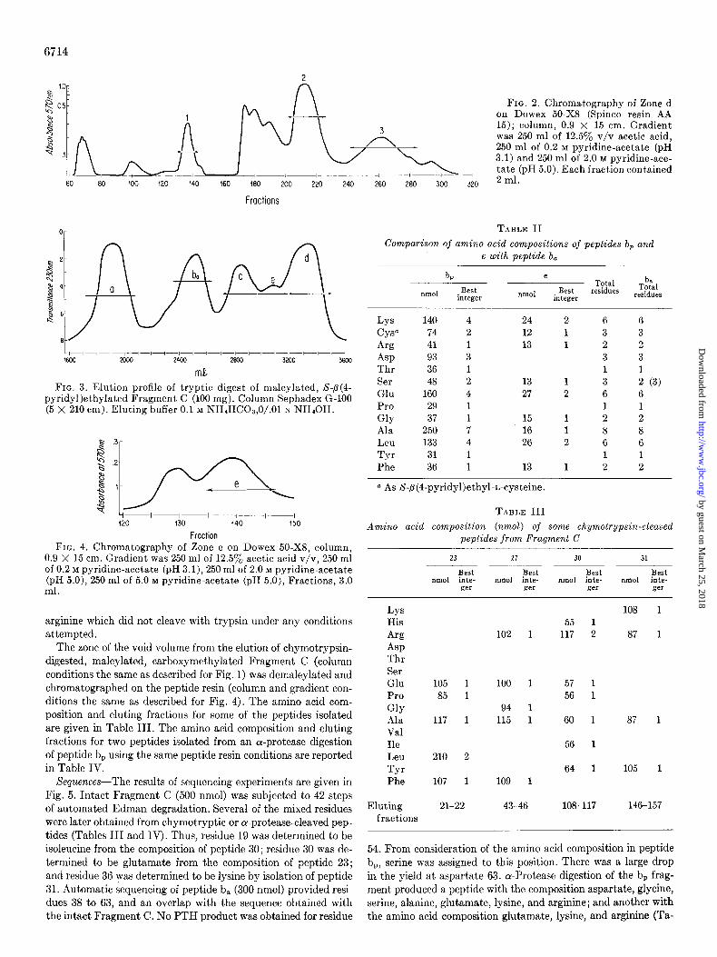
FIG. 2. Chromatography of Zone d on Dowex 50-X8 (Spinco resin AA 15); column, 0.9 X 15 cm. Gradient was 250 ml of 12.5y0 v/v acetic acid, 250 ml of 0.2 M pyridine-acetate (pH 3.1) and 250 ml of 2.0 M pyridine-ace- tate (pH 5.0). Each fraction contained 2 ml.
TABLE II
Comparison of amino acid compositions of peptides b, and e with peptide b,
b, e Total b,
Total IllllOl Best
integer nmol Best residues integer residues
LYS 140 4 24 2 6 6 cyso 74 2 12 1 3 3
Arg 41 1 13 1 2 2
Asp 93 3 3 3 Thr 36 1 1 1 Ser 48 2 13 1 3 2 (3) Glu 160 4 27 2 6 6 Pro 29 1 1 1
GUY 37 1 15 1 2 2 Ala 250 7 16 1 8 8 Leu 133 4 26 2 6 6 Tyr 31 1 1 1 Phe 36 1 13 1 2 2
a As S-~(4-pyridyl)ethyl-I~-cysteine.
TABLE III
Amino acid composition (nmol) of some chymotrypsin-cleaved peptides from Fragment C
23 27 30 31
Best Best Best Best nmol inte- IlRlOl inte- IIlIlOl inte- nmol inte-
ger .ser ger ger
LYS His
Arg Asp Thr Ser Glu Pro
GUY Ala Val Ile Leu Tyr Phe
108 55 1
102 1 117 2 87
105 1 100 1 57 1 85 1 56 1
94 1 117 1 115 1 60 1 87
56 1 210 2
64 1 105 107 1 109 1
Eluting fractions
21-22 43-46 108-117 146-157
54. From consideration of the amino acid composition in peptide b,, serine was assigned to this position. There was a large drop in the yield at aspartate 63. cy-Protease digestion of the b, frag- ment produced a peptide with the composition aspartate, glycine, serine, alanine, glutamate, lysine, and arginine; and another with the amino acid composition glutamate, lysine, and arginine (Ta-
by guest on March 25, 2018
http://ww
w.jbc.org/
Dow
nloaded from

ble IV). Neither of these peptides would sequence by the Edman procedure, presumably because blocking of the NHz-terminal residues had occurred in their isolation. Carboxypeptidase 13 re- acted with b, producing arginine. Carboxypeptidase A subse- quently released glutamine. A mixture of carboxypeptidases A and B released arginine, glutamine, lysine, and alanine with a slow release of serine. On this basis, the order of assigned residues 64 to 69 seems reasonably correct. No peptide was isolated to es- tablish evidence of overlap between residues 69 and 70. However, all other overlaps of arginine-cleaved peptides were accounted for,
TABLE IV
Amino acid composition (nmol) of some ol-protease-cleaved peptides from peptide b,
ba W nmol B&integer nmol Best integer
LYS 81 1 9 1 Arg 60 1 10 1 ASP 66 1 Ser 76 1 Glu 60 1 9 1 GUY 56 1 Ala 62 1
Eluting fractions 40-43 125-133
Intact
Intact
ba
Intact
ba
b a
e
27
e
27
dl
d2
NBS-C
a
NBS-C
a
6715
leading one to conclude that this assignment is correct. Peptides e, di, and dz were sequenced manually. Negative results were ob- tained for residue 74, and serine was assigned by difference. A chymotryptic peptide containing glycine, glutamate, arginine, alanine, and phenylalanine was isolated (Table III), which, by manual sequence, was shown to be that of residues 79 to 83, con- firming the overlap of this region. The sequence of n-bromosuc- cinimide-reacted Fragment C (500 nmol) provided an overlap for peptides d1 and dz as well as for peptide a. The amino acid analysis of peptide dS is consistent with that of residues 22 to 37. Peptide a (500 nmol) sequenced poorly. The yield after the first residue dropped markedly, presumably due to the presence of proline at the second position. Table V provides a summary of residue identification.
Location of Afinity Labels in Fragment C-Combined tryptic and chymotryptic digestion of acetyl-L-tryptophan-albumin (la- beled at I : 1 molar ratio of bromoacetyl-L-tryptophan to albumin) produced peptide 1 (Fig. 6) with the composition on hydrolysis of arginine, 3-carboxymethylhistidine, proline, and tyrosine. When compared with the total 3H label in albumin, 60% or more could be accounted for in this peptide zone. No carboxymethyl- histidine (or N’-carboxymethyllysine for that matter) was found on hydrolysis of any of the other zones. Upon sequencing, the
123 4 following residues were obtained: ArL-Pro-Tyr. The acetyl- tryptophan-labeled histidine was assigned to the second residue.
12 3 4 5 6 7 8 9 10 11 12 13 14 15 16 17 18 19 20 Cys-Thr-Ala-Phe-His-Asp-Asn-Gln-Glu-Thr-Phe-Leu-Lys-Lys-Tyr-Leu-Tyr-Glu-Ile-Ala-
-+ + * -f + + + -+ + + -+ + + + + + -+ * .a* *
21 22 23 24 25 26 27 28 29 30 31 32 33 34 35 36 37 38 39 40 Arg-Arg-IIis-Pro-Tyr-Phe-Tyr-Ala-Pro-Glu-Leu-Leu-Phe-Phe-Ala-Lys-Arg-Tyr-Lys-Ala-
* -+ -t -f + + + -+ -+ . . . -f * -+ -f + . . . -f * -f -+
Tyr-Lys-Ala-
41 42 43 44 45 46 47 48 49 50 51 52 53 54 55 56 57 58 59 60 Ala-Phe
+ +
Ala-Phe-Thr-Glu-Cys-Cys-Gln-Ala-Ala-Asp-Lys-Ala-Ala-Ser-Leu-Leu-Pro-Lys-Leu-Asp -f + + + -+ + -+ * * * + -t -f . . . -+ -+ -+ + -+ *
61 62 63 64 65 66 67 68 69 70 71 72 73 74 75 76 77 78 79 80 Glu-Lzu-Azp
*
Asp-Gly-Ser-Ala-Lys-Gln-Arg . . . f f f c c
Leu-Lys-Cys-Ala-Ser-Leu-Gln-Lys-Phe-Gly-Glu- + * -+ -+ . . . -+ i + + + +
Gly-G;u- *
81 82 83 84 85 86 87 88 89 90 91 92 93 94 95 96 97 98 99 100
Arg
Arg-Ala-Phe + * *
Ala-Phe-Lys-Ala-Trp-Ala-Val-Al+a-Arg -+ -f * + + + -+ +
Leu-Ser-Glu-Arg -f + -+ -f
Ala-Val-Ala-Arg-Leu-Ser-Glu-Arg-Phe-Pro- - - - - - -Phe- -f -f -+ 3 -+ * -+ -+ + -f . . . . . . . . . . . *
Phe-Pro-Lys-Ala-Glu-Phe- + -f + * * *
101 102 103 104 105 106 107 108 109 110 Ala-Glu-Val- - - - -Leu-Val- - - - -Leu
+ + -f . . . . . . . -f + . . . . . . . +
Ala-Glu-Val-Lys-Ala-Leu-Val- - - - -Leu * + + + + * + . . . . . . . -f
FIG. 5. Sequence summary of first 107 residues in Fragment C. Peptides isolated by chymotrypsin or a-protease digestions are in- dicated by p. Arrows (-) indicate direction from which sequence was conducted.
by guest on March 25, 2018
http://ww
w.jbc.org/
Dow
nloaded from

6716
TABLE V
Residue identification summary
1 cys a
2 Thr b
3 Ala b
4 Phe b
5 His b
6 Asp b
7 Asn b
8 Glnb
9 GIUb
10 Thr b
11 Phe b
12 Leu b
13 LYS b
14 =Ys b
15 Wr b
16 Leu b
17 Wr b
18 Glub
19 Ilee
20 Ala b
21 Arg b
22 Arg b
23 His b
24 Pro b
25 Tyr b
26 Phe b
27 Tyr b
E 28
TLC 29
TLC 30
TLC 31
ColorC 32
TLC 33
TLC 34
TLC 35
TLC 36
TLC 37
TLC 38
TLC d 39
TLC 40
TLC 41
TLC 42
TLC d 43
TLC 44
TLC 45
AA 46
TLC 47
Color f 48
Color f 49
ColorC 50
TLC d 51
TLC 52
TLC 53
TLC 54
Ala b
Pro b
Glug
Leub
Leu b
Phe b
Phe b
Alab
Lysh
Arg b
Tyr b
LYS b
Alab
Ala b
Phe b
Thr b
Glub
CYS b
CYS b
Glnb
Ala b
Alab
Asp'
LYS b
Ala b
Alab
Serl
TLC
TLC
AA
TLC d
TLC d
TLC
TLC
TLC
AA
HI
TLC
HI
TLC
TLC
TLC
TLC
TLC
HI
HI
TLC
TLC
TLC
TLC
TLC
TLC
TLC
AA
55 Leu b
56 Leu b
57 Pro b
58 LYS b
59 Leub
60 71sp b
61 Glub
62 Leu b
63 ASP b
64 Glyj
65 Ser k
66 Ala k
67 LYS k
68 Gln k
69 Argk
70 Leul
71 Lys 1
72 CYS 1
75 Ala'
74 Serm
75 Leu 1
76 Gin'
77 LYS 1
78 Phe 1
79" GUY 1
80" Glu'
81" Arg'
TLC d 82n
TLC d 83"
TLC d 84
HI 85
TLC d 86
TLC 87"
TLC 88n
TLC d 89"
TLC 90"
AA 91n
AA 92”
AA 93”
AA 94”
AA 95
AA 96
TLC d
97
TLC 98
HI 99
TLC 100
AA 101
TLCd 102
TLC 103
TLC 104
TLC 105
TLC 106
TLC 107
HI ‘ii0’ . .
Alaa
Phea
Lysa
Alaa
Trpa
Ala b
Val b
Ala b
Arg b
Leu b
Ser b
Glub
Aw b
Phe b
Prob
Lys b
Ala b
Glu b
Phe b
Alab
Glub
Val b
LYS b
Alab
Leub
Val b
- TLC
TLC
TLC
TLC
TLC
TLC
TLC
TLC
Colorf
TLC d
TLC
TLC
Colorf
TLC
TLC d
TLC
TLC
TLC
TLC
TLC
TLC
TLC d
TLC
TLC
TLC d
TLC d
. . . . . . . . . . . Leub TLC
a*
Abbreviations: E, flat bed electrophoresis; TLC, thin layer chromatography; AA, amino acid analysis of a peptide residue; HI, PTH derivative hydrolyzed and identification made by amino acid analysis.
a, manual-dansyl derivative; b, automatic sequetitor; c, p-anisidine test; d, confirmed by gas chromatography; e, identified as either leucine or isoleucine by PTH derivative, confirmed as isoleucine by amino acid analysis of peptide containing residues 18-25; f,phenanthrenequinone test; q, identified as either glutamate or lysine by PTH derivative, confirmed as glutamate by amino acid analysis of peptide containing residues 28-33; h, identified as lysine by isolation of peptide with residues 35-38; i, b
obtained by difference from amino acid analysis of peptide . j,
w?;;h composition of residues 63-69; obtained by difference from amino acid analysis of peptide bp and isolation of peptide
k, carboxypeptidase A and B; 1, manual sequencing, identi- fication via PTH derivative; m, by difference from amino acid analysis of peptide e; n, these residues were identified from two different peptides.
On comparison with the sequence in Fig. 4, this residue was found to be histidine 23. This position was further confirmed when the label, after maleylation and tryptic digestion of Fragment C, was found to be located in Zone d. The composition of the peptides in the latter permit the label to be present only at residue 23.
Chromatography of the tryptic digest of performic acid-oxi- dized, maleylated, dansyl-Fragment C (albumin dansylated at a molar ratio of 1: 1) and a similar treatment of pyridosal-P-Frag- ment C (albumin reacted with pyridoxal 5’.phosphate at a molar
ratio of 1 :I) produced the results in Fig. 7. These labels were clearly shown to be concentrated in peptide b,. The dansyl con- tent was estimated as 0.35 moljmol of peptide b and the pyri- doxal 5’-phosphate content as 0.25 mol/mol of peptide b. Amino acid analyses demonstrated that the labels were present only in N’-lysyl derivatives.
Reduced S-P(4-pyridyl)ethylated Fragment C from albumin after labeling with dansyl chloride in the presence of indolepropi- onate was also maleylated and reacted with trypsin. The profile
by guest on March 25, 2018
http://ww
w.jbc.org/
Dow
nloaded from

Volume (ml )
FIG. 6. Chromatography profile for Dowex 50-X8 column (0.9 X 15 cm) of tryptic-chymotryptic digest of Fragment C obtained from acetyltryptophan-HSA, labeled at a 1:l molar ratio of bromoacetyl-L-tryptophan to albumin. Gradient 250 ml of 0.2 M pyridine-acetate, pH 3.1, 250ml of 2.0 M pyridine-acetate, pH 5.0.
Or -
1200 1600 2000 2400 2000 3200 3600
Volume ( ml) FIG. 7. Elution profiles of tryptic digest of maleylated, per-
formic acid-oxidized C fragments obtained from pyridoxal-P- HSA and dansvl-HSA (conditions of labeling agent to HSA 1:l
_I
molar ratios). Column and eluting conditions Gere the same as Fig. 3. Transmittance was the same for all digests. Solid bars indi- cate the per cent of pyridoxal 5’-phosphate label per fragment; speckled bars indicate the per cent dansyl label per fragment; open bars show the per cent of dansyl label present per fragment in a tryptic digest of S-p(4-pyridyl)ethylated Fragment C obtained from albumin labeled in the presence of indolepropionate.
obtained was similar to that in Fig. 3. In this instance, the b, zone was clearly much less labeled than when labeling was con- ducted in the absence of indolepropionate (for comparative pur- poses these data are also indicated in Fig. 7). Furthermore, the label increased considerably in Zones c and d when indolepropi- onate was present, but was unchanged in Zone a.
When albumin was labeled with pyridoxal S/-phosphate in the presence of indolepropionate, the amount of label incorporated into Fragment b, was approximately one-half that incorporated in the absence of indolepropionate. The behavior was similar to dansyl chloride in this respect. From analogy with the dansyl- labeling studies, the fact that pyridoxal 5’-phosphate was earlier found to react with a lysyl residue in albumin which inhibited acetyl-L-tryptophan binding, it is most likely that pyridoxal 5’- phosphate also reacts at lysine 67 in the same manner as dansyl chloride.
DISCUSSION
The specific residues to which the dansyl labels were attached The indole site is inhibited by labeling at two positions in were identified when the labeling was conducted both in the Fragment C, histidine 23 and lysine 67. The peptide chain un- presence and absence of indolepropionate. Trypsin digestion of doubtedly folds back on itself in this region in development of peptide b, (labeled with dansyl chloride 1: 1 in the absence of the binding site. It had earlier been found that a tyrosyl OH indolepropionate) followed by descending chromatography in group in another major albumin fragment (Fragment A, M, = 1-butanol/glacial acetic acid/water, 4/l/5, v/v for 44 hours pro- -34,000) was also involved in the indole binding site. More re- duced one strong fluorescent zone (-15 cm from origin) which on cent evidence indicates that this label is located in A-Phe, a sub- amino acid analysis was found to contain residues 59 to 69. This fragment of Fragment A with 92 residues. Thus, these three posi- identified the major labeling position as lysine 67. Other dans- tions on albumin, although much removed from each other in ylated zones on the chromatogram were of much lower intensity. the peptide sequence, have been identified as within or in close In a similar experiment with peptide b, isolated from albumin proximity of the indole binding site. One perhaps cannot rule labeled with dansyl chloride in the presence of indolepropionate, out an effect induced by labeling at some distance from the site.
6717
two dansyl zones of moderate intensity were present, one at lysine 67, identified as described above, and another which could not be identified by elution from the paper chromatogram. ol-Protease digestion of peptide b, followed by resin column chromatography provided three zones containing dansyl residues. On amino acid analysis, they corresponded to residues 39 to 40, 63 to 68, and 63 to 67. Both lysine 39 and 67 were, therefore, dansylated when labeling was conducted in the presence of in- dolepropionate. Furthermore, lysine 39 was found to be dans- Slated only in the presence of indolepropionate. In view of the difficulties in elution of dansyl zones from the chromatogram, no quantitation was attempted. However, one should note that the total dansyl label in peptide b, was approximately twice as high when labeled in the absence of indolepropionate, compared to when indolepropionate was present. Labeling at residue 67, there- fore, must be much reduced by the presence of indolepropionate occupying the indole binding site.
Trypsin digestion of peptide c followed by chromatography and electrophoresis (pH 6.0) provided separation of a large peptide- containing residues 1 to 14 which strongly fluoresced. Since tryp- sin would not be expected to cleave at NC-dansyl-lysyl residues, the labeling position in this peptide was assigned to lysine 13. When labeling was conducted in the presence of inclolepropionate, labeling again was found in residue 13 which, on the basis of increased dansylation indicated in peptide c uuder these dansyla- tion conditions (Fig. 7), led to the conclusion that labeling at lysine 13 was increased approximately Z-fold by the presence of indolepropionate at the binding site.
Zone d obtained from Fragment C isolated from albumin dans- ylated in the presence of indolepropionate was also analyzed for peptides with dansyl labels. Two dansyl zones, one very strong, and one of moderate fiuorescent strength, appeared on a chro- matogram developed 24 hours in the 1-butanol/glacial acetic acid/water solvent. The strongly fluorescent zone contained resi- dues 82 to 90, the other weaker zone had an amino acid composi- tion which did not correspond to any known peptide or amino acid sequence found in Fragment C (in nmol, lysine 6.6, proline 13.4, alanine 22, and phenylanine 12). The source of this peptide was not further investigated at this time.
by guest on March 25, 2018
http://ww
w.jbc.org/
Dow
nloaded from

6718
Since, however, blockage at the site had earlier been shown to the absence of the acetyl group, this fragment was cleaved into be noncompetitive (only 12, the amount of free site, was affected- two subfragments, with amino acid compositions leucine, lysine; not the binding constant itself), a distance effect such as may occur in allosteric regulation seems unlikely.
The position of residue 23 in Fragment C is adjacent to 2 arginine residues, in support of other evidence that arginine is involved in major anionic binding sites on serum albumin (17- 20). There is a clustering of types of amino acids in regions of Fragment C. Thus, 5 out of 6 tgrosines, 5 out of 8 phenylalanines, 4 leucines, and 1 isoleucine fall within residues 10 to 43. Serine, glycine, threonine, aspartate, valine, and cysteine are missing in this region. There are 2 proline residues near residue 23, suggest- ing the absence of helical structure in this part of the site. In the
and serine, glutamate, alanine, leucine, and lysine. The leucine- lysine peptide is apparently the only one in the trvptic digest of albumin. Except for cysteine, which, it appears, was not assayed for by the authors, these residues correspond with our own residues 70 to 76.
The presence of an indole ligand on the indole site makes avail- able other c-amino groups in the general site region for reaction with dansyl chloride. This is seen at lysines 13,39, and 84 (Fig. 7), where a large increase in labeling occurred when indolepropionate was present. This latter is also consistent with the previous evi- dence that the binding of indolepropionate-enhanced association
sequence 10 to 43, there are 8 basic residues and only 2 acidic of L-tryptophan at secondary sites (25). Presumable indole li- residues. In this segment, amino acids predominate with hydro- gands binding at the primary site induce a rearrangement of the phobic and basic side chains, which, if proper conformation albumin structure exposing hydrophobic and positively charged exists, should provide good binding sites for anions with hydro- groups for the accommodation of further ligands. A very small phobic components. The latter is a property albumin strongly entropy change was found on the association of skatole, a pre- possesses. dominantly hydrophobic molecule, with albumin (26) ; to esplain
Skatole was found to bind to albumin only in the presence of such a small value, one must allow for an alteration in the struc- anions which also bind (21). Presumably, the high positively ture of albumin, which would counter the entropy change caused charged density of the site must be neutralized by an anion such by the removal of skatole from the aqueous solution. This ther- as chloride, or thiocyanate before it can accept the hydrophobic modynamically anticipated structural change would also be compound, skatole. The 2 positively charged arginines ad- consistent with the exposure of additional hydrophobic areas, jacent to the site, which would strongly favor anionic binding, presumably to become secondary binding sites for further ligands. are consistent with this concept. Interestingly, acetyl-n-trypto- Finally, evidence suggests that the region consisting of residues phan which has its OWII anionic charge, binds strongest to albumin 1 to 86 of the C fragment and certain residues of the A-Phe frag- in the absence of chloride or thiocyanate. One is led to speculate ment is the region for all major binding sites of ligands 011 al- that there may be carboxyl groups on the A-Phe fragment (the bumin. The indole binding siteis in this region, and it is seen to latter we know is adjacent to the indole site in Fragment C) accommodate a variety of ligands. In unreported experiments, which bind to the positively charged groups in the 10 to 43 residue Mr. Douglas Karrel found that the primary binding site of region of Fragment C, furnishing the forces for forming the site bilirubin was most likely in the A-Phe fragment, probably ad- between the A-Phe and C fragments. In the affinity labeling ex- jacent to the C fragment. Salicylic acid binding, as described periments, no reactive lysine groups were evident in the A-Phe above, is taken to involve lysine 71 of the C fragment. Farr’s fragment, rather, they were overwhelmingly present in the C group (27) showed that the binding of 3-acetamido-2,4,6-tri- fragment. The C fragment, on the other hand, contained a num- iodobenzoate (acetrizoate) was enhanced when the e-amino group ber of tyrosines which, however, were generally unreactive with present at residue 71 was acetylated. This latter observation im- dansyl chloride. The indole site is destroyed in the N-F transition plied that this region of the peptide involves the association site (22), concurrent with the titration of a large number of carboxyl of acetrizoate. Chignell and Starkweather (28) found that the groups. Removal of the charges on Fragment A-Phe would, thus binding of phenylbutazone was similarly enhanced on the acet- remove the electrostatic attractions holding it to Fragment C. ylation of albumin with acetylsalicylic acid. Furthermore,
In the vicinity of residue 67, no such dominance of hydrophobic Pinckard et al. (24) found that a number of drugs, dyes, and other or basic residues exists. There are 6 acidic and 6 basic residues, compounds, including bilirubin, inhibited acetrizoate binding. 1 phenylalanine, 6 leucines, and no tyrosine, isoleucine, or Tryptophan did not inhibit acetrizoate binding, nor did bilirubin valine between residues 43 and 81. This area has a number of when added to albumin at a 1: 1 molar ratio inhibit tryptophan amino acids with hydrophilic side chains. 111 addition to the binding. In some unreported experiments, the primary site for acidic and basic amino acids, there are 3 serines, 2 glycines, 3 fatty acid binding is indicated to be in the A-Phe fragment. The half-cystines, and 6 alanines. Unless the folding of the chain clustering of major binding sites in this limited area on albumin fortuitously positions certain residues close to each other, this where interactions between associations are evident may be im- region would not appear capable of providing hydrophobic forces portant in physiological regulation, such as, for example, plasma for binding ligands. This region probably makes up a third side fatty acid effects on tryptophan binding leading to changes in of the binding site, providing mostly positions for accommodating serotonin levels in the brain (5). the anionic or hydrophilic segments of the ligands. Tryptophan is located at position 86. Others have noted that it is near major binding sites on albumin. There is no evidence of involvement of residues 87 to 159 of Fragment C in the indole site. Under all conditions studied, this part of Fragment C was labeled only weakly with dansyl chloride and pyridoxal phosphate and not at all with bromoacetyl-L-tryptophan.
Lysine 71 is most likely the residue at which the acetyl group derived from acetylsalicylic acid is attached. Hawkins et al. (23, 24) isolated a fragment with the acetyl group attached, con- taining 2 leucines, 2 lysines, serine, glutamate, and alanine. In
REFERENCES
1. GAMBHIR, K. K., AND MCMENAMY, R. H. (1973) J. Biol. Chem. 248, 1956- 1960
2. TRITSCH. G. L.. AND TRITSCH. N. 13. (1963) J. Biol. Chem. 238, 138-142 ’
3. MCMENAMY, It. H. (1965) J. Biol. Chem. 240,4235-4243 4. MARKUS. G.. MCCLINTOCK. D. K.. AND CASTELL.ANI. B. A.
(1967) k. &ol. Chem. 242, 4402-4408 5. FERNSTROM, J. D., AND WURTMAN, R. J. (1974) Sci. Am. 230,
84-91 6. HERMODSON, M. A., ERICKSON, L. H., TITANI, K., AND NEU-
RATH, H. (1972) Biochemistry 11, 4492-4502
by guest on March 25, 2018
http://ww
w.jbc.org/
Dow
nloaded from

6719
7. HILL, R. L., AND DELANEY, R. (1967) Methods Enzymol. 11, 339-351
8. BRAUNITZER, G., SCHRANK, B., AND RUHFUS, A. (1970) Hoppe- Seyler’s 2. Physiol. Chemie 361, 1589- 1590
9. GRAY, W. R. (1967) Methods Enzymol. 11, 469-478 10. SAUER, R. T., NIALL, H. D., HOGAN, M. L., KEUTMANN, H.
T., O’RIORDAN, J. L. H., AND POTTS, J. T., JR. (1974) Bio- chemistry 13, 1994-1999
11. JEPPSSON, J. O., AND SJOQUIST, J. (1967) Anal. Biochem. 18, 264-269
12. INAGANI, T., AND MURAKAMI, K. (1972) Anal. Biochem. 47, 501-504
13. PISANO, J. J., AND BRONZERT, T. J. (1969) J. Biol. Chem. 244, 5597-5607
14. YAMADA, S., AND ITANO, H. A. (1966) Biochim. Biophys. Acta 130, 538-540
15. SANGER, F., AND TUPPY, H. (1951) Biochem. J. 49, 463-490 16. MCMENAMY, R. H., DINTZIS, H. M., AND WATSON, F. (1971)
J. Biol. Chem. 246, 4744-4750 17. PANDE, C. S., AND MCMENAMY, R.. H. (1970) Arch. Biochem.
Biophys. 136, 260-267
18. MCMENAMY, R. H., MADEJA, M. I., AND WATSON, F. (1968) J. Biol. Chem. 243, 2328-2336
19. GROSSBERG, A. L., AND PRESSMAN, D. (1968) Biochemistry 7, 272- 279
20. JONAS, A., AND WEBER, G. (1971) Biochemistry 10, 4492-4496 21. MCMENAMY, R. H., MADEJA, M. I., AND WATSON, F. .(1968) J.
Biol. Chem. 243, 2625-2630 22. MCMENAMY, R. H. (1964) J. Biol. Chem. 239, 2835-2841 23. HAWKINS, D. R., PINCKARD, N., CRAWFORD, C. P., AND FARR,
R. S. (1969) J. Clin. Invest. 48, 536-542 24. PINCKARD, R. N., HAWICINS, D., AND FARR, R. A. (1974) Ann.
iv. Y. Acad. Sci. 226, 341-354 25. MCMENAMY, R. H., AND SEDER, R. H. (1963) J. Biol. Chem.
238, 3241-3248 26. KRASNER, J., AND MCMENAMY, R. H. (1965) J. Biol. Chem. 241,
4186-4196 27. FARR, R. S., RADMAN, G. P., AND LASSER, E. C. (1961) J. Clin.
Invest. 40, 1037 28. CHIGNELL, C. F., AND STARKWEATHER, D. K. (1971) Mol. Phar-
macol. 7. 229-237
by guest on March 25, 2018
http://ww
w.jbc.org/
Dow
nloaded from

K K Gambhir, R H McMenamy and F Watsonof 107-residue fragment.
Positions in human serum albumin which involve the indole binding site. Sequence
1975, 250:6711-6719.J. Biol. Chem.
http://www.jbc.org/content/250/17/6711Access the most updated version of this article at
Alerts:
When a correction for this article is posted•
When this article is cited•
to choose from all of JBC's e-mail alertsClick here
http://www.jbc.org/content/250/17/6711.full.html#ref-list-1
This article cites 0 references, 0 of which can be accessed free at
by guest on March 25, 2018
http://ww
w.jbc.org/
Dow
nloaded from