physical therapy in sport - wichita state...
TRANSCRIPT

lable at ScienceDirect
Physical Therapy in Sport xxx (2010) 1e12
Contents lists avai
Physical Therapy in Sport
journal homepage: www.elsevier .com/ptsp
Masterclass
Superior labrum anterior to posterior (SLAP) rehabilitation in the overheadathleteq
Robert Manske a,*, Daniel Prohaskab b
aDepartment of Physical Therapy, Wichita State University, 1845 North Fairmount, Campus Box 210, Wichita, KS 67260-0210, United StatesbAdvanced Orthopedic Associates, Wichita, Kansas, United States
a r t i c l e i n f o
Article history:Received 16 December 2009Received in revised form8 June 2010Accepted 9 June 2010
Keywords:SLAPGlenoid labrumShoulderAthlete
q We would like to thank you for asking us to wrirehabilitation of SLAP Lesions for Physical Therapy in* Tel.: þ1 316 978 3702, þ1 316 680 8096 (cell); fa
E-mail address: [email protected] (R. M
1466-853X/$ e see front matter � 2010 Elsevier Ltd.doi:10.1016/j.ptsp.2010.06.004
Please cite this article in press as: Manske, RPhysical Therapy in Sport (2010), doi:10.101
a b s t r a c t
Due to the complexity of shoulder pathomechanics in the overhead athlete, injuries located in thesuperior aspect of the glenoid, known as superior labral anterior to posterior (SLAP) lesions, are oftena surgical and rehabilitation challenge. In an effort to determine surgical versus conservative care of SLAPlesions a thorough clinical examination and evaluation are necessary. If surgery is identified as thetreatment of choice, post operative rehabilitation will vary pending surgical findings including the extentand location of the SLAP lesion, and other concomitant findings and procedures. This manuscript willprovide an overview of the pathology, examination and evaluation of SLAP lesions, surgical managementand post operative rehabilitation following various SLAP categories.
� 2010 Elsevier Ltd. All rights reserved.
1. Introduction
Arthroscopic techniques have enabled a continual increasedunderstanding of the complicated anatomical region known as thesuperior biceps labral complex. The advancement in surgical tech-niques has subsequently enabled a progression of physical reha-bilitation of overhead athletes with labral injuries. A successfulreturn to full unrestricted activities requires an integrated teamapproach. Andrews et al. in the mid 1980s originally describeddetachment of the superior labrumebiceps complex as a set ofpathologies in the overhead throwing athlete (Andrews, Carson, &McCleod, 1985; Andrews, Carson, & McLeaod, 1984). A short fiveyears later, Snyder et al. divided these bicepselabral complexlesions into four distinct subtypes and coined the now commonlabel of superior labral anterior to posterior or “SLAP Lesions”(Snyder, Karzel, Del Pizzo, Ferkel, & Friedman, 1990) (Fig. 1). Later,others have expanded the classification system based on subtledifferences that were felt to have critical implications on surgeryand outcomes (Gartsman, & Hammerman, 2000; Maffet, Gartsman,& Moseley, 1995; Morgan, Burkhart, Palmeri & Gillespie, 1998).Despite the overwhelming attention the evaluation and treatmentof SLAP lesions demands, the true incidence of these injuries is
te this manuscript regardingSports.x: þ1 316 878 3025.anske).
All rights reserved.
., Prohaskab, D., Superior labru6/j.ptsp.2010.06.004
relatively low with an incidence of between 6 and 20% of allarthroscopic shoulder cases (Barber, Field, & Ryu, 2007; Field &Savoie, 1993; Handelberg, Willems, Shahabpour, Huskin, Kuta1998; Hawkins, & Kennedy, 1980; Ide, Maeda, & Takagi, 2005;Kim, Ha, Kim, & Choi, 2002; Maffet et al., 1995; Mileski & Snyder,1998; Resch, Golser, Thoeni, & Sperner, 1993; Snyder, Banas, &Belzer, 1996; Snyder, Banas, & Karzel, 1995; Snyder et al., 1990;Snyder, Rames, & Wolber, 1991; Stetson, Snyder, Karzel, Banas &Rahhal, 1998). The purpose of this article is to discuss the anatomyand classification of SLAP tears, the mechanism of injury, anevidence-based examination, surgical procedures and subsequentrehabilitation of these complicated lesions.
2. Anatomy
Because the glenohumeral joint is one of the most mobile in thehuman body, stability is provided as the result of a complex inter-action between capsule, tendons, muscles, osseous configurationand the glenoid labrum. The biceps anchor is located at thesupraglenoid tubercle. At this same area is an intermingling offibers from the glenoid labrum. The glenoid labrum is composed ofeither fibro cartilaginous (Bost & Inman, 1942; Codman, 1934;DePalma, Callery, & Bennett, 1993), dense cartilaginous fibroustissue with chondrocytes (Nishida, Hashizume, Toda, et al., 1996;Prodromos, Ferry, Schiller, & Zarins, 1990; Williams, Bannister, &Berry, 1995), or dense fibrous collagen tissue with a small transi-tional zone between the hyaline cartilage and fibrous labral tissue(Cooper, et al., 1992; Huber, & Putz, 1997; Moseley, & Overgaard,
m anterior to posterior (SLAP) rehabilitation in the overhead athlete,
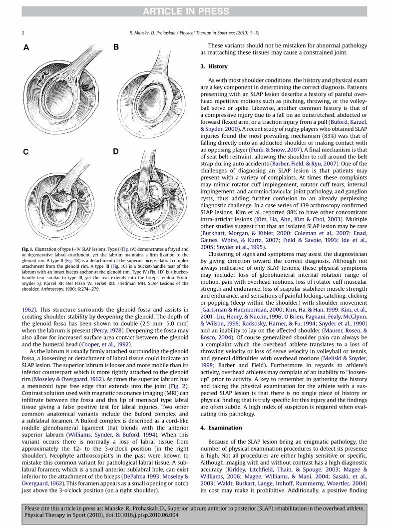
Fig. 1. Illustration of type IeIV SLAP lesions. Type I (Fig. 1A) demonstrates a frayed andor degenerative labral attachment, yet the labrum maintains a firm fixation to theglenoid rim. A type II (Fig. 1B) is a detachment of the superior bicepselabral complexattachment from the glenoid rim. A type III (Fig. 1C) is a bucket-handle tear of thelabrum with an intact biceps anchor at the glenoid rim. Type IV (Fig. 1D) is a bucket-handle tear similar to type III, yet the tear extends into the biceps tendon. From:Snyder SJ, Karzel RP, Del Pizzo W, Ferkel RD, Friedman MH. SLAP Lesions of theshoulder. Arthroscopy. 1990; 6:274e279.
R. Manske, D. Prohaskab / Physical Therapy in Sport xxx (2010) 1e122
1962). This structure surrounds the glenoid fossa and assists increating shoulder stability by deepening the glenoid. The depth ofthe glenoid fossa has been shown to double (2.5 mme5.0 mm)when the labrum is present (Perry, 1978). Deepening the fossa mayalso allow for increased surface area contact between the glenoidand the humeral head (Cooper, et al., 1992).
As the labrum is usually firmly attached surrounding the glenoidfossa, a loosening or detachment of labral tissue could indicate anSLAP lesion. The superior labrum is looser andmoremobile than itsinferior counterpart which is more tightly attached to the glenoidrim (Moseley & Overgaard, 1962). At times the superior labrum hasa meniscoid type free edge that extends into the joint (Fig. 2).Contrast solution used with magnetic resonance imaging (MRI) caninfiltrate between the fossa and this lip of meniscal type labraltissue giving a false positive test for labral injuries. Two othercommon anatomical variants include the Buford complex anda sublabral foramen. A Buford complex is described as a cord-likemiddle glenohumeral ligament that blends with the anteriorsuperior labrum (Williams, Synder, & Buford, 1994). When thisvariant occurs there is normally a loss of labral tissue fromapproximately the 12- to the 3-o’clock position (in the rightshoulder). Neophyte arthroscopist’s in the past were known tomistake this common variant for pathological labral tissue. A sub-labral foramen, which is a small anterior sublabral hole, can existinferior to the attachment of the biceps (DePalma 1993; Moseley &Overgaard,1962). This foramen appears as a small opening or notchjust above the 3-o’clock position (on a right shoulder).
Please cite this article in press as: Manske, R., Prohaskab, D., Superior labruPhysical Therapy in Sport (2010), doi:10.1016/j.ptsp.2010.06.004
These variants should not be mistaken for abnormal pathologyas reattaching these tissues may cause a constrained joint.
3. History
Aswithmost shoulder conditions, the history and physical examare a key component in determining the correct diagnosis. Patientspresenting with an SLAP lesion describe a history of painful over-head repetitive motions such as pitching, throwing, or the volley-ball serve or spike. Likewise, another common history is that ofa compressive injury due to a fall on an outstretched, abducted orforward flexed arm, or a traction injury from a pull (Buford, Karzel,& Snyder, 2000). A recent study of rugby players who obtained SLAPinjuries found the most prevailing mechanism (83%) was that offalling directly onto an adducted shoulder or making contact withan opposing player (Funk, & Snow, 2007). A final mechanism is thatof seat belt restraint, allowing the shoulder to roll around the beltstrap during auto accidents (Barber, Field, & Ryu, 2007). One of thechallenges of diagnosing an SLAP lesion is that patients maypresent with a variety of complaints. At times these complaintsmay mimic rotator cuff impingement, rotator cuff tears, internalimpingement, and acromioclavicular joint pathology, and ganglioncysts, thus adding further confusion to an already perplexingdiagnostic challenge. In a case series of 139 arthroscopy confirmedSLAP lesions, Kim et al. reported 88% to have other concomitantintra-articlar lesions (Kim, Ha, Ahn, Kim & Choi, 2003). Multipleother studies suggest that that an isolated SLAP lesion may be rare(Burkhart, Morgan, & Kibler, 2000; Coleman et al., 2007; Enad,Gaines, White, & Kurtz, 2007; Field & Savoie, 1993; Ide et al.,2005; Snyder et al., 1995).
Clustering of signs and symptoms may assist the diagnosticianby giving direction toward the correct diagnosis. Although notalways indicative of only SLAP lesions, these physical symptomsmay include: loss of glenohumeral internal rotation range ofmotion, pain with overhead motions, loss of rotator cuff muscularstrength and endurance, loss of scapular stabilizer muscle strengthand endurance, and sensations of painful locking, catching, clickingor popping (deep within the shoulder) with shoulder movement(Gartsman & Hammerman, 2000; Kim, Ha, & Han, 1999; Kim, et al.,2001; Liu, Henry, & Nuccin, 1996; O’Brien, Pagnani, Fealy, McGlynn,& Wilson, 1998; Rodsosky, Harner, & Fu, 1994; Snyder et al., 1990)and an inability to lay on the affected shoulder (Maurer, Rosen, &Bosco, 2004). Of course generalized shoulder pain can always bea complaint which the overhead athlete translates to a loss ofthrowing velocity or loss of serve velocity in volleyball or tennis,and general difficulties with overhead motions (Meliski & Snyder,1998; Barber and Field). Furthermore in regards to athlete’sactivity, overhead athletes may complain of an inability to “loosen-up” prior to activity. A key to remember in gathering the historyand taking the physical examination for the athlete with a sus-pected SLAP lesion is that there is no single piece of history orphysical finding that is truly specific for this injury and the findingsare often subtle. A high index of suspicion is required when eval-uating this pathology.
4. Examination
Because of the SLAP lesion being an enigmatic pathology, thenumber of physical examination procedures to detect its presenceis high. Not all procedures are either highly sensitive or specific.Although imaging with and without contrast has a high diagnosticaccuracy (Kirkley, Litchfield, Thain, & Spouge, 2003; Magee &Williams, 2006; Magee, Williams, & Mani, 2004; Sasaki, et al.,2003; Waldt, Burkart, Lange, Imhoff, Rummerny, Woertler, 2004)its cost may make it prohibitive. Additionally, a positive finding
m anterior to posterior (SLAP) rehabilitation in the overhead athlete,

Fig. 2. Normal meniscoid type labrum. From Getelman MH, Snyder SJ. SLAP Lesions and lesions of the long head of the biceps tendon. Treatment considerations. In: Warren RF, CraigEV, Altchek DW (eds). The Unstable Shoulder. Lippincott-Raven Publishers, Philadelphia, 1999.
R. Manske, D. Prohaskab / Physical Therapy in Sport xxx (2010) 1e12 3
may not even result in an alteration of patient care since surgerymay be required regardless. In the author’s opinion a consistentthorough physical examination is the best way to determine anaccurate diagnosis for labral lesions.
Multiple tests have historically been used to diagnose SLAPlesions. Table 1 describes the respective studies and findingsregarding sensitivity, specificity, and positive/negative predictivevalues, while Table 2 includes specifics regarding each of thestudies. Several of the main special tests used to diagnose an SLAPlesion are the following:
4.1. Anterior slide test
Kibler’s anterior slide test (Kibler, 1995) is performed with thepatient standing with arms at side, hands on hips, thumbs facingposterior to thorax. Standing behind the patient the examinerpushes the arm forward and slightly superior at the elbowalong thelong axis of the humerus. The patient is asked to resist the anteriorsuperior force applied by the examiner. A positive test is indicatedby a painful popping or clicking sensation.
4.2. Biceps load I
Kim (Kim et al., 1999) and colleagues described placing thepatients arm in 90� of abduction and full external rotation as in theapprehension test. Once in that position the patient’s forearm issupinated and they are asked to resist elbow flexion (Fig. 3). If thepatient’s apprehension or symptoms are decreased with thatmaneuver, the test is considered negative. If however, the patienthas no change or aworsening of symptoms with resistance, the testis positive.
4.3. Biceps load II
Kim and colleagues (Kim et al., 2001) later described the bicepsload II test by placing the patient supine with the shoulder in 120�
of elevation. With full external rotation, elbow flexion of 90� and
Please cite this article in press as: Manske, R., Prohaskab, D., Superior labruPhysical Therapy in Sport (2010), doi:10.1016/j.ptsp.2010.06.004
full forearm supination, the patient is asked to flex the elbowagainst resistance. Similar to the biceps load I test, a positive resultis indicated by a worsening of symptoms.
4.4. Biceps tension test
Snyder (Snyder et al., 1990) described the biceps tension test forlabral tears in which the examiner resists shoulder flexion with thepatients elbow fully extended and externally rotated. This test isvery similar to the Speeds test.
4.5. Compression rotation
The compression rotation test (Snyder et al., 1990) is per-formed with the patient supine. The affected shoulder is manu-ally compressed through its long axis while rotated both ina clockwise and counterclockwise direction in an attempt toentrap a piece of torn labral tissue. Popping, catching, snappingpain are considered positive. Although the direction of pressureis usually into the superior portion of the labrum, it can also bedirected to other regions including the anterior inferior portionfor a bankart tear or the posterior inferior for a reverse bankarttear.
4.6. Clunk test
Andrews (Andrews et al., 1985) described the clunk test inwhich the patient is supine and while the examiner grasps theproximal portion of the patients affected elbow. While stabi-lizing the proximal hand the examiner horizontally adducts/abducts the shoulder in a motion back and forth across thefrontal plane. This movement is done in an attempt to entrapa torn piece of labral tissue. As the examiner moves theshoulder back and forth, movement of the elbow into moreabduction can be done to selectively test various portion of thelabrum. Clicking, popping, catching and pain reproduction areconsidered at positive test.
m anterior to posterior (SLAP) rehabilitation in the overhead athlete,

Table 1Biceps tendon and SLAP special tests sensitivity, specificity, and positive and negative predictive values, and positive and negative likelihood Ratios.
Study Study Type Test Sensitivity Specificity PPV/NPV PLR/NLR
Bennett et al., 1998 Prosp case series Speed 90 13.8 23/83 1.1/0.7Berg and Ciullo, 1998 Retro e cons rev Slapprehension 50 Type I NR NR NR
87.5 Type 2e4 NA NR NRGuanche and Jones, 2003 Cons sample Speed 18 87 80/26 0.4/1.2
Yergason’s test 9 93 80/25 3.3/0.9O’Brien’s test 63 73 87/40 1.1/0.9Jobe relocation 44 87 91/34 1.0/1.0Crank test 40 73 82/29 1.2/0.9Biccipital groove tend 44 40 69/19 1.0/1.0
Holtby and Razmjou, 2004 Prosp blinded study Speed 32 79? 60/65 1.3/0.9Yergason’s test 43 75 50/58 2.0/0.7
Kibler 1995 Retro e case series Anterior slide 78.4 91.5 84/87 9.7/0.3Kibler, Sciascia, Hester,
Dome, & Jacobs, 2009Prosp case control Yergason’s 26 70 48/48 0.86/0.0
Speed’s 29 69 48/49 0.94/1.03Dynamic labral shear 72 98 97/77 36/0.027Anterior slide 48 82 73/60 2.66/0.63O’Brien’s test 61 84 80/67 3.8/0.46
Kim et al., 1999 Cons case series Biceps load 90.9 96.9 83/98 29.1/0.1Kim et al., 2001 Double blind Biceps load II 89.7 96.6 92.1/95.5 30.0/0.1Kim, Queale, Cosgarea,
and McFarland, 2003Prosp case control Speed p ¼ 0.004 NA NA
Active compression p ¼ 0.146 NA NAKim, Kim, Ha, Choy, Joo,
and Chung, 2007Case Series Passive compression 81.8 85.7 87.1/80.0 5.72/0.2
Liu et al., 1996 Retro chart review Combination 90 85 0.95/0.73 6.0/0.12Liu et al., 1996 Case series Crank test 91 93 94/90 13.52/0.1McFarland et al., 2002 Case control Active compression 47 55 10/91 1.044/0.96
Anterior slide 8 84 5/90 0.5/1.095Mimori et al., 1992 Pros case series Pain provocation 100 90 97/NR 10.0/0.0Morgan et al., 1998 Retro case series Jobe
Ant 4 27 NR/NR 0.05/3.5Post 85 68 NR/NR 2.65/0.22Combo 59 54 NR/NR 1.28/0.76SpeedsAnt 100 70 NR/NR 3.33/0.0Post 29 11 NR/NR 0.32/6.45Combo 78 37 NR/NRBicepital groove tendAnt 100 47 NR/NR 1.88/0.0Post 32 13 NR/NR 0.36/5.2Combo 74 35 NR/NR 1.14/0.74Active compressionAnt 88 42 NR/NR 1.51/0.28Post 32 13 NR/NR 0.36/5.23Combo 85 41 NR/NR 1.44/0.36
Myers et al., 2005 Non-rand prosp O’Brien 54 11 70/14 0.875/2.0Myers et al., 2005 Non-rand prosp Resisted supination ER 83 82 92/64 4.55/0.21Nakagawa et al., 2005 Speeds test 4 1.0 100/57 0.0/0.6
Yergasons test 13 100 100/59 1.92/0.94Active compression 63 60 52/62 1.35/0.767Anterior slide 5 93 33/56 0.714/1.021Clunk 44 68 53/59 1.375/0.824Compression rot 25 100 100/58 0.00/0.75Crank 58 72 63/68 2.072/0.583
O’Brien et al., 1998 Prosp Active compression 100 98.5 94.6/100 66.66/0.00Parentis et al., 2006 Yergasons test 12.5 93.5 45/71 1.92/0.9636
Active compression 54 50 35.5/75 1.25/0.75Anterior slide 10 81.5 19/67.6 0.55/1.104Crank test 12.5 82.6 23.8/68.4 1.436/1.059Pain Provocation 15 90.2 40/70.9 1.53/0.94Speeds test 47.8 67.4 34.8/72.1 1.466/0.774
Stetson and Tremplin, 2002 Non-rand prosp Crank 46 56 41/61 1.364/0.964O’Brien 54 31 34/50 0.783/1.48
Cons ¼ Consecutive; Non-rand ¼ Non-randomized; Prosp ¼ prospective; Retro ¼ retrospective; PPV ¼ Positive Predictive Value; NPV ¼ Negative Predictive.Value; PLR ¼ Positive Likelihood Ration; NLR ¼ Negative Likelihood Ratio.
R. Manske, D. Prohaskab / Physical Therapy in Sport xxx (2010) 1e124
4.7. Crank test
In performing the Crank test as described by Liu (Liu et al., 1996),the examiner stands beside the supine or sitting patients affectedextremity with the shoulder in 160� of elevation. The examinercompresses the humerus via an axial load in an attempt to scour thejoint and potentially entrap a piece of torn labral tissue. The arm isthen taken through internal and external rotation while still
Please cite this article in press as: Manske, R., Prohaskab, D., Superior labruPhysical Therapy in Sport (2010), doi:10.1016/j.ptsp.2010.06.004
compressing the two joint surfaces. Reproduction of pain, catching,or clicking are all positive tests.
4.8. Dynamic labral shear
Kibler (Kibler, et al. 2009) has most recently described thedynamic labral shear test. This test is performed with the patientin standing and the examiner alongside the extremity to be
m anterior to posterior (SLAP) rehabilitation in the overhead athlete,

Table 2Patient demographics for biceps tendon and SLAP lesion special testing studies.
Study Test Age (mean/range/SD) Patient population
Bennett et al., 1998 Speed M:16e76; F:30e80 45 patients (31 M, 14 F), 46 shoulders (26 D, 20 ND)Berg and Ciullo, 1998 Slapprehension NR 66 patientsGuanche and Jones, 2003 Speed, Yergason’s 38(15e76) 59 patients (48 M, 11 F), 60 shoulders
O’Brien’s test, Jobe relocation,Crank test, Biccipital groove tend
Holtby and Razmjou, 2004 Speed,Yergason’s 50(24e79) 50 patients (34 M, 16 F)Kibler 1995 Anterior slide 24.6(18e32) 46 patients (30 M, 16 W) Athletes in tennis, baseball, VolleyballKibler et al., 2009;Kim et al., 1999
Yergason’s, Speed’s, 43.2(þ/�12.6) 325 patients (232 M 93 W)Dnamic labral shear,Anterior slide, O’Brien’sBiceps load 24.8(16e41) 75 patients (64 M, 11 F) Unilateral chronic dislocation, 61 D, 14 N)
Kim et al., 2001 Biceps load II 30.6(15e52) 127 patients (89 M, 31 F) 36 Athletics, 91 D, 36 N)Kim et al., 2003 Speed, Active compression 44.2(12e86) 544 patients (309 M, 235 F)Kim et al, 2007 Passive compression 32.6(19e54) 61 patients (57 M, 4 W) 50 Trauma, (55 D, 6 N)Liu et al., 1996 Apprehension, relocation 34(17e55) 54 patients (37 M, 17 F)Liu et al., 1996 Crank test 28(18e57) 62 patients (40 M, 22 F) 50 Recreational, (53 D, 9 N)McFarland et al., 2002 Active compression, Anterior slide 45 (SD 17.8) 426 patients (252 M, 174 F)Mimori et al., 1992 Pain provocation 20.9(17e29) 32 patients (30 M, 2 F) Baseball playersMorgan et al., 1998 Jobe, Speeds, Bicepital groove tend, 33(27e72) 102 patients
Active compressionMyers et al., 2005 O’Brien, Resisted Supination ER 23.9(17e50) 40 (39 M, 1 F) AthletesNakagawa et al., 2005 Speeds, Yergasons, Active 23(14e40) 54 (52 M; 2 F) Overhead athletes
Compression, Anterior slide,Clunk, Compression rot, Crank
O’Brien et al., 1998 Active compression NR 268 patientsParentis et al., 2006 Yergasons, Active compression, 42(15e71) 132 patients (98 M, 34 F)
Anterior slide, Crank test, PainProvocation, Speeds test
Stetson and Tremplin, 2002 Crank, O’Brien 45.9(18e75) 65 patients (45 M, 20 F) (45 D, 20 N)
M ¼ Males; F ¼ Females; D ¼ Dominant; N ¼ Non dominant; SD ¼ Standard deviation; NR ¼ Not reported.
R. Manske, D. Prohaskab / Physical Therapy in Sport xxx (2010) 1e12 5
tested. The patients elbow is flexed to 90� while the shoulder isabducted in the scapular plane to greater than 120�. Whileexternally rotating the shoulder to tightness, the arm is guidedinto maximum horizontal abduction. The examiner impartsa shear load to the joint via lowering the arm from approximately120e60� of abduction. It is imperative to not place the shoulderinto maximal horizontal abduction until the arm is in greater than120 of abduction. Placing the arm into horizontal abduction firstcreated a high percentage of false-positives. A positive test isindicated by reproduction of pain and or painful click or catchbetween the motion of 120 and 60� of abduction.
Fig. 3. The biceps load test I.
Please cite this article in press as: Manske, R., Prohaskab, D., Superior labruPhysical Therapy in Sport (2010), doi:10.1016/j.ptsp.2010.06.004
4.9. O’Brien test (active compression test)
The O’Brien test (O’Brien, et al., 1998) is actually a combinationof two tests in one, assessing both the biceps/labral attachment andthe acromioclavicular joint (ACJ). The O’Brien test is performedwith the examiner standing behind or beside the patient on theside of the involved shoulder. The affected shoulder is placed ina position of 90� of flexion and 15� of horizontal adduction towardthe midline of the thorax. With the patients arm in full internalrotation, a downward force is applied to the distal forearm (Fig. 4),followed by the same resistance from a position of full supinationand external rotation. A positive test occurs when pain is worse
Fig. 4. Active Compression Test of O’Brien.
m anterior to posterior (SLAP) rehabilitation in the overhead athlete,

Fig. 5. Speed Test.
R. Manske, D. Prohaskab / Physical Therapy in Sport xxx (2010) 1e126
during internal rotation and lessens or resolves with the position ofsupination. The key is the location of pain. Pain on the “top” of theshoulder indicates ACJ pathology, while pain “deep” in the shoulderwould indicate an SLAP tear. Since pain is the “provocation” for thistest determination of an SLAP tear can be difficult as overheadthrowers often have other conditions which may mimick a labraltear when placed in this position. Shoulder impingement can occurwhen placed in shoulder internal rotation and 90� of shoulderabduction producing shoulder pain. Additionally, posteriorshoulder pain with this maneuver may be indicative of posteriorcapsulitis or posterior cuff muscle strain or tension while in theadducted position.
4.10. Pain provocation test
Mimori (Mimori, Muneta, & Nakagawa, 1992) described placingthe supine patients shoulder in 90� of abduction and full externalrotation. The patients hand is placed in either full supination, thenfull pronation. The patient is then asked which position provokesthe most pain. If the patient has more pain with the forearm inpronation the test is considered positive.
4.11. Pronated load test
The pronated load test was recently described by Wilk, Reinold,Dugas, Arrigo, Moser, and Andrews (2005). In the pronated loadtest the patient is either seated or supine. The affected shoulder isplaced in 90� of abduction with the forearm fully pronated. Whenat end range in this position the patient is asked to resist anisometric contraction of the biceps, which will simulate a peel-backlesion of the labral anchor.
4.12. Resisted supination external rotation test
The resisted supination external rotation test as describedrecently by Myers (Myers, Zemonavic & Andrews, 2005) positionsthe patient in supine with the shoulder in 90� of abduction andapproximately 65� of elbow flexion. The examiner passivelyexternally rotates the shoulder while resisting the patients attemptto maximally supinate the forearm. Pain is considered a positivetest.
4.13. SLAP-rehension test
This test is a modification of the O’Brien test, inwhich the arm ishorizontally adducted 45� versus only 15 in the original test. Theincrease in adduction motion is thought to place more stress on thebiceps anchor. It must be remembered however, that this position isalso much more likely to compress the ACJ.
4.14. Speed’s test
Speeds test is performed with the examiner standing alongsidethe patients arm to be tested. The patient’s arm is place inapproximately 90� of flexion, with forearm in full supination.Resistance is given to a downward force on the distal forearm(Fig. 5). Pain produces either anterior shoulder pain or pain in thelong head of the biceps and is indicative of a positive test for longhead biceps pathology.
4.15. Yergason’s test
Yergason’s test is performed with the examiner standing besidethe patients arm to be tested. With the patients arm flexed 90� atthe elbow, the examiner grasps the inside of the patients arm at the
Please cite this article in press as: Manske, R., Prohaskab, D., Superior labruPhysical Therapy in Sport (2010), doi:10.1016/j.ptsp.2010.06.004
wrist. The patient is asked to supinate the hand, flex the elbow andexternally rotate the shoulder. Careful attention should be paid tothe long head of the biceps proximally for any subluxation occur-ring. Pain, popping or combination of the two are indicative ofa positive test of some form of pathology to the biceps long head.
Calvert et al., (Calvert, Chambers, Regan, Hawkins & Lieth, 2009)recently performed a systematic review of various special physicalexamination tests for SLAP tears and have concluded that there areserious limitations to past studies examining sensitivities andspecificities for physical tests implicating these labral lesions. It istheir contention that current literature used to teach medicalschools and continuing education courses lacks validity, and that atpresent there are no good single physical examination tests thatexist for effectively diagnosing an SLAP lesion.
Oh and colleagues (Oh, Kim, Kim, Gong & Lee, 2008) datasuggests that combinations of 2 relatively sensitive tests(compression rotation, O’Brien’s test, and anterior apprehension),and 1 relatively specific test (either Speeds test, Yergason’s test, orbiceps load II) increases the diagnostic efficacy of SLAP lesions.Requiring 1 of the 3 tests to be positive will result in a sensitivity ofabout 75%, where requiring all 3 to be positive will result ina specificity of about 90%.
A recent meta-analysis (Meserve, Cleland, & Boucher, 2009)performed examining the clinical utility of examination of SLAPlesions has concluded that the active compression, crank andSpeeds test are more accurate than anterior slide at detectinga labral tear in the shoulder. Active compression appears to be themost sensitive of tests while Speeds test is most specific.
Since other concurrent pathologies can complicate examination,multiple other special tests may need to be applied to detect thepresence of injuries at the acromioclavicular joint, rotator cuff, andbicipital tendon.
5. Conservative treatment
Because labral tears are common place in the athlete (Andrewset al., 1985) a course of conservative treatment is always indicated.Conservative care should focus on endurance and strength trainingof the rotator cuff and scapular stabilizer muscles. Posteriorshoulder stretching/mobilization of capsular and cuff tightness thatlimits internal rotation motion should be performed and given ashome exercise program. It is thought that stretching to attain full,symmetrical internal rotation may alleviate pain and symptoms
m anterior to posterior (SLAP) rehabilitation in the overhead athlete,

Fig. 6. Arthroscopic view of type III lesion.
R. Manske, D. Prohaskab / Physical Therapy in Sport xxx (2010) 1e12 7
associated with SLAP lesions. Relative rest from aggravating activ-ities helps to decrease inflammation and associated symptoms sothat stretching and strengthening can begin sooner. As alwaysassessment should include a holistic approach of viewing the bodyand any proximal cervicothoracic and postural impairments ordistal referral sources to the shoulder should be addressed. Iftherapy does not resolve symptoms a course of anti-inflammatoryor a corticosteroid injectionmay give the athlete relief. When this isto no avail a diagnostic arthroscopy is usually in order.
6. Surgical treatment
Various surgical treatments for SLAP lesions can be clearly seenin Table 3. The rehabilitation described in this manuscript willdiscuss all SLAP lesions, but most emphasis will be placed on thetype II SLAP lesion because of its acceptance as being the primarySLAP lesion repaired in overhead athletes. Although all types ofSLAP tears can be treated surgically, the overwhelming amount oftype II repairs significantly outnumber those of the other categories(types I, III, and IV).
7. Post surgical rehabilitation
7.1. Type I and type III lesions
There are more than likely, many overhead athletes thatdemonstrate fraying or a type I SLAP lesion and are still able toparticipate in sporting and recreational activities. Because of theloose labral tissue in the type III lesion they are probably going to bemore symptomatic. At times due to instability or shoulder symp-toms surgical intervention to debride the fraying labrum back tothe glenoid rim for the type I lesion is often required. The type IIIlesion (Fig. 6) will typically need to have the loose tissue removedand any fraying labrum should be debrided back to the stable rim.As long as there are no other confounding variables or concomitantprocedures, rehabilitation following surgical intervention of a type Iand III lesion can be fairly aggressive regarding attainment of rangeof motion. Since the biceps anchor has not been altered or reat-tached tension can be safely placed through the biceps and softtissue healing constraints are generally lifted. Passive, activeassistive and active range of motion can be initiated early, althoughsince there may have been some debridement to the biceps anchorloading of elbow flexion may be held for about 1 week. Manualtherapy treatments such as joint mobilization can be started earlyand should address restricted areas of the capsule including theposterior and inferior regions. Other concomitant procedures per-formed at the same time (capsular plication, rotator cuff tears) willrequire appropriately placed restrictions.
A sling should be worn for several days for comfort althoughalmost always is discontinued by the end of the first post operativeweek. Active assisted range of motion (AAROM) and passive rangeof motion (PROM) are initiated on first post operative visit.Approximately 1 week after surgery light isotonic glenohumeraland scapular exercises are initiated. In an attempt to rest thedebrided site, light biceps activity is started at the end of the second
Table 3Surgical treatment of SLAP lesions.
Lesion Surgical treatment
Type I Debridement of frayed edgesType II Repair biceps anchorType III Debridement of bucket-handle tearType IV Debridement of bucket-handle tear, repair biceps anchor,
biceps tenodesis, biceps tenotomy
Please cite this article in press as: Manske, R., Prohaskab, D., Superior labruPhysical Therapy in Sport (2010), doi:10.1016/j.ptsp.2010.06.004
week. Isotonic exercises with bands and weights are progressed astolerated, while more aggressive exercises such as shoulder plyo-metrics dedicated to acceleration and deceleration are not beganuntil week 4e5, pending satisfactory clinical course to that point.
7.2. Type IV lesion
Surgical intervention for the type IV SLAP lesion requiresdebridement of the fraying tissue if it cannot be reattached. In somecases a repair similar to the type II lesion by reattaching the tornlabrum back to the glenoid is done. In other cases a biceps tenodesisis required. Unless simple tissue resection is required, the postoperative protocol for the type IV repair will be very similar to thatof the type II SLAP lesion. If a biceps tenodesis is performeda minimum of 10 weeks is recommended without biceps activity toallow the repaired soft tissue to fully incorporate into the bonetunnels. (Fig. 7)
7.3. Type II lesion
The rehabilitation following surgical reconstruction of the SLAPII lesion will follow a more restrictive protocol devised in Table 4.Evidence for an exact optimal treatment protocol is still not avail-able, therefore this protocol will utilize a mixture of current bestevidence from existing literature and empirically based recom-mendations. As usual this protocol should be used solely as a set of
Fig. 7. Arthroscopic view of a type IV lesion.
m anterior to posterior (SLAP) rehabilitation in the overhead athlete,

Table 4Rehabilitation following arthroscopic type II SLAP repair.
PHASE I: Protective Phase (day 1 to week 6)
Goals:Protect the surgical repairDecrease pain and inflammationPrevent the negative effects of immobilizationRestore normal arthrokinematicsPrevent primary/secondary capsular hypomobilityPromote dynamic stabilityPrevent reflex inhibition and secondary muscle atrophyDevelop neuromuscular control of shoulder complexPerformance of core stability
Weeks 0e2Shoulder sling � 4 weeksSleep in sling � 4 weeksRemoval of sling for exercises onlyShoulder, elbow and hand ROMNO active isolated biceps activity (elbow flexion or forearmsupination � 6 weeks)NO active external shoulder rotation, extension, or abduction
Hand-gripping exercisesPassive and gentle active assisted ROM exercisesCodmans exercisesFlexion to 90�
Scapular plane to 90�
External rotation to 30� � 4 weeks (approx. 10� per weekafter 2 nd week)Internal rotation to 45�
Scapulothoracic AROM in all planesSubmaximal isometrics for shoulder musculatureCryotherapy and modalities as needed for pain control
Weeks 3e4Discontinue sling use at 4 weeks (Per physician approval)Discontinue sling at night at 4 weeksContinue shoulder, elbow and hand ROMFlexion to 90�
Scapular plane to 90�
External rotation to 30�
Internal rotation to 60�
NO active external rotation, extension, or elevationInitiate scapulothoracic isometricsInitiate proprioceptive training (rhythmic stabilization drills)Gentle submaximal shoulder isometricsContinue use of cryotherapy as needed
Weeks 5e6Return to light work activitiesContinue to gradually improve ROMFlexion to 145�
Elevation in scapular plane to 145�
External rotation to 50�
Internal rotation to 60�
Full ROM should be achieved at weeks 8e10Initiate limited AROM/AAROM of shoulder to 90� flexion or abductionContinue submaximal shoulder isometricsCan begin AROM supination (no resistance/elbow flexed)No biceps loading until week 10
Clinical Milestones to Progress to Phase IIFlexion to 125�
Abduction to 70�
Scapular plane internal rotation to 40�
External rotation to 40�
*3e4/5 manual muscle test for scapular/rotator cuff muscles*AROM in appropriate ranges without pain
Phase II Moderate Protection Phase (Weeks 7e12)
Goals:Progressively restore ROM (Full by week 10)Maintain repairProgressively restore strength and balance
Weeks 7e9Continue to progress AROM/PROM (full by 8e10 weeks)Begin isotonic rotator cuff internal/external strengthening with bands/weights
Table 4 (continued )
Phase II Moderate Protection Phase (Weeks 7e12)
ProgressionsSubmaximal to maximalSlow speeds to fast speedsKnown patterns to random patternsEyes open to eyes closedOpen kinetic chain to closed kinetic chain
ExercisesScapular plane elevationSide lying external rotationStanding rotator cuff seriesProne horizontal abduction/extension
Manual resistance to shoulderNO biceps loading until week 10
Weeks 10e12Initiate stretching exercises if ROM not full by 10 weeksFlexion to 180�
Scapular plane elevation to 180�
External rotation at 90� abduction to 90�
Internal rotation at 90� abduction to 79�
Begin submaximal isometrics and AROM to bicepsBegin more aggressive exercises for rotator cuff and scapulothoracic
musculatureContinue isotonic progressive resistive exercises and manually resisted
exercisesProgress external rotation motion to 90/90 positionBegin submaximal exercises above 90� of elevationInitiate “throwers ten” exercises
Clinical Milestones to Progress to Phase IIIFlexion to 160�
Scapular plane external rotation to 65�
Abduction to 70�
Scapular plane internal rotation to 40�
External rotation to 40�
External rotation at 90�e45�
Scapular plane internal rotation fullInternal rotation at 90� abduction to 45�
Abduction to 150�
Near full symmetrical posterior shoulder mobility*4/5 manual muscle test for scapular/rotator cuff muscles*Active range of motion in appropriate ranges without pain
Phase III Minimum Protection Phase (Weeks 13e20)
Goals:Full non painful AROM/PROMRestoration of muscle strength, power and enduranceNo pain or tendernessGradual initiation of functional activities
Weeks 13e16Continue stretching exercises if neededMaintain full ROMExternal rotation @ 90� abduction up to 120� (throwers)
Initiate throwers motionContinue phase II exercise progression and principlesIsotonic elbow flexion and forearm supinationCan increase intensity and decrease repetitionsInitiate light plyometric activities (2 handed, progressing to one)
Week 16e 20Initiate modified throwing progressions from level surfaceContinue to progress resistive exercisesContinue to progress plyometric exercisesContinue stretching exercises as needed
Clinical Milestones to Progress to Phase IVWithin 10� of full active range of motion from opposite side in allplanes of motionFull symmetrical posterior shoulder mobility5/5 isometric shoulder manual muscle test5/5 scapulothoracic and rotator cuff manual muscle test
Phase IV Advanced Strengthening Phase (21 weeks to 26)GoalsEnhance muscle strength, power and enduranceMaintain shoulder AROM/PROM
R. Manske, D. Prohaskab / Physical Therapy in Sport xxx (2010) 1e128
Please cite this article in press as: Manske, R., Prohaskab, D., Superior labrum anterior to posterior (SLAP) rehabilitation in the overhead athlete,Physical Therapy in Sport (2010), doi:10.1016/j.ptsp.2010.06.004

Table 4 (continued )
Phase III Minimum Protection Phase (Weeks 13e20)
Goals:Progress functional activities
Weeks 21e26Initiate single arm plyometric trainingProgress interval sports programsBegin throwing from mound (weeks 24e28)
Clinical milestones to progress to phase VFull AROM from opposite side in all planes of motion e especiallyOverhead motionsFull symmetrical posterior shoulder mobility5/5 isometric shoulder manual muscle test5/5 scapulothoracic and rotator cuff manual muscle test
Phase IV return to activity (6 months to 9 months)GoalsReturn to unrestricted sports activityMaintain full shoulder AROM/PROMMaintain full shoulder muscle strength, power and endurance
6 monthsþAllow full velocity throwing from moundContinue capsular stretches (especially posterior indefinitely)
Fig. 8. Supine rhythmic stabilization drills working on early neuromuscular control,strength and endurance of shoulder musculature.
R. Manske, D. Prohaskab / Physical Therapy in Sport xxx (2010) 1e12 9
guidelines as clinical judgment should supersede pending anyother co-morbidities or concomitant surgical procedures (e.g., grossligamentous laxity, capsular plication, rotator cuff tears).
7.3.1. Phase I: Protective Phase7.3.1.1. Weeks 0e2. The protective phase is incorporated to main-tain safety of the operated extremity for a short duration. This isdone initially in an effort to control swelling, maintain onlyminimal to slight tension on the repair site and allow the normalinflammatory response to run its expected course. A shoulder slingis worn continuously for the first week (Burkhart & Morgan, 2001;Burkhart, Morgan & Kibler, 2003a, 2003b; Gartsman &Hammerman, 2000; Morgan, et al., 1998). Sling use continues,including at night for up to 4 weeks. Formal rehabilitation beginsusually after the first week unless there is concern that stiffnessmay result at which time rehabilitation can begin as early as the 3rdto 5th post operative day. At week one gentle PROM is begun inboth the scapular plane and flexion to 90�. Codman’s passivependulum exercises are allowed to help decrease the risk ofcapsular adhesions and to nourish the articular cartilage of thehumeral head and glenoid fossa. Although Burkhart and Morgan(Burkhart & Morgan, 1998) have suggested that external shoulderrotation not be taken past 0� early, due to what they describe as the“peel-back phenomenon”, we typically allow a gradual increase ofexternal rotation of about 10� per week after week one, not toexceed 30� by week 4. Internal shoulder rotation is allowed totolerance up to 45�. Active elbow, forearm and hand motions areallowed with the exception of elbow flexion and supination whichwould create active tension into the biceps anchor. Scapular muscleactivation can be initiated early in side lying by moving the scapulathrough all available planes. Gentle submaximal isometrics canbegin for the glenohumeral muscles after the 2nd week. Theseisometric contractions can be performed via rhythmic stabilizationdrills for internal/external shoulder rotation, glenohumeral flexion/extension and abduction/adduction. This form of strengtheninghelps to promote dynamic stabilization, joint proprioception, andneuromuscular control.
Modalities can be utilized early on in an attempt to decreaseinflammation, swelling and muscle inhibition. These could includepulsed non-thermal ultrasound, electrical stimulation, and cryo-therapy. There remains limited evidence that these forms of
Please cite this article in press as: Manske, R., Prohaskab, D., Superior labruPhysical Therapy in Sport (2010), doi:10.1016/j.ptsp.2010.06.004
modalities actually alter healing times, so their use should be usedaccordingly.
Because most of the patient population having repair of theSLAP lesion are active individuals, an implementation of lowerextremity, and core training program should also be added.
7.3.1.2. Weeks 3e4. The shoulder sling is slowly discontinued at oraround week 4 as long as symptoms continue to decrease. Duringweeks 3e4, internal rotation motion can be increased up to 50�, inan attempt to stretch and mobilize the posterior capsule. Onehypothesis for progression of SLAP tears, described by Burkhart,Morgan and Kibler (Burkhart, Morgan & Kibler, 2003a), is thata posterior e superior shear of the humeral head occurs duringglenohumeral elevation and external rotation. This could theoret-ically be caused due to obligate translations as described by Har-ryman (Harryman, Sidles, Clark, McQuade, Gibb & Matsen 1990)and Karduna (Karduna, Williams, Williams, & Iannotti, 1996). Anobligate translation is one that occurs following a tightness incapsular tissues that causes motion of a given joint segment tooccur in the direction opposite the tight structures. Thus an isolatedtight inferior glenohumeral ligament complex will create an obli-gate translation of the humeral head in a superior direction withelevation. While a tight posterior capsule will create an anteriorshear with internal rotation. All other shoulder motions remain thesame as week 0e2.
Although actual strengthening is not a concern at this point inthe rehabilitation sequence, muscle firing and decreasing muscleinhibition is always a concern. Submaximal shoulder and gleno-humeral isometrics via rhythmic stabilization drills (Fig. 8) cancontinue to progress, as can isometrics for the scapulothoracicmusculature.
7.3.1.3. Weeks 5e6. At week 5 and 6 glenohumeral ROM is nowprogressed. Elevation (flexion and scapular plane) can be increasedto 145� as tolerated. External rotation can progress to 50�, whileinternal to 60�. It is anticipated that full ROM should be achievedaround 10 week or 12 weeks. Limited active assisted range ofmotion (AAROM) and active range of motion (AROM) can begin to90� of elevation. This is typically progressed from a supine positionwhere the effects of gravity are minimized, progressing to a seatedor standing position where the full effects of gravity are felt. Thesemotions should be pain free with no abnormal contortions noted.
m anterior to posterior (SLAP) rehabilitation in the overhead athlete,

Fig. 10. Posterior capsule joint mobilizations are done to decrease risk of obligatetranslation in superior posterior direction which can occur if posterior inferior capsulemobility is restricted.
R. Manske, D. Prohaskab / Physical Therapy in Sport xxx (2010) 1e1210
Resistance exercises or active loading of the biceps is still discour-aged and will not begin until 10 weeks post operative. To maintaina decreased load on the biceps tendon even exercise such as rowingshould be altered by performing with a straight arm (Fig. 9) and notallowing the arm to pass posterior to the frontal plane (Durall,Manske & Davies, 2001).
7.3.2. Phase II: Moderate Protection Phase (Weeks 7e12).Goals change slightly in the moderate protection phase to
continue progression of ROM, to maintain surgical repair and tostart restoration of strength and balance of the rotator cuff andscapulothoracic muscles.
7.3.2.1. Weeks 7e9. A gradual push for full ROM of the shoulderbegins at week 7 if there are remaining limitations. As long asmotion is progressing nicelymore aggressive treatment approachessuch as stretching and joint mobilizations are not needed. Becausea tight posterior capsule is thought to be a precipitator of thispathology, it should be constantly assessed. If a restriction remainsthe addition of posterior capsule joint mobilization techniques arebeneficial. Manske et al. have shown in a randomized controlledtrial that both stretching and posterior capsule joint mobilizations(Fig.10) together aremore effective at increasing posterior shouldermobility than stretching in isolation (Manske, Meschke, Porter,Smith, & Reiman, 2010). At this point stretching can occur inhigher levels of elevation and external rotation. Begin at 45� ofabduction progressing to 90� of abduction for positioning thehumerus during stretching of the anterior capsule. By the end ofweek 9, ROM for elevation should be full, external rotation shouldbe 90�, and internal rotation should be approximately 70�.
More aggressive isotonic exercises can also be performed andincludes scapular plane elevation, side lying external rotation, allrotator cuff series, and prone rowing and horizontal abduction.These exercises have been shown to demonstrate high levels ofelectromyographic activity in the scapular and rotator cuff muscles(Manske, 2006).
7.3.2.2. Weeks 10e12. It is at this point that submaximal isometricscan be used with the repaired biceps tendon. The initiation of the“thowers ten” can also be started (Wilk et al., 2005). Full shoulderROM should be achieved and includes up to approximately 120� ofexternal rotation for the thrower. Strengthening can also be
Fig. 9. Safe method to begin scapular retractor strength by keeping elbow straight tonot allow tension into the biceps anchor.
Please cite this article in press as: Manske, R., Prohaskab, D., Superior labruPhysical Therapy in Sport (2010), doi:10.1016/j.ptsp.2010.06.004
progressed to the 90/90 position with manual resistance or tubing.As before these exercises in the 90/90 position should be startedusing submaximal resistance forces then progressing to higherlevels as the patient tolerates.
7.3.3. Phase III: Minimal Protection Phase (weeks 13e20)The goals of the minimal protection phase include achieving full
unrestricted, non painful AROM/PROM of the glenohumeral joint.Also the patient should have a restoration of muscle strength,power and endurance. The athlete should have no pain or tender-ness in the shoulder and be able to begin a gradual restoration offunctional activities.
7.3.3.1. Weeks 13e16. A continued emphasis should be placed onjoint mobilization with particular attention to the posteriorcapsule. Self home stretches such as the cross arm stretch andthe sleeper stretch should be used judiciously if limitations ofmotion exist. External rotation ROM should now be around 120�
for throwers. Isotonic resisted exercises can commence for bothelbow flexion and forearm supination. In this time frame plyo-metric exercises can be progressed starting with two armthrows such as chest pass, chopping motions (Fig. 11). Single-arm throws from the side for internal rotation will performedbefore overhead single arm or overhead double arm tosses.Weight should gradually increase starting with 2 poundsincreasing to tolerance.
7.3.3.2. Week 17e21. Although there is no absolute evidence thatexists describing an exact date at which throwing can commence,the 16 week point is typically the date that is used. As long asROM is full, both rotator cuff and scapular strength are tested atfull (5/5), there is no pain with overhead movements, and theathlete has passed a satisfactory clinical examination, the athletecan begin a throwing progression on level ground. Around week14e15 very easy tossing can begin indoors from about 20 feetwith a tennis ball to make sure that the athlete can transition toa starting distance of about 45 feet with an actual baseball. Allthrowing is to be done with a crow hop and with no pain orsymptoms. When the throwing progression or interval sportsprogram begins the athlete is usually asked to decrease theirworkout regimen for the shoulder to 2e3 times per week.Shoulder resistance workouts are done on the same days as the
m anterior to posterior (SLAP) rehabilitation in the overhead athlete,

Fig. 11. Bilateral chopping motion plyometrics with weighted ball performed late inrehabilitation to work on acceleration/deceleration activities that are required inpowerful overhead motions for a full return to sporting activities.
R. Manske, D. Prohaskab / Physical Therapy in Sport xxx (2010) 1e12 11
athlete throws to ensure adequate rest interval with at least onefull day between activities.
7.3.4. Phase IV: Advanced Strengthening Phase (weeks 21e26)The main goals of the advanced strengthening phase are to
maintain AROM/PROM, to enhance strength, power, and enduranceof the rotator cuff and scapulothoracic muscles, and to progress thefunctional activities including sports and recreation.
7.3.4.1. Weeks 21e26. Single arm plyometric exercise should beable to be performed at the 21 week time frame. The throwingprogression or interval sports program should be progressed astolerated. Throwing from the mound usually occurs between 24and 28 weeks.
7.3.5. Phase V: Return to Activity (6 monthsþ).Themain goal in the return to activity phase is for a full return to
unrestricted athletic participation.
8. Conclusions
Despite the evasiveness of diagnosing SLAP lesions therapistsand physicians alike are recognizing signs and symptoms of thispathology. Due to abundance of type II lesions that are symptom-atic a thorough understanding of anatomy, mechanism of injury,signs and symptoms are a pre requisite for proper conservative andsurgical management. Successful treatment for SLAP lesionsrequires a delicate balance of regaining shoulder mobility andprogression of strengthening exercises for the rotator cuff andscapulothoracic musculature. This paper has outlined pertinentanatomy, examination methods, and treatment strategies to opti-mize a beneficial outcomes following superior labral injury.
Ethical approvalWe understand that work on human beings that is submitted to
Physical Therapy in Sport should comply with the principles laiddown in the declaration of Helsinki; Recommendations guidingphysicians in biomedical research involving human subjects.Adopted by the 18th World Medical Assembly, Helsinki, Finland,June 1964, amended by the 29th World Medical Assembly, Tokyo,Japan, October 1975, the 35th World Medical Assembly, Venice,Italy, October 1983, and the 41st World Medical Assembly, Hong
Please cite this article in press as: Manske, R., Prohaskab, D., Superior labruPhysical Therapy in Sport (2010), doi:10.1016/j.ptsp.2010.06.004
Kong, September 1989. The manuscript should contain a statementthat has been approved by the appropriate ethical committeesrelated to the institution(s) in which it was performed and thatsubjects gave informed consent to the work. Studies involvingexperiments with animals must state that their care was in accor-dance with institution guidelines. Patients’ and volunteers’ names,initials, and hospital numbers should not be used.
In a case report, the subject’s written consent should beprovided. It is the author’s responsibility to ensure all appropriateconsents have been obtained.
This is a descriptive article related to rehabilitation followingSLAP lesions therefore this information does not apply.
Conflict of interestWe have no conflicts of interest to report related to this article
submission.
References
Andrews, J. R., Carson, W. G., Jr., & McCleod, W. D. (1985). Glenoid labrum tears relatedto the long head of the biceps. American Journal of Sports Medicine, 13, 337e341.
Andrews, J. R., Carson, W. G., & McLeaod, W. D. (1984). The arthroscopic treatmentof glenoid labrum tears e the throwing athlete. Orthopaedic Transactions, 8,44e54.
Barber, F. A., Field, L. D., & Ryu, R. K. (2007). Biceps tendon and superior labruminjuries: decision-making. Journal of Bone & Joint Surgery, American, 89,1844e1855.
Bennett, W. F. (1998). Specificity of the Speed's test: arthroscopic technique forevaluating the biceps tendon at the level of the bicipital groove. Arthroscopy, 14,789e796.
Berg, E. E., & Ciullo, J. V. (1998). A clinical test for superior glenoid labral or ‘SLAP’lesions. Clinical Journal of Sport Medicine, 8(2), 121e123.
Bost, F., & Inman, V. (1942). The pathological changes in recurrent dislocation of thesholder. A report of Bankart’s operative procedures. Journal of Bone & JointSurgery, American, 24, 595e613.
Buford, D. A., Karzel, R. P., & Snyder, S. J. (2000). SLAP lesions: history, diagnosis,treatment and results. Techniques in Shoulder & Elbow Surgery, 1, 2002e2208.
Burkhart, S. S., & Morgan, C. (2001). SLAP lesions in the overhead throwing athlete.Orthopedic Clinics of North America, 32, 431e441.
Burkhart, S. S., & Morgan, C. D. (1998). The peel-back mechanism: its role producingand extending posterior type II SLAP lesions and its effect on SLAP repairmechanics. Arthroscopy, 14, 637e640.
Burkhart, S. S., Morgan, C. D., & Kibler, W. B. (2000). Shoulder injuries in overheadathletes. The “dead arm” revisited. Clinical Journal of Sport Medicine, 19,125e158.
Burkhart, S. S., Morgan, C. D., & Kibler, W. B. (2003a). The disabled throwingshoulder: spectrum of pathology part I: pathoanatomy and biomechanics.Arthroscopy, 19, 404e420.
Burkhart, S. S., Morgan, C. D., & Kibler, W. B. (2003b). The disabled throwingshoulder: spectrum of pathology part II: evaluation and treatment of SLAPlesions in throwers. Arthroscopy, 19, 531e539.
Calvert, E., Chambers, G. K., Regan, W., Hawkins, R. H., & Lieth, J. M. (2009). Specialphysical examination tests for superior labrum anterior posterior shoulder tearsare clinically limited and invalid: a diagnostic systematic review. Journal ofClinical Epidemiology, 62, 558e563.
Codman, E. A. (1934). The shoulder. Boston: MA. Thomas Todd.Coleman, S. H., Cohen, D. B., Drakos, M. C., Allen, A. A., O’Brien, S. J., Altchek, D. W.,
et al. (2007). Arthroscopic repair of type II superior labral anterior posteriorlesions with and without acromioplasty: a clinical analysis of 50 patients.American Journal of Sports Medicine, 35, 749e753.
Cooper, D. E., Arnoczky, S. P., O'Brien, S. J., Warren, R. F., DiCarlo, E., & Allen, A. A.(1992). Anatomy, histology, and vascularity of the glenoid labrum. Ananatomical study. Journal Bone Joint Surgery, 74A, 46e52.
DePalma, A., Callery, G., & Bennet, G. (1993). Variational anatomy and degenerativelesions of the shoulder joint. Ann Arbor, MI. In W. Blount, & S. Banks (Eds.), TheAAOS instructional course lectures, Vol. VI (pp. 255).
Durall, C., Manske, R. C., & Davies, G. J. (2001). Avoiding shoulder injury fromresistance training. Journal of Strength & Conditioning, 20, 10e18.
Enad, J. G., Gaines, R. J., White, S. M., & Kurtz, C. A. (2007). Arthroscopic superiorlabrum anterioreposterior repair in military patients. Journal of Shoulder andElbow Surgery, 16, 300e305.
Field, L. D., & Savoie, F. H., III (1993). Arthroscopic suture repair of superior labraldetachment lesions of the shoulder. American Journal of Sports Medicine, 21,783e790.
Funk, L., & Snow, M. (2007). SLAP tears of the glenoid labrum in contact athletes.Clinical Journal of Sports Medicine, 17, 1e4.
Gartsman, R., & Hammerman, S. M. (2000). Superior labrum, anterior and posteriorlisions. When and how to treat them. Clinical Sports Medicine, 19, 115e124.
Guanche, C. A., & Jones, D. C. (2003). Clinical tests for tears of the glenoid labrum.Arthroscopy, 19, 517e523.
m anterior to posterior (SLAP) rehabilitation in the overhead athlete,

R. Manske, D. Prohaskab / Physical Therapy in Sport xxx (2010) 1e1212
Handelberg, F., Willems, S., Shahabpour, M., Huskin, J. P., & Kuta, J. (1998). SLAPlesions: a retrospective multicenter study. Arthroscopy, 14, 856e862.
Harryman, D. T., Sidles, J. A., Clark, J. M., McQuade, K. J., Gibb, T. D. , & Matsen, F. A.(1990). Translation of the humeral head on the glenoid with passive gleno-humeral motion. Journal of Bone & Joint Surgery, American, 72, 1334e1343.
Hawkins, R. J., & Kennedy, J. C. (1980). Impingement syndromes in athletes. Amer-ican Journal of Sports Medicine, 8, 151e158.
Huber, W. P, & Putz, R. V. (1997). Periarticular fiber system of the shoulder joint.Arthroscopy, 13, 680e691.
Holtby, R, & Razmjou, H (2004). Accuracy of the Speed's and Yergason's test indetecting biceps pathology and SLAP lesions: comparison with arthroscopicfindings. Arthroscopy, 20, 231e236.
Ide, J., Maeda, S., & Takagi, K. (2005). Sports activity after arthroscopic superiorlabral repair using suture anchors in overhead-throwing athletes. AmericanJournal of Sports Medicine, 33, 507e514.
Karduna, A. R., Williams, G. R., Williams, J. L., & Iannotti, J. P. (1996). Kinematics ofthe glenohumeral joint: influences of muscle forces, ligamentous constraintsand articular geometry. Journal Orthopedic Research, 14, 986e993.
Kibler, W. B. (1995). Sensitivity and specificity of the anterior slide test in throwingathletes with superior glenoid labral tears. Arthroscopy, 14, 447e457.
Kibler, W. B., Sciascia, A. D., Hester, P., Dome, D., & Jacobs, C. (2009). Clinical utility oftradional and new tests in the diagnosis of biceps tendon injuries and superiorlabrum anterior and posterior lesions in the shoulder. American Journal of SportsMedicine, 37, 1840e1847.
Kim, S. H., Ha, K., Ahn, J., Kim, S. H., & Choi, H. J. (2003). Biceps load test II: a clinicaltest for SLAP lesions of the shoulder. Arthroscopy, 17, 160e164.
Kim, S. H., Ha, K., & Han, K. (1999). Biceps load test: a clinical test for superiorlabrum anterior and posterior lesions in shoulders with recurrent anteriordislocation. American Journal of Sports Medicine, 27, 300e303.
Kim, S. H., Ha, K. I., Kim, S. H., & Choi, H. J. (2002). Results of arthroscopic treatmentof superior labral lesions. Journal of Bone & Joint Surgery, American, 84, 981e985.
Kim, S. Y., Kim, J. M., Ha, K. Y., Choy, S., Joo, M. W., & Chung, Y. J. (2007). The passivecompression test. A new clinical test for superior labral tears of the shoulder.American Journal of Sports Medicine, 35, 1489e1494.
Kim, T. K., Queale,W. S., Cosgarea, A. J., &McFarland, E. G. (2003). Clinical features of thedifferent types of SLAP lesions. Journal of Bone Joint Surgery, American, 85, 66e71.
Kim, Y. S., Kim, J. M., Ha, K. Y., Choy, S., Joo, M. W., & Chung, Y. G. (2001). The passivecompression test: a new clinical test for superior labral tears of the shoulder.American Journal of Sports Medicine, 35, 1489e1494.
Kirkley, A., Litchfield, R., Thain, L., & Spouge, A. (2003). Agreement betweenmagneticresonance imaging and arthroscopic evaluation of the shoulder joint in primaryanteriordislocationof the shoulder.Clinical Journal of SportsMedicine,13,148e151.
Liu, S. H., Henry, M. H., & Nuccin, S. L. (1996). A prospective evaluation of a newphysical examination in predicting glenoid labral tears. American Journal ofSports Medicine, 24, 721e725.
Maffet, M. W., Gartsman, G. M., & Moseley, B. (1995). Superior labrum-biceps tendoncomplex lesions of the shoulder. American Journal of Sports Medicine, 23, 93e98.
Magee, T., & Williams, D. (2006). Sensitivity and specificity in detection of labral tearswith 3.0-TMRI of the shoulder. American Journal of Roentgenology,187,1448e1452.
Magee, T., Williams, D., & Mani, N. (2004). Shoulder MR arthrography: whichpatient group benefits most? American Journal of Roentgenology, 183, 969e974.
Manske, R. C. (2006). Electromyographically assessed exercises for the scapularmuscles. Athletic Therapy Today, 11, 19e23.
Manske, R. C., Meschke, M., Porter, A., Smith, B. S., & Reiman, M. A. (2010).Randomized, controlled, single-blind comparison of stretching versus stretch-ing and joint mobilization for posterior shoulder tightness. Submitted To: SportsHealth: A Multidisciplinary Approach, 2, 94e100.
Maurer, S. G., Rosen, J. E., & Bosco, J. A. (2004). SLAP lesions of the shoulder. Hospitalfor Joint Diseases, 61(3&4), 186e192.
McFarland, E. G., Kim, T. K., & Savino, R. M. (2002). Clinical assessment of threecommon tests for superior labral anterior-posterior lesions. American Journal ofSports Medicine, 30, 810e815.
Meserve, B. B., Cleland, J. A., & Boucher, T. R. (2009). A meta-analysis examiningclinical test utility for assessing superior labral anterior posterior lesions.American Journal of Sports Medicine, 37, 2252e2258.
Mileski, R. A., & Snyder, S. J. (1998). Superior labral lesions in the shoulder: path-oanatomy and surgical management. Journal of American Academy of OrthopedicSurgeons, 6, 121e131.
Mimori, K.,Muneta, T., &Nakagawa,T. (1992). Anewpainprovocation test for superiorlabral tears of the shoulder. American Journal of Sports Medicine, 27, 137e142.
Please cite this article in press as: Manske, R., Prohaskab, D., Superior labruPhysical Therapy in Sport (2010), doi:10.1016/j.ptsp.2010.06.004
Morgan, C. D., Burkhart, S. S., Palmeri, M., & Gillespie, M. (1998). Type II SLAPlesions: three subtypes and their relationships to superior instability androtator cuff tears. Arthroscopy, 14, 553e565.
Moseley, H. F., & Overgaard, B. (1962). The anterior capsular mechanism in recurrentanterior dislocation of the shoulder: morphological and clinical studies withspecial reference to the glenoid labrum and gleno-humeral ligaments. Journal ofBone and Joint Surgery, American, 443, 913e927.
Myers, T. H., Zemonavic, J. R., & Andrews, J. R. (2005). The resisted supinationexternal rotation test: a new test for diagnosis of SLAP lesions. American Journalof Sports Medicine, 33, 1315e1320.
Nishida, K., Hashizume, H., Toda, K., & Inoue, H. (1996). Histologic and scanningelectron microscopic study of the glenoid labrum. Journal of Shoulder ElbowSurgery, 5, 132e138.
Nakagawa, S., Yoneda, M., Hayashida, K., Obata, M., Fukushima, S., & Miyazaki, Y.(2005). Forced shoulder abduction and elbow flexion test: a new simple clinicaltest to detect superior labral injury in the throwing shoulder. Arthroscopy, 21,1290e1295.
O’Brien, S. J., Pagnani, M. J., Fealy, S., McGlynn, S., & Wilson, J. B. (1998). The activecompression test: a new and effective test for diagnosing labral tears andacromio-clavicular joint abnormality. American Journal of Sports Medicine, 26,610e613.
Oh, J. H., Kim, J. Y., Kim, W. S., Gong, H. S., & Lee, J. H. (2008). The evaluation ofvarious physical examinations for the diagnosis of type II superior labrumanterior and posterior lesion. American Journal of Sports Medicine, 36, 353e359.
Parentis, M. A., Glousman, R. E., Mohr, K. S., & Yocum, L. A. (2006). An evaluation ofthe provocative tests for superior labral anterior posterior lesions. AmericanJournal of Sports Medicine, 34, 265e268.
Perry, J. (1978). Normal upper extremity kinesiology. Physical Therapy, 58,265e278.
Prodromos, C. C., Ferry, J. A., Schiller, A. L., & Zarins, B. (1990). Histological studies ofthe glenoid labrum from fetal life to old age. Journal of Bone and Joint Surgery,72, 1344e1348.
Resch, H., Golser, K., Thoeni, H., & Sperner, G. (1993). Arthroscopic repair of superiorglenoid labral detachment (the SLAP lesions). Journal of Shoulder Elbow Surgery,2, 147e155.
Rodsosky, M. W., Harner, C. D., & Fu, F. H. (1994). The role of the long head of thebiceps muscle and superior glenoid labrum in anterior instability of theshoulder. American Journal of Sports Medicine, 22, 121e130.
Sasaki, T., Saito, Y., Yodono, H., Prado, G. L., Miura, H., Itabashi, Y., et al. (2003).Labral-ligamentous complex of the shoulder: evaluation with double obliqueaxial MR arthrogrography. Acta Radiology, 44, 435e439.
Snyder, S. J., Banas, M. P., & Belzer, J. P. (1996). Arthroscopic evaluation and treat-ment of injuries to the superior glenoid labrum. Instructional Course Lectures,45, 65e70.
Snyder, S. J., Banas, M. P., & Karzel, R. P. (1995). An analysis of 140 injuries to thesuperior glenoid labrum. Journal of Shoulder Elbow Surgery, 4, 243e248.
Snyder, S. J., Karzel, R. P., Del Pizzo, W., Ferkel, R. D., & Friedman, M. J. (1990). SLAPlesions of the shoulder. Arthroscopy, 6, 274e279.
Snyder, S. J., Rames, R. D., & Wolber, E. (1991). Labral lesioins. In J. B. McGinty, &Operative arthroscopy, 1991. (Eds.), Operative arthroscopy (pp. 491e499). NewYork: Raven Press.
Stetson, W.B., Snyder, S.J., Karzel, R.P., Banas, M.P., Rahhal, S.E. (1998). Long-termclinical follow-up of isolated SLAP lesions of the shoulder. Presented at the 65thAnnual Meeting of the American Academy of Orthopaedic Surgeons, NewOrleans, March 23, 1998.
Stetson, W. B., & Tremplin, K. (2002). The crank test, the O’Brien test, and routinemagnetic resonance imaging scans in the diagnosis of labral tears. AmericanJournal of Sports Medicine, 30, 806e815.
Waldt, S., Burkhart, A., Lange, P., Imhoff, A. B., Rummerny, E. J., & Woertler, K. (2004).Diagnostic performance of MR arthrography in the assessment of superiorlabral anteroposterior lesions of the shoulder. American Journal of Roentgen-ology, 182, 1271e1278.
Wilk, K. E., Reinold, M. M., Dugas, J. R., Arrigo, C. A., Moser, M. W., & Andrews, J. R.(2005). Current concepts in recognition and treatment of superior labral (SLAP)lesions. Journal of Orthopedic & Sports Physical Therapy (35), 273e291.
Williams, M. M., Snyder, S. J., & Buford, D., Jr. (1994). The Buford complex e the“cord-like” middle glenohumeral ligament and absent anterosuperior labrumcomplex: a normal anatomic capsulolabral variant. Arthroscopy, 10, 241e247.
Williams, P. L., Bannister, L. H., Berry, M. M., Collins, P., Dyson, M., Dussek, J. E., et al.(1995). Gray's Anatomy (8th ed.). Edinburgh: Churchill Livingstone.
m anterior to posterior (SLAP) rehabilitation in the overhead athlete,