phenotypic analysis of the rat placenta - kumc€¦ · · 2008-09-09hematoxylin and eosin-stained...
TRANSCRIPT
Phenotypic Analysis of Rat Placenta 295
295
From: Methods in Molecular Medicine, Vol. 121: Placenta and Trophoblast: Methods and Protocols, Vol. 1Edited by: M. J. Soares and J. S. Hunt © Humana Press Inc., Totowa, NJ
21
Phenotypic Analysis of the Rat Placenta
Rupasri Ain, Toshihiro Konno, Lindsey N. Canham,and Michael J. Soares
SummaryThe rat is an important model for studying the biology of trophoblast-uterine development.
This chapter describes methods that are useful for the characterization of the rat uteroplacentalcompartment.
Key Words: Rat; chorioallantoic placenta; choriovitelline placenta; junctional zone; laby-rinth zone; metrial gland; spongiotrophoblast; trophoblast giant cells; trophoblast cell inva-sion; prolactin gene family.
1. IntroductionIn this chapter, we discuss the rat as an experimental model for investiga-
tions directed toward pregnancy and the uteroplacental interface. We presentan overview on the organization of the rat maternal–fetal interface and presentmethods for studying the rat placenta and trophoblast cells.
1.1. Merits of the Rat as an Animal Model for Placental Research
Historically, the rat has been a valuable model for studying most aspects ofreproduction and, in many areas, still remains the preferred model system. Therat is the dominant preclinical model system used by the pharmaceutical andagro-chemical industries. Genetic approaches for analysis of the rat are rapidlyadvancing. The first draft of the rat genome is completed (1) and significantprogress is being made in developing strategies for the genetic manipulation ofthe rat, including transgenic (2–7), in vivo mutagenesis (8), and nuclear trans-fer (9,10). Finally, panels of consomic rat strains have been established, whichwill permit a systematic and physiologically relevant dissection of the ratgenome (11).
21_Ain_295_314_F 8/29/05, 11:22 AM295

296 Ain et al.
The use of animal model systems provides an essential tool for dissectingmolecular mechanisms controlling cellular development. The maternal–fetalinterface is no exception. The premise of employing any animal model systemis that if the process being studied is fundamental it will likely demonstrateconservation across species. Although there are some differences in the orga-nization of the rodent vs the primate maternal–fetal interface, there are over-riding similarities in the functions and lineages of cells comprising thematernal–fetal interface. Among these are events transpiring during the lastweek of gestation in the rat. During this latter phase of pregnancy in the rat,trophoblast cells exit the chorioallantoic placenta and invade into the uterineendometrium, where they establish intimate relationships with the uterine vas-culature (12). These invasive events in the rat are remarkably similar to inva-sive events occurring during the latter stages of the first trimester andthroughout the second trimester of pregnancy in the human (13).
If we can understand and appreciate biological processes at the maternal–fetal interface in species that can be experimentally manipulated (e.g., rat,mouse), then we can more intelligently study the development of the humanmaternal–fetal interface and identify pivotal junctures of cellular control, fa-cilitating diagnosis and therapeutic intervention.
In some instances, cross-species similarities may prevail, whereas in othercases, the differences may be most compelling. Nonetheless, our appreciationfor the biology of pregnancy increases. Animal models provide us with a meansof studying biological phenomena at levels that are not ethically permissiblewith humans. Viviparity is vital to the success of our species. Mechanisms thatunderlie viviparity will exhibit some level of conservation.
1.2. Overview of the Organization of the Uteroplacental Compartmentof the Rat
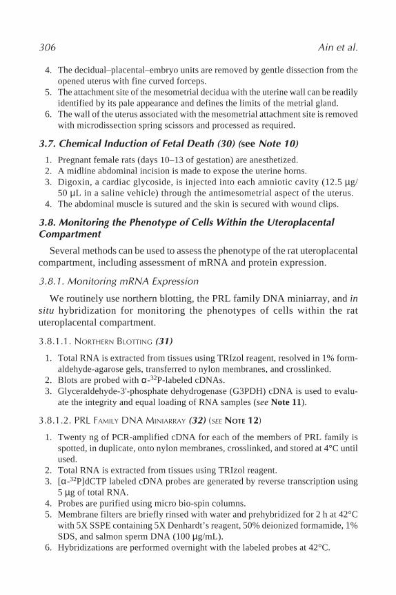
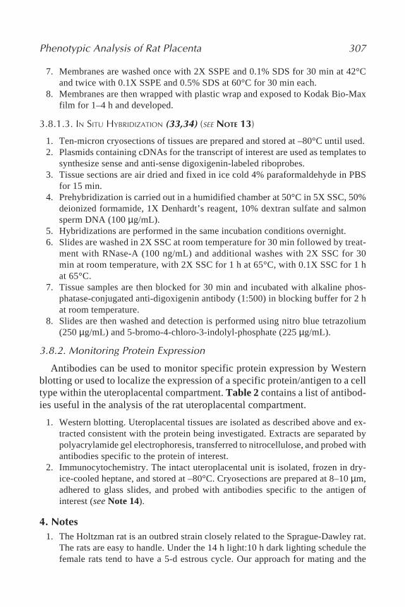
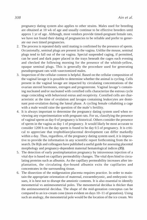
The uteroplacental compartment of the rat is similar to the mouse and pos-sesses similarities and differences to the organization of the uteroplacentalcompartment of other species with hemochorial placentation (14–17). This hasled to the utilization of an assortment of terms to describe components of theuteroplacental compartment. Schematic representations of the rat uteroplacentalcompartment are presented in Figs. 1 and 2.
The site where blood enters the uterus determines the orientation of theuteroplacental compartment. This region is referred to as the mesometrial com-partment, and the opposite end is termed the antimesometrial compartment.The uterine mesometrial compartment is prominently comprised of stromalcells, blood vessels (endothelial cells, smooth muscle cells), immune/inflam-matory cells (natural killer cells, macrophages), smooth muscle cells of themyometrium, and trophoblast cells. Cellular composition of this compartment
21_Ain_295_314_F 8/29/05, 11:22 AM296

Phenotypic Analysis of Rat Placenta 297
is dynamic and has gestation-stage dependent and species-specific characteris-tics. Following implantation, natural killer cells expand in number, and infil-trate the mesometrial decidua, located adjacent to the developingchorioallantoic placenta. Decidual cells are derived from uterine stromal cellsand exhibit functional differences depending upon their location (18,19). Me-sometrial decidua is the site of extensive vascular remodeling, whereasantimesometrial decidual cells are conspicuous in their elaboration ofcytokines, including members of the prolactin (PRL) family (20). A triangle-shaped area rich in blood vessels is situated between the mesometrial deciduaand the surface of the uterus (21,22). This region has been referred to by theterms “mesometrial triangle,” “metrial gland,” and several others (23). As ges-tation advances, extraembryonic and embryonic structures expand in size andthe decidua thins. Accompanying these events, natural killer cells vacate themesometrial decidua and infiltrate the mesometrial triangle where they associ-ate with the resident vasculature. Subsequently, the antimesometrial deciduumand mesometrial-associated natural killer cells degenerate. As natural killercells depart, a specialized population of trophoblast cells exits the chorioallan-toic placenta and invades into the mesometrial decidua (12). In the mouse,trophoblast invasion is limited to the mesometrial decidua, whereas in the rat,trophoblast cells penetrate through the mesometrial decidua and infiltrate themesometrial triangle.
Fig. 1 (see companion CD for color version). Hematoxylin and eosin-stained tissuesection of the midgestation rat uteroplacental compartment (left panel, day 11 of ges-tation) and a corresponding schematic diagram (right panel).
21_Ain_295_314_F 8/29/05, 11:22 AM297

298 Ain et al.
298
Fig
. 2 (s
ee c
ompa
nion
CD
for c
olor
ver
sion
). H
emat
oxyl
in a
nd e
osin
-sta
ined
tiss
ue s
ecti
on o
f the
late
ges
tati
onra
t ut
erop
lace
ntal
com
part
men
t (l
eft
pane
l, da
y 18
of
gest
atio
n) a
nd a
cor
resp
ondi
ng s
chem
atic
dia
gram
(ri
ght
pane
l) w
ith
high
ligh
ted
expa
nded
vie
ws
of th
e la
byri
nth
and
junc
tion
al z
ones
(lo
wer
pan
els)
.
21_Ain_295_314_F 8/29/05, 11:22 AM298

Phenotypic Analysis of Rat Placenta 299
The chorioallantoic placenta is situated at the base of the mesometrial com-partment. It develops from trophoblast stem cells present in the ectoplacentalcone and generates two recognizable structures: (a) labyrinth zone and (b) junc-tional zone. The labyrinth zone arises from the interaction of allantoic meso-derm with the trophoblast stem cell population (24), yielding trophoblast cellsyncytialization and establishment of the barrier for maternal–fetal exchange(17). Once the barrier is established, endocrinologically active trophoblastgiant cells appear within the labyrinth zone. Four trophoblast cell lineagesdifferentiate from trophoblast stem cells within the junctional zone: (a) tro-phoblast giant cells, (b) spongiotrophoblast cells, (c) glycogen cells, and (d)invasive trophoblast cells. Trophoblast giant cells are the first trophoblast celllineage to develop and, until the last week of gestation, are the most distallylocated trophoblast cell types within the uterus. Spongiotrophoblast cells arethe main constituents of the junctional zone. Trophoblast giant cells,spongiotrophoblast cells, and invasive trophoblast cells are the major endo-crine cells of the rat and mouse placenta. Glycogen cells appear during the lastweek of pregnancy, notably accumulate glycogen, and disappear before theend of pregnancy. The invasive trophoblast cell population first appears at mid-gestation. It consists of trophoblast cells that penetrate and surround the uter-ine vasculature present within the developing chorioallantoic placenta. Asgestation advances, invasive trophoblast cells exit the junctional zone andenter the mesometrial compartment. Three terms have been used to describethe invasive trophoblast cells (25). Endovascular trophoblast cells replace theendothelium, intramural trophoblast cells are embedded within the vascularwall, and interstitial trophoblast cells are situated between the vasculature.
1.3. Investigation of the Rat Placenta
In this chapter, we describe methods for (1) mating and gestational staging;(2) detection of pregnancy during early postimplantation stages; (3) dissectionof the midgestation uteroplacental compartment; (4) dissection of the chorioal-lantoic placenta; (5) establishment of spongiotrophoblast cell primary cultures;(6) isolation of the metrial gland; (7) chemical induction of fetal death; and (8)monitoring the phenotype of cells within the uteroplacental compartment. Theoutlined protocols are based on our experience in working with the rat placentaover the past two decades.
2. Materials2.1. Mating and Gestational Staging
1. Holtzman rats are obtained from Harlan Sprague-Dawley (Indianapolis, IN).2. Saline solution (0.9% NaCl).3. Glass slide with wells.4. Microscope (×40–100 magnification).
21_Ain_295_314_F 8/29/05, 11:22 AM299
300 Ain et al.
2.2. Detection of Pregnancy During Early Post Implantation Stages
Chicago Blue B (1% solution; Matheson Coleman & Bell ManufacturingChemists, Norwood, OH, cat. no. CX685 B364).
2.3. Mid-Gestation Placental Dissection1. Dissecting microscope (×10–20 magnification).2. Hank’s balanced salt solution (HBSS; Sigma Chemical Company, St. Louis, MO,
cat. no. H-387).3. Fine forceps and microdissecting spring scissors (Roboz Surgical Instrument Co.,
Gaithersburg, MD, cat. nos. RS-5155 and RS-5602, respectively).
2.4. Chorioallantoic Placental Dissection1. Dissecting microscope (×10–20 magnification).2. HBSS (Sigma).3. Fine forceps and microdissecting spring scissors (Roboz).4. 23-gauge needles (BD Biosciences, Franklin Lakes, NJ, cat. no. 305134).
2.5. Primary Culture of Spongiotrophoblast Cells
1. Fine forceps and microdissecting spring scissors (Roboz).2. Dispase II (Roche Diagnostic Corporation Indianapolis, IN, cat. no. 295825).3. DNase I (Sigma, cat. no. D4263).4. HBSS (Sigma).5. Dulbecco’s modified Eagle’s medium (DMEM) culture medium (Mediatech
Cellgro, Herdon, VA, cat. no. 10-017-CV) supplemented with penicillin andstreptomycin (Mediatech Cellgro) and 10% fetal bovine serum (FBS; AltantaBiologicals, Norcross, GA , cat. no. S11150).
6. Nylon mesh cell strainers (70 µm; BD Biosciences, cat. no. 352350).7. Percoll (Amersham Biosciences, Uppsala, Sweden, cat. no. 17-089-02).
2.6. Metrial Gland Isolation1. Fine forceps and microdissecting spring scissors (Roboz).2. HBSS (Sigma).
2.7. Chemical Induction of Fetal Death
Digoxin (Elkin-Sinn, Cherry Hill, NJ).
2.8. Monitoring the Phenotype of Cells Within the UteroplacentalCompartment
1. TRIzol reagent (Invitrogen Life Technologies, Carlsbad, CA, cat. no. 15596-018).2. 1% Formaldehyde-agarose gels. Formaldehyde (Fisher Scientific, Pittsburgh, PA,
cat. no. F79-4); agarose (Sigma, cat. no. A-9539).3. Nylon membranes (Nytran Super Charge, Schleicher & Schuell Biosciences, Inc.,
Keene, NH, cat. no. 10416296).
21_Ain_295_314_F 8/29/05, 11:22 AM300

Phenotypic Analysis of Rat Placenta 301
4. Crosslinker (Model XL-1000, Spectronics Corporation, Westbury, NY).5. [α-P32]dCTP (Perkin Elmer, Boston, MA, cat. no. Blu/NEG/0134).6. Plasmids containing cDNAs for the transcripts of interest (Table 1).
2.8.1. PRL Family DNA Miniarray (32) (see Note 12)
1. Polymerase chain reaction (PCR)-amplified cDNA for each of the members ofPRL family is spotted, in duplicate, onto nylon membranes (Schleicher & SchuellBiosciences, Inc.), crosslinked, and stored at 4°C until used.
2. TRIzol reagent (Invitrogen).3. [αP32]dCTP (Perkin Elmer, Boston, MA).4. Micro bio-spin columns (Bio-Rad Laboratories, Hercules, CA, cat. no. 732-
6223).5. Denhardt’s reagent (50X Denhard’s reagent): 1% ficoll (Sigma, cat. no. F-4375),
1% polyvinylpyrrolidone (Sigma, cat. no. P-5288), 1% bovine serum albumin(BSA; Fraction V, Fisher, cat. no. BP1600-100) diluted in distilled water.
6. Deionized formamide (Sigma, cat. no. F9037).7. Salmon sperm DNA (Invitrogen, cat. no. 15632-011).8. SSPE buffer (20X SSPE): add 175 g sodium chloride, 27.6 g sodium phosphate
monobasic, 7.4 g ethylenediamine tetraacetic acid (EDTA) to 1 L of distilledwater.
9. 0.1% sodium dodecyl sulfate (SDS; Fisher, cat. no. BP 166-500).10. Kodak Bio-Max film (Kodak, Rochester, NY, cat. no. 829-4985).
2.8.2. In Situ Hybridization (33,34) (see Note 13)
1. Plasmids containing cDNAs for the transcript of interest are used as templates tosynthesize sense and anti-sense digoxigenin-labeled riboprobes (Table 1).
2. Dry-ice-cooled heptane (Fisher, cat. no. 03008-1).3. Cryostat (Model No. CM 1850-3-1, Lieca Microsystems, Germany).4. 4% paraformaldehyde (P-6148, Sigma) in phosphate-buffered saline (PBS) at 4°C.5. Deionized formamide (Sigma).6. 1X Denhardt’s reagent.7. 10% Dextran sulfate (Fisher, cat. no. BP 1585-100).8. Salmon sperm DNA (Invitrogen).9. Standard saline citrate (SSC; 20X SSC): 175.3 g sodium chloride, 88.2 g sodium
citrate in 1 L of distilled water.10. RNase-A (Sigma, cat. no. R-6513).11. Dig-High Prime DNA Labeling and Detection Starter Kit II (Roche Diagnostic
Corporation, cat. no. 1585614).
2.9. Monitoring Protein Expression
2.9.1. Western Blotting
1. Reagents for polyacrylamide gel electrophoresis and electrophoretic transfer(BioRad Laboratories).
21_Ain_295_314_F 8/29/05, 11:22 AM301

302 Ain et al.
Table 1cDNAs Used in the Phenotypic Analysis of Cells Within the Uteroplacental Com-partment
GenBankCell type Gene Gestation stage accession no. Reference
TrophoblastEctoplacental cone PLF-RP Early NM_053364 37Trophoblast giant cell PL-I Early to mid D21103 31
PL-II Mid to late M13749 31,38PLP-Fα Early to mid NM_022530 37,
unpublisheda
P450scc Early to late J05156 39,40P450c17 Mid to late NM_012753 40,413βHSD Early to late L17138 Unpublishedb
Spongiotrophoblast PLP-B Mid to late M31155 42,43PLP-Fβ Mid to late AY741310 Unpublisheda
SSP Mid to late NM_172073 44Labyrinthine TGC PL-II Mid to late M13749 42,45
PLP-K Mid to late NM_138861 32PLF-RP Mid to late NM_053364 37
Syncytial trophoblast FABP3 Mid to late NM_024162 46Alk Phos Mid to late NM_013059 47
Invasive trophoblast PLP-L Mid to late NM_138527 12PLP-M Mid to late NM_053791 12PLP-N Mid to late NM_153738 48IGF-II Mid to late X17012 49,
unpublishedc
Decidual cellsMesometrial α2-MG Early to mid NM_012488 50Antimesometrial dPRP Early to mid NM_022846 51,52
PLP-J Early to mid NM_031316 32Natural killer cells Osteopontin Mid NM_012881 Unpublishedd
Abbreviations: TGC, Trophoblast giant cell; PLF-RP, proliferin-related protein; PL, placental lacto-gen, PLP, prolactin-like protein; P450scc, side chain cleavage; P450c17, 17α hydroxylase; 3βHSD, 3βhydroxysteroid dehydrogenase; SSP, spongiotrophoblast-specific protein; FABP3, fatty acid bindingprotein-3; IGF-II, insulin-like growth factor-II; α2-MG, α2-macroglobulin; dPRP, decidual prolactin-related protein.
aHo-Chen, J., Bustamante, J. J., and Soares, M. J., unpublished results.bCanham, L. N. and Soares, M. J., unpublished results.cAin, R. and Soares, M.J., unpublished results.dLiu, B. and Soares, M.J., unpublished results.
21_Ain_295_314_F 8/29/05, 11:22 AM302
Phenotypic Analysis of Rat Placenta 303
2. Nitrocellulose (Optitran, Schleicher & Schuell Biosciences, Inc., Cat. No. BA-S 85).3. Antibodies to the protein of interest (Table 2).
2.9.2. Immunocytochemistry
1. Dry-ice-cooled heptane (Fisher).2. Cryostat (Leica).3. Antibodies to the antigen(s) of interest (Table 2).
Table 2Antibodies Used in the Phenotypic Analysis of Cells Within the UteroplacentalCompartment
Cell type Antigen Gestation stage Source (Cat. No.) Reference
Trophoblast Cytokeratin All stages Sigma Chemical Co., 12St. Louis, MO (C2931)
Trophoblast giant cell PL-I Early to mid Chemicon International, 31,53Temecula, CA (AB1288)
PL-II Mid to late Chemicon (AB1289) 31,38,54PLP-A Mid to late Chemicon (AB1290) 55,56P450scc Early to late Chemicon (AB1244, 39,57
AB1294)P450c17 Mid to late see refs. 40,58
Spongiotrophoblast PLP-B Mid to late Chemicon (AB1291) 43SSP Mid to late see ref. 44
Labyrinthine TGC PL-II Mid to late Chemicon (AB1289) 38
Syncytial trophoblast FABP3 Mid to late see ref. 46Alk Phos Mid to late see ref. 47
Decidual cellsMesometrial α2-MG Early to mid see ref. 50Antimesometrial dPRP Early to mid Chemicon (AB1293) 52,59
PLP-J Early to mid see ref. Unpublisheda
Natural killer cells Perforin Mid Torrey Pines Biolabs, 12Houston, TX (TP251)
Macrophages ED-1/ED-2 All stages Serotec Inc., Raleigh, NC Unpublishedb
(MCA341R/ MCA342R)Smooth muscle cells Sm actin All stages Sigma (A2547) Unpublishedb
Abbreviations: TGC, trophoblast giant cells; PL, placental lactogen, PLP, prolactin-like protein; P450scc,side chain cleavage; P450c17, 17α hydroxylase; SSP, spongiotrophoblast-specific protein; FABP3, fatty acidbinding protein-3; β2-MG, α2-macroglobulin; dPRP, decidual prolactin-related protein; Sm, smooth muscle.
aAlam, S. M. K., Konno, T., and Soares, M. J., unpublished results.bAin, R. and Soares, M. J., unpublished results.
21_Ain_295_314_F 8/29/05, 11:22 AM303
304 Ain et al.
3. Methods3.1. Mating and Gestational Staging
1. The rats are maintained on a 14 h light:10 h dark lighting schedule with lights onat 0600 h (see Note 1).
2. Adult males, preferably older than 10 wk of age, are placed one per cage.3. Adult females, generally 7–10 wk of age, are transferred to a cage with a male
(no more than two females per male). The fur on one of the females is generallymarked with a dye to distinguish it from the other female.
4. Every morning between 0800 and 0900 h, each female cohabiting a cage with amale, is removed from the cage for the purpose of obtaining a vaginal lavage.
5. A few hundred microliters of saline are delivered with a pipette into the vagina ofthe female and recovered with the same pipet.
6. The contents of each saline lavage are transferred to a well within a multi-wellglass plate.
7. After all of the vaginal lavages are collected, they are examined with the aid of amicroscope (×40–100 magnification).
8. The presence of sperm in the lavage is recorded, as is the cellular content of thelavage (see Notes 2 and 3).
9. The sperm positive females are transferred to separate cages. The presence ofsperm in the vaginal lavage is considered day 0 of pregnancy (see Note 4).
3.2. Detection of Pregnancy During Early Post Implantation Stages (26)(see Note 5)
1. Pregnancy detection within the first 48 h post implantation requires intravenousinjection of a vital blue dye, such as Chicago Blue B.
2. A volume of 0.25–0.5 mL/100 g body weight of a 1.0% solution of Chicago BlueB can be injected into the tail vein of the rat.
3. Implantation reactions are identified by the accumulation of blue bands withinthe uterus after 15 min.
3.3. Mid-Gestation Placental Dissection (27) (see Note 6)1. Embryos with their encapsulating decidual tissues (conceptuses) are retrieved
from the uterus from days 10–13 of gestation.2. Conceptuses are dissected with the aid of a dissecting microscope (×10–20 mag-
nification).3. Tissues are collected and washed with HBSS.4. The overlying decidua basalis and decidua capsularis are removed with fine for-
ceps and gentle dissection.5. A cut through the mural pole of the trophoblast layer is made and the trophoblast
retracted. Be careful not to cut through the yolk sac/amnion.6. The visceral yolk, amnion, and embryo are separated from the developing chorio-
allantoic placenta by cutting at the insertion site of the allantois with microdis-section spring scissors.
7. The entire trophoblast component is flattened (allantoic insertion site is up) andcan be further separated into chorioallantoic and choriovitelline layers with the
21_Ain_295_314_F 8/29/05, 11:22 AM304

Phenotypic Analysis of Rat Placenta 305
aid of microdissection spring scissors. The inner dark circle of tissue (more vas-cular) comprising the chorioallantoic tissue is cut away from the lighter surround-ing tissue (less vascular) consisting of the choriovitelline tissue.
8. Dissected decidua basalis, decidua capsularis, and trophoblast components caneach be processed as required, and/or stored at –80°C, until further use.
3.4. Chorioallantoic Placental Dissection (28) (see Note 7)
1. Embryos with their encapsulating decidual tissues (conceptuses) can be retrievedfrom the uterus on days 13 to 21 of gestation.
2. Conceptuses are dissected with the aid of a dissecting microscope (×10–20 mag-nification).
3. The tissues are collected into and washed with HBSS.4. The overlying decidual basalis tissue and underlying yolk sac/umbilical insertion
are removed with fine forceps and microdissection spring scissors.5. The junctional zone is identified by its pale appearance, due to the absence of
fetal blood, and separated from the labyrinth zone, a richly vascularized tissue,with fine forceps and 23-gauge needles.
6. Recovered tissues are rinsed in HBSS, processed as required, and/or stored at–80°C, until further use.
3.5. Primary Culture of Spongiotrophoblast Cells (29) (see Note 8)
1. Junctional zones from day-13 rat chorioallantoic placentas are dissected understerile conditions (see Subheading 3.4.).
2. Tissues are cut into small pieces with microdissection spring scissors and disso-ciated with Dispase II (4.8 mg/mL) and DNase I for 1 h at 37°C with continuousshaking.
3. At the end of the digestion, the suspension of cells and tissue fragments are mixedseveral times with the aid of a Pasteur pipet, and centrifuged.
4. The harvested cells are then resuspended in DMEM culture medium supple-mented with 10% FBS and filtered through a nylon mesh (70 µm).
5. The cell suspension is then centrifuged through a 60% cushion of Percoll for15 min at room temperature.
6. Cells at the interface are collected, washed with DMEM supplemented with 10%FBS, and cultured in the same medium.
7. The cells can be maintained in DMEM medium containing FBS (1–10%) for7–10 d.
3.6. Metrial Gland Isolation (see Note 9)
The metrial gland can be isolated from midgestation onward (Fig. 1).
1. The uterus is removed from the female rat.2. Fat and mesenteries are removed from the uterus.3. Microdissection spring scissors are used to cut each uterine horn along its
antimesometrial surface.
21_Ain_295_314_F 8/29/05, 11:22 AM305
306 Ain et al.
4. The decidual–placental–embryo units are removed by gentle dissection from theopened uterus with fine curved forceps.
5. The attachment site of the mesometrial decidua with the uterine wall can be readilyidentified by its pale appearance and defines the limits of the metrial gland.
6. The wall of the uterus associated with the mesometrial attachment site is removedwith microdissection spring scissors and processed as required.
3.7. Chemical Induction of Fetal Death (30) (see Note 10)
1. Pregnant female rats (days 10–13 of gestation) are anesthetized.2. A midline abdominal incision is made to expose the uterine horns.3. Digoxin, a cardiac glycoside, is injected into each amniotic cavity (12.5 µg/
50 µL in a saline vehicle) through the antimesometrial aspect of the uterus.4. The abdominal muscle is sutured and the skin is secured with wound clips.
3.8. Monitoring the Phenotype of Cells Within the UteroplacentalCompartment
Several methods can be used to assess the phenotype of the rat uteroplacentalcompartment, including assessment of mRNA and protein expression.
3.8.1. Monitoring mRNA Expression
We routinely use northern blotting, the PRL family DNA miniarray, and insitu hybridization for monitoring the phenotypes of cells within the ratuteroplacental compartment.
3.8.1.1. NORTHERN BLOTTING (31)
1. Total RNA is extracted from tissues using TRIzol reagent, resolved in 1% form-aldehyde-agarose gels, transferred to nylon membranes, and crosslinked.
2. Blots are probed with α-32P-labeled cDNAs.3. Glyceraldehyde-3'-phosphate dehydrogenase (G3PDH) cDNA is used to evalu-
ate the integrity and equal loading of RNA samples (see Note 11).
3.8.1.2. PRL FAMILY DNA MINIARRAY (32) (SEE NOTE 12)
1. Twenty ng of PCR-amplified cDNA for each of the members of PRL family isspotted, in duplicate, onto nylon membranes, crosslinked, and stored at 4°C untilused.
2. Total RNA is extracted from tissues using TRIzol reagent.3. [α-32P]dCTP labeled cDNA probes are generated by reverse transcription using
5 µg of total RNA.4. Probes are purified using micro bio-spin columns.5. Membrane filters are briefly rinsed with water and prehybridized for 2 h at 42°C
with 5X SSPE containing 5X Denhardt’s reagent, 50% deionized formamide, 1%SDS, and salmon sperm DNA (100 µg/mL).
6. Hybridizations are performed overnight with the labeled probes at 42°C.
21_Ain_295_314_F 8/29/05, 11:22 AM306
Phenotypic Analysis of Rat Placenta 307
7. Membranes are washed once with 2X SSPE and 0.1% SDS for 30 min at 42°Cand twice with 0.1X SSPE and 0.5% SDS at 60°C for 30 min each.
8. Membranes are then wrapped with plastic wrap and exposed to Kodak Bio-Maxfilm for 1–4 h and developed.
3.8.1.3. IN SITU HYBRIDIZATION (33,34) (SEE NOTE 13)
1. Ten-micron cryosections of tissues are prepared and stored at –80°C until used.2. Plasmids containing cDNAs for the transcript of interest are used as templates to
synthesize sense and anti-sense digoxigenin-labeled riboprobes.3. Tissue sections are air dried and fixed in ice cold 4% paraformaldehyde in PBS
for 15 min.4. Prehybridization is carried out in a humidified chamber at 50°C in 5X SSC, 50%
deionized formamide, 1X Denhardt’s reagent, 10% dextran sulfate and salmonsperm DNA (100 µg/mL).
5. Hybridizations are performed in the same incubation conditions overnight.6. Slides are washed in 2X SSC at room temperature for 30 min followed by treat-
ment with RNase-A (100 ng/mL) and additional washes with 2X SSC for 30min at room temperature, with 2X SSC for 1 h at 65°C, with 0.1X SSC for 1 hat 65°C.
7. Tissue samples are then blocked for 30 min and incubated with alkaline phos-phatase-conjugated anti-digoxigenin antibody (1:500) in blocking buffer for 2 hat room temperature.
8. Slides are then washed and detection is performed using nitro blue tetrazolium(250 µg/mL) and 5-bromo-4-chloro-3-indolyl-phosphate (225 µg/mL).
3.8.2. Monitoring Protein Expression
Antibodies can be used to monitor specific protein expression by Westernblotting or used to localize the expression of a specific protein/antigen to a celltype within the uteroplacental compartment. Table 2 contains a list of antibod-ies useful in the analysis of the rat uteroplacental compartment.
1. Western blotting. Uteroplacental tissues are isolated as described above and ex-tracted consistent with the protein being investigated. Extracts are separated bypolyacrylamide gel electrophoresis, transferred to nitrocellulose, and probed withantibodies specific to the protein of interest.
2. Immunocytochemistry. The intact uteroplacental unit is isolated, frozen in dry-ice-cooled heptane, and stored at –80°C. Cryosections are prepared at 8–10 µm,adhered to glass slides, and probed with antibodies specific to the antigen ofinterest (see Note 14).
4. Notes1. The Holtzman rat is an outbred strain closely related to the Sprague-Dawley rat.
The rats are easy to handle. Under the 14 h light:10 h dark lighting schedule thefemale rats tend to have a 5-d estrous cycle. Our approach for mating and the
21_Ain_295_314_F 8/29/05, 11:22 AM307
308 Ain et al.
pregnancy dating system also applies to other strains. Males used for breedingare obtained at 10 wk of age and usually continue to be effective breeders untilapprox 1 yr of age. Although, most vendors provide timed-pregnant female rats,we have not found their dating of pregnancies to be reliable and prefer to gener-ate our own timed pregnancies.
2. The process is repeated daily until mating is confirmed by the presence of sperm.Occasionally, seminal plugs are present in the vagina. Unlike the mouse, seminalplugs tend to fall out of the rat vagina. Special suspended caging, if permitted,can be used and dark paper placed in the trays beneath the cages each eveningand checked the following morning for the presence of the whitish-yellow,opaque seminal plugs. This is generally the procedure used for generatingpseudopregnant rats with vasectomized males.
3. Inspection of the cellular content is helpful. Based on the cellular composition ofthe vaginal lavage it is possible to determine whether the animal is cycling. Cellspresent in the vaginal lavage are impacted by circulating concentrations of theovarian steroid hormones, estrogen and progesterone. Vaginal lavage’s contain-ing nucleated and/or nucleated with cornified cells characterize the estrous cyclestage coinciding with behavioral estrus and receptivity. Cornified cells are char-acteristic at the time of ovulation and lavages containing leukocytes are domi-nant post-ovulation during the luteal phase. A cycling female cohabiting a cagewith a male would raise the question of the male’s fertility.
4. It is always important to determine the pregnancy dating system used when re-viewing any experimentation with pregnant rats. For us, classifying the presenceof vaginal sperm as day 0 of pregnancy is historical. Others consider the presenceof sperm in the vagina as day 1 of pregnancy. It would likely be most accurate toconsider 1200 h on the day sperm is found to be day 0.5 of pregnancy. It is criti-cal to appreciate that trophoblast/placental development can differ markedlywithin a day. Thus, regardless, of the pregnancy dating system used, it is impera-tive to provide the information in any scientific report forthcoming from the re-search. De Rijk and colleagues have published a useful guide for assessing placentalmorphology and pregnancy-dependent maternal hematological indices (35).
5. The detection of early postimplantation pregnancy by intravenous injection of avital dye is based on capillary permeability changes. The vital dyes bind to circu-lating proteins such as albumin. As the capillary permeability increases after im-plantation, the circulating dye-bound albumin exits the capillaries andconcentrates in the surrounding tissue.
6. The dissection of the midgestation placenta requires practice. In order to main-tain the appropriate orientation of maternal, extraembryonic, and embryonic tis-sues, it is best not to disrupt the amniotic contents. It is also essential to identifymesometrial vs antimesometrial poles. The mesometrial decidua is thicker thanthe antimesometrial decidua. The shape of the mid-gestation conceptus can becompared to an ice-cream cone (more evident on days 10–11 of gestation). Usingsuch an analogy, the mesometrial pole would be the location of the ice cream. We
21_Ain_295_314_F 8/29/05, 11:22 AM308
Phenotypic Analysis of Rat Placenta 309
have used chorioallantoic and choriovitelline to discriminate between the polarand mural trophoblast components of the midgestation conceptus. Some haveargued that the relationship between the mural trophoblast and the yolk sac doesnot constitute a true choriovitelline or yolk sac placenta (36).
7. Prior to day 13 of gestation, it is difficult to separate the junctional and labyrinthzones. After day 15, it is difficult to completely remove the thinning deciduabasalis, which becomes firmly adherent to the junctional zone. Beginning on day15 of gestation, it also becomes possible to dissect the chorioallantoic zones withfine forceps; needles are not required. Separation of junctional and labyrinthzones is not readily achieved in the mouse and hamster as a result of extensiveinterdigitation of the zones. It is important to appreciate that even in the rat, thedissection of the junctional and labyrinth zones is not perfect and usually, a smallamount of residual contamination is evident.
8. Dissection of the junctional zone from tissue obtained earlier than day 13 of ges-tation is more time-consuming and yields a nominal amount of starting tissue forthe enzymatic dissociation. Junctional zones from later in gestation are easy todissect but do not yield a good starting cell population. In a limited series ofexperiments, we have found that the establishment of junctional zone culturesfrom placentas obtained later during gestation is difficult and the cells show poorviability. The rat day-13 junctional zone cell cultures show no evidence of prolif-eration of any cell type. These cultures also show modest contamination ofvimentin-positive cells (2–3%). The rat day-13 junctional zone cells in primaryculture spontaneously differentiate as demonstrated by their expression of membersof the placental PRL gene family (29). Most of the cells exhibit a spongiotrophoblastcell phenotype; however, some trophoblast giant cells and syncytial trophoblast cellsare also detected.
9. The metrial gland is a heterogeneous tissue. Its landmarks are not well defined.Thus consistency in dissection is a necessity.
10. Digoxin is an effective tool for inducing fetal death at midgestation but does notwork well as gestation advances. We have had some success using intra-amnioticinjections of potassium chloride to kill fetuses from later in gestation.
11. Interpretation of RNA measurements in the uterus or placenta is directly depen-dent upon the quality of tissue dissection. Simply labeling a sample containingsome placental tissue as placenta does not make it placenta. Most placentalsamples contain varying amounts of decidual tissue and yolk sac. We have uti-lized an assortment of different housekeeping genes to monitor RNA integrityand loading efficiency. These have included β-actin, G3PDH, β-tubulin, and 28Sribosomal RNA.
12. The PRL family miniarray assay represents an effective screening tool formonitoring trophoblast cell development. Information can be retrieved regard-ing decidual and trophoblast cell lineages and temporal aspects of differentia-tion. The assay is most effective when complementary procedures are employedfor monitoring changes in gene expression.
21_Ain_295_314_F 8/29/05, 11:22 AM309

310 Ain et al.
13. In our hands, in situ hybridization is a reliable method for identifying cell typescontributing to the expression of a specific gene within the uteroplacental com-partment. An appreciation of the dynamic changes in uteroplacental morphologyis essential to maximize the benefit of the approach.
14. Antibodies are effective tools for identifying and localizing proteins. Each antibody–antigen interaction needs to be optimized for the specific technique employed.
AcknowledgmentsWe would like to thank past and current members of our laboratory for their
efforts in developing and characterizing the methods described in this chapter.This work was supported by grants from the National Institutes of Health (NIH)(HD20676, HD39878, HD48861) and the Hall Family Foundation.
References1. Rat Genome Sequencing Project Consortium. (2004) Genome sequence of the
Brown Norway rat yields insights into mammalian evolution. Nature 428, 493–521.2. Hammer, R. E., Maika, S. D., Richardson, J. A., Tang, J. P., and Taurog, J. D.
(1990) Spontaneous inflammatory disease in transgenic rats expressing HLA-B27and human beta 2m: an animal model of HLA-B27-associated human disorders.Cell 63, 1099–1112.
3. Mullins, J. J., Peters, J., and Ganten, D. (1990) Fulminant hypertension intransgenic rats harbouring the mouse Ren-2 gene. Nature 344, 541–544.
4. Hamra, F. K., Gatlin, J., Chapman, K. M., et al. (2002) Production of transgenicrats by lentiviral transduction of male germ-line stem cells. Proc. Natl. Acad. Sci.USA 99, 14,931–14,936.
5. Hasuwa, H., Kaseda, K., Einarsdottir, T., and Okabe, M. (2002) Small interferingRNA and gene silencing in transgenic mice and rats. FEBS Lett. 532, 227–230.
6. Lois, C., Hong, E. J., Pease, S., Brown, E. J., and Baltimore, D. (2002) Germlinetransmission and tissue-specific expression of transgenes delivered by lentiviralvectors. Science 295, 868–872.
7. Orwig, K. E., Avarbock, M. R., and Brinster, R. L. (2002) Retrovirus-mediatedmodification of male germline stem cells in rats. Biol. Reprod. 67, 874–879.
8. Zan, Y., Haag, J. D., Chen, K.-S., et al. (2003) Production of knockout rats usingENU mutagenesis and a yeast-based screening assay. Nat. Biotechnol. 21, 645–651.
9. Mullins, L. J., Wilmut, I., and Mullins, J. J. (2004) Nuclear transfer in rodents. J.Physiol. 554, 4–12.
10. Zhou, Q., Renard, J.-P., Le Friec, G., et al. (2003) Generation of fertile cloned ratsby regulating oocyte activation. Science 302, 1179.
11. Cowley, A. W., Roman, R. J., and Jacob, H. J. (2004) Application of chromo-somal substitution techniques in gene-function discovery. J. Physiol. 554, 46–55.
12. Ain, R., Canham, L. N., and Soares, M. J. (2003) Gestational stage-dependentintrauterine trophoblast cell invasion in the rat and mouse: novel endocrine phe-notype and regulation. Dev. Biol. 260, 176–190.
21_Ain_295_314_F 8/29/05, 11:22 AM310

Phenotypic Analysis of Rat Placenta 311
13. Pijnenborg, R., Robertson, W. B., Brosens, I., and Dixon, G. (1981) Trophoblastinvasion and the establishment of haemochorial placentation in man and labora-tory animals. Placenta 2, 71–92.
14. Bridgman, J. (1949) A morphological study of the development of the placenta ofthe rat. II. An histological and cytological study of the development of the chorio-allantoic placenta of the white rat. J. Morphol. 83, 195–224.
15. Davies, J. and Glasser, S. R. (1968) Histological and fine structural observationson the placenta of the rat. Acta Anat. 69, 542–608.
16. Georgiades, P., Ferguson-Smith, A. C., and Burton, G. J. (2002) Comparativedevelopmental anatomy of the murine and human definitive placentae. Placenta23, 3–19.
17. Rossant, J. and Cross, J. C. (2002) Extraembryonic lineages, in Mouse Develop-ment (Rossant, J. and Tam, P. P. L., eds.). Academic, New York: pp. 155–180.
18. De Feo, V. J. (1967) Decidualization, in Biology of the Uterus (Wynn, R. M., ed.).Appleton-Century-Crofts, New York: pp. 191–290.
19. Bell, S. C. (1983). Decidualization: regional differentiation and associated func-tion. Oxford Rev. Reprod. Biol. 5, 220–271.
20. Orwig, K. E., Rasmussen, C. A., and Soares, M. J. (1997) Decidual signals in theestablishment of pregnancy: the prolactin family. Trophoblast Res. 10, 329–343.
21. Selye, H. and McKeown, T. (1935) Studies on the physiology of the maternalplacenta in the rat. Proc. R. Soc. Lond. Biol. 119, 1–31.
22. Peel, S. (1989) Granulated metrial gland cells. Adv. Anat. Embryol. Cell Biol.115, 1–112.
23. Ain, R. and Soares, M. J. (2004) Is the metrial gland really a gland? J. Reprod.Immunol. 61, 129–131.
24. Downs, K. M. (2002) Early placental ontogeny in the mouse. Placenta 23, 116–131.25. Kaufmann, P., Black, S., and Huppertz, B. (2003) Endovascular trophoblast inva-
sion: implications for the pathogenesis of intrauterine growth retardation andpreeclampsia. Biol. Reprod. 69, 1–7.
26. Psychoyos, A. (1971) Methods for studying changes in capillary permeability ofthe rat endometrium, in Methods in Mammalian Embryology (Daniel, J. C., ed.).W.H. Freeman, San Francisco: pp. 334–338.
27. Soares, M. J., Julian, J. A. and Glasser, S. R. (1985). Trophoblast giant cell re-lease of placental lactogens: temporal and regional characteristics. Dev. Biol. 107,520–526.
28. Soares, M. J. (1987). Developmental changes in the intraplacental distribution ofplacental lactogen and alkaline phosphatase in the rat. J. Reprod. Fertil. 79, 93–98.
29. Lu, X.-J., Deb, S., and Soares, M. J. (1994) Spontaneous differentiation of tro-phoblast cells along the spongiotrophoblast pathway: expression of members ofthe placental prolactin gene family and modulation by retinoic acid. Dev. Biol.163, 86–97.
30. Roby, K. F. and Soares, M. J. (1993) Trophoblast cell differentiation and organi-zation: role of fetal and ovarian signals. Placenta 14, 529–545.
21_Ain_295_314_F 8/29/05, 11:22 AM311

312 Ain et al.
31. Faria, T. N., Deb, S., Kwok, S. C. M., Talamantes, F., and Soares, M. J. (1990).Ontogeny of placental lactogen-I and placental lactogen-II expression in the de-veloping rat placenta. Dev. Biol. 141, 279–291.
32. Dai, G., Lu, L., Tang, S., Peal, M. J., and Soares, M. J. (2002) The prolactinfamily miniarray: a tool for evaluating uteroplacental/trophoblast endocrine cellphenotypes. Reproduction 124, 755–765.
33. Braissant, O. and Wahli, W. (1998) A simplified in situ hybridization protocolusing non-radioactively labeled probes to detect abundant and rare mRNAs ontissue sections. Biochemica 1, 10–16.
34. Wiemers, D.O., Shao, L.-J., Ain, R., Dai, G., and Soares, M. J. (2003) The mouseprolactin gen e family locus. Endocrinology 144, 313–325.
35. De Rijk, E. P. C. T., van Esch, E., and Flik, G. (2002) Pregnancy dating in the rat:placental morphology and maternal blood parameters. Toxicol. Pathol. 30, 271–282.
36. Wooding, F. B. P. and Flint, A. P. F. (1994) Placentation, in Marshall’s Physiol-ogy of Reproduction, Fourth Edition, Volume 3 Pregnancy and Lactation (Lam-ming, G. E., ed.). Chapman & Hall, London: pp. 235–460.
37. Sahgal, N., Knipp, G. T., Liu, B., Chapman, B. M., Dai, G., and Soares, M. J.(2000) Identification of two new nonclassical members of the rat prolactin family.J. Mol. Endocrinol. 24, 95–108.
38. Campbell, W.J., Deb, S., Kwok, S. C. M., Joslin, J., and Soares, M. J. (1989).Differential ex pression of placental lactogen-II and prolactin-like protein-A inthe rat chorioallantoic placenta. Endocrinology 125, 1565–1574.
39. Yamamoto, T., Roby, K. F., Kwok, S. C. M., and Soares, M. J. (1994) Transcrip-tional activation of cytochrome P450 side chain cleavage enzyme expression dur-ing trophoblast cell differentiation. J. Biol. Chem. 269, 6517–6523.
40. Durkee, T. J., McLean, M. P., Hales, D. B., et al. (1992) P450 (17α) and P450sccgene expression and regulation in the rat placenta. Endocrinology 130, 1309–1317.
41. Yamamoto, T., Chapman, B.M., Johnson, D.C., Givens, C.R., Mellon, S.H., andSoares, M.J. (1996) Cytochrome P450 17α-hydroxylase gene expression in dif-ferentiating rat trophoblast cells. J. Endocrinol. 150, 161–168.
42. Duckworth, M. L., Schroedter, I. C., and Friesen, H. G. (1990) Cellular localiza-tion of rat placental lactogen-II and rat prolactin-like proteins A and B by in situhybridization. Placenta 11, 143–155.
43. Cohick, C. B., Xu, L., and Soares, M. J. (1997) Prolactin-like protein-B: heterolo-gous expression and characterization of placental and decidual species. J.Endocrinol. 152, 291–302.
44. Iwatsuki, K. Shinozaki, M., Sun, W., Yagi, S., Tanaka, S., and Shiota, K. (2000)A novel secretory protein produced by rat spongiotrophoblast. Biol. Reprod. 62,1352–1359.
45. Deb, S., Faria, T. N., Roby, K. F., et al. (1991) Identification and characterizationof a new member of the placental prolactin family: placental lactogen-I variant. J.Biol. Chem. 266, 1605–1610.
21_Ain_295_314_F 8/29/05, 11:22 AM312

Phenotypic Analysis of Rat Placenta 313
46. Knipp, G. T., Liu, B., Audus, K. L., Fujii, H., Ono, T., and Soares, M. J. (2000)Fatty acid transport regulatory proteins in the developing rat placenta and in tro-phoblast cell culture models. Placenta 21, 367–375.
47. Campbell, W. J., Larsen, D., Deb., S., Kwok, S. C. M., and Soares, M. J. (1991)Expression of alkaline phosphatase in differentiated rat labyrinthine trophoblasttissue. Placenta 12, 227–237.
48. Wiemers, D. O., Ain R, Ohboshi, S., and Soares, M. J. (2003) Migratory tropho-blast cells express a newly identified member of the prolactin gene family. J.Endocrinol. 179, 335–346.
49. Correia-da-Silva, G., Bell, S. C., Pringle, J. H., and Teixeira, N. (1999) Expres-sion of mRNA encoding insulin-like growth factors I and II by uterine tissues andplacenta during pregnancy in the rat. Mol. Reprod. Dev. 53, 294–305.
50. Gu, Y., Jayatilak, P. G., Parmer, T. G., Gauldie, J., Fey, G. H., and Gibori, G.(1992) Alpha 2-macroglobulin expression in the mesometrial decidua and its regu-lation by decidual luteotropin and prolactin. Endocrinology 131, 1321–1328.
51. Roby, K. F., Deb, S., Gibori, G., et al (1993) Decidual prolactin related protein:identification, molecular cloning and characterization. J. Biol. Chem. 268, 3136–3142.
52. Rasmussen, C. A., Orwig, K. E., Vellucci, S., and Soares, M. J. (1997) Dual ex-pression of prolactin-related protein in decidua and trophoblast tissues duringpregnancy. Biol. Reprod. 55, 647–654.
53. Hamlin, G. P., Lu, X.-J., Roby, K. F., and Soares, M. J. (1994) Recapitulation ofthe pathway for trophoblast giant cell differentiation in vitro: stage-specific ex-pression of members of the prolactin gene family. Endocrinology 134, 2390–2396.
54. Deb, S., Hashizume, K., Boone, K., et al. (1989) Antipeptide antibodies revealstructural and functional characteristics of rat placental lactogen-II. Mol. Cell.Endocrinol. 63, 45–56.
55. Deb, S., Youngblood, T., Rawitch, A., and Soares, M. J. (1989) Placental prolac-tin-like protein-A: identification and characterization of two major glycoproteinspecies with antipeptide antibodies. J. Biol. Chem. 264, 14,348–14,353.
56. Deb, S., Hamlin, G. P., Kwok, S. C. M., and Soares, M. J. (1993) Heterologousexpression and characterization of prolactin-like protein-A: identification of se-rum binding proteins. J. Biol. Chem. 268, 3298–3305.
57. Roby, K. F., Larsen, D., Deb, S., and Soares, M. J. (1991) Generation and charac-terization of antipeptide antibodies to rat cytochrome P-450 side chain cleavageenzyme. Mol. Cell. Endocrinol. 79, 13–20.
58. Johnson, D. C. (1992) Cellular localization and factors controlling rat placentalcytochrome P45017 alpha (CYP17): 17 alpha-hydroxylase/C17,20-lyase activity.Biol. Reprod. 46, 30–38.
59. Rasmussen, C. A., Hashizume, K., Orwig, K. E., Xu, L., and Soares, M. J. (1996)Decidual prolactin-related protein: heterologous expression and characterization.Endocrinology 137, 5558–5566.
21_Ain_295_314_F 8/29/05, 11:22 AM313

21_Ain_295_314_F 8/29/05, 11:22 AM314