pathological, bio-chemical and molecular diversity amongst...
TRANSCRIPT
International Journal of Environment, Agriculture and Biotechnology (IJEAB) Vol-1, Issue-2, July-Aug- 2016
ISSN: 2456-1878
www.ijeab.com Page | 1
Pathological, Bio-chemical and Molecular
diversity amongst the isolates of Xanthomonas
axonopodis pv. citri causing Citrus canker in acid
lime from different agro-climatic region of India Madhuri Katkar, K.S.Raghuwanshi, V. P. Chimote, S.G.Borkar
Department of Plant Pathology and Agriculture Microbiology, Biotechnology Centre, MPKV, Rahuri - 413722, Dist
Ahmednagar , Maharashtra
Abstract— In present investigation isolates of
Xanthomonas axonopodis pv. citri (Xac)causing citrus
canker were collected from fourteen agro climatic regions
of India. The pathogenic variability of Xac was studied on
four different varieties of acid lime viz. Sai sarbati, Phule
sarbati, Pramalini and PKM-1 by using detached leaf
assay. The isolates showed varied reaction in the symptoms
development. The isolates viz. Xac- III, Xac- V, Xac- VII,
Xac- XI, Xac- XIII and Xac- XIV found highly virulent and
showed of typical symptoms at the point of inoculation
within 7 to 9 days. The isolates Xac- I, Xac- II and Xac- IV
were found less virulent, developed symptoms after 13 to 16
days of inoculation. The isolates Xac- I and Xac- II failed to
develop symptom on variety PKM-1. Further all 15 selected
isolates were subjected to biochemical characterization; all
isolates were found rod shaped, gram –ve, with colony
colour ranging from pale yellow to dark yellow. The isolate
were positive for Catalase, KOH and H2S production,
hydrolyse starch and gelatin liquefaction. All isolates
produce acid from Trahalose. Whereas all isolates fails to
produce Indol. The Random Amplified Polymorphic DNA
(RAPD) was used to study the variation amongst the 15
isolates of Xac. A total of 27 RAPD primers were screened.
Off which 19 primers showed amplification and produced
scorable bands with high degree of polymorphism. A total
220 amplicons were obtained of which 218 amplicons were
polymorphic with 99.52% level of polymorphism. The
banding profile varied from minimum 5 band types (OPB-
1) to maximum 21 band type (REP) indicating the high
molecular variability amongst all the fifteen isolates of
Xac.The similarity coefficient ranged from 0.27 to 0.68. The
maximum genetic similarity was found amongst the isolate
from Uttar Pradesh (Xac- V) and Shriganganagar (Xac-
XIV) i.e. 0.68
Keywords— Biochemical test , ERIC, Detached leaf assay,
REP, Xanthomonas axonopodis pv. Citri.
I. INTRODUCTION
The Acid lime is an important fruit crop and grown in
varying tropical or subtropical regions in the world. It has
enormous therapeutic values (Chaudhry et al., 1992). It
belongs to family Rutaceae. The worldwide production of
acid lime is threatened by a number of biotic and abiotic
factors. The citrus canker which is one of the major
constraints in cultivation was first reported from Punjab
(Luthra and Sattar, 1942). Its occurrence was further
recorded in Tamil Nadu (Ramakrishnan, 1954), Andhra
Pradesh (Rao,G.P.,1954), Karnataka
(Venkatakrishnaiah,1957),Rajasthan (Prasad,1959),
Madhya Pradesh (Parsai,1959), Assam(Chowdhury,1951)
and Uttar Pradesh (Nirvan, 1960).The bacterium,
Xanthomonas causes different types of the symptoms
ranging from pustules to necrotic lesions consisting of
erumpent corky tissue surrounded by water soaked tissues
and yellow halo on leaves, stems and fruits, results in
defoliation, dieback, premature fruit drop and blemished
fruit, which consequently decrease fruit production and
market value of the fruits both qualitatively and
quantitatively (Zekri et al., 2005; Graham et al.,2004; Das,
2003). There are many types in citrus canker disease caused
by various pathovars and variants of the bacterium
Xanthomonas axonopodis (Graham et al., 2004). Recently
canker has been detected in kinnow mandarin nursery in
Punjab state (Anonymous, 2000). In India, occurrence of
strains (pathotypes) of the pathogen has been reported by
Rangaswami and Soumini (1957) and Hamlin(1967). Khan
and Hingorani (1970) grouped 15 isolates of the pathogens
into 3 strains by their reaction on Murraya exotica. Kishore
International Journal of Environment, Agriculture and Biotechnology (IJEAB) Vol-1, Issue-2, July-Aug- 2016
ISSN: 2456-1878
www.ijeab.com Page | 2
and Chand (1972) studied the reaction of eight isolates on
C. aurantifolia, C. sinensis and C. jambhiri and showed the
presence of more than one strain of the pathogens in
Harayana. Recently Das (2002) reported the existence of
pathogenic variability within the 'A' strain of X. axonopodis
pv. citri. The molecular variability amongst the X.
axonopodis pv. Citri can be detected by the Serology
(Alvarez et al., 1991), plasmid fingerprints(Pruvost et al.,
1992), DNA-DNA homology(Egel et al.,1991) and by
various RFLP (Restriction Fragment Length
Polymorphism) and PCR (Polymerase Chain Reaction)
analyses (Miyoshi et al., 1998; Cubero and Graham, 2002).
When the DNA-based assays were unavailable, strains of X.
axonopodis pv. citri can be distinguished from other
pathovars by infecting a panel of susceptible and resistant
citrus hosts or as a bioassay on detached-leaves or leaf-disks
(Gottwald et al., 1993). Such pathogenecity test is an
essential component in diagnostic programmes for
regulation of citrus canker diseases (Schubert et al., 2001).
Genetic diversity analyses were performed using two
marker systems; Repetitive Polymerase Chain Reaction
(Rep-PCR) and Random Amplified Polymorphic DNA
(RAPD), (Rezaei, et al., 2012). Hence the present study
aimed to understand the Pathogenic and molecular
variability amongst the Xanthomonas axonopodis pv. citri
in different agro-climatic regions of India
II. MATERIAL AND METHOD
Collection of symptomatic samples and Isolation of
causal agent
The symptomatic samples of Citrus canker were collected
from the 14 Agro-climatics region of the India (Table-1).
The different plant parts like infected leaves, twigs and
fruits were used for isolation of pathogen by tissue isolation
method. The isolation of Xanthomonas axonopodis pv.citri
was done on Nutrient Agar (NA) medium.The typical
bacterial colonies showing characteristics of Xanthomonas
spp. were maintained on the slant containing Yeast Extract
Glucose Chalk Agar (YGCA) medium and subsequently
sub cultured at regular intervals. The fourteen pure bacterial
isolates of Xanthomonas axonopodis pv.citri were
inoculated on NA medium.The cultures were incubated at
27±20C for 48 hrs. after 48 hrs bacterial suspention of 1 x
108 cfu/ml was prepared for each isolate.The pathogenic
variation amongst all fifteen isolates of Xanthomonas
axonopodis pv.citri; there reaction were tested by detached
leaf method on four different cultivers of acid lime viz. Sai
Sarbati, Phule Sarbati,Pramalini and PKM-1 and the
isolates were catagerized on the basis of days requried for
the development of symptoms.The fully expanded leaves of
all four cultivers of acid lime were collected separatly and
washed under running water for about 10 min to remove the
dirt on the leaves then leaves were soaked in 1% sodium
hypochloride for 1 min., after that leaves were washed for 3
times with sterilized distilled water to remove the traches of
chemical and leaves were kept for air drying. For –ve
control 10µl of sterilized distilled water was placed
aseptically onto three leaves of each cultivar at six different
sites on each leaf with the help of sterilized syring. For +ve
control 10µl of each bacterial suspension was placed onto
three leaves of each cultivar at six different sites on each
leaf with the help of sterilized syring. Separate syring was
used for each isolate. plate incling was wrap and plates were
placed at 27±20C in a growth cabinate equipped with white
light for 12 hrs exposure to white light and 12 hrs for dark.
Observations were recorded from 4th
day of inoculation
upto 25th
day of inoculation to record development of
symptoms.
Biochemical characterization of Xanthomonas
axonopodis pv. citri:
All the isolates of X. axonopodis pv. citri were
characterized on the basis of their biochemical reactions as
per by Aneja (2003).The different biochemical tests
performed viz. Gram staining, KOH test, Starch hydrolysis,
Gelatin liquefaction, H2S Production, Indol Production,
Acid and gas production, Catalase test.
Molecular differentiation among the different isolates of
Xanthomonas axonopodis pv. citri.
The Random amplified polymorphic DNA (RAPD) analysis
was used to detect the variations among the different
isolates of Xanthomnas axonopodis pv. citri. The DNA
obtained after extraction was confirmed by running it on
0.8% agarose gel containing ethidium bromide @ 0.5
mg/ml in a horizontal gel electrophoresis system. Genomic
DNA (2 µl) of each isolate + 3µl loading dye + 5µl sterile
water loaded in each well. After completion of 5 cm run, the
gel was observed under UV light and the DNA yield and
quality was confirmed. The master mix for each primer was
prepared by dissolving PCR Reaction 10X without Mgcl2
2.0 µl, MgCl2 25 Mm 2.0 µl , DNTPs10 mM each 1 µl Taq
DNA Polymerase 5 unit µl-1
0.50 µl, Primer 10 uM 1.0 µl,
DNA (10ng)10ng/ µl 1.0 µl, Sterile milli Q water 12.50 µl
to make the final volum 20 µl.The PCR was performed in
Thermo cycler (Applied Biosystem) using a programme for
the RAPD primer. The master mix was distributed to PCR
tubes and later 10 ng of template DNA of each isolate was
added separately to each of the master mix tube. Final
volume was made upto 20 μl.The PCR programme consist
International Journal of Environment, Agriculture and Biotechnology (IJEAB) Vol-1, Issue-2, July-Aug- 2016
ISSN: 2456-1878
www.ijeab.com Page | 3
of initial denaturation at 940C for 5 min for one cycle,
fallowed by denaturation at 940C for 1 min.Anneling at
370C for 1 min and extension at 72
0C for 2 min for a total of
40 cycle,with the final elongation at 750C for 5 min and
retension of PCR-RAPD product at 40C. The PCR-RAPD
product analysis was carried out in horizontal gel
electrophoresis. The PCR products were separated
electrophoretically in 1.5% agarose gel using 1X TBE
buffer. The gel was stained with ethidium bromide
(Sambrook et al.2001). The gel was run for 2 hrs. at 80v.
After the run, the gel was removed carefully from the unit
and observed under Gel Doc instrument to visualize the
amplification.The amplified profiles for all the primers were
compared with each other and bands of DNA fragments
were scored as ‘1’ for presence and ‘0’ for absence, generating ‘0’ and ‘1’ matrix. Per cent polymorphism was calculated by using the formula.
Number of polymorphic
bands
Per cent polymorphism = -----------------------------------------
------ × 100
Total number of bands
The data was used to generate similarity
coefficient using simple matching coefficient based on
RAPD bands scoring. The Dice similarity coefficient
between each pair of accessions were then used to construct
a dendrogram using the Unweighted Pair Group Method
with Arithmetic Average (UPGMA).
III. RESULT AND DISCUSSION
Collection of diseased samples
A total of fifteen symptomatic samples of acid lime
infected with citrus canker were collected from fourteen
agro climatic regions of India as listed in Table -1.The
isolation of causal agent was done from various infected
plant parts viz. leaf, twig and fruit. The causal agent thus
isolated from each location was designated as an ‘isolate’ viz., Xac- I, Xac- II, Xac- III, Xac- IV, Xac- V, Xac- VI, Xac-
VII, Xac- VIII, Xac- IX, Xac- X, Xac- XI, Xac- XIIA, Xac-
XIIB, Xac-XIII and Xac- XIV. Two isolates were taken from
12th
agro climatic region of India (West Cost Plane and
Ghat Region) so they are designated as Xac- XIIA and Xac-
XIIB (Table 1). Valenchia et al. (2004) obtained 123
Isolates of Xanthomonas axonopodis pv. dieffenbachiae
(Xad) from Los Banos.Islam et al. (2014) who collected 9
disease samples of Citrus canker from different regions of
Bangladesh and the isolates were identified based on
morphological, cultural and biochemical characteristics.
Similarly;
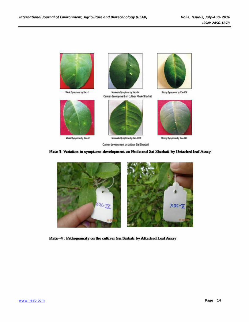
The pathogenic variability among Xanthomonas
axonopodis pv. citri
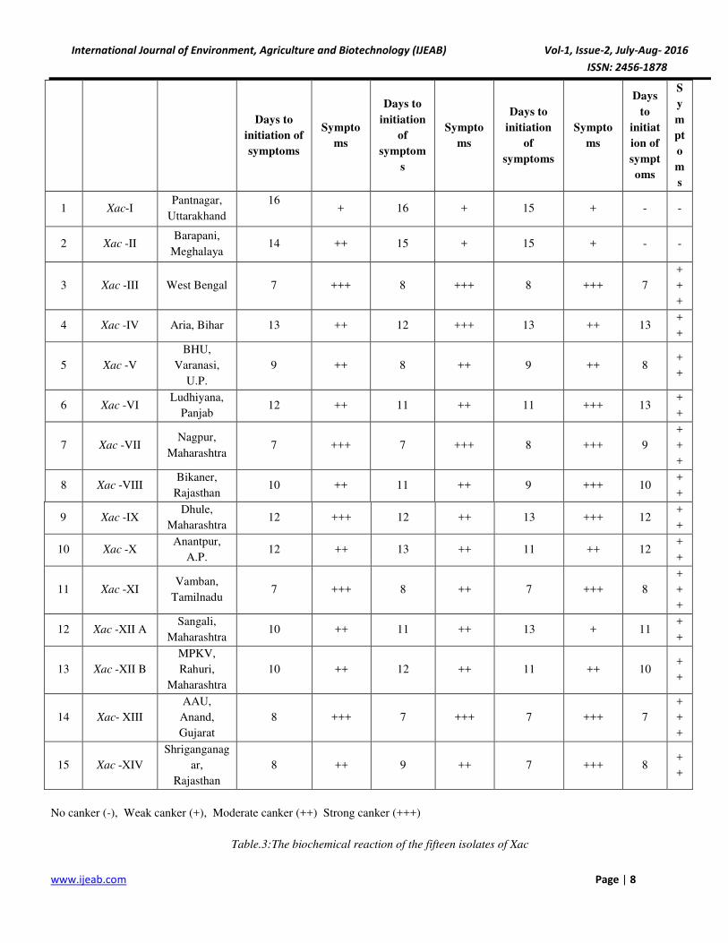
The pathogenic variability amongst the fifteen isolates
Xanthomonas axonopodis pv. citri, was studying (Table-2
,Plate-3 )inoculating on four different varieties of acid lime
viz. Sai sharbati, Phule sharbati, Pramalini and PKM-1 by
using detached leaf assay technique (Tuite, 1969). All
varieties were found susceptible to all the fifteen isolates
Xanthomonas axonopodis pv. citri. The isolates
Xanthomonas axonopodis pv. citri showed varied reaction
in the symptoms development. The isolates viz. Xac- III,
Xac- V, Xac- VII, Xac- XI, Xac- XIII and Xac- XIV found
highly virulent in development of typical symptoms i.e.
white crystalline callus formation at the point of inoculation
within 7 to 9 days. The isolates Xac- I, Xac- II and Xac- IV
were found less virulent as they developed symptoms after
13 to 16 days of inoculation. The isolates Xac- I and Xac- II
failed to develop symptom on variety PKM-1. The
categorization of isolates of Xac was done on the basis of
symptoms development on leaves and days taken for
appearance of the symptoms as No canker (-), Weak canker
(+), Moderate canker (++) and Strong canker (+++) as
presented in Table. 2. Atiq et al. (2007) who screened
fifteen citrus cultivars for resistance against citrus canker
disease incited by X. axonopodis pv. citri and reported that
no immune response was exhibited by any variety in the
experiment. Ismail et al. (2014) studied the reaction of Xac
on 5 different host of Rutaceace family by detached leaf
assay and reported that the pathogen also produced water
soaking, followed by necrosis around the wound inoculated
surface on grape fruit, Rough lemon followed by Lime.
Biochemical Characterization of Xanthomonas
axonopodis pv. citri
All the fifteen isolates of Xac were rod shaped produce
typical mucoid colonies with the color variation among all
the isolates from pale yellow to dark yellow. They are
Gram’s positive and showed string formation in KOH test
(Table-3). All the fifteen isolates of Xac were found
positive for hydrolysis of starch in variable degree. The
isolates Xac- 1, Xac- III, Xac- IV, Xac- V, Xac-VII, Xac-IX,
Xac-XIV, Xac- XIIB and Xac-XIII showed strong reaction
with clear zone around hydrolyzed area when lugol iodine
was poured. The isolates Xac- II, Xac- VI, Xac- VIII, Xac-
X, Xac- XI, Xac- XIIA and Xac- XIV showed moderate
reaction, showed slow zone formation around the area of
hydrolysis when lugol iodine was poured. For the
liquefaction of gelatin by fifteen isolates of Xanthomonas
International Journal of Environment, Agriculture and Biotechnology (IJEAB) Vol-1, Issue-2, July-Aug- 2016
ISSN: 2456-1878
www.ijeab.com Page | 4
axonopodis pv. citri, showed variable reaction. The isolates
Xac- V and Xac- XIIA showed strong reaction for gelatin
liquefaction as they produced strong proteolytic exo-
enzyme due to which gelatin was hydrolyzed. The isolate
Xac- III, Xac- IV ,Xac- VI, Xac- VIII, Xac- IX, Xac- XI,
Xac- X, Xac- XIIB showed moderate reaction for gelatin
liquefaction. The isolate Xac- I, Xac- II, Xac- VII, Xac- XIII
and Xac- XIV showed weak reaction for gelatin liquefaction
(Table-3). All the fifteen isolates of Xac showed variable
reaction for H2S Production. The isolates Xac- I, Xac-II,
Xac- III, Xac- IV, Xac- V, Xac- VI, Xac- IX, Xac- X, Xac-
XIIA, Xac- XIIB, Xac- XIII and Xac- XIV were strong H2S
producer, showed black coloration along the line of stab
inoculation within 3 to 4 days after inoculation. The isolate
Xac- VII, Xac-VIII and Xac- XI were found weak H2S
producer as the black coloration along the line of stab
inoculation was formed after 7 days of inoculation.
Similarly all isolates produce acid from carbon source
Trahalose but they are fails to produce gas from the same
carbon source. All isolates were negative for Indol
production. All the fifteen isolates of Xac were found
positive for Catalase test as bubble formation was observed
after addition of 3% hydrogen peroxide in the 48 hrs old
incubated bacterial culture. All the isolates were rod shaped,
Gram negative showed circular pale yellow colonies on
nutrient agar as earlier reported by Patel (1950) who
observed bacterial colonies of X .malvacearum as flat
glistering pale yellow on nutrient agar also identified the
organism is short rod with rounded ends, Gram negative.
The results obtained are in confirmation with those reported
by Manjula (2002) who reported that, the bacterium
Xanthomonas axonopodis pv. punicae causing oily spot
pomegranate were small rods, appeared singly, Gram
negative. Gottwald et al. (2002) who reported that
Xanthomonas axonopodis is a rod shaped Gram negative
bacterium.The cultures showed variable reaction among the
isolates of Xanthomonas axonopodis pv. citri. Similar
variation among the isolates has been earlier noted by Raut
(1990) he studied 15 isolates of Xanthomonas axonopodis
pv. mangiferae indicae for different physiological and
biochemical properties viz. H2S production , action on
carbohydrates, gelatin test, KOH test etc. Das (2003)
reported that the bacterial cells of Xanthomonas citri are
positive for hydrolysis of starch, liquefaction of gelatin,
catalase. Das (2005) studied different isolates of
Xanthomonas axonopodis pv. citri for different
physiological and biochemical properties viz. H2S
production, gelatin liquefaction, KOH test, catalase test,
acid production from different sugars.Bhardwaj et al.
(2014) collected 20 isolates of Xanthomonas axonopodis
pv. citri were collected from various regions of Varanasi.
Isolates were characterized with the help of morphological,
pathogenicity and biochemical analysis. All the isolates
showed similar morphological and biochemical
characteristic and all were found pathogenic on citrus, thus
confirming the identity of isolates as belonging to those of
Xanthomonas axonopodis pv. citri.
Molecular characterization of Xanthomonas axonopodis
pv. citri
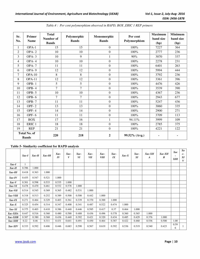
Out of 27 primers screened, 19 primers showed
amplification and produced scorable bands with high degree
of polymorphism. A total 220 amplicons were obtained of
which 218 amplicons were polymorphic with 99.52% level
of polymorphism was observed (Table-4). The banding
profile varied from minimum 5 band types (OPB- 1) to
maximum 21 band (REP) indicating the high molecular
variability amongst all the fifteen isolates of Xac. The
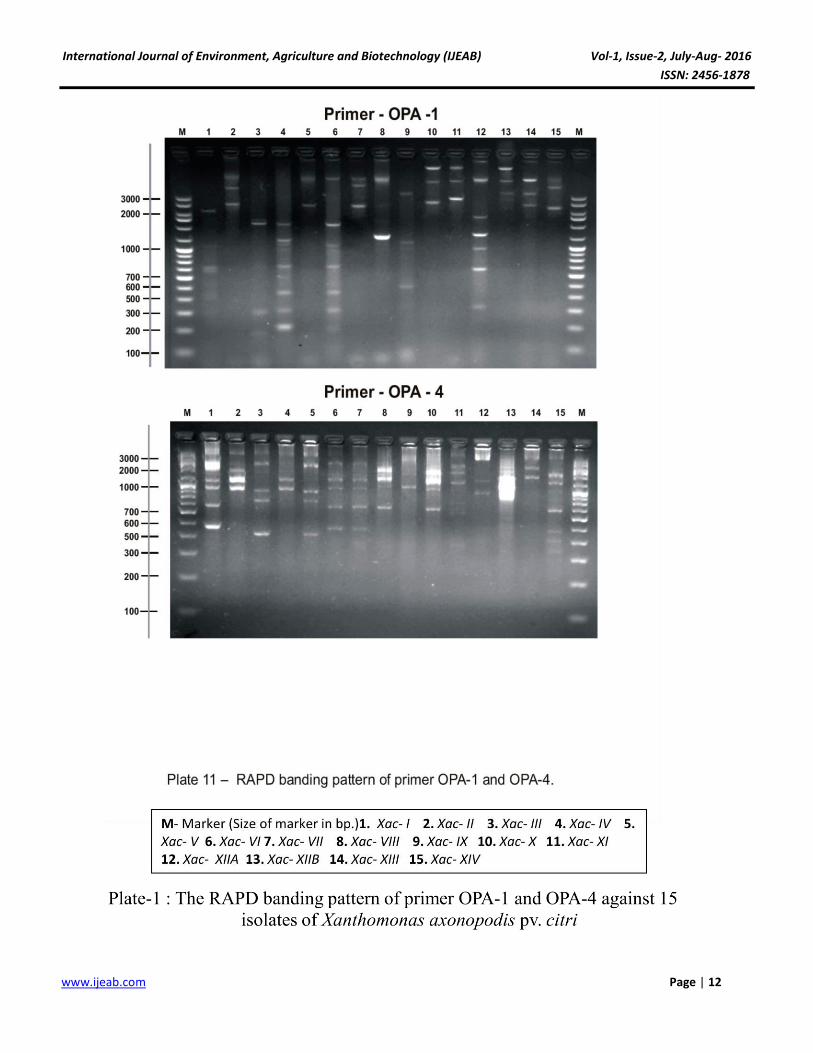
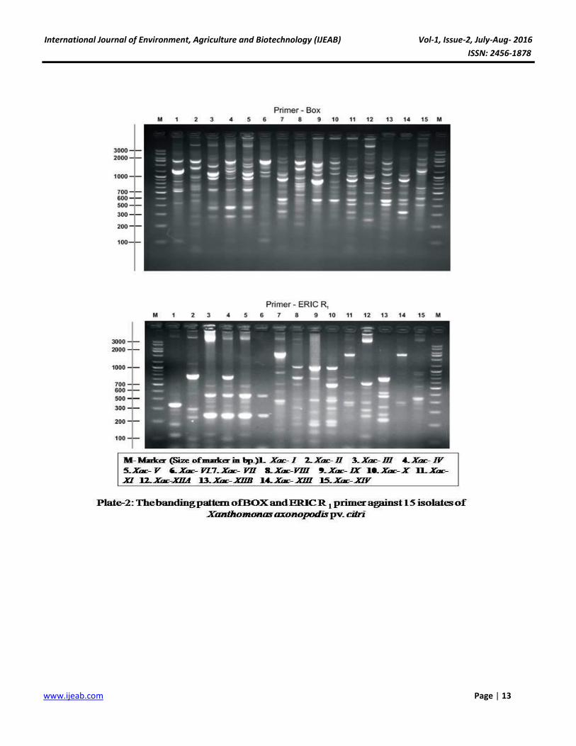
amplification profile of selected primer on 1.5% agarose gel
were showed in Plate-1and 2. The information on banding
pattern of all primers was used to determine genetic
distance between the fifteen isolates of Xac and the
dendrogram was constructed by using Un-weighted Pair
Group Arithmetic Mean method (UPGMA). Based on
simple matching coefficient a genetic similarity matrix was
constructed to access the genetic relatedness amongst the
fifteen isolates of Xac. The genetic similarity coefficient of
fifteen isolates given in Table 5. The similarity coefficient
ranged from 0.27 to 0.68 showed high genetic diversity.
The maximum genetic similarity was found between the
isolate from Uttar Pradesh (Xac- V) and Shriganganagar
(Xac- XIV) i.e 0.68 and both isolates in a same cluster B1.
The least similarity was found between the isolates from
Uttarakhand (Xac- I) and Dhule, Maharashtra (Xac- IX)
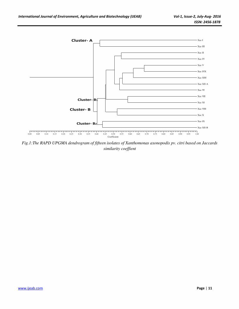
0.27.The dendrogram (Fig-1) showed that both isolates
falls in different cluster i.e. Xac- I in cluster A and Xac- IX
in sub cluster B2 of cluster B. Further the dendrogram
constructed by UPGMA clearly showed the main two
clusters viz. Cluster A and Cluster B. the Cluster B was
again divided into two sub clusters namely Cluster B1 and
Cluster B2. Two isolates Xac- I and Xac- III were falls
under cluster A as they are more similar to each other and
much differs with other isolates of Xac. Isolate Xac- II, Xac-
IV, Xac- V, Xac- XIV , Xac- XIII, Xac- XIIA ,Xac- VI, Xac-
VII, Xac- XI, Xac- VIII and Xac- X were falls under same
cluster in cluster B1 and remaining two isolates Xac- IX and
Xac- XIIB were falls under same cluster B2. The dendrogram
showed matched results with similarity coefficient values;
as the isolates showed high similarity coefficient value falls
International Journal of Environment, Agriculture and Biotechnology (IJEAB) Vol-1, Issue-2, July-Aug- 2016
ISSN: 2456-1878
www.ijeab.com Page | 5
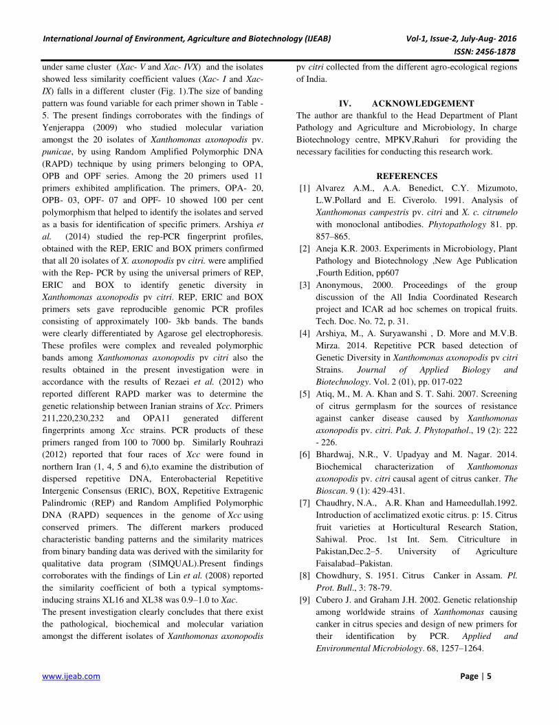
under same cluster (Xac- V and Xac- IVX) and the isolates
showed less similarity coefficient values (Xac- I and Xac-
IX) falls in a different cluster (Fig. 1).The size of banding
pattern was found variable for each primer shown in Table -
5. The present findings corroborates with the findings of
Yenjerappa (2009) who studied molecular variation
amongst the 20 isolates of Xanthomonas axonopodis pv.
punicae, by using Random Amplified Polymorphic DNA
(RAPD) technique by using primers belonging to OPA,
OPB and OPF series. Among the 20 primers used 11
primers exhibited amplification. The primers, OPA- 20,
OPB- 03, OPF- 07 and OPF- 10 showed 100 per cent
polymorphism that helped to identify the isolates and served
as a basis for identification of specific primers. Arshiya et
al. (2014) studied the rep-PCR fingerprint profiles,
obtained with the REP, ERIC and BOX primers confirmed
that all 20 isolates of X. axonopodis pv citri. were amplified
with the Rep- PCR by using the universal primers of REP,
ERIC and BOX to identify genetic diversity in
Xanthomonas axonopodis pv citri. REP, ERIC and BOX
primers sets gave reproducible genomic PCR profiles
consisting of approximately 100- 3kb bands. The bands
were clearly differentiated by Agarose gel electrophoresis.
These profiles were complex and revealed polymorphic
bands among Xanthomonas axonopodis pv citri also the
results obtained in the present investigation were in
accordance with the results of Rezaei et al. (2012) who
reported different RAPD marker was to determine the
genetic relationship between Iranian strains of Xcc. Primers
211,220,230,232 and OPA11 generated different
fingerprints among Xcc strains. PCR products of these
primers ranged from 100 to 7000 bp. Similarly Rouhrazi
(2012) reported that four races of Xcc were found in
northern Iran (1, 4, 5 and 6),to examine the distribution of
dispersed repetitive DNA, Enterobacterial Repetitive
Intergenic Consensus (ERIC), BOX, Repetitive Extragenic
Palindromic (REP) and Random Amplified Polymorphic
DNA (RAPD) sequences in the genome of Xcc using
conserved primers. The different markers produced
characteristic banding patterns and the similarity matrices
from binary banding data was derived with the similarity for
qualitative data program (SIMQUAL).Present findings
corroborates with the findings of Lin et al. (2008) reported
the similarity coefficient of both a typical symptoms-
inducing strains XL16 and XL38 was 0.9–1.0 to Xac.
The present investigation clearly concludes that there exist
the pathological, biochemical and molecular variation
amongst the different isolates of Xanthomonas axonopodis
pv citri collected from the different agro-ecological regions
of India.
IV. ACKNOWLEDGEMENT
The author are thankful to the Head Department of Plant
Pathology and Agriculture and Microbiology, In charge
Biotechnology centre, MPKV,Rahuri for providing the
necessary facilities for conducting this research work.
REFERENCES
[1] Alvarez A.M., A.A. Benedict, C.Y. Mizumoto,
L.W.Pollard and E. Civerolo. 1991. Analysis of
Xanthomonas campestris pv. citri and X. c. citrumelo
with monoclonal antibodies. Phytopathology 81. pp.
857–865.
[2] Aneja K.R. 2003. Experiments in Microbiology, Plant
Pathology and Biotechnology ,New Age Publication
,Fourth Edition, pp607
[3] Anonymous, 2000. Proceedings of the group
discussion of the All India Coordinated Research
project and ICAR ad hoc schemes on tropical fruits.
Tech. Doc. No. 72, p. 31.
[4] Arshiya, M., A. Suryawanshi , D. More and M.V.B.
Mirza. 2014. Repetitive PCR based detection of
Genetic Diversity in Xanthomonas axonopodis pv citri
Strains. Journal of Applied Biology and
Biotechnology. Vol. 2 (01), pp. 017-022
[5] Atiq, M., M. A. Khan and S. T. Sahi. 2007. Screening
of citrus germplasm for the sources of resistance
against canker disease caused by Xanthomonas
axonopodis pv. citri. Pak. J. Phytopathol., 19 (2): 222
- 226.
[6] Bhardwaj, N.R., V. Upadyay and M. Nagar. 2014.
Biochemical characterization of Xanthomonas
axonopodis pv. citri causal agent of citrus canker. The
Bioscan. 9 (1): 429-431.
[7] Chaudhry, N.A., A.R. Khan and Hameedullah.1992.
Introduction of acclimatized exotic citrus. p: 15. Citrus
fruit varieties at Horticultural Research Station,
Sahiwal. Proc. 1st Int. Sem. Citriculture in
Pakistan,Dec.2–5. University of Agriculture
Faisalabad–Pakistan.
[8] Chowdhury, S. 1951. Citrus Canker in Assam. Pl.
Prot. Bull., 3: 78-79.
[9] Cubero J. and Graham J.H. 2002. Genetic relationship
among worldwide strains of Xanthomonas causing
canker in citrus species and design of new primers for
their identification by PCR. Applied and
Environmental Microbiology. 68, 1257–1264.
International Journal of Environment, Agriculture and Biotechnology (IJEAB) Vol-1, Issue-2, July-Aug- 2016
ISSN: 2456-1878
www.ijeab.com Page | 6
[10] Das, A.K. 2002. Pathogenic variability in
Xanthomonas axonopodis pv. citri causal agent of
citrus canker. J. Mycol. Pl. Patho.54, 274-279.
[11] Das A.K .2003. Citrus canker-A review. J. Appl. Hort.
5(1): 52-60.
[12] Das, S. 2005. Variability among the isolates of
Xanthomonas axonopodis pv. citri. M.Sc. Thesis
(Unpub.) Dr. P.D.K.V.Akola.23- 29.
[13] Egel, D.S., J.H. Graham and R.E. Stall. 1991.
Genomic relatedness of Xanthomonas compestris
strains causing diseases of citrus. Appl. Environ.
Microbiol. 57: 2724-2730.
[14] Graham, J.H, T.R. Gottwald, J. Cubero and D.S.
Achor.2004. Xanthomonas axonopodis pv. citri:
factors affecting successful eradication of citrus
canker. Mol. Plant Pathol. 5(1):1-15.
[15] Gottwald T.R., Graham, J.H., Civerolo, E.L., Barrett,
H.C., 1993, Differential host renge reaction of citrus
and citrus relative to citrus canker and citrus bacterial
spot determined by leaf mesophyll susceptibility.
Plant dis. 77:1004-1009.
[16] Gottwald T.R, J.H. Graham and T.S. Schubert. 2002.
Citrus canker the Pathogen and its impact. Online.
Plant helth progress. 812-01RV.
[17] Hamlin, S.A. 1967. Studies on occurrence of
pathotypes in Xanthomonas citri (Hasse) Dowson.
Punjab Hort. J., 7: 90-93.
[18] Islam MA, Mazumdar RM, Islam S, Alam MJ, Urmee
SA., 2014, Isolation, identification and in-vitro
antibiotic sensitivity pattern of citrus canker causing
organism Xanthomonas axonopodis.. Adv. life sci.,
1(4), pp. 215-222.
[19] Ismail M., M.I. Haque, A. Raiz, M.A. Abro, M.H.
Khan, 2014 Pathogenic variability among different
isolates of Xanthomonas axonopodis pv. citri. Pak J.
Agri., Agri. Engg., Vet Sci., 30 (2) 187-194.
[20] Kishore, V. and J.N. Chand, 1972. Citrus Canker in
Haryana. Haryana Agric. Univ. J. Res., 27: 124-127.
[21] Khan, L.D. and M.K. Hingorani, 1970. Strain studies
on Xanthomonas citri (Hasse) Dowson . J. Hort. Sci.,
45: 15-17.
[22] Lin, H. C.,H. Chang, and K. C. Tzeng. 2008.
Characterization of novel strains of citrus canker
bacteria from citrus in Taiwan. J. Taiwan Agric. Res.
57:265–278.
[23] Luthra, J.C. and A. Sattar.1942. Citrus canker and its
control in Punjab. Punjab Fruit J.,6 (1):
[24] Manjula, C. P.2002, Studies on bacterial blight of
pomegranate (Punica granatum L.) caused by
Xanthomonas axonopodis pv. punicae. M.Sc. (Agri.)
Thesis, Univ. Agric. Sci., Bangalore, Karnataka
(India).
[25] Miyoshi, T., H.Sawada, Y. Tachibana, I. Matsuda.
1998. Detection of Xanthomonas
campestris pv. citri by PCR using primers from the
spacer region between the 16S and 23S rRNA genes.
Annals of the Phytopathological Society of Japan 64 :
249-254.
[26] Nirvan, R.S. 1960. Effect of antibiotic sprays on citrus
canker. Hort.Adv., 4:155-160.
[27] Patel, M.K. and Y.S. Kulkarni. 1950. Bacterial leaf
spot of cotton.Indian Phytopath 3: 51-62.
[28] Prasad, N. 1959. Citrus canker. Proc. Seminar on
Disease of Horticultural Plants, Simla,pp. 87-88.
[29] Parsai, P.S.1959. Citrus canker. Proc. Seminar on
Diseases of Horticultural Plants. Simla, pp. 91-95.
[30] Pruvost, O., J.S.Hartung, E.L. Civerolo, C. Dubois and
X. Perrier. 1992. Plasmid DNA fingerprints
distinguish pathotypes of Xanthomans campestris pv.
citri, the causal agent of citrus bacterial canker
disease. Phytopathology 82:485-490. Punjab. Punjab
Hort. J., 2: 89-91
[31] Ramakrishnan, T.S. 1954. Common diseases of citrus
in Madras state.Govt of Madras publication.
[32] Rangaswami, G. and R.C.K. Soumini,1957.Disease of
citrus canker in Madras State. Indian Hort 5:50-57.
[33] Rao, G.P. 1954. Citrus diseases and their control in
AndhraState. Andhra Agric. J., 1: 187-192.
[34] Rezaei, M K., M. Shams-Bakhsh and A. Alizadeh.
2012. Genetic diversity among Xanthomonas citri
subsp. citri strains in Iran. J.Pl. Prot.Res.52: 1, 1-9.
[35] Raut B.T. (1990) Studied on leaf spot of mango
caused by Xanthomonas campestreis pv. mangiferae
indicae. Ph.D.Thesis U.A.S Dharwad, pp104.
[36] Sambrook and Russell. 2001. Molecular cloning and
laboratory manual 3rd
edition. Cold spring horbor
lobotary press, New York pp. 2231
[37] Schubert, T. S., Rizvi, S. A., Sun, X., Gottwald, T. R.,
Graham, J. H., and Dixon, W. N. 2001. Meeting the
challenge of eradicating citrus canker in Florida-again.
Plant Dis. 85:340-356.
[38] Tuite, J., 1969, Plant Pathological Methods : Fungi
and Bacteria. Burgess Publishing Co., Minneapolis,
U. S. A., p. 239.
[39] Valenchia, L.D , M.P. Natural, G.G. Divinagaracia
and V.N Villegas. 2004.Streptomycin resistance to
anthuriums and sources of host resistance to
International Journal of Environment, Agriculture and Biotechnology (IJEAB) Vol-1, Issue-2, July-Aug- 2016
ISSN: 2456-1878
www.ijeab.com Page | 7
Xanthomonas axonopodis pv. diefenbachiae Indian J.
Exptl. Biol., 29: 180-181.
[40] Venkatakrishnaiah, N.S. 1957.Canker disease ofsour
lime and its control. J. Mysore Hort. Sci., 2(2, 3):
40-44.
[41] Yenjerappa, S.T., 2009. Epedemiology and
management of bacterial blight of pomegranate caused
by Xanthomonas axonopodis pv.punicae (Hingorani
and Singh) Vuterrin et al. Ph. D. Thesis, Univ. Agri.
Sci. Dharwad (India).147.
[42] Zekri, M., Chamberlain, H., Timmer, P., Roberts, P.,
and Muchove, R. 2005. Field identification of citrus
canker symptoms and decontamination procedures.
Uni.Florida. IFAS extension.
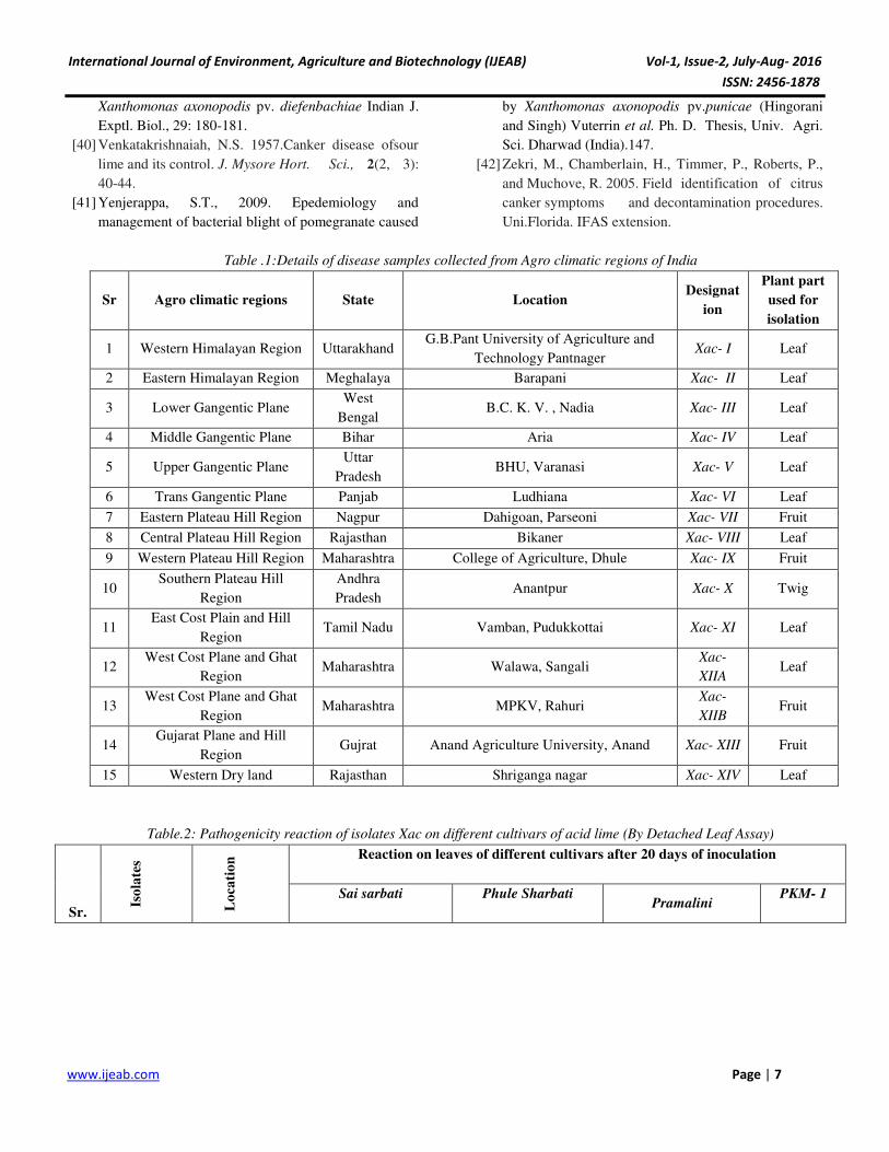
Table .1:Details of disease samples collected from Agro climatic regions of India
Sr Agro climatic regions State Location Designat
ion
Plant part
used for
isolation
1 Western Himalayan Region Uttarakhand G.B.Pant University of Agriculture and
Technology Pantnager Xac- I Leaf
2 Eastern Himalayan Region Meghalaya Barapani Xac- II Leaf
3 Lower Gangentic Plane West
Bengal B.C. K. V. , Nadia Xac- III Leaf
4 Middle Gangentic Plane Bihar Aria Xac- IV Leaf
5 Upper Gangentic Plane Uttar
Pradesh BHU, Varanasi Xac- V Leaf
6 Trans Gangentic Plane Panjab Ludhiana Xac- VI Leaf
7 Eastern Plateau Hill Region Nagpur Dahigoan, Parseoni Xac- VII Fruit
8 Central Plateau Hill Region Rajasthan Bikaner Xac- VIII Leaf
9 Western Plateau Hill Region Maharashtra College of Agriculture, Dhule Xac- IX Fruit
10 Southern Plateau Hill
Region
Andhra
Pradesh Anantpur Xac- X Twig
11 East Cost Plain and Hill
Region Tamil Nadu Vamban, Pudukkottai Xac- XI Leaf
12 West Cost Plane and Ghat
Region Maharashtra Walawa, Sangali
Xac-
XIIA Leaf
13 West Cost Plane and Ghat
Region Maharashtra MPKV, Rahuri
Xac-
XIIB Fruit
14 Gujarat Plane and Hill
Region Gujrat Anand Agriculture University, Anand Xac- XIII Fruit
15 Western Dry land Rajasthan Shriganga nagar Xac- XIV Leaf
Table.2: Pathogenicity reaction of isolates Xac on different cultivars of acid lime (By Detached Leaf Assay)
Sr.
Iso
late
s
Lo
ca
tio
n
Reaction on leaves of different cultivars after 20 days of inoculation
Sai sarbati
Phule Sharbati
Pramalini
PKM- 1
International Journal of Environment, Agriculture and Biotechnology (IJEAB) Vol-1, Issue-2, July-Aug- 2016
ISSN: 2456-1878
www.ijeab.com Page | 8
Days to
initiation of
symptoms
Sympto
ms
Days to
initiation
of
symptom
s
Sympto
ms
Days to
initiation
of
symptoms
Sympto
ms
Days
to
initiat
ion of
sympt
oms
S
y
m
pt
o
m
s
1 Xac-I Pantnagar,
Uttarakhand
16
+ 16 + 15 + - -
2 Xac -II Barapani,
Meghalaya 14 ++ 15 + 15 + - -
3 Xac -III West Bengal 7 +++ 8 +++ 8 +++ 7
+
+
+
4 Xac -IV Aria, Bihar 13 ++ 12 +++ 13 ++ 13 +
+
5 Xac -V
BHU,
Varanasi,
U.P.
9 ++ 8 ++ 9 ++ 8 +
+
6 Xac -VI Ludhiyana,
Panjab 12 ++ 11 ++ 11 +++ 13
+
+
7 Xac -VII Nagpur,
Maharashtra 7 +++ 7 +++ 8 +++ 9
+
+
+
8 Xac -VIII Bikaner,
Rajasthan 10 ++ 11 ++ 9 +++ 10
+
+
9 Xac -IX Dhule,
Maharashtra 12 +++ 12 ++ 13 +++ 12
+
+
10 Xac -X Anantpur,
A.P. 12 ++ 13 ++ 11 ++ 12
+
+
11 Xac -XI Vamban,
Tamilnadu 7 +++ 8 ++ 7 +++ 8
+
+
+
12 Xac -XII A Sangali,
Maharashtra 10 ++ 11 ++ 13 + 11
+
+
13 Xac -XII B
MPKV,
Rahuri,
Maharashtra
10 ++ 12 ++ 11 ++ 10 +
+
14 Xac- XIII
AAU,
Anand,
Gujarat
8 +++ 7 +++ 7 +++ 7
+
+
+
15 Xac -XIV
Shriganganag
ar,
Rajasthan
8 ++ 9 ++ 7 +++ 8 +
+
No canker (-), Weak canker (+), Moderate canker (++) Strong canker (+++)
Table.3:The biochemical reaction of the fifteen isolates of Xac

International Journal of Environment, Agriculture and Biotechnology (IJEAB) Vol-1, Issue-2, July-Aug- 2016
ISSN: 2456-1878
www.ijeab.com Page | 9
Parameter
s
Xac- I Xac-
II
Xac-
III
Xac-
IV Xac- V
Xac-
VI Xac- VII
Xac-
VIII
Xac-
IX Xac- X
Xac-
XI
Xac-
XIIA
Xac-
XIIB Xac- XIII
Xa
c-
XI
V
Shape Rod Rod Rod Rod Rod Rod Rod Rod Rod Rod Rod Rod Rod Rod Ro
d
Colon
y
colour
Pale
yellow
Pale
yello
w
yello
w
Pale
yello
w
Dark
yellow yellow
Dark
yellow
Dark
yellow
Pale
yellow Yellow
Dark
Yellow
Yellow
Yellow
Dark
Yellow
Da
rk
Ye
llo
w
Gram
reacti
on
-ve -ve -ve -ve -ve -ve -ve -ve -ve -ve -ve -ve -ve -ve -ve
Catala
se +++ +++ +++ +++ +++ +++ +++ +++ +++ +++ +++ +++ +++ +++
++
+
Indole
produ
ction
- - - - - - - - - - - - - - -
KOH +++ +++ +++ +++ +++ +++ +++ +++ +++ +++ +++ +++ +++ +++ ++
+
H2S
Produ
ction
+++ +++ +++ +++ +++ +++ + + +++ +++ + +++ +++ +++ ++
+
Acid
and
gas
produ
ction
T
r
a
h
a
l
o
s
e
+++ +++ +++ ++ + +++ ++ ++ +++ +++ ++ +++ +++ +++ ++
+
Hydro
lysis
G
e
l
e
t
i
n
e
+ + ++ ++ +++ ++ + ++ ++ ++ ++ +++ ++ + +
S
t
a
r
c
h
+++ ++ +++ +++ +++ ++ +++ ++ +++ ++ ++ ++ +++ +++ ++
-
:
Negative Reaction
+ : Weak Reaction
+
+ : Moderate Reaction
+
+
+
: Strong Reaction
International Journal of Environment, Agriculture and Biotechnology (IJEAB) Vol-1, Issue-2, July-Aug- 2016
ISSN: 2456-1878
www.ijeab.com Page | 10
Table.4 : Per cent polymorphism observed in RAPD, BOX ,ERIC 1 REP primers
Sr.
No.
Primer
Name
Total
Number of
Bands
Polymorphic
Bands
Monomorphic
Bands
Per cent
Polymorphism
Maximum
band size
(bp)
Minimum
band size
(bp)
1 OPA-1 15 15 0 100% 7227 364
2 OPA- 2 10 10 0 100% 2777 236
3 OPA- 3 10 9 1 90% 3070 337
4 OPA- 4 10 10 0 100% 2278 231
5 OPA- 7 11 11 0 100% 6401 263
6 OPA- 9 12 12 0 100% 5994 444
7 OPA-10 8 8 0 100% 3792 236
8 OPA-11 12 12 0 100% 3361 396
9 OPB- 1 5 5 0 100% 4476 426
10 OPB- 4 7 7 0 100% 3539 390
11 OPB- 5 10 10 0 100% 4367 236
12 OPB- 6 7 7 0 100% 2943 677
13 OPB- 7 11 11 0 100% 5247 436
14 OPF- 2 13 13 0 100% 3060 335
15 OPF- 4 14 14 0 100% 2900 271
16 OPF- 6 11 11 0 100% 3709 113
17 BOX 17 16 1 94.11% 3999 109
18 ERIC 1 18 18 0 100% 2785 375
19 REP 21 21 0 100% 4221 122
Total No. of
Bands 220 218 2 99.52% (Avg.) - -
Table 5- Similarity coefficient for RAPD analysis
Xac-I Xac-II Xac-III
Xac-
IV
Xac-
V
Xac-
VI
Xac-
VII
Xac-
VIII
Xac-
IX Xac-X
Xac-
XI
Xac-XII
A
Xac-XII
B
Xac
-
XIII
Xa
c-
XI
V
Xac-I 1
Xac-II 0.396 1.000
Xac-III 0.418 0.363 1.000
Xac-IV 0.455 0.547 0.521 1.000
Xac-V 0.301 0.598 0.533 0.535 1.000
Xac-VI 0.478 0.470 0.461 0.532 0.578 1.000
Xac-VII 0.514 0.545 0.369 0.365 0.482 0.531 1.000
Xac-VIII 0.318 0.513 0.252 0.389 0.500 0.500 0.442 1.000
Xac-IX 0.271 0.464 0.329 0.403 0.361 0.339 0.370 0.300 1.000
Xac-X 0.325 0.454 0.314 0.347 0.400 0.341 0.487 0.522 0.474 1.000
Xac-XI 0.375 0.403 0.410 0.386 0.440 0.446 0.585 0.417 0.37 0.464 1.000
Xac-XIIA 0.447 0.516 0.360 0.480 0.588 0.488 0.436 0.496 0.378 0.360 0.365 1.000
Xac-XIIB 0.387 0.380 0.368 0.436 0.440 0.392 0.421 0.320 0.434 0.485 0.429 0.376 1.000
Xac-XIII 0.22 0.46 0.311 0.431 0.641 0.491 0.450 0.464 0.387 0.422 0.460 0.536 0.500 1.00
Xac-XIV 0.333 0.592 0.406 0.446 0.683 0.590 0.567 0.619 0.392 0.536 0.519 0.540 0.423 0.59
6 1
International Journal of Environment, Agriculture and Biotechnology (IJEAB) Vol-1, Issue-2, July-Aug- 2016
ISSN: 2456-1878
www.ijeab.com Page | 11
Coefficient
0.00 0.05 0.10 0.15 0.20 0.25 0.30 0.35 0.40 0.45 0.50 0.55 0.60 0.65 0.70 0.75 0.80 0.85 0.90 0.95 1.00
Xac-I
Xac-III
Xac-II
Xac-IV
Xac-V
Xac-IVX
Xac-XIII
Xac-XII-A
Xac-VI
Xac-VII
Xac-XI
Xac-VIII
Xac-X
Xac-IX
Xac-XII-B
Cluster- A
Cluster- B
Cluster- B1
Cluster- B2
Fig.1:The RAPD UPGMA dendrogram of fifteen isolates of Xanthomonas axonopodis pv. citri based on Jaccards
similarity coeffient
International Journal of Environment, Agriculture and Biotechnology (IJEAB) Vol-1, Issue-2, July-Aug- 2016
ISSN: 2456-1878
www.ijeab.com Page | 12
International Journal of Environment, Agriculture and Biotechnology (IJEAB) Vol-1, Issue-2, July-Aug- 2016
ISSN: 2456-1878
www.ijeab.com Page | 13
International Journal of Environment, Agriculture and Biotechnology (IJEAB) Vol-1, Issue-2, July-Aug- 2016
ISSN: 2456-1878
www.ijeab.com Page | 14