pancreatic tumor organoid protocol -...
TRANSCRIPT

Muthuswamy Laboratory Tumor Organoid Protocol . 1
Pancreatic Tumor Organoid Protocol
The following protocol is optimized for generating Patient-Derived tumor Organoid (PDO) cultures either directly from surgical resections of human Pancreatic adenocarcinoma (PDAC) or from Patient-derived xenografts of human PDAC tumors. We usually achieve about 80 - 90% success of establishing organoid cultures when starting with tumor resections, and greater than 90% success rate when starting from patient-derived xenograft (PDX)-derived tumors. Important factors contributing to success is the amount of the input tissue and the tumor cell content in the tissue. Tumor chunks larger than 5.0 mm3 will increase the chance of success, whereas, smaller chunks will give variable results. In addition, the tumor cells content is also a critical determinant of success, if the tumor chunk happens to be primarily stroma-rich, it will negatively affect on the ability to generate organoid cultures.
The initial organoid culture that develops after plating of the processed tissue is referred to Passage 0 (P0), all subsequent passages should be kept track of and be serially numbered. We recommend using cultures within the P6 – P8 for experiments to avoid clonal drift. Starting P2, the aliquots of the cultures should be passaged to maintain a supply of early passage cultures.
Materials Needed
Materials Vendor Catalog #
Collagenase/Dispase (100 mg/ml) Sigma 10269638001
Accutase Sigma A6964
DMEM-high glucose Sigma 5796
Rock Inhibitor (Y267632) Tocris 1254
Matrigel (Growth factor reduced)
(Concentration ~8.5mg/ml)
BD 354230
Penicillin-Strepomycin
10,000 U/mL
Gibco 15140-122
Bovine Serum Albumin Sigma A7906

Muthuswamy Laboratory Tumor Organoid Protocol . 2
Media Components & Recipes:
1. Digestion Media:
DMEM 1:100 dilution of 100mg/ml stock Collagenase/ Dispase
1.0 % Penicillin-streptomycin
Preparation Tip: Prepare only the needed amount, fresh just before use, and discard the unused portion of the media.
2. Resuspension Media: DMEM 1.0 % Penicillin-streptomycin 1.0 % BSA Preparation Tip: Add 1% BSA by weight to DMEM+1.0% Pencillin-streptomycin containing media and stir to dissolve the BSA. Once dissolved, filter sterilize using 0.2µ filter and store at 4oC.
Heat shock fraction
Growth Media See recipe below
PaTOM Growth Factor Cocktail Muthuswamy laboratory (Mix A)
Cryostar CS10 Freezing media Sigma C2874
Hydrocortizone Muthuswamy laboratory (Mix B)
EASYstrainer (100µm) Greiner Bio-one 542000
4 well chamber slides
P1000 pipette with tips cut to make wide bore
12 well multi-well dish
Swing-bucket, tabletop 4oC centrifuge

Muthuswamy Laboratory Tumor Organoid Protocol . 3
3. PaTOM Growth Media: DMEM: 500 ml 5.0 ml of PaTOM Growth factors cocktail * (Tube A) 1.0 % Penicillin-streptomycin 250 µl Hydrocortizone (1.0 mg/ml) (Tube B) • Contains: B27, ascorbic acid, insulin, hydrocortisone, FGF2, and all-
trans retinoic acid (Obtained as a premixed, qual i ty contro l l ed , cocktai l f rom the Muthuswamy laboratory)
Preparation Tip:
• Do NOT mix Tube A and Tube B by themselves as the alcohol in Tube B solution will denature the growth factors in Tube A.
• After adding all ingredients filter the media through a 0.2µ filter • Store prepared media at 40C, for NO more than one month.
4. Culture Media: PaTOM growth media 5.0 % GFR-Matrigel 10 µM Y267632, Rock inhibitor (10mM, a 1000x stock). Rock inhibitor is prepared in sterile Phosphate Buffered Saline. Preparation Tip: Prepare fresh. You may premix PaTOM growth media and ROCK inhibitor and keep on ice. Add Matrigel just before use. Discard unused portion of the media.
5. Freezing Media: Cryostar freezing media 10 µM Y267632, Rock inhibitor
Reference:
Huang L et al, Ductal pancreatic cancer modeling and drug screening using human pluripotent stem cell– and patient-derived tumor organoids. Nat Med. 2015
Nov;21(11):1364-71.

Muthuswamy Laboratory Tumor Organoid Protocol . 4
PROCEDURE FOR PROCESSING TISSUES
1. Place the tumor tissue received from the surgery in a 35mm petri dish. You may choose to take a small section of the tissue and fix it for histology processing. And, another piece of the tissue can be snap frozen in liquid nitrogen for future use to isolate DNA/RNA. These are optional and will depend on the amount of surgical material received.
2. Add 1.0-3.0 ml of Resuspension media in the dish depending on the size of the tumor. It would be advisable to use the smallest amount of media possible (~1.0 ml), which would provide better control during the tissue mincing process.
3. Mince tumor tissue with a sterile surgical scalpel (#21) into 0.5-1.0 mm fragments.
It would be better to use two scalpel blades and move them in anti-parallel direction to generate a finely minsed tissue preparation. Be careful to not apply too much pressure during this step, if you do, you will end up scraping the plastic from the 35mm tissue culture plate. When the process is complete, the tissue pulp should look like it is shown in the image below.
4. Using a P1000 tip transfer the tissue pulp in to a 15.0 ml conical tube. You will not be able to collect all the tissue chunks using a regular P1000 tip, so use a fresh P1000 tip with its end cut to widen the bore. Add 1.0 ml of Resuspension media

Muthuswamy Laboratory Tumor Organoid Protocol . 5
in a rinsing motion and transfer the digested tissue pulp to the 15.0 ml conical tube. Pellet the tissue suspension at 1500 rpm for 5 minutes at 4oC. Remove the supernatant carefully. Be aware the tissue pellet will not be firm so care should be taken to not disturb the tissue pellet. It would be advisable to first remove majority of the supernatant with a P1000 tip and then use a P200 tip to remove the remaining supernatant. See below for an image of the tissue pellet.
5. Add 2.0 ml of Digestion media. Using a P1000 tip with a tip cut (for wide bore)
gently re-suspend the tissue pellet and transfer the pellet to a fresh 35 mm tissue culture plate. Incubate in 37oC incubator for no more than 30 minutes. Check every 10 minutes for progress. After about 20 minutes you should be able to see a distinctive pink/purple hallow around the tissue chunks, when observed under a microscope.
6. After 30 minutes, transfer the digested tissue slurry to a 15 ml conical tube. To remove all digested tissues, add an additional 2.0 ml of Resuspension media and transfer to the conical tube using a P1000 tip that has a wide bore, as performed above.
7. Centrifuge at 1500 rpm for 5 minutes at 4oC.
8. Aspirate the supernatant gently using a P1000 tip taking care not to disturb the pellet. Add 2.0 ml of Accutase to the pellet. Mix well using a P1000 tip with wide
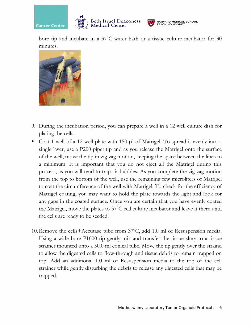
Muthuswamy Laboratory Tumor Organoid Protocol . 6
bore tip and incubate in a 37oC water bath or a tissue culture incubator for 30 minutes.
9. During the incubation period, you can prepare a well in a 12 well culture dish for plating the cells.
• Coat 1 well of a 12 well plate with 150 µl of Matrigel. To spread it evenly into a single layer, use a P200 pipet tip and as you release the Matrigel onto the surface of the well, move the tip in zig zag motion, keeping the space between the lines to a minimum. It is important that you do not eject all the Matrigel during this process, as you will tend to trap air bubbles. As you complete the zig zag motion from the top to bottom of the well, use the remaining few microliters of Matrigel to coat the circumference of the well with Matrigel. To check for the efficiency of Matrigel coating, you may want to hold the plate towards the light and look for any gaps in the coated surface. Once you are certain that you have evenly coated the Matrigel, move the plates to 37oC cell culture incubator and leave it there until the cells are ready to be seeded.
10. Remove the cells+Accutase tube from 37oC, add 1.0 ml of Resuspension media.
Using a wide bore P1000 tip gently mix and transfer the tissue slury to a tissue strainer mounted onto a 50.0 ml conical tube. Move the tip gently over the straind to allow the digested cells to flow-through and tissue debris to remain trapped on top. Add an additional 1.0 ml of Resuspension media to the top of the cell strainer while gently disturbing the debris to release any digested cells that may be trapped.

Muthuswamy Laboratory Tumor Organoid Protocol . 7
11. Centrifuge the flow-through at 1500 rpm for 5 minutes at 4oC. You should be able to observe a small but distinct pellet as shown below.
12. Gently resuspend the pellet in 2.0 ml of freshly prepared Culture media. You should not make Culture media with 5.0% Matrigel and let it sit on ice for long periods of time as it will result in sedimentation of the Matrigel. To save time, you may prepare Culture media without Matrigel and keep on ice until ready – just before you need to resuspend cells add 5% Matrigel and use immediately.

Muthuswamy Laboratory Tumor Organoid Protocol . 8
13. Gently, in drops, transfer the 2.0 ml of the resuspended cells to the Matrigel
coated well in the 12 well dish. Be careful not to squirt the suspension as you will disturb the underlying Matrigel layer.
14. Monitor growth. You should take representative images every 4 days to document growth. These images can be used to determine growth rate by morphometric analysis.
15. Changing media. It is important to change media every 3-4 days. As noted earlier – Matrigel containing culture media must be prepared just before addition to the well to avoid sedimentation of the Matrigel.
16. Aspirate the old media gently using a P1000 tip, taking care not to disturb the Matrigel pellet or the Organoids. Add Matrigel containing Culture media to the well, gently, in drops. Take care not to add is too fast as it will disturb the organoids and the Matrigel layer.

Muthuswamy Laboratory Tumor Organoid Protocol . 9
PROCEDURE FOR MAINTAINING, PASSAGING AND CRYOPRESERVING ORGANOID CULTURES
The time to passage the organoids can vary depending on the organoid line. In our experience we have found some PDO lines grow rapidly and are ready for passage by day 12-14, whereas other take much longer. In general, the time to passage is identified by culture that have stopped growing for more than 4 days.
The protocol below is optimized for 1 well of an organoid line in a 12 well plate
1. Aspirate the old media using a pipet tip. Do not use an aspirator. Be careful not to disturb the Matrigel layer or the organoids.
2. Add 0.5 ml/well Digestion media. Add gently and in drops, working close to the Matrigel surface to avoid generation of turbulence by the media being added.
3. Incubate at 37oC for 2.5 to 3.0 hours. By 2.5 hours you should be able to observe the Matrigel digested into a pulp/slury and the organoid dispersed into small clumps or clusters. This would indicate completion of the digestion. If these changes are not obvious, you could extend the digestion for another 30 minutes. 4. Re-suspend the cells by adding 1.0 ml of Resuspension media. The media can be cold, preferably not warmed to 37oC. Add the media in drops. Gently resuspend using a p1000 tip several times until the Matrigel is a smooth suspension and the

Muthuswamy Laboratory Tumor Organoid Protocol . 10
organoids have dispersed into single cells or smaller clumps. Transfer it to a 15.0 ml conical tube.
5. Centrifuge at 1500 rpm for 5 minutes at 4oC. The pellet that contains Matrigel and cells, will not be prominent, can be only seen as a difference in texture and not necessarily as a difference in color. Remove the supernatant gently using a P100 tip. This is a very critical step as it is very easy to aspirate off the Matrigel/cell pellet. Pay attention to difference in consistency while removing. To be safe, you may choose to remove the supernatant in two stages – first remove ~1200 microlitres using P1000 and remove the rest using a P200 tip. The P200 tip will allow you to readily feel the difference in consistency between the supernatant and the Matrigel/cell pellet. The image below shows the Matrigel pellet with few microliters of the supernatant remaining.
6. Re-suspend the Matrigel pellet by adding 500 µl of Accutase (kept on ice). Accutase comes as a 100.00 ml from the vendor. This should be aliquoted into smaller volume (1.0, 5.0 or 10.0 ml) to avoid repeated freeze-thaw. Pipette up and down until the Matrigel/cell pellet is resuspended into a smooth slury (see image below).
7. Incubate cells+accutase tubes at 37oC for 30-40 minutes.

Muthuswamy Laboratory Tumor Organoid Protocol . 11
8. During the above incubation period, prepare wells in a 12 well plate for
reseeding cells. We typically split the culture at a ratio of 1:2 to 1:4. The choice of split ratio will depend on the growth rate (fast growing versus slow growing organoid lines) or other need for cells (part of the cells to used for isolating DNA/RNA or for cryopreservation). We have found that some lines grow very well and can be split at 1:4 ratio, whereas, some are slow growing and require a 1:2 split. It may be wise to perform a 1:2 split during the early passages until you become familiar with the organoid line.
Once your determine the split ratio, coat the required number of wells in a 12 well plate with 150 µl of Matrigel. To spread it evenly into a single layer, use a P200 pipet tip and as you release the Matrigel onto the surface of the well, move the tip in zig zag motion, keeping the space between the lines to a minimum. It is important that you do not eject all the Matrigel during this process, as you will tend to trap air bubbles. As you complete the zig zag motion from the top to bottom of the well, use the remaining few microliters of Matrigel to coat the circumference of the well with Matrigel. To check for the efficiency of Matrigel coating, you may want to hold the plate towards the light and look for any gaps in the coated surface. Once you are certain that you have evenly coated the Matrigel, move the plates to 37oC cell culture incubator and leave it there until the cells are ready to be seeded.
9. Remove the cells+accutase tubes from 37oC and add 500 µl of Resuspension
media. Gently pipette up and down to generate a smooth suspension of cells. Use a small aliquot to determine cell count in a coulter counter or hemocytometer. If you need cells for generating a cell pellet (for DNA/RNA isolation), an aliquot of this cell suspension can be used. It is also wise to freeze down (see below for details) as many vials of cells as possible during early passages to have stocks of early passage organoid cultures. Once you have taken all the cells needed for generating a pellet or cryopreservation, pellet the rest of the cells at 1500 rpm for 5.0 minutes at 4oC. Aspirate the media using a P1000 tip and leave a few microliters of media to be safe to not disturb the cell pellet (see image below).

Muthuswamy Laboratory Tumor Organoid Protocol . 12
10. For Re-seeding: • Prepare Culture media only just before you need and keep it on ice. It would not be advisable to add the 5.0% Matrigel to the media and let it sit for long periods of time as it will result in sedimentation of the Matrigel. You should also bring the Matrigel coated 12 well dish to the hood, so resuspended cells (see below) can be plated immediately. • If you are re-seeding a defined number of cells per well, you can re-seed them at a density of 50,000 to 150,000 cells per well in a 12 well plate. The choice of cell number will depend on the organoid line, fast growing lines can be plated at lower density whereas slow-growing lines need to be plated at high density. Re-suspend pellet @ 1.0 ml of Culture media for each well of the 12 well dish. Gently resuspend the cells using a P1000 tip and plate them on the Matrigel coated well of the 12 well dish immediately.
• Changing media. It is important to change media every 3-4 days. As noted earlier – Matrigel containing culture media must be prepared just before addition to the well to avoid sedimentation of the Matrigel. You may choose to prepare Culture media without Matrigel, but containing ROCK inhibitor, and store it on ice until you are ready to add it to the culture dish.

Muthuswamy Laboratory Tumor Organoid Protocol . 13
• Aspirate the old media gently using a P1000 tip, taking care not to disturb the Matrigel pellet or the Organoids. Add Matrigel containing Culture media to the well, gently, in drops. Take care not to add is too fast as it will disturb the organoids and the Matrigel layer.
• You should take representative images every 4 days to document growth. These images can be used to determine growth rate by morphometric analysis.
11. Histogel blocks: This is an effective method to prepare tissue blocks of
organoids that can be used for various immunohistochemical analysis. Having a bank of this histogel blocks from different organoid cultures can empower your discovery and validation studies.
• Prepare 4 well chamber slide. Coat 100 ul of Matrigel in each well of the
slide. The procedure for spreading the Matrigel is the same as outlined above. Store the coated Chamber slide in 37oC cell culture incubator for a minimum of 20 minutes or until you are ready to plate the cells.
• Re-suspend the cell pellet in 3.2 ml Culture media. Remember to follow the precautions in preparing Culture media with 5% Matrigel
• Add, as drops, 800 ml of cells in culture media into each well of the chamber slide.
• You may choose to stop culturing the cells at any point during the culture to prepare them for histogel blocks. Typically, we prepare histogel blocks prepared from cultures at different time points – days 4, 8, 12, 16 etc. during the early stages of our studies. For later studies, we tend to use a time point at which the organoids are fully developed.
12. Cryopreservation:
• Re-suspend pellet from step 9 in freshly prepared Freezing media • Freeze at 200,000 cells/ml @ 500 µl / 1.5 ml Cryotube • Store at -80oC over night and transfer to liquid Nitrogen tank next day
(within 24 hours of placing in -80oC)