protocol for the examination of specimens from patients ... · tumor extension (select all that...
TRANSCRIPT

© 2017 College of American Pathologists (CAP). All rights reserved. For Terms of Use please visit www.cap.org/cancerprotocols.
Protocol for the Examination of Specimens From Patients With Carcinoma of the Pancreas Version: PancreasExocrine 4.0.0.1 Protocol Posting Date: June 2017 Includes pTNM requirements from the 8th Edition, AJCC Staging Manual For accreditation purposes, this protocol should be used for the following procedures AND tumor types: Procedure Description Resection Includes specimens designated pancreatectomy, partial or total, and
pancreaticoduodenectomy (Whipple resection) Tumor Type Description Carcinoma Invasive carcinomas including small cell and large cell (poorly
differentiated) neuroendocrine carcinoma. This protocol is NOT required for accreditation purposes for the following: Procedure Biopsy Enucleation (excisional biopsy) Primary resection specimen with no residual cancer (eg, following neoadjuvant therapy) Cytologic specimens
The following tumor types should NOT be reported using this protocol: Tumor Type Intraductal papillary mucinous neoplasm without associated invasive carcinoma Mucinous cystic neoplasm without associated invasive carcinoma Well-differentiated neuroendocrine tumor (consider Pancreas Endocrine protocol) Ampullary tumors (consider Ampulla of Vater protocol) Lymphoma (consider the Hodgkin or non-Hodgkin Lymphoma protocols) Sarcoma (consider the Soft Tissue protocol)
Authors Sanjay Kakar, MD*; Chanjuan Shi, MD, PhD*; N. Volkan Adsay, MD; Patrick Fitzgibbons, MD; Wendy L. Frankel, MD; David S. Klimstra, MD; Alyssa M. Krasinskas, MD; Mari Mino-Kenudson, MD; Timothy Pawlik, MD, MPH, PhD;, Jean-Nicolas Vauthey, MD; Mary K. Washington, MD, PhD With guidance from the CAP Cancer and CAP Pathology Electronic Reporting Committees. * Denotes primary author. All other contributing authors are listed alphabetically.

Gastrointestinal • Pancreas (Exocrine) PancreasExocrine 4.0.0.1
2
Accreditation Requirements This protocol can be utilized for a variety of procedures and tumor types for clinical care purposes. For accreditation purposes, only the definitive primary cancer resection specimen is required to have the core and conditional data elements reported in a synoptic format. • Core data elements are required in reports to adequately describe appropriate malignancies. For
accreditation purposes, essential data elements must be reported in all instances, even if the response is “not applicable” or “cannot be determined.”
• Conditional data elements are only required to be reported if applicable as delineated in the protocol. For instance, the total number of lymph nodes examined must be reported, but only if nodes are present in the specimen.
• Optional data elements are identified with “+” and although not required for CAP accreditation purposes, may be considered for reporting as determined by local practice standards.
The use of this protocol is not required for recurrent tumors or for metastatic tumors that are resected at a different time than the primary tumor. Use of this protocol is also not required for pathology reviews performed at a second institution (ie, secondary consultation, second opinion, or review of outside case at second institution). Synoptic Reporting All core and conditionally required data elements outlined on the surgical case summary from this cancer protocol must be displayed in synoptic report format. Synoptic format is defined as: • Data element: followed by its answer (response), outline format without the paired "Data element:
Response" format is NOT considered synoptic. • The data element should be represented in the report as it is listed in the case summary. The response for
any data element may be modified from those listed in the case summary, including “Cannot be determined” if appropriate.
• Each diagnostic parameter pair (Data element: Response) is listed on a separate line or in a tabular format to achieve visual separation. The following exceptions are allowed to be listed on one line:
o Anatomic site or specimen, laterality, and procedure o Pathologic Stage Classification (pTNM) elements o Negative margins, as long as all negative margins are specifically enumerated where applicable
• The synoptic portion of the report can appear in the diagnosis section of the pathology report, at the end of the report or in a separate section, but all Data element: Responses must be listed together in one location
Organizations and pathologists may choose to list the required elements in any order, use additional methods in order to enhance or achieve visual separation, or add optional items within the synoptic report. The report may have required elements in a summary format elsewhere in the report IN ADDITION TO but not as replacement for the synoptic report i.e. all required elements must be in the synoptic portion of the report in the format defined above. CAP Laboratory Accreditation Program Protocol Required Use Date: March 2018* * Beginning January 1, 2018, the 8th edition AJCC Staging Manual should be used for reporting pTNM. CAP Pancreas Exocrine Protocol Summary of Changes Version 4.0.0.1 Added Procedure - enucleation Added Margins - enucleation specimens Version 4.0.0.0 The following data elements were modified: Pathologic Stage Classification (pTNM, AJCC 8th Edition)

CAP Approved Gastrointestinal • Pancreas (Exocrine) PancreasExocrine 4.0.0.1
+ Data elements preceded by this symbol are not required for accreditation purposes. These optional elements may be clinically important but are not yet validated or regularly used in patient management.
3
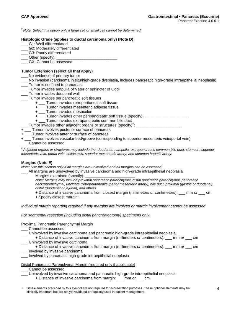
Surgical Pathology Cancer Case Summary Protocol posting date: June 2017 PANCREAS (EXOCRINE): Select a single response unless otherwise indicated. Procedure (Note A) ___ Excisional biopsy (enucleation) ___ Pancreaticoduodenectomy (Whipple resection), partial pancreatectomy ___ Total pancreatectomy ___ Partial pancreatectomy, pancreatic body ___ Partial pancreatectomy, pancreatic tail ___ Other (specify): ____________________________ ___ Not specified Tumor Site (select all that apply) (Note B) ___ Pancreatic head ___ Uncinate process ___ Pancreatic body ___ Pancreatic tail ___ Other (specify): ____________________________ ___ Cannot be determined ___ Not specified Tumor Size Greatest dimension (centimeters): ___ cm + Additional dimensions (centimeters): ___ x ___ cm ___ Cannot be determined (explain): _____________________________ Histologic Type (select all that apply) (Note C) ___ Ductal adenocarcinoma ___ Colloid carcinoma (mucinous noncystic carcinoma) ___ Signet-ring cell carcinoma ___ Adenosquamous carcinoma ___ Intraductal papillary-mucinous neoplasm with an associated invasive carcinoma ___ Intraductal tubulopapillary neoplasm with an associated invasive carcinoma ___ Mucinous cystic neoplasm with an associated invasive carcinoma ___ Large cell neuroendocrine carcinoma ___ Small cell neuroendocrine carcinoma ___ Neuroendocrine carcinoma (poorly differentiated)#
___ Undifferentiated (anaplastic) carcinoma ___ Undifferentiated carcinoma with osteoclast-like giant cells ___ Acinar cell carcinoma ___ Acinar cell cystadenocarcinoma ___ Serous cystadenocarcinoma ___ Mixed acinar-ductal carcinoma ___ Mixed ductal-neuroendocrine carcinoma ___ Mixed acinar-neuroendocrine carcinoma ___ Mixed acinar-neuroendocrine-ductal carcinoma ___ Solid-pseudopapillary neoplasm ___ Hepatoid carcinoma ___ Medullary carcinoma ___ Other histologic type not listed (specify): ____________________________
CAP Approved Gastrointestinal • Pancreas (Exocrine) PancreasExocrine 4.0.0.1
+ Data elements preceded by this symbol are not required for accreditation purposes. These optional elements may be clinically important but are not yet validated or regularly used in patient management.
4
# Note: Select this option only if large cell or small cell cannot be determined. Histologic Grade (applies to ductal carcinoma only) (Note D) ___ G1: Well differentiated ___ G2: Moderately differentiated ___ G3: Poorly differentiated ___ Other (specify): ____________________________ ___ GX: Cannot be assessed Tumor Extension (select all that apply) ___ No evidence of primary tumor ___ No invasion (carcinoma in situ/high-grade dysplasia, includes pancreatic high-grade intraepithelial neoplasia) ___ Tumor is confined to pancreas ___ Tumor invades ampulla of Vater or sphincter of Oddi ___ Tumor invades duodenal wall ___ Tumor invades peripancreatic soft tissues + ___ Tumor invades retroperitoneal soft tissue + ___ Tumor invades mesenteric adipose tissue + ___ Tumor invades mesocolon + ___ Tumor invades other peripancreatic soft tissue (specify): ____________________ + ___ Tumor invades extrapancreatic common bile duct ___ Tumor invades other adjacent organs or structures (specify)#: _____________________ + ___ Tumor involves posterior surface of pancreas + ___ Tumor involves anterior surface of pancreas + ___ Tumor involves vascular bed/groove (corresponding to superior mesenteric vein/portal vein) ___ Cannot be assessed # Adjacent organs or structures may include the. duodenum, ampulla, extrapancreatic common bile duct, stomach, superior mesenteric vein, portal vein, celiac axis, superior mesenteric artery, and common hepatic artery. Margins (Note E) Note: Use this section only if all margins are uninvolved and all margins can be assessed. ___ All margins are uninvolved by invasive carcinoma and high-grade intraepithelial neoplasia
Margins examined (specify): _______________________ Note: Margins may include proximal pancreatic parenchymal, distal pancreatic parenchymal, pancreatic neck/parenchymal, uncinate (retroperitoneal/superior mesenteric artery), bile duct, proximal (gastric or duodenal), distal (duodenal or jejunal), and others. + Distance of invasive carcinoma from closest margin (millimeters or centimeters): ___ mm or ___ cm + Specify closest margin: __________________________
Individual margin reporting required if any margins are involved or margin involvement cannot be assessed For segmental resection (including distal pancreatectomy) specimens only: Proximal Pancreatic Parenchymal Margin ___ Cannot be assessed ___ Uninvolved by invasive carcinoma and pancreatic high-grade intraepithelial neoplasia
+ Distance of invasive carcinoma from margin (millimeters or centimeters): ___ mm or ___ cm ___ Uninvolved by invasive carcinoma
+ Distance of invasive carcinoma from margin (millimeters or centimeters): ___ mm or ___ cm ___ Involved by invasive carcinoma ___ Involved by pancreatic high-grade intraepithelial neoplasia Distal Pancreatic Parenchymal Margin (required only if applicable) ___ Cannot be assessed ___ Uninvolved by invasive carcinoma and pancreatic high-grade intraepithelial neoplasia
+ Distance of invasive carcinoma from margin: ___ mm or ___ cm

CAP Approved Gastrointestinal • Pancreas (Exocrine) PancreasExocrine 4.0.0.1
+ Data elements preceded by this symbol are not required for accreditation purposes. These optional elements may be clinically important but are not yet validated or regularly used in patient management.
5
___ Uninvolved by invasive carcinoma + Distance of invasive carcinoma from margin (millimeters or centimeters): ___ mm or ___ cm
___ Involved by invasive carcinoma ___ Involved by pancreatic high-grade intraepithelial neoplasia Other Margin(s) (required only if applicable) Specify margin(s): _____________________________ ___ Cannot be assessed ___ Uninvolved by invasive carcinoma ___ Involved by invasive carcinoma For enucleation specimens only Pancreatic Parenchymal Margin ___ Cannot be assessed ___ Uninvolved by tumor
+ Distance of tumor from margin (millimeters or centimeters): ___ mm or ___ cm ___ Involved by tumor Other Margin(s) (required only if applicable) Specify margin(s): _____________________________ ___ Cannot be assessed ___ Uninvolved by tumor ___ Involved by tumor For pancreaticoduodenal resection specimens only Pancreatic Neck/Parenchymal Margin ___ Cannot be assessed ___ Uninvolved by invasive carcinoma and pancreatic high-grade intraepithelial neoplasia
+ Distance of invasive carcinoma from margin (millimeters or centimeters): ___ mm or ___ cm ___ Uninvolved by invasive carcinoma
+ Distance of invasive carcinoma from margin (millimeters or centimeters): ___ mm or ___ cm ___ Involved by invasive carcinoma ___ Involved by pancreatic high-grade intraepithelial neoplasia Uncinate (Retroperitoneal/Superior Mesenteric Artery) Margin ___ Cannot be assessed ___ Uninvolved by invasive carcinoma
+ Distance of invasive carcinoma from margin (millimeters or centimeters): ___ mm or ___ cm ___ Involved by invasive carcinoma Bile Duct Margin ___ Cannot be assessed ___ Uninvolved by invasive carcinoma and high-grade intraepithelial neoplasia
+ Distance of invasive carcinoma from margin (millimeters or centimeters): ___ mm or ___ cm ___ Uninvolved by invasive carcinoma
+ Distance of invasive carcinoma from margin (millimeters or centimeters): ___ mm or ___ cm ___ Involved by invasive carcinoma ___ Involved by high-grade intraepithelial neoplasia Proximal Margin (Gastric or Duodenal) ___ Cannot be assessed ___ Uninvolved by invasive carcinoma and high-grade dysplasia ___ Uninvolved by invasive carcinoma ___ Involved by invasive carcinoma

CAP Approved Gastrointestinal • Pancreas (Exocrine) PancreasExocrine 4.0.0.1
+ Data elements preceded by this symbol are not required for accreditation purposes. These optional elements may be clinically important but are not yet validated or regularly used in patient management.
6
___ Involved by high-grade dysplasia Distal Margin (Distal Duodenal or Jejunal) ___ Cannot be assessed ___ Uninvolved by invasive carcinoma and high-grade dysplasia ___ Uninvolved by invasive carcinoma ___ Involved by invasive carcinoma ___ Involved by high-grade dysplasia Other Margin(s) (required only if applicable) Specify margin(s): _____________________________ ___ Cannot be assessed ___ Uninvolved by invasive carcinoma ___ Involved by invasive carcinoma Treatment Effect (Note F) ___ No known presurgical therapy ___ Present + ___ No viable cancer cells (complete response, score 0) + ___ Single cells or rare small groups of cancer cells (near complete response, score 1) + ___ Residual cancer with evident tumor regression, but more than single cells or rare small groups of
cancer cells (partial response, score 2) __ Absent
+ ___ Extensive residual cancer with no evident tumor regression (poor or no response, score 3) ___ Cannot be determined Lymphovascular Invasion (Note G) ___ Not identified ___ Present ___ Cannot be determined Perineural Invasion (Note H) ___ Not identified ___ Present ___ Cannot be determined Regional Lymph Nodes ___ No lymph nodes submitted or found Lymph Node Examination (required only if lymph nodes are present in the specimen) Number of Lymph Nodes Involved: ____ ___ Number cannot be determined (explain): ______________________ Number of Lymph Nodes Examined: ____ ___ Number cannot be determined (explain): ______________________ Pathologic Stage Classification (pTNM, AJCC 8th Edition) (Note I) Note: Reporting of pT, pN, and (when applicable) pM categories is based on information available to the pathologist at the time the report is issued. Only the applicable T, N, or M category is required for reporting; their definitions need not be included in the report. The categories (with modifiers when applicable) can be listed on 1 line or more than 1 line. TNM Descriptors (required only if applicable) (select all that apply) ___ m (multiple primary tumors) ___ r (recurrent)

CAP Approved Gastrointestinal • Pancreas (Exocrine) PancreasExocrine 4.0.0.1
+ Data elements preceded by this symbol are not required for accreditation purposes. These optional elements may be clinically important but are not yet validated or regularly used in patient management.
7
___ y (posttreatment) Primary Tumor (pT)# ___ pTX: Primary tumor cannot be assessed ___ pT0: No evidence of primary tumor ___ pTis: Carcinoma in situ (This includes high-grade pancreatic intraepithelial neoplasia (PanIN-3), intraductal
papillary mucinous neoplasm with high-grade dysplasia, intraductal tubulopapillary neoplasm with high-grade dysplasia, and mucinous cystic neoplasm with high-grade dysplasia)
___ pT1: Tumor ≤2 cm in greatest dimension ___ pT1a: Tumor ≤0.5 cm in greatest dimension ___ pT1b: Tumor >0.5 cm and <1 cm in greatest dimension ___ pT1c: Tumor 1–2 cm in greatest dimension ___ pT2: Tumor >2 cm and ≤4 cm in greatest dimension ___ pT3: Tumor >4 cm in greatest dimension ___ pT4: Tumor involves the celiac axis, superior mesenteric artery, and/or common hepatic artery # Size of invasive component should be used for determining the T category. Regional Lymph Nodes (pN) ___ pNX: Regional lymph nodes cannot be assessed ___ pN0: No regional lymph node metastasis ___ pN1: Metastasis in one to three regional lymph nodes ___ pN2: Metastasis in four or more regional lymph nodes Distant Metastasis (pM) (required only if confirmed pathologically in this case) ___ pM1: Distant metastasis Specify site(s), if known: ____________________________ + Additional Pathologic Findings (select all that apply) (Note J) + ___ None identified + ___ Pancreatic intraepithelial neoplasia (highest grade: PanIN ___) + ___ Chronic pancreatitis + ___ Acute pancreatitis + ___ Other (specify): ____________________________ + Ancillary Studies + Specify: ___________________________________ + Comment(s)

Background Documentation Gastrointestinal • Pancreas (Exocrine) PancreasExocrine 4.0.0.1
8
Explanatory Notes A. Tumors This protocol applies to epithelial tumors of the exocrine pancreas. It excludes endocrine tumors and tumors of the ampulla of Vater. More than 90% to 95% of malignant tumors of the pancreas are exocrine carcinomas. For these tumors, surgical resection remains the only potentially curative approach, and the prognosis is primarily dependent on the anatomic extent of disease and performance status. B. Definition of Location The anatomic subdivisions defining location of tumors of the pancreas (Figure 1) are as follows1: • Tumors of the head of the pancreas are those arising to the right of the left border of the superior mesenteric
vein. The uncinate process is part of the head. • Tumors of the body of the pancreas are those arising between the left border of the superior mesenteric vein
and the left border of the aorta. • Tumors of the tail of the pancreas are those arising between the left border of the aorta and the hilum of the
spleen.
Figure 1. Anatomic subsites of the pancreas. From Greene et al.30 Used with permission of the American Joint Committee on Cancer (AJCC), Chicago, Illinois. The original source for this material is the AJCC Cancer Staging Atlas (2006) published by Springer Science and Business Media LLC, www.springerlink.com. C. Histologic Type A classification of malignant epithelial tumors of the exocrine pancreas recommended by the World Health Organization (WHO) is shown below.2 However, this protocol does not preclude the use of other histologic types or systems of classification. WHO Classification of Epithelial Tumors of the Exocrine Pancreas Malignant Tumors Ductal adenocarcinoma Colloid carcinoma (mucinous noncystic carcinoma) Signet-ring cell carcinoma Adenosquamous carcinoma

Background Documentation Gastrointestinal • Pancreas (Exocrine) PancreasExocrine 4.0.0.1
9
Mucinous cystic neoplasm with an associated invasive carcinoma Intraductal papillary-mucinous neoplam with an associated invasive carcinoma Intraductal tubulopapillary neoplasm with an associated invasive carcinoma Neuroendocrine carcinoma (poorly differentiated) Large cell neuroendocrine carcinoma Small cell neuroendocrine carcinoma Undifferentiated (anaplastic) carcinoma Undifferentiated carcinoma with osteoclast-like giant cells Acinar cell carcinoma Acinar cell cystadenocarcinoma Serous cystadenocarcinoma Mixed acinar-ductal carcinoma Mixed ductal-neuroendocrine carcinoma Mixed acinar-neuroendocrine carcinoma Mixed acinar-neuroendocrine-ductal carcinoma Solid-pseudopapillary neoplasm Hepatoid carcinoma Medullary carcinoma ### These histologic types are not usually graded. By definition, neuroendocrine carcinomas are high grade (grade 3) based on WHO 2010 grading scheme for neuroendocrine neoplasms. Invasive carcinoma with an associated mucinous cystic neoplasm (MCN) can be used if the invasive component is substantial. D. Histopathologic Grade For adenocarcinomas, a histologic grade based on the extent of glandular differentiation is shown below3: Grade X Cannot be assessed Grade 1 Well-differentiated (greater than 95% of tumor composed of glands) Grade 2 Moderately differentiated (50% to 95% of tumor composed of glands) Grade 3 Poorly differentiated (49% or less of tumor composed of glands) Certain histologic subtypes, including acinar cell carcinoma, acinar cell cystadenocarcinoma, serous cystadenocarcinoma, and solid-pseudopapillary neoplasm, are not assigned a grade. By convention, signet-ring cell carcinomas are assigned grade 3. Undifferentiated carcinomas lack morphologic or immunohistochemical evidence of glandular, squamous, or neuroendocrine differentiation. This grading scheme is not applicable to poorly differentiated neuroendocrine carcinomas. For pancreatic ductal carcinoma, histologic grade has been shown to have prognostic significance, with high grade (grade 3) being an unfavorable prognostic factor.3,4 Kloeppel grading scheme uses a combination of glandular differentiation, mucin production, mitoses, and nuclear pleomorphism. No differences in predictive value have been demonstrated in comparisons between the Klöppel grading system and the grading system based on glandular differentiation alone. 4 Other systems based on patterns of infiltration of predominant and secondary tumor patterns have been proposed3 but have not been widely adopted. E. Margins The nonperitonealized surface of the uncinate process (uncinate margin) constitutes the inferior-posterior retroperitoneal margin of pancreaticoduodenectomy specimens (Figure 2) and should be inked; sections through the tumor at its closest approach to this margin should be submitted.5 This margin has also been referred to as retroperitoneal margin and superior mesenteric artery margin.

Background Documentation Gastrointestinal • Pancreas (Exocrine) PancreasExocrine 4.0.0.1
10
Figure 2. Posterior view of tumor arising in the pancreatic head, with dotted line indicating the location of the confluence of the portal and superior mesenteric veins. The hatched area shows the retroperitoneal (uncinate process) margin. From Greene et al.33 Used with permission of the American Joint Committee on Cancer (AJCC), Chicago, Illinois. The original source for this material is the AJCC Cancer Staging Atlas (2006) published by Springer Science and Business Media LLC, www.springerlink.com. Because local recurrences of invasive pancreatic adenocarcinoma arise in the pancreatic bed corresponding to the uncinate margin and vascular groove of, Inking of the vascular groove corresponding to portal and superior mesenteric veins and submission of sections through the tumor at its closest approach to this surface is recommended. Reporting of tumor involvement of anterior and non-uncinate posterior surfaces is recommended, but not required. The vascular groove, anterior surface and the non-uncinate posterior surface are not considered as resection margins.1,5 When dealing with an intraductal tumor, the pancreatic (neck/parenchymal) resection margin and the common bile duct margin (Whipple resection) are the most critical. Complete en face sections through the pancreatic resection margin and the common bile duct margin should be taken.5 The presence of tumor at or within 1 mm of resection margin constitutes a positive margin.6,7 Margin status can be reported as negative (R0, no residual disease), R1 (positive, microscopic residual disease) and R2 (positive, macroscopic residual disease).1 F. Treatment Effect Response of tumor to previous chemotherapy or radiation therapy should be reported. Several scoring systems have been described, and a modified Ryan scheme8 is recommended, as below: Modified Ryan Scheme for Tumor Regression Score8 Description Tumor Regression Score No viable cancer cells (complete response) 0
Single cells or rare small groups of cancer cells (near complete response) 1 Residual cancer with evident tumor regression, but more than single cells or rare small groups of cancer cells (partial response)
2
Extensive residual cancer with no evident tumor regression (poor or no response)
3
Sizable pools of acellular mucin may be present after chemoradiation but should not be interpreted as representing residual tumor. The size of the viable tumor should be used to assign the ypT category, and requires a combined assessment of gross and microscopic findings. Multiple foci of viable tumor within the same tumor mass can be added to obtain the maximum linear dimension for staging.
Background Documentation Gastrointestinal • Pancreas (Exocrine) PancreasExocrine 4.0.0.1
11
This protocol does not preclude the use of other systems for assessment of tumor response,.9,10 A modification of the above scoring scheme into a 3-tier scheme has been shown to correlate better with outcome: no residual carcinoma (grade 0), minimal residual carcinoma defined as single cells or small groups of cancer cells, <5% residual carcinoma (grade 1), 5% or more residual carcinoma (grade 2). 11,12 G. Venous/Lymphatic Vessel Invasion Venous as well as lymphatic (small vessel) invasion has been shown to be an adverse prognostic factor.13,14 H. Perineural Invasion Perineural invasion has been shown to be an adverse prognostic factor. 14,15 I. Pathologic Stage Classification The TNM staging system for carcinoma of the exocrine pancreas of the American Joint Committee on Cancer (AJCC) and the International Union Against Cancer (UICC) is recommended and shown below.1 The postresection prognosis of a patient with pancreatic carcinoma is primarily determined by the anatomic extent of disease as defined by the TNM stage groupings. According to AJCC/UICC convention, the designation “T” refers to a primary tumor that has not been previously treated. The symbol “p” refers to the pathologic classification of the TNM, as opposed to the clinical classification, and is based on gross and microscopic examination. pT entails a resection of the primary tumor or biopsy adequate to evaluate the highest pT category, pN entails removal of nodes adequate to validate lymph node metastasis, and pM implies microscopic examination of distant lesions. Clinical classification (cTNM) is usually carried out by the referring physician before treatment during initial evaluation of the patient or when pathologic classification is not possible. Pathologic staging is usually performed after surgical resection of the primary tumor. Pathologic staging depends on pathologic documentation of the anatomic extent of disease, whether or not the primary tumor has been completely removed. If a biopsied tumor is not resected for any reason (eg, when technically infeasible) and if the highest T and N categories or the M1 category of the tumor can be confirmed microscopically, the criteria for pathologic classification and staging have been satisfied without total removal of the primary cancer. TNM Descriptors For identification of special cases of TNM or pTNM classifications, the “m” suffix and “y,” “r,” and “a” prefixes are used. Although they do not affect the stage grouping, they indicate cases needing separate analysis. The “m” suffix indicates the presence of multiple primary tumors in a single site and is recorded in parentheses: pT(m)NM. The “y” prefix indicates those cases in which classification is performed during or after initial multimodality therapy (ie, neoadjuvant chemotherapy, radiation therapy, or both chemotherapy and radiation therapy). The cTNM or pTNM category is identified by a “y” prefix. The ycTNM or ypTNM categorizes the extent of tumor actually present at the time of that examination. The “y” categorization is not an estimate of tumor before multimodality therapy (ie, before initiation of neoadjuvant therapy). The “r” prefix indicates a recurrent tumor when staged after a documented disease-free interval and is identified by the “r” prefix: rTNM. The “a” prefix designates the stage determined at autopsy: aTNM. Vessel Invasion According to AJCC/UICC convention, vessel invasion (lymphatic or venous) does not affect the T category indicating local extent of tumor unless specifically included in the definition of a T category. T Category Considerations (Figures 3 and 4) If more than 1 tumor is present in the pancreas, the tumor with the highest T category should be classified according to the pT definitions and either the multiplicity (“m”) or the actual number of simultaneous multiple

Background Documentation Gastrointestinal • Pancreas (Exocrine) PancreasExocrine 4.0.0.1
12
tumors (eg, “3”) should be indicated in parentheses after the T category of the primary tumor (eg, pT3[m] or pT3[2]).
This applies only to grossly recognizable, synchronous primary carcinomas and not to a single, grossly detected tumor with multiple separate microscopic foci.14 Tis includes high-grade pancreatic intraepithelial neoplasia (PanIn-3), intraductal papillary mucinous neoplasm with high-grade dysplasia, intraductal tubulopapillary neoplasm with high-grade dysplasia and mucinous cystic neoplasm with high-grade dysplasia. The T categories T1-T3 are defined by tumor size as it provides better prognostic stratification than classification based on extension into peripancreatic tissue. 17-21 Tumor size is determined by measurement of the gross lesion and should be corroborated on microscopic assessment. For invasive carcinoma associated with intraductal papillary mucinous neoplasms, intraductal tubulopapillary neoplasms and mucinous cystic neoplasms, only the size of the invasive component should be used to determine the T category. The synoptic report is not required for intraductal papillary mucinous neoplasms, intraductal tubulopapillary neoplasms and mucinous cystic neoplasms in the absence of an invasive component. The invasive portion in these cases can be multifocal. It is currently not clear whether size of the largest tumor focus or combined size of all invasive foci determines tumor outcome. Both measurements can be included in the pathology report, and the the maximum linear dimension of the largest invasive focus is used for staging. Extension beyond the pancreas may include invasion of peripancreatic soft tissue, peritoneum (including mesocolon, greater/lesser omentum), extrapancreatic biliary system, and/or duodenum (including the ampulla of Vater) for pancreatic head tumors, while stomach, spleen, left adrenal, and peritoneum can be involved by direct extension of body/tail tumors. Tumor extension in these areas does not affect staging, but should be noted in the pathology report. Invasion of the portal vein does not affect staging, but has been shown to be an independent prognostic factor.22 T4 tumors are characterized by involvement of superior mesenteric artery, celiac axis and/or common hepatic artery. In most instances, these tumors are considered unresectable and hence T4 category is determined by radiologic studies and is not usually assigned by pathologists.
Figure 3. T1 (left of dotted line) is defined as tumor measuring 2 cm or less in greatest dimension and limited to the pancreas. T2 (right of dotted line) is defined as tumor measuring more than 2 cm in greatest dimension and less than 4 cm in greatest dimension. From Greene et al.33 Used with permission of the American Joint Committee on Cancer (AJCC), Chicago, Illinois. The original source for this material is the AJCC Cancer Staging Atlas (2006) published by Springer Science and Business Media LLC, www.springerlink.com.

Background Documentation Gastrointestinal • Pancreas (Exocrine) PancreasExocrine 4.0.0.1
13
Figure 4. T4 tumor involves the celiac axis (above dotted line) or the superior mesenteric artery (below dotted line). T4 tumors are considered unresectable and are rarely encountered in surgical pathology specimens. From Greene et al.33 Used with permission of the American Joint Committee on Cancer (AJCC), Chicago, Illinois. The original source for this material is the AJCC Cancer Staging Atlas (2006) published by Springer Science and Business Media LLC, www.springerlink.com. N Category Considerations The regional lymph nodes for head and neck cancers include lymph nodes along common bile duct, common hepatic artery, portal vein, pyloric, anterior and posterior pancreaticoduodenal arcades, superior mesenteric vein and right lateral wall of superior mesenteric artery (Figures 5 and 6). The regional lymph nodes for the pancreatic body and tail cancers include lymph nodes along common hepatic artery, celiac axis, splenic artery, and splenic hilum. Tumor involvement of other nodal groups is considered distant metastasis. Anatomic division of lymph nodes is not necessary, but separately submitted lymph nodes should be individually reported. Lymph node metastasis is an independent adverse prognostic factor.13,17,19,23-25 Microscopic evaluation of at least 12 lymph nodes is recommended for Whipple resections.26,27 Based on outcome data, tumors with positive lymph nodes are now categorized as N1 or N2.28,29
Figure 5. Regional lymph nodes of the pancreas (anterior view). From Greene et al.33 Used with permission of the American Joint Committee on Cancer (AJCC), Chicago, Illinois. The original source for this material is the AJCC Cancer Staging Atlas (2006) published by Springer Science and Business Media LLC, www.springerlink.com.

Background Documentation Gastrointestinal • Pancreas (Exocrine) PancreasExocrine 4.0.0.1
14
Figure 6. Regional lymph nodes of the pancreas (anterior view with pancreatic body removed to reveal retroperitoneal vessels and lymph nodes). From Greene et al.32 Used with permission of the American Joint Committee on Cancer (AJCC), Chicago, Illinois. The original source for this material is the AJCC Cancer Staging Atlas (2006) published by Springer Science and Business Media LLC, www.springerlink.com. M Category Considerations Peritoneal seeding or positive peritoneal cytology is considered M1.1,16 J. Additional Pathologic Findings Pancreatic Intraepithelial Neoplasia (PanIN) Noninvasive lesions of the ductal epithelium often are found in the pancreatic parenchyma surrounding ductal adenocarcinoma. These lesions are collectively known as pancreatic intraepithelial neoplasia (PanIN). PanINs were previously classified into 3 grades.30 The most recent consensus recommends a 2-tier grading scheme for better reproducibility and for better alignment of the grades with treatment options.31 A similar 2-tier scheme is recommended for noninvasive MCN and intraductal papillary mucinous neoplasm (IPMN).32 Normal Nonmucinous flattened or cuboidal epithelium without dysplasia PanIN, low grade Includes flat mucinous epithelium without dysplasia (PanIN-1A), papillary mucinous
epithelium without dysplasia (PanIN-1B) and flat or papillary mucinous epithelium with mild-to-moderate dysplasia featuring mild-to-moderate nuclear irregularity, hyperchromasia, and loss of polarity (PanIN-2)
PanIN, high grade Flat or papillary mucinous epithelium with severe dysplasia (marked nuclear irregularity, hyperchromasia, and loss of polarity), often with cribriforming and intraluminal blebbing (budding off of noncohesive cells), corresponds to carcinoma in situ
High-grade PanIN at the resection margins of an otherwise completely resected malignancy should be noted in the pathology report. In this setting, the biologic significance of PanIN of any grade remains unclear. The presence of dysplasia at the margin of a noninvasive IPMN is also uncertain. The highest grade even if focal determines the final grade. For IPMN and MCN, the extent of high-grade dysplasia can be recorded, but does not currently have clinical relevance. Other Findings In addition to the examination of other tissues and organs that are part of pancreaticoduodenectomy specimens, pathologic evaluation may also include examination of the gastric antrum for gastritis (eg, Helicobacter pylori gastritis or chemical gastritis) and the duodenum for duodenitis, peptic ulcer disease, and ampullitis.

Background Documentation Gastrointestinal • Pancreas (Exocrine) PancreasExocrine 4.0.0.1
15
References 1. Amin MB, Edge SB, Greene FL, et al, eds. AJCC Cancer Staging Manual. 8th ed. New York, NY: Springer;
2017. 2. Bosman FT, Carneiro F, Hruban RH, Theise ND, eds. WHO Classification of Tumours of the Digestive
System. Geneva, Switzerland: WHO Press; 2010. 3. Adsay NV, Basturk O, Bonnett M, et al. A proposal for a new and more practical grading scheme for
pancreatic ductal adenocarcinoma. Am J Surg Pathol. 2005;29(6):724-733. 4. Giulianotti PC, Boggi U, Fornaciari G, et al. Prognostic value of histological grading in ductal adenocarcinoma
of the pancreas: Kloppel vs TNM grading. Int J Pancreatol. 1995;17(3):279-289. 5. Adsay NV, Basturk O, Saka B, et al. Whipple made simple for surgical pathologists: orientation, dissection,
and sampling of pancreaticoduodenectomy specimens for a more practical and accurate evaluation of pancreatic, distal common bile duct, and ampullary tumors. Am J Surg Pathol. 2014;38(4):480-493.
6. Campbell F, Smith RA, Whelan P, et al. Classification of R1 resections for pancreatic cancer: the prognostic relevance of tumour involvement within 1 mm of a resection margin. Histopathol. 2009;55(3):277-283.
7. Verbeke CS, Menon KV. Redefining resection margin status in pancreatic cancer. HPB. 2009;11(4):282-289. 8. Ryan R, Gibbons D, Hyland JMP, et al. Pathological response following long-course neoadjuvant
chemoradiotherapy for locally advanced rectal cancer. Histopathology. 2005;47:141-146. 9. Evans DB, Rich TA, Byrd DR, et al. Preoperative chemoradiation and pancreaticoduodenectomy for
adenocarcinoma of the pancreas. Arch Surg. 1992;127:1335-1339. 10. Breslin TM, Hess KR, Harbison DB, et al. Neoadjuvant chemoradiotherapy for adenocarcinoma of the
pancreas: treatment variables and survival duration. Ann Surg Oncol. 2001;8(2):123-132. 11. Chatterjee D, Katz MH, Rashid A, et al. Histologic grading of the extent of residual carcinoma following
neoadjuvant chemoradiation in pancreatic ductal adenocarcinoma: a predictor for patient outcome. Cancer. 2012;118(12):3182-3190.
12. Lee SM, Katz MH, Liu L, et al. Validation of a proposed tumor regression grading scheme for pancreatic ductal adenocarcinoma after neoadjuvant therapy as a prognostic indicator for survival. Am J Surg Pathol. 2016;40(12):1653-1660.
13. Garcea G, Dennison AR, Ong SL, et al. Tumour characteristics predictive of survival following resection for ductal adenocarcinoma of the head of pancreas. Eur J Surg Oncol. 2007;33(7):892-897.
14. Chen JW, Bhandari M, Astill DS, et al. Predicting patient survival after pancreaticoduodenectomy for malignancy: histopathological criteria based on perineural infiltration and lymphovascular invasion. HPB (Oxford). 2010;12(2):101-108.
15. Chatterjee D, Katz MH, Rashid A, et al. Perineural and intra-neural invasion in posttherapy pancreaticoduodenectomy specimens predicts poor prognosis in patients with pancreatic ductal adenocarcinoma. Am J Surg Pathol. 2012;36(3):409.
16. Wittekind C, Greene FL, Hutter RVP, Sobin LH, Henson DE, eds. TNM Supplement: A Commentary on Uniform Use. 3rd ed. New York, NY: Wiley-Liss; 2003.
17. Lim JE, Chien MW, Earle CC. Prognostic factors following curative resection for pancreatic adenocarcinoma: a population-based, linked database analysis of 396 patients. Ann Surg. 2003;237(1):74-85.
18. Matsumoto G, Muta M, Tsuruta K, Horiguchi S, Karasawa K, Okamoto A. Tumor size significantly correlates with postoperative liver metastases and COX-2 expression in patients with resectable pancreatic cancer. Pancreatology. 2007;7(2-3):167-173.
19. Moon HJ, An JY, Heo JS, Choi SH, Joh JW, Kim YI. Predicting survival after surgical resection for pancreatic ductal adenocarcinoma. Pancreas. 2006;32(1):37-43.
20. Saka B, Balci S, Basturk O, et al. Pancreatic ductal adenocarcinoma is spread to the peripancreatic soft tissue in the majority of resected cases, rendering the AJCC T-stage protocol (7th Edition) inapplicable and insignificant: a size-based staging system (pT1: ≤2, pT2: >2-≤4, pT3: >4 cm) is more valid and clinically relevant. Ann Surg Oncol. 2016;23(6):2010-2018.
21. Allen PJ, Kuk D, Castillo CF, et al. Multi-institutional validation study of the American Joint Commission on Cancer (8th Edition) changes for T and N staging in patients with pancreatic adenocarcinoma. Ann Surg. 2017;265(1):185-191.
22. Nakagohri T, Kinoshita T, Konishi M, Inoue K, Takahashi S. Survival benefits of portal vein resection for pancreatic cancer. Am J Surg. 2003;186(2):149-153.
23. Geer RJ, Brennan MF. Prognostic indicators for survival after resection of pancreatic adenocarcinoma. Am J Surg. 1993;165(1):68-73.

Background Documentation Gastrointestinal • Pancreas (Exocrine) PancreasExocrine 4.0.0.1
16
24. House MG, Gonen M, Jarnagin WR, et al. Prognostic significance of pathologic nodal status in patients with resected pancreatic cancer. J Gastrointest Surg. 2007;11(11):1549-1555.
25. Pawlik TM, Gleisner AL, Cameron JL, et al. Prognostic relevance of lymph node ratio following pancreaticoduodenectomy for pancreatic cancer. Surgery. 2007;141(5):610-618.
26. Tomlinson JS, Jain S, Bentrem DJ, et al. Accuracy of staging node-negative pancreas cancer: a potential quality measure. Arch Surg. 2007;142(8):767-773; discussion 773-774.
27. Schwarz RE, Smith DD. Extent of lymph node retrieval and pancreatic cancer survival: information from a large US population database. Ann Surg Oncol 2006;13(9):1189-1200.
28. Strobel O, Hinz U, Gluth A, et al. Pancreatic adenocarcinoma: number of positive nodes allows to distinguish several N categories. Ann Surg. 2015;261(5):961-969.
29. Olca B, Burcu S, Serdar B, et al. Substaging of lymph node status in resected pancreatic ductal adenocarcinoma has strong prognostic correlations: proposal for a revised N classification for TNM staging. Ann Surg Oncol. 2015;22:1187-1195.
30. Hruban RHMD, Adsay NVMD, Albores-Saavedra JMD, et al. Pancreatic intraepithelial neoplasia: a new nomenclature and classification system for pancreatic duct lesions. Am J Surg Pathol. 2001;25:579-586.
31. Basturk O, Hong SM, Wood LD, et al. A revised classification system and recommendations from the Baltimore consensus meeting for neoplastic precursor lesions in the pancreas. Am J Surg Pathol. 2015;39:1730-1741.
32. Adsay V, Mino-Kenudson M, Furukawa T, et al; Members of Verona Consensus Meeting, 2013. Pathologic evaluation and reporting of intraductal papillary mucinous neoplasms of the pancreas and other tumoralintraepithelial neoplasms of pancreatobiliary tract: recommendations of Verona Consensus Meeting. Ann Surg. 2016;263(1):162-177.
33. Greene FL, Compton, CC, Fritz AG, et al, eds. AJCC Cancer Staging Atlas. New York, NY: Springer; 2006.