original article calcium signaling is implicated in the ... · original article calcium signaling...
TRANSCRIPT
Int J Clin Exp Pathol 2015;8(5):4388-4397www.ijcep.com /ISSN:1936-2625/IJCEP0007238
Original Article Calcium signaling is implicated in the diffuse axonal injury of brain stem
Jiao Mu1,2, Yucheng Song3, Ji Zhang1, Wei Lin1, Hongmei Dong1
1Department of Forensic Medicine, Huazhong University of Science and Technology, Tongji Medical College, No. 13 Hangkong Lu, Hankou, Wuhan 430030, Hubei, PR China; 2Department of Pathology, Hebei North University, No. 11 Zuanshinan Lu, Zhangjiakou 075000, Hebei, PR China; 3Criminal Investigation Brigade of Taicang, No. 128 Loujiangbei Lu, Taicang, Suzhou 215400, Jiangsu, PR China
Received February 21, 2015; Accepted April 13, 2015; Epub May 1, 2015; Published May 15, 2015
Abstract: Evaluating diffuse axonal injury (DAI) remains challenging in clinical sciences since the physiopathologic mechanism of DAI is still unclear. The calcium overload in the axoplasm is considered to be crucial for secondary axonal injury. The present study use calcium channel blocker, nimodipine, to explore the influence of Ca2+ in the pathogenesis of rat DAI. In the DAI group, the expressions of β-APP and NF-L in axons were increased from 12 to 72 h. The ultrastructural observation indicated the axon and vessel injury appeared at 12 h post-injury and severely aggravated from 24 to 72 h. The expression of vWF and brain water content was increased at 12 h after injury and further increased at 24 h. Nimodipine decreased the expression of β-APP, NF-L and vWF, and also attenuated the ul-trastructural damage of vascular wall and axons. Furthermore, Ca-dependent enzyme, the calcineurin activity were increased in DAI and nimodipine suppressed the activity of calcineurins (CaN). However, the amount of CaN expres-sion was not changed. Our results showed that disturbances of axonal calcium homeostasis play an important role in the secondary damage of the axon, neuron and capillary vessel which may be related with activating CaN during the acute phase of DAI. Nimodipine can alleviate the secondary damage by suppressing the calcineurin activity.
Keywords: Diffuse axonal injury, calcium signal, nimodipine, animal study
Introduction
Traumatic brain injury (TBI) leading to death or hospitalization is estimated to afflict over 10 million people each year worldwide. It is pre-dicted that TBI will surpass many diseases as the major cause of death and disability by the year 2020 [1]. Diffuse axonal injury (DAI) is one of the most common and important pathologic features of TBI [2]. DAI encompasses a spec-trum of pathological changes including axo-plasmic transport interruption, proteolysis, cell swelling, breaking of the axonal cytoskeleton and tear of the small blood vessels [3]. Clinically, there aren’t characteristic images for DAI, and most patients only have a normal imaging or minimal changes such as small petechial hem-orrhages within the white matter at first [4]. The histopathological identification of DAI at post-mortem is dependent upon the existence of axonal varicosities and axonal retraction ball,
which is easily neglected and leads to missed diagnosis. Although the immunostaining for beta-amyloid precursor protein (β-APP) was regarded as sensitive method for diagnosis, it can also be expressed in axons in other neuro-logical disorders including hematomas and infarcts [5]. The reason why the diagnosis of DAI remains challenging in clinical sciences is that the physiopathologic mechanism of DAI is still unclear.
DAI is not only caused by primary axotomy from mechanical forces, but also from secondary axotomy due to a series of biochemical events after initial injury [6]. The intracellular accumu-lation of free calcium in the axoplasm is consid-ered to be crucial for secondary axonal injury. [7, 8]. By using calcium imaging technique, cal-cium accumulation had been showed in injury axons 2 h after injury and persisting over 48 h [9]. Moreover, calcium overload initiates a cas-
Preliminary study of calcium signal in DAI
4389 Int J Clin Exp Pathol 2015;8(5):4388-4397
cade of Ca-dependent enzymies and the sub-sequent biochemical responses which resulted in pathological changes [10]. A large amount of literatures have displayed that Ca-dependent enzyme, such as calcineurins and calpain, could impair or degrade a broad spectrum of proteins, including cytoskeletal proteins (e.g. spectrin, neurofilament, actin, tubulin) and the major myelin proteins [11]. At last, cytoskeletal dissolution, focal axoplasmic perturbation and blood-brain barrier damage, even cell death occurred.
In the present study, we used nimodipine, L-type calcium channel blocker, to explore the influence of Ca2+ in the pathogenesis and mechanism of rat DAI by observing the activity and content of calcineurin, the injury of the axon and capillary vessel, the water content, and the ultrastructure in the brain stem. Simultaneously, we hope to explore the poten-tial diagnosis significance of some indicators for DAI.
Materials and methods
Animal model
All procedures involving animals were approved and monitored by the Animal Care Committee of Tongji Medical College of Huazhong University of Science and Technology. Sprague-Dawley rats (male and female) weighting 220 to 240 g, were randomly divided into the control group; DAI group and ND group (the DAI+ nimodipine group). DAI model was made by linear accelera-tion load referring to Marmarou method [12]. In the DAI group, the rats were anaesthetized by intraperitoneal injection of 0.4% pentobarbital sodium (30 mg/kg). The scalp of the anesthetic rat was shaved, a midline incision was per-formed, and the periosteum covering the vertex was exposed. A steel disk of 10 mm in diameter and 3 mm in thickness was fixed at the center of vertex. Subsequently, rats were prostrated and fixed onto a sponge bed. Injury was deliv-ered by dropping a 500 g weight freely onto the coin from a height of 1 m. Then, the rat was immediately removed and the scalp was sutured after gently removing the steel disk from the skull. In the control group, rats under-went the surgical procedure without impact. The ND group: DAI rat was induced by intragas-tric administration of nimodipine (5.4 mg/kg) 20 minutes before hit and three times a day after impact (every 8 h) until it was sacrificed.
The rats were sacrificed at 12 h, 24 h, and 72 h after hit.
Histology and immunohistochemistry
The brain were sampled and bisected along the sagittal fissure into two equal. The half-brain-stem were immersed in 4% paraformaldehyde for 24 h, and then to undertake dehydration, transparent, and embedding. The paraffin-embedded tissues were cut using microtome to provide 5 μm thickness tissue sections for VWF immunohistochemical stain (1:100, Boster, Wuhan, China).
The other half- brainstem were fixed with 4% paraformaldehyde for 24 h and then immersed in 25% sucrose and embedded in an optimal cutting tool. The sections (14-μm-thickness) were taken using a cryotome and mounted on gelatin-covered microscope slides for β-APP immunohistochemical stain (1:4000, Abcam, Cambridge, UK) and NF-L immunofluorescence stain (1:50, Boster, Wuhan, China).
Transmission electronic microscopy
The rats were deeply anesthetized with 10% chloral hydrate and perfused through the heart with 2% paraformaldehyde and 0.4% glutaral-dehyde in 0.1 M phosphate buffer. The brain-stem were trimmed into blocks of 2 × 1 × 0.5 mm and conventionally fixed, rinsed, dehydrat-ed, and embedded in epoxy resin. The samples were then cut into 70-nm-thick sections, stained with uranyl acetate and bismuth subni-trite, and examined by Transmission electronic microscope.
Evaluation of brain edema
Brain edema was measured by the wet and dry weight method as described previously [13]. Briefly, brains were quickly removed, the wet weight of hemispheres were measured. The tis-sues were then dried in an oven at 85°C for 24 h, and weighed again to obtain the dry weight. The water content of each hemisphere was cal-culated using the following formula: water con-tent = [(wet weight-dry weight)/wet weight] × 100%.
CaN phosphatase activity
CaN phosphatase activity was measured using a CaN cellular activity assay kit (Genmed, Shanghai, China) according to the manufactur-
Preliminary study of calcium signal in DAI
4390 Int J Clin Exp Pathol 2015;8(5):4388-4397
er’s directions. The RII phosphopetide was used as a highly specific substrate for CaN. The detection of free inorganic phosphate released from RII by CaN was based on the ferrous sul-fate method. After homogenates of the brain-stem were incubated at 30°C for 10 min, the reactions were terminated, and the optical den-sity at 660 nm was determined for each sam-ple using a microplate reader (Dyne Technology). The amount of free phosphate was determined using a phosphate standard curve. The activity was normalized by protein concentration, which was determined by BCA method. A final activity of CaN was obtained according to the following formula:
CaN activity per sample (nmol per mg per min) = {[(total phosphate conc. - background phos-phate conc.) × 10] × sample dilution}/10/sam-ple protein concentration.
Western blot analysis
The homogenates of the brainstem were resolved on SDS PAGE and transferred to a
nitrocellulose membrane using the Geni blot system. The membrane was blocked with 5% milk in Tris-buffered saline+tween-20 (TBST) for 2 h at room temperature and then incubat-ed with the Anti-CaN antibody (1:300) overnight at 4°C. After washing 3 times for 5 min with TBST, the membrane was incubated with a sec-ondary antibody (1:6000) for 60 min at room temperature. After washing, blots were devel-oped with a solution containing 10 ml PBS, 0.025% (v/v) H2O2, and 8 mg 4-chloro-1-napthol dissolved in 2 ml of methanol. Specific immu-noreactive bands were quantified by computer-assisted densitometry.
Statistical analysis
For all the experimental data, we used Prizm 5.0 (GraphPad Software Inc., 108 La Jolla, CA, USA) to perform a one-way analysis of variance (ANOVA). P values <0.05 were considered sta-tistically significant. The data are expressed as mean ± standard deviation.
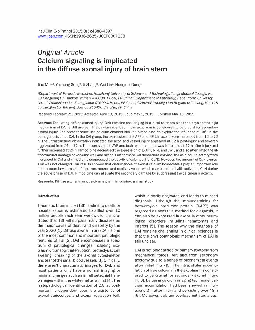
Figure 1. Effect of nimodipine on the expression of β-APP in the brain-stem. No β-APP expression in the control group (C); β-APP expression was increased in the axons of DAI following 24-72 h post-injury (A1-3); The expression of β-APP was decreased in the ND group compared to the DAI group at the same time point (B1-3).
Preliminary study of calcium signal in DAI
4391 Int J Clin Exp Pathol 2015;8(5):4388-4397
Results
General physiological findings
The injured rats were presented with shallow breathing, severe decortication flexion defor-mity of the forelimbs, with loss of muscle tone after impact immediately and maintained an unconscious state for 45 min. After recovery of consciousness, the DAI rats were slow-moving and had a sluggish response to stimuli over hours after injury. By comparison, the control rats recovering from anesthesia appeared bright, alert, and responsive to stimuli within 30 min.
Expression of β-APP and NF-L
Axonal injury was observed by β-APP immuno-histochemical stain. No β-APP-stained axons were found in the brainstem of the control rats. At 12 h after injury, β-APP-stained axon was presented with dot-like or swellings profiles, and markedly increased at 24-72 h post-injury. Meanwhile, the expression of β-APP in axons
were decreased in the ND group compared to the DAI group at the same time point (Figure 1).
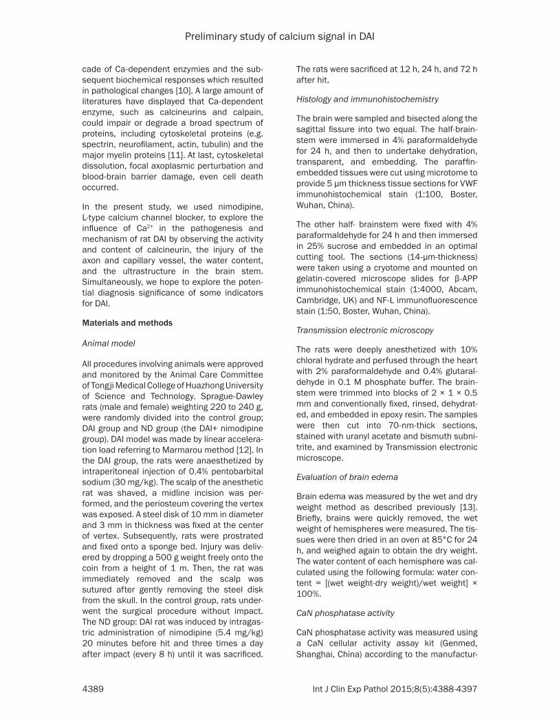
The NF-L expression of brainstem sections were further used to observe the axonal dam-age. In the control group, there was no NF-L staining in the brainstem. While in the DAI group, the number of NF-L stained axons were increased from 12 to 72 h. Meanwhile, the number of NF-L positive axons was decreased in ND group at the same time compared with DAI group (Figure 2).
Ultrastructural change
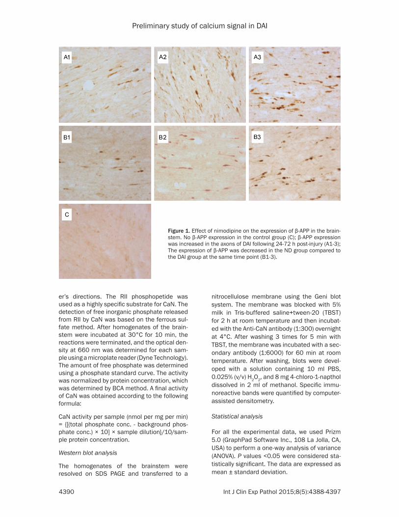
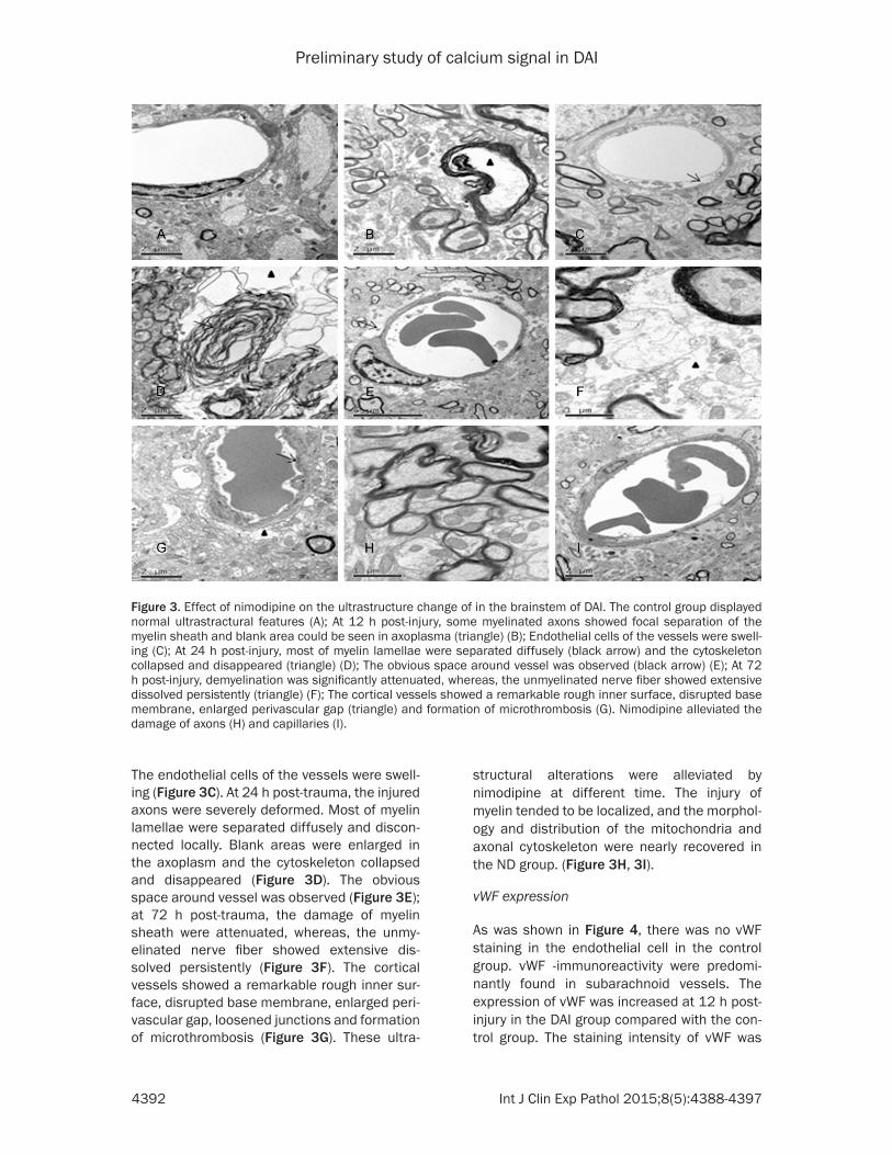
The myelin lamellae in the brainstem of the control rat were stuck to each other closely, axolemma adhered tightly to the inner layer of myelin sheath, and the intra-axonal organells such as mitochondria, microtubes and neuro-filaments were distributed evenly (Figure 3A). At 12 h post-trauma, the microtubes and neuro-filaments had irregular arrangement and blank areas occurred in the axoplasm. Myelin sheath was separated at different degrees (Figure 3B).
Figure 2. Effect of nimodipine on the expression of NF-L in the brain-stem. No NF-L expression in the control group (G); NF-L expression was increased from 12 to 72 h in the DAI group (A-C). The expression of NF-L was decreased in the ND group compared to the DAI group at the same time (D-F).
Preliminary study of calcium signal in DAI
4392 Int J Clin Exp Pathol 2015;8(5):4388-4397
The endothelial cells of the vessels were swell-ing (Figure 3C). At 24 h post-trauma, the injured axons were severely deformed. Most of myelin lamellae were separated diffusely and discon-nected locally. Blank areas were enlarged in the axoplasm and the cytoskeleton collapsed and disappeared (Figure 3D). The obvious space around vessel was observed (Figure 3E); at 72 h post-trauma, the damage of myelin sheath were attenuated, whereas, the unmy-elinated nerve fiber showed extensive dis-solved persistently (Figure 3F). The cortical vessels showed a remarkable rough inner sur-face, disrupted base membrane, enlarged peri-vascular gap, loosened junctions and formation of microthrombosis (Figure 3G). These ultra-
structural alterations were alleviated by nimodipine at different time. The injury of myelin tended to be localized, and the morphol-ogy and distribution of the mitochondria and axonal cytoskeleton were nearly recovered in the ND group. (Figure 3H, 3I).
vWF expression
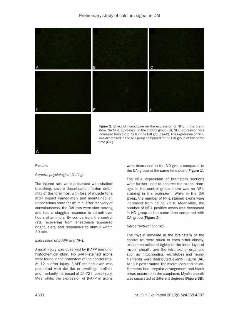
As was shown in Figure 4, there was no vWF staining in the endothelial cell in the control group. vWF -immunoreactivity were predomi-nantly found in subarachnoid vessels. The expression of vWF was increased at 12 h post-injury in the DAI group compared with the con-trol group. The staining intensity of vWF was
Figure 3. Effect of nimodipine on the ultrastructure change of in the brainstem of DAI. The control group displayed normal ultrastractural features (A); At 12 h post-injury, some myelinated axons showed focal separation of the myelin sheath and blank area could be seen in axoplasma (triangle) (B); Endothelial cells of the vessels were swell-ing (C); At 24 h post-injury, most of myelin lamellae were separated diffusely (black arrow) and the cytoskeleton collapsed and disappeared (triangle) (D); The obvious space around vessel was observed (black arrow) (E); At 72 h post-injury, demyelination was significantly attenuated, whereas, the unmyelinated nerve fiber showed extensive dissolved persistently (triangle) (F); The cortical vessels showed a remarkable rough inner surface, disrupted base membrane, enlarged perivascular gap (triangle) and formation of microthrombosis (G). Nimodipine alleviated the damage of axons (H) and capillaries (I).

Preliminary study of calcium signal in DAI
4393 Int J Clin Exp Pathol 2015;8(5):4388-4397
further increased at 24 h post-injury and decreased at 72 h. The expression of vWF in the ND group was decreased than that in the DAI group. However, the expression of vWF was extremely weak in the small vessels within the brain parenchyma.
Water content
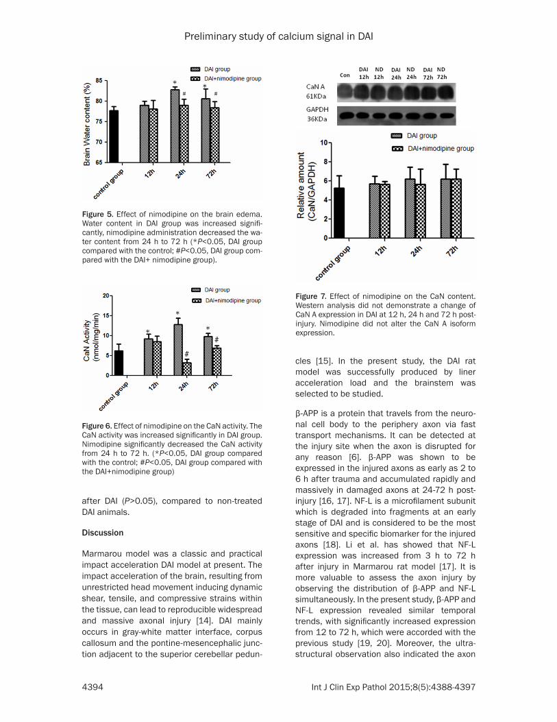
The brain water content was slightly increased at 12 h after injury compared with the control group, but no significant difference was found (79.06±0.93% versus 77.70±1.06%, P>0.05). It was further increased at 24 h and decreased at 72 h (24 h: 82.81±0.71%, 72 h: 80.57±2.40% versus 77.70±1.06%, P<0.05). Nimodipine administration decreased the water content at 24 h (79.06±1.46% versus 82.81±0.71%, P<0.05) and 72 h (78.44±1.43% versus 80.57±2.40%, P<0.05) but not 12 h (78.07± 2.12% versus 79.06±0.93%, P>0.05) com-pared to the DAI group (Figure 5).
CaN activity
At 12 h post-injury, a significant increase in CaN activity was observed (9.18±1.26 versus 6.18±1.72, P<0.05), CaN activity was further increased at 24h post-injury (24 h: 12.79±1.65, 72 h: 9.77±0.85, versus 6.18±1.72, P<0.05). The administration of nimodipine significantly decreased the CaN activity from 24 h (3.18±0.91 versus 12.79±1.65, P<0.05) to 72 h (6.91±0.59 versus 9.77±0.85, P<0.05) after injury, but not 12 h (8.48±1.46 versus 9.18±1.26, P>0.05) (Figure 6).
CaN amount
As was shown in Figure 7, there was no signifi-cant difference on the expression of CaN A iso-form at 12 h, 24 h and 72 h post-injury com-pared with the control (P>0.05). Additionally, the administration of nimodipine can not alter the CaN A isoform expression from 12 h to 72 h
Figure 4. Effect of nimodipine on the expression of vWF. There was no vWF staining in the endothelial cell in the control group (G). The stain-ing intensity of vWF was increased at 12 h (A), further increased at 24 h (B) and decreased at 72 h (C) after injury. The expression of vWF in the ND group was decreased compared to that in the DAI group at the same time (D-F).
Preliminary study of calcium signal in DAI
4394 Int J Clin Exp Pathol 2015;8(5):4388-4397
after DAI (P>0.05), compared to non-treated DAI animals.
Discussion
Marmarou model was a classic and practical impact acceleration DAI model at present. The impact acceleration of the brain, resulting from unrestricted head movement inducing dynamic shear, tensile, and compressive strains within the tissue, can lead to reproducible widespread and massive axonal injury [14]. DAI mainly occurs in gray-white matter interface, corpus callosum and the pontine-mesencephalic junc-tion adjacent to the superior cerebellar pedun-
cles [15]. In the present study, the DAI rat model was successfully produced by liner acceleration load and the brainstem was selected to be studied.
β-APP is a protein that travels from the neuro-nal cell body to the periphery axon via fast transport mechanisms. It can be detected at the injury site when the axon is disrupted for any reason [6]. β-APP was shown to be expressed in the injured axons as early as 2 to 6 h after trauma and accumulated rapidly and massively in damaged axons at 24-72 h post-injury [16, 17]. NF-L is a microfilament subunit which is degraded into fragments at an early stage of DAI and is considered to be the most sensitive and specific biomarker for the injured axons [18]. Li et al. has showed that NF-L expression was increased from 3 h to 72 h after injury in Marmarou rat model [17]. It is more valuable to assess the axon injury by observing the distribution of β-APP and NF-L simultaneously. In the present study, β-APP and NF-L expression revealed similar temporal trends, with significantly increased expression from 12 to 72 h, which were accorded with the previous study [19, 20]. Moreover, the ultra-structural observation also indicated the axon
Figure 5. Effect of nimodipine on the brain edema. Water content in DAI group was increased signifi-cantly, nimodipine administration decreased the wa-ter content from 24 h to 72 h (*P<0.05, DAI group compared with the control; #P<0.05, DAI group com-pared with the DAI+ nimodipine group).
Figure 6. Effect of nimodipine on the CaN activity. The CaN activity was increased significantly in DAI group. Nimodipine significantly decreased the CaN activity from 24 h to 72 h. (*P<0.05, DAI group compared with the control; #P<0.05, DAI group compared with the DAI+nimodipine group)
Figure 7. Effect of nimodipine on the CaN content. Western analysis did not demonstrate a change of CaN A expression in DAI at 12 h, 24 h and 72 h post-injury. Nimodipine did not alter the CaN A isoform expression.
Preliminary study of calcium signal in DAI
4395 Int J Clin Exp Pathol 2015;8(5):4388-4397
injury appeared at 12 h post-injury and severely aggravated from 24 to 72 h. These findings supported that secondary process contributes to further axonal injury and axonotmesis after the initial injury.
The ultrastructural findings on the brainstem in this study indicated the endothelial cell swelling at the early stage and further exacerbated at 72 h after injury, presented with remarkable rough inner surface, disrupted base mem-brane, enlarged perivascular gap, loosened junctions and formation of microthrombosis. vWF are endothelial injury markers [21]. The expression of vWF was increased in the endo-thelial cell after injury in our study. The disrup-tion of the blood brain barrier (BBB) resulted in electrolyte and plasma protein leaking out of vessels which plays an important role in the vasogenic brain edema. In the study, the brain water content was increased at 12 h after inju-ry and reached a peak at 24 h. The cerebrovas-cular damage degree and brain edema were not completely synchronized. It is most likely that the other kind of mechanism such as cyto-toxic edema also contributed to the brain edema at the earlier stage.
Nimodipine is a dihydropyridine-derived calci-um antagonist and it is highly-lipophilic and can easily enter into the brain. The receptors of nimodipine were existed in both neurons and cerebral vessels [22]. Therefore, in our study, we use nimodipine to explore the influence of Ca2+ in the DAI pathogenesis. Staal et al. showed that intracellular calcium concentra-tion was increased as early as 2 h after injury and reach a peak at 24 h with no further increase at 48 h [9]. Our study indicated that axon and vessel damage occurs at 12 h post-injury and severely aggravated from 24 to 72 h. Xiao-Sheng et al. [23] showed that ultra-struc-tural distribution of Ca2+ precipitates in the damaged axons, and moreover, calcium depos-its was closely associated with axonal damage in structures by cytochemical electron-micro-scopic technique. It is controversial that wheth-er the application of a calcium antagonist in DAI is effective to lighten the symptoms of DAI [24, 25]. In our study, nimodipine was demonstrat-ed to alleviate axonal damage, the BBB disrup-tion and traumatic brain edema simultaneous-ly, which suggests that the disturbance of cal-cium homeostasis is a key factor for DAI and the protective effects of nimodipine on DAI may
be associated with intervention time and dose. In addition, we considered that the role of cal-cium is through activating calcium-dependent enzymes in axon injury.
Calcineurin, a neuronally enriched, calcium-stimulated phosphatase, is an important mod-ulator of many neuronal processes [26]. Jonathan et al. found a TBI-dependent increase of calcineurin activity in both the cortex and hippocampus [27, 28]. Additionally, the calci-neurin inhibitors cyclosporin A and FK-506 was shown have neuroprotective effect in several models of TBI [29-32]. In this study, a signifi-cant increase in calcineurin activity following DAI in the brainstem was observed. However, the calcineurin protein amount was not changed. Thus, a significant increase in calci-neurin activity was not due to the increased expression. CaN is highly sensitive to an increase of intracellular free calcium concen-trations. Therefore, the study suggests that increased intracellular calcium contents acti-vate CaN activity in DAI. Moreover, nimodipine can alleviate the axonal injury by blocking cal-cium overloading and suppressing the calcineu-rin activity.
Conclusion
Our results have shown that disturbance of axo-nal calcium homeostasis and subsequent increased CaN activity play an important role in the secondary damage of the axon, neuron and vessel during the acute phase of DAI. Calcium channel blocker nimodipine alleviate the sec-ondary damage by suppressing CaN activity.
Acknowledgements
Funded by National Nature Science Foundation of China (No. 81102298 and No. 81471821) and Fundamental Research Funds for the Central Universities (No. 2012TS002).
Disclosure of conflict of interest
None.
Address correspondence to: Dr. Hongmei Dong, Department of Forensic Medicine, Tongji Medical College of Huazhong University of Science and Technology, 13 Hangkong Road, Wuhan 430020, Hubei, P. R. China. Tel: 86-18062128672; Fax: 86-27-83692638; E-mail: [email protected]; [email protected]
Preliminary study of calcium signal in DAI
4396 Int J Clin Exp Pathol 2015;8(5):4388-4397
References
[1] Hyder AA, Wunderlich CA, Puvanachandra P, Gururaj G and Kobusingye OC. The impact of traumatic brain injuries: a global perspective. NeuroRehabilitation 2007; 22: 341-53.
[2] Povlishock JT and Katz DI. Update of neuropa-thology and neurological recovery after trau-matic brain injury. J Head Trauma Rehabil 2005; 20: 76-94.
[3] Johnson VE, Stewart W and Smith DH. Axonal pathology in traumatic brain injury. Exp Neurol 2013; 246: 35-43.
[4] Thomas M and Dufour L. Challenges of diffuse axonal injury diagnosis. Rehabil Nurs 2009; 34: 179-80.
[5] Yang T, He G, Zhang X, Chang L, Zhang H, Ripple MG, Fowler DR and Li L. Preliminary study on diffuse axonal injury by fourier trans-form infrared spectroscopy histopathology im-aging. J Forensic Sci 2014; 59: 231-5.
[6] Maxwell WL, Povlishock JT and Graham DL. A mechanistic analysis of nondisruptive axonal injury: a review. J Neurotrauma 1997; 14: 419-40.
[7] Stirling DP and Stys PK. Mechanisms of axonal injury: internodal nanocomplexes and calcium deregulation. Trend Mol Med 2010; 16: 160-70.
[8] Stys PK. General mechanisms of axonal dam-age and its prevention. J Neurol Sci 2005; 233: 3-13.
[9] Staal JA, Dickson TC, Gasperini R, Liu Y, Foa L and Vickers JC. Initial calcium release from in-tracellular stores followed by calcium dysregu-lation is linked to secondary axotomy following transient axonal stretch injury. J Neurochem 2010; 112: 1147-55.
[10] Tsutsui S and Stys PK. Metabolic injury to ax-ons and myelin. Exp Neurol 2013; 246: 26-34.
[11] Park E, Bell JD and Baker AJ. Traumatic brain injury: can the consequences be stopped? CMAJ 2008; 178: 1163-70.
[12] Marmarou A, Foda MA, Brink W, Campbell J, Kita H and Demetriadou K. A new model of dif-fuse brain injury in rats: Part I: Pathophysiology and biomechanics. J Neurosurg 1994; 80: 291-300.
[13] Fukui S, Fazzina G, Amorini AM, Dunbar JG and Marmarou A. Differential effects of atrial natri-uretic peptide on the brain water and sodium after experimental cortical contusion in the rat. J Cereb Blood Metab 2003; 23: 1212-8.
[14] Abd-Elfattah Foda MA and Marmarou A. A new model of diffuse brain injury in rats: Part II: Morphological characterization. J Neurosurg 1994; 80: 301-13.
[15] Meythaler JM, Peduzzi JD, Eleftheriou E and Novack TA. Current concepts: diffuse axonal
injury [ndash] associated traumatic brain inju-ry. Arch Phys Med Rehabil 2001; 82: 1461-71.
[16] Farkas O, Tamás A, Zsombok A, Reglődi D, Pál J, Büki A, Lengvári I, Povlishock JT and Dóczi T. Effects of pituitary adenylate cyclase activating polypeptide in a rat model of traumatic brain injury. Regul Pept 2004; 123: 69-75.
[17] Li S, Sun Y, Shan D, Feng B, Xing J, Duan Y, Dai J, Lei H and Zhou Y. Temporal profiles of axonal injury following impact acceleration traumatic brain injury in rats-a comparative study with diffusion tensor imaging and morphological analysis. Int J Legal Med 2013; 127: 159-67.
[18] Li J, Li XY, Feng DF and Pan DC. Biomarkers associated with diffuse traumatic axonal inju-ry: exploring pathogenesis, early diagnosis, and prognosis. J Trauma 2010; 69: 1610-8.
[19] Marmarou CR, Walker SA, Davis CL and Povlishock JT. Quantitative analysis of the rela-tionship between intra-axonal neurofilament compaction and impaired axonal transport fol-lowing diffuse traumatic brain injury. J Neurotrauma 2005; 22: 1066-80.
[20] Stone JR, Singleton RH and Povlishock JT. Intra-axonal neurofilament compaction does not evoke local axonal swelling in all traumati-cally injured axons. Exp Neurol 2001; 172: 320-31.
[21] Wagner DD, Olmsted J and Marder VJ. Immunolocalization of von Willebrand protein in Weibel-Palade bodies of human endothelial cells. J Cell Biol 1982; 95: 355-60.
[22] Ercan M, Inci S, Kilinc K, Palaoglu S and Aypar Ü. Nimodipine attenuates lipid peroxidation during the acute phase of head trauma in rats. Neurosurg Rev 2001; 24: 127-30.
[23] Xiao-Sheng H, Xiang Z, Zhou F, Luo-An F and Shuang W. Calcium overloading in traumatic axonal injury by lateral head rotation: a mor-phological evidence in rat model. J Clin Neurosci 2004; 11: 402-7.
[24] Ismailoglu O, Atilla P, Palaoglu S, Cakar N, Yasar U, Kilinc K and Kaptanoglu E. The thera-peutic effects of melatonin and nimodipine in rats after cerebral cortical injury. Turk Neurosurg 2011; 22: 740-6.
[25] Vergouwen MD, Vermeulen M and Roos YB. Effect of nimodipine on outcome in patients with traumatic subarachnoid haemorrhage: a systematic review. Lancet Neurol 2006; 5: 1029-32.
[26] Rusnak F and Mertz P. Calcineurin: form and function. Physiol Revi 2000; 80: 1483-521.
[27] Kurz JE, Hamm RJ, Singleton RH, Povlishock JT and Churn SB. A persistent change in subcel-lular distribution of calcineurin following fluid percussion injury in the rat. Br Res 2005; 1048: 153-60.

Preliminary study of calcium signal in DAI
4397 Int J Clin Exp Pathol 2015;8(5):4388-4397
[28] Kurz JE, Parsons JT, Rana A, Gibson CJ, Hamm RJ and Churn SB. A significant increase in both basal and maximal calcineurin activity follow-ing fluid percussion injury in the rat. J Neurotrauma 2005; 22: 476-90.
[29] DiLeonardi AM, Huh JW and Raghupathi R. Differential Effects of FK506 on structural and functional axonal deficits following diffuse brain injury in the immature rat. J Neuropathol Exp Neurol 2012; 71: 959.
[30] Okonkwo DO, Melon DE, Pellicane AJ, Mutlu LK, Rubin DG, Stone JR and Helm GA. Dose-response of cyclosporin A in attenuating trau-matic axonal injury in rat. Neuroreport 2003; 14: 463-6.
[31] Singleton RH, Stone JR, Okonkwo DO, Pellicane AJ and Povlishock JT. The immunophilin ligand FK506 attenuates axonal injury in an impact-acceleration model of traumatic brain injury. J Neurotrauma 2001; 18: 607-14.
[32] Suehiro E, Singleton RH, Stone JR and Povlishock JT. The immunophilin ligand FK506 attenuates the axonal damage associated with rapid rewarming following posttraumatic hypo-thermia. Exp Neurol 2001; 172: 199-210.