nsls-ii user workshop, july 2007 nsls-ii environmental sciences breakout session antonio lanzirotti...
TRANSCRIPT

NSLS-II User Workshop, July 2007
NSLS-II Environmental Sciences Breakout Session
Antonio Lanzirotti (U. Chicago) Welcoming Remarks
Review of NSLS Molecular Environmental Sciences statistics. How are MES researchers utilizing the NSLS.
Overview of U.Chicago CARS MES research and future directions (10 minutes)
Richard Reeder (Stony Brook U.) EnviroSync MES community efforts
Overview of Stony Brook MES synchrotron research and future directions
(10 minutes)
Jeff Fitts (BNL Env. Sciences) BNL EnviroSuite initiative (10 minutes)
Satish Myneni (Princeton U.) Overview of Princeton U. Molecular Environmental Chemistry synchrotron research and future directions
(10 minutes)
Matt Ginder-Vogel (U. Delaware) Overview of U. Delaware Environmental Soil Chemistry synchrotron research and future directions
(10 minutes)
Unscheduled Presentations Open Presentations by Attendees (15 minutes)
NSLS-II Techincal Review Brief Overview of proposed capabilities of NSLS-II. Impact for the MES community
(10 minutes)
Open Discussion What are the requirements of the community for continued high quality MES research at NSLS-II?What new research that will be made possible by NSLS-II?Of the techniques potentially available at NSLS-II, which are the highest priorities? What is the preferred mode of access for the community? Dedicated beamlines? General User access only? How should planning/design/proposal writing/operations teams be organized/supported? What existing equipment is likely to be transferable?
(30 minutes)
Closeout Recommendations of the community to NSLS-II (15 minutes)

NSLS-II User Workshop, July 2007

NSLS-II User Workshop, July 2007
NSLS MES Publications 1998-2007 % by Technique
0.0
5.0
10.0
15.0
20.0
25.0
30.0
35.0
40.0
Hard X-RayMicroprobe
Soft TransmissionX-Ray Microscopy
X-Ray AbsorptionSpectroscopy
Infra-RedMicrospectroscopy
X-Ray Diffraction
% o
f P
ub
lica
tio
ns

NSLS-II User Workshop, July 2007
Total Publications for NSLS% of NSLS MES Publications
by Beamline 1998-2007
0.0
5.0
10.0
15.0
20.0
25.0
30.0
35.0
40.0
Beamline Port
% o
f to
tal M
ES
Pu
bs
Hard X-Ray MicroprobeSoft Transmission X-Ray MicroscopyX-Ray Absorption SpectroscopyInfra-Red MicrospectroscopyX-Ray Diffraction

NSLS-II User Workshop, July 2007
% Oversubscription by TechniqueCycle 2, FY 2007, January through April 2007
0.0
50.0
100.0
150.0
200.0
250.0
Hard X-RayMicroprobe
Soft TransmissionX-Ray Microscopy
X-Ray AbsorptionSpectroscopy
Infra-RedMicrospectroscopy
X-Ray Diffraction
% O
ve
rsu
bs
cri
pti
on
2
2
8
56

NSLS-II User Workshop, July 2007
NSLS-II Workshop DeliverablesNSLS-II Workshop Deliverables
• What are the key scientific drivers? What experiments will NSLS-II enable that are not presently possible?
• What technical capabilities will these require? (Beamlines, endstations, undulators…)
• Estimate of community size. • What detector requirements does this field have? Do these
require R+D?• What software and computing infrastructure requirements are
there? (Control, data acquisition, analysis)• Any particular accelerator requirements?• Any particular conventional facility requirements?
Report Summarizing what was learned will be sent to DOE by COB August 5th.

NSLS-II User Workshop, July 2007
Why an X-ray microprobe: Distinct advantages over many analytical techniques by allowing analyses to be done in-situ and/or in-vivo, for example being the ability to determine chemical speciation of a wide variety of toxic elements in moist soils and biological specimens with little or no chemical pretreatment, low detection limits, and minimal beam interaction.
Multiple Complimentary TechniquesµXRF and elemental mapping: Spot XRF analyses of trace element composition.µXAFS: Spot XANES and EXAFS determinations of oxidation state and speciation.µXRD: In-situ phase identification and correlation with elemental and speciation information.Fluorescence Microtomography: Internal 2D and 3D elemental imaging.
Synchrotron Hard X-Ray Microprobe in Environmental SciencesSynchrotron Hard X-Ray Microprobe in Environmental Sciences
Two KB-Mirror Based X-Ray Microprobes
Beam
LineSource
Beam Size
(µm)Flux @ 10 keV
(ph/s)µXRF µXANES µEXAFS fCMT
NSLS
X26A
Bending Magnet
(2.8 GeV)5-8 2 x 10⁸ 1 ppm 10-100 ppm 1000 ppm -1 % 10-100 ppm
APS
13-ID
Undulator(7 GeV)
1 4 x 10¹¹ 100 ppb 1-10 ppm 100-1000 ppm 1-100 ppm

NSLS-II User Workshop, July 2007
X-ray optic Diffractive Optics Reflective Optics Refractive Optics
Numerical aperture
High NA possible Limited NA Limited NA
Resolution limit
< 1 nm? − KB: ~ 16 nm
− Wolter:~3nm
CRL: ~ 20 nm
A-CRL: ~ 2 nm
Efficiency 20% - 30% (60%-80%)
70% - 90% 20% - 30%
Chromaticity f ~ 1/λ Non-chromatic f ~ 1/λ2
Features • Monochromatic beam
• On-axis geometry
• Any x-ray energy
• White (pink) beam (non-ML)
• Grazing inc. geometry
• Any x-ray energy
• KB: working distance!
• Monochromatic beam
• On-axis geometry
• Limited energy range
• Long lenses
Limitations •(High aspect ratio/tilt)
•Positioning-alignment
Figure errors Small working distance at high resolution
Modified from Jörg Maser, 2006
X-ray Focusing and Imaging – Current State of the Art

NSLS-II User Workshop, July 2007
State of the art in x-ray imaging and focusing (2D focus):• Refractive Optics: δ ~ 50 nm (E = 21 keV) (Schroer, APL, 2005)• Reflective Optics: δ ~ 40 nm (E ~ 15 keV) (Mimura, JJAP 2005)• Diffractive Optics: δ ~ 15 nm, (E = 0.8 keV) (Chao, Nature, 2005)
What is the ultimate resolution limit for x-ray focusing?• Diffractive optics: ~ 1 nm (Kang, 2006); Å feasible?• Reflective Optics: ~ 16 nm (KB), 3 nm (Wolter) (non-ML)• Refractive Optics: ~ 2 nm (β = 0, Schroer, 2005)
X-ray Focusing and Imaging – Current State of the Art

NSLS-II User Workshop, July 2007
Combined capabilities for small spot size, achromatic focusing, large gain and long working distance.
Achromatic focusing - focus/beam position is retained during an energy scan
Large gain - gains of > 105 achievable, high elemental sensitivity
Long working distance - simplifies use of detectors, optical viewing systems, special sample chambers, etc.
Disadvantage - beam sizes ~ 0.1 micrometer currently unachievable for hard x-rays
Advantages of KB MicrofocusingAdvantages of KB Microfocusing
KB Microfocusing System Designed by P. Eng (U. Chicago)

NSLS-II User Workshop, July 2007
KB Optics NSLS Bend (X26A)
f1 9 m source to optic distance
f2 (H) 0.075 m optic to sample distance
f2 (V) 0.2 m optic to sample distance
m(H) 120 horizontal demag
m(V) 45 vertical demag
x 0.000464 m horizontal source size
y 0.00009 m vertical source size
S' (H) 5.16E-05 m source size
S' (V) 0.00001 m source size'T 1.00E-06 radians 1.00E-07 radians total angular RMS deviation from perfect ellipse
D(H) 1.000752 1.00001 deviation from perfect in horizontal
D(V) 1.019804 1.0002 deviation from perfect in vertical
FWHM(H) 9.09 microns 9.09 microns
FWHM(V) 4.79 microns 4.70 micronsagrees with observations. Also, the mirror imperfections are negligible compared to the angular source size

NSLS-II User Workshop, July 2007
KB Optics NSLS-II Hard X-Ray Undulator (U19)
f1 40 m source to optic distance
f2 (H) 0.075 m optic to sample distance
f2 (V) 0.2 m optic to sample distance
m(H) 533.3333 horizontal demag
m(V) 200 vertical demag
x 0.000028 m horizontal source size
y 2.6E-06 m vertical source size
S' (H) 7E-07 m source size
S' (V) 6.5E-08 m source size 'T 1.00E-06 radians 1.00E-07 radians total angular RMS deviation from perfect ellipse
D(H) 3.027089 1.04002 deviation from perfect in horizontal
D(V) 30.78548 3.23534 deviation from perfect in vertical
FWHM(H) 0.37microns 0.13 microns
FWHM(V) 0.94microns 0.10 microns
both vertical and horizontal are improved with better mirrors and benders; close to linear esp. vertical. Improve deviation by factor of 3 and beam gets smaller by ~ that amount

NSLS-II User Workshop, July 2007
KB Optics NSLS-II Three Pole Wiggler
f1 40 m source to optic distance
f2 (H) 0.075 m optic to sample distance
f2 (V) 0.2 m optic to sample distance
m(H) 533.3333 horizontal demag
m(V) 200 vertical demag
x 0.000136 m horizontal source size
y 1.57E-05 m vertical source size
S' (H) 3.4E-06 m source size
S' (V) 3.93E-07 m source size 'T 1.00E-06 radians 1.00E-07 radians total angular RMS deviation from perfect ellipse
D(H) 1.160181 1.00173deviation from perfect in horizontal
D(V) 5.192739 1.12234 deviation from perfect in vertical
FWHM(H) 0.70 microns 0.60 microns FWHM(V) 0.96 microns 0.21 microns

NSLS-II User Workshop, July 2007
Zone plate based HXR µprobeZone plate based HXR µprobe
An example is beamline 2ID-D at the APS
•100 nm-width tin oxide nanobelt

NSLS-II User Workshop, July 2007
NSLS II Sources

NSLS-II User Workshop, July 2007
NSLS II Sources

NSLS-II User Workshop, July 2007
Technique: X-ray Micro- Fluorescence, Spectroscopy, DiffractionResearchers:H. Jamieson, S. Walker, C. Andrade, (Queen’s University, Canada), A. Lanzirotti and S. Sutton (U. Chicago, CARS)Publication: Walker, S.R., Jamieson, H.E., Lanzirotti, A. and Andrade, C.F. (2005) Determining arsenic speciation in iron oxides: Application of synchrotron micro-XRD and micro-XANES at the grain scale. Canadian Mineralogist, v. 43, p. 1205-1224
Synchrotron-based µ-XRF mapping, µ-XANES and µ-XRD of arsenic-rich gold mine tailings and lacustrine sediments from Yellowknife
Bay, Canada
Characterize As bearing solids in roaster residue and roaster-derived iron oxides in a subareal weathered tailings horizon
Oxidation state and bonding mechanisms at the scale of individual particle (µ-XANES).
Phase identification (µ-XRD) of individual grains. Objective is to distinguish hematite (-Fe2O3) and maghemite (-Fe2O3).
Chemical mapping of individual grains (µ-XRF).
Understand bioavailability, predict long-term stability, design remediation to ensure As immobility.
Field of view 0.16mm x 0.10mm
Complex zoning at micron scales
hematite (-Fe2O3) and maghemite (-Fe2O3)

NSLS-II User Workshop, July 2007
Synchrotron-based µ-XRF mapping, µ-XANES and µ-XRD of arsenic-rich gold mine tailings and lacustrine sediments from Yellowknife
Bay, Canada
•H. Jamieson, S. Walker, C. Andrade, (Queen’s University, Canada), A. Lanzirotti and S. Sutton (U. Chicago, CARS)
Characterize As bearing solids in roaster residue and roaster-derived iron oxides in a subareal weathered tailings horizon
Oxidation state and bonding mechanisms at the scale of individual particle (µ-XANES).
Phase identification (µ-XRD) of individual grains. Objective is to distinguish hematite (-Fe2O3) and maghemite (-Fe2O3).
Chemical mapping of individual grains (µ-XRF).
Understand bioavailability, predict long-term stability, design remediation to ensure As immobility.

NSLS-II User Workshop, July 2007
Influence of Plutonium Oxidation State on Long-Term Transport Influence of Plutonium Oxidation State on Long-Term Transport through a Subsurface Sedimentthrough a Subsurface Sediment
Pu(IV) and Pu(VI) were placed in Savannah River Site lysimeters (1980) exposed to natural weather conditions, with the intent of evaluating the long-term environmental fate of Pu.
Pu in the Pu(VI)-amended Pu in the Pu(VI)-amended lysimeter traveled ~10 times lysimeter traveled ~10 times faster (12.5 cm yr-1) than the faster (12.5 cm yr-1) than the Pu(IV)-amended lysimeter (1.1 Pu(IV)-amended lysimeter (1.1 cm yr-1)cm yr-1)
MicroXANES showed Pu oxidation state was IV in IV-amended system (undetectable oxidized species)Yucca Mtn Tuff soak experiment
Optical ImageOptical Image
Pu “hot spots” located by Pu “hot spots” located by microXRF mappingmicroXRF mapping
Pu L MicroXANESPu L MicroXANES
•M. C. Duff, D. Kaplan, Savannah River National Laboratory (SRNL), B. Powell, Clemson U., A. Lanzirotti and S. Sutton (U. Chicago, CARS)

NSLS-II User Workshop, July 2007
0.1
1
10
100
Ni Fe Cr Mn Cu Ga Ge Se Zn Br
Element (>volatility)
Ab
un
da
nc
e/C
I
IDPs
CI C2
C3
• Potentially most primitive solar system solids • Meteoritic material least altered by atmospheric entry • Hosts of interstellar grains
1 µm
• Total mass ~ 30 picogram (trillionths of a gram)
Interplanetary Dust ParticlesInterplanetary Dust Particles
NSLS-II User Workshop, July 2007
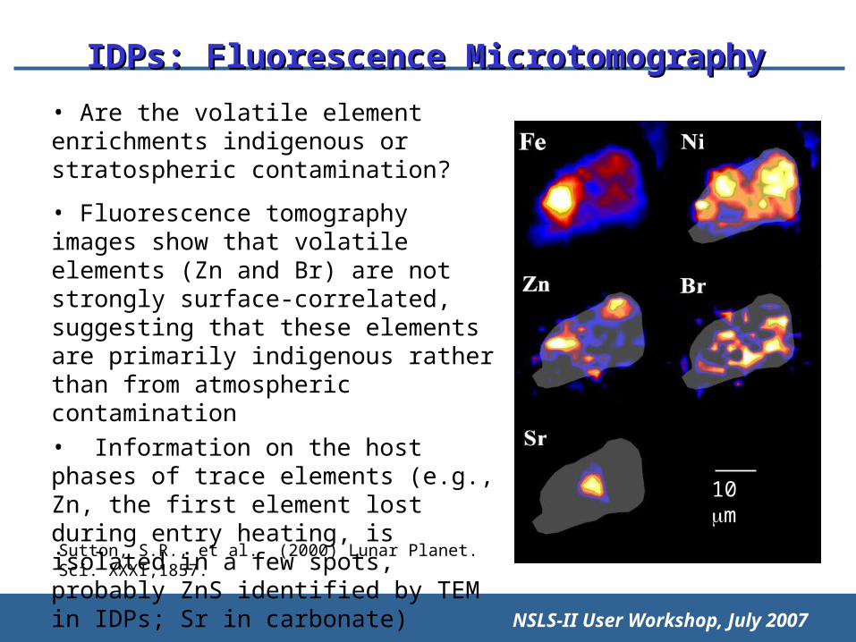
IDPs: Fluorescence MicrotomographyIDPs: Fluorescence Microtomography
• Are the volatile element enrichments indigenous or stratospheric contamination?
• Fluorescence tomography images show that volatile elements (Zn and Br) are not strongly surface-correlated, suggesting that these elements are primarily indigenous rather than from atmospheric contamination• Information on the host phases of trace elements (e.g., Zn, the first element lost during entry heating, is isolated in a few spots, probably ZnS identified by TEM in IDPs; Sr in carbonate)
10 m
Sutton, S.R., et al. (2000) Lunar Planet. Sci. XXXI,1857.
NSLS-II User Workshop, July 2007
Ionomics: the study of how genes regulate ions in cells.
Fe deficiency most common nutritional disorder in the world
2 billion (mainly in developing countries) are anemic
A number of the key genes involved in iron uptake in plants have been identified. Armed with this knowledge, it should now be possible to engineer or breed plants with improved iron uptake abilities and in more bioavailable forms.
Use synchrotron x-ray microprobe techniques to assign functions to metal homeostasis genes whose phenotypes could not be observed using volume-averaged metal analysis techniques
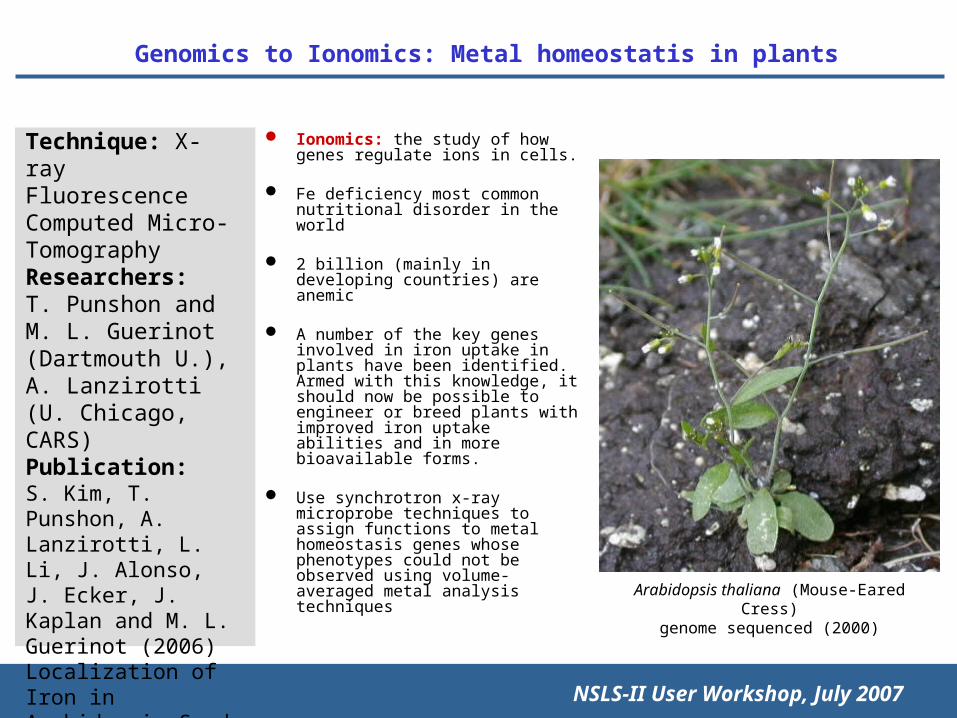
Genomics to Ionomics: Metal homeostatis in plantsGenomics to Ionomics: Metal homeostatis in plants
Technique: X-ray Fluorescence Computed Micro-TomographyResearchers:T. Punshon and M. L. Guerinot (Dartmouth U.), A. Lanzirotti (U. Chicago, CARS)Publication:S. Kim, T. Punshon, A. Lanzirotti, L. Li, J. Alonso,J. Ecker, J. Kaplan and M. L. Guerinot (2006) Localization of Iron in Arabidopsis Seed Requires the Vacuolar MembraneTransporter VIT1, Science.
Arabidopsis thaliana (Mouse-Eared Cress)genome sequenced (2000)

NSLS-II User Workshop, July 2007
Absorption Tomograms
cotyledonsradicle
seed coat

NSLS-II User Workshop, July 2007
Fluorescence TomogramsFluorescence Tomograms
Col-O atvit1-1
Fe Fe MnMn Zn Zn
• Synchrotron x-ray fluorescence microtomography shows that the majority of iron is preciselylocalized to the provascular strands of the embryo. This localization of iron is completelyabolished when the vacuolar iron uptake transporter VIT1 is disrupted, making vacuolesa promising target for increasing the iron content of seeds.