novel salts of sunitinib an anticancer drug with improved ... · groups that favor the formation of...
TRANSCRIPT

_____________________________________________________________________________________________________ *Corresponding author: E-mail: [email protected];
International Research Journal of Pure & Applied Chemistry
5(4): 352-365, 2015, Article no.IRJPAC.2015.028 ISSN: 2231-3443
SCIENCEDOMAIN international
www.sciencedomain.org
Novel Salts of Sunitinib an Anticancer Drug with Improved Solubility
Sangeeta Sangwan1*, Tapas Panda2, Ram Thiamattam1, Sharwan K. Dewan3
and Rajesh K. Thaper1*
1Ranbaxy Laboratories Limited, Chemical Research, Research and Development Centre, Sarhaul,
Sector-18, Gurgaon – 122015, Haryana, India. 2Ranbaxy Laboratories Limited, Analytical Research, Research and Development Centre, Sarhaul,
Sector-18, Gurgaon – 122015, Haryana, India. 3Department of Chemistry, Maharshi Dayanand University, Rohtak – 124001, Haryana, India.
Authors’ contributions
This work was carried out in collaboration between all authors. All authors read and approved the final
manuscript.
Article Information
DOI: 10.9734/IRJPAC/2015/13578 Editor(s):
(1) Rafat Milad Mohareb, Department of Organic Chemistry, Cairo University, Egypt. (2) Lichun Sun, Department of Medicine, Tulane University Health Sciences Center, USA.
(3) Ichiro Imae, Division of Chemistry and Chemical Engineering, Faculty of Engineering, Hiroshima University, Japan. Reviewers:
(1) Anonymous, Utrecht University, Netherlands. (2) Pavan Balmukund Rathi, Department of Pharmaceutics, Head of Department, Shri Bhagwan College of Pharmacy, N-6,
CIDCO, Aurangabad, Dr. Babasaheb Ambedkar Marathwada University, Auranganad, Maharashtra, India. (3) Fleming Martínez, Department of Pharmacy, National University of Colombia, Colombia.
Complete Peer review History: http://www.sciencedomain.org/review-history.php?iid=809&id=7&aid=7436
Received 23rd
August 2014 Accepted 4
th December 2014
Published 19th December 2014
ABSTRACT
Polymorph, co-crystal and salt screening experiments were carried out to identify novel solid forms with the improved physicochemical properties, particularly water solubility in the present case. Co-crystal formation was evaluated with urea and nicotinamide. These coformers do not have any ionic groups that favor the formation of salts. Sunitinib malate salt is being currently sold in the market. It is poorly soluble in water. Salt screening experiments were conducted with adipic acid, glutaric acid, nicotinic acid, 4-hydroxy benzoic acid and saccharin. The salts with 1:1 ratios were obtained with these acids, except for adipic acid, which yielded a 2:1 solid form. The solubility of these salts in deionized water was found to be 6 to 10 times greater than that of the marketed salt (sunitinib malate).
Original Research Article

Sangwan et al.; IRJPAC, 5(4): 352-365, 2015; Article no.IRJPAC.2015.028
353
Reversible Z-E isomerization was also well thought-out especially in presence of light and the isomerization was checked by HPLC. Formation of undesired E-isomer was additionally confirmed by
1H NMR. This observation has implications in the solubility study of sunitinib salt samples using
HPLC method.
Keywords: Sunitinib; photosensitive; novel salts; conformers; solubility; HPLC.
1. INTRODUCTION An important goal of solid-state pharmaceutical development is to increase drug solubility while maintaining a stable form. This objective is critical because solubility and permeability are the major factors used to describe oral absorption according to the biopharmaceutics classification system (BCS). The use of salts in the pharmaceutical industry is a well-known fact. Salts modify properties of the solid forms such as melting temperature, solubility, stability, dissolution rate and bioavailability without altering the desired effect of drug [1]. Salt formation is essentially a three component system involving an acid (A), a base (B) and one or more solvents. A salt is formed by the transfer of a proton (H+) from an acid (A) to a base (B): A-H + B → (A‒
) (B+-H). In order to assist salt
selection a number of empirical rules have been proposed, such as the ‘rule of three’. This states that salt formation generally requires a difference of at least three pKa units between the conjugate base and the conjugate acid, pKa (Base) - pKa (Acid) ≥ 3, where pKa is the ability of an ionisable group to donate a proton (H+) in an aqueous medium and is often referred to as the dissociation constant. When the difference is ≤ 3, the product can be either salt or co-crystal.
API co-crystals, like API salts, have demonstrated the ability to modify physicochemical properties of the APIs [2a-f]. Cocrystals can be made for nonionizable drugs, which are restricted from salt formation. Also, for ionizable drugs, the number of suitable cocrystal ligands (i.e. coformers) can exceed the number of suitable counter ions. Solvates are crystal forms containing either stoichiometric or nonstoichiometric amounts of a solvent [3]. If the incorporated solvent is water, the solvate is commonly known as a hydrate. Different polymorphs of a given crystalline compound (salt or co-crystal) can have different physicochemical properties [4]. Systematic screening for polymorphs has therefore become an essential step in drug development to select a factual form in order to avoid problems caused by polymorphic transitions during processing. [5]
The crystallization of polymorphs can be affected by numerous factors, which include, the nature of solvents [6a-d] and the presence of polymers [7] or other additives [8].
Sunitinib is a new vascular endothelial growth factor receptor inhibitor, has demonstrated high activity in renal cell carcinoma (RCC) and is now extensively used for patients with metastatic disease [9,10]. It is sold as malate salt by Pfizer under the brand name Sutent. Sunitinib is a yellow to orange powder with solubility of 25 mg/ml in acidic solutions (pH 1.2 - 6.8) [11] and 10-50 µM in plain water. The solubility of sunitinib rapidly decreases at pH greater than 6.8. Hence Sunitinib malate may be considered class III drug, a high solubility and a low permeability compound according to the biopharmaceutical classification. Consequently there is a need to obtain new salts of sunitinib with improved water solubility, though several salts have already been identified for this API [12,13] (Scheme 1).
Sunitinib molecule has an exocyclic double bond at 3-position of the oxindole ring and consequently it can exhibit Z-E isomerism. Anand and Narmada have revealed that SU5416, closely related to sunitinib, undergoes photo induced conversion into the less stable E isomer in solution [14] (Scheme 2). Present study deals with the synthesis of sunitinib, Z-E isomerization and identification of novel salts with improved water solubility.
2. MATERIALS AND METHODS
All the key intermediates for synthesis of sunitinib were procured from Shanghai Parling pharma co. ltd. China. Solvents and reagents were used by way of, obtained from Rankem, Qualigens and Ranbaxy Labs. Ltd., India. Waters HPLC 2695 alliance separation module (with customized syringe and loop volume of 2.5 mL) with PDA 2998 detector was used for investigation. Perkin Elmer CHNS/O analyzer 2400 was used for CHN analysis. Melting point was recorded on Buchi b 545. IR spectra were recorded on Perkin-Elmer Spectrum One FT-IR spectrometer.
1H NMR
spectra was recorded in DMSO-d6 at 400 MHz

Sangwan et al.; IRJPAC, 5(4): 352-365, 2015; Article no.IRJPAC.2015.028
354
using TMS as an internal standard. Mass/Ms-Ms data was generated by using QTRAP LC/MS/MS system (Applied Biosystems). The XRPD of the samples were determined by using Instrument: PANalytical; Model X’pert PRO; Detector: X’celerator; Scan Range: 3-40; Step size: 0.02; Range: 3-40 degree 2 theta; CuKα radiation at 45 kV and 40 mA. DSC of the samples was determined by using Mettler-Toledo 821e. Data collection parameters: Scanning rate: 10 ºC/min; Temperature: 30 ºC to 300 ºC. TGA of the samples were determined by using TA Q500 between 30 ºC to 300 ºC at 10 ºC/min scan rate. In the Z-E isomerization studies, the solutions (in 100 ml volumetric flasks) - prepared by means of dissolving 25 mg of sunitinib in 100 ml of each solvent - were either directly exposed to halogen lamp (150 W) or sunlight.
3. EXPERIMENTAL
3.1 Z-E Isomerization
Photo induced Z-E isomerization study in sunitinib molecule was carried out by HPLC method using Kromasil C8 (250 × 4.6 mm; 5 µm particle size) (procured from Akzonobel) stationary phase. The gradient elution consisted of potassium dihydrogen orthophosphate buffer (pH 4.5) and acetonitrile (1:1 v/v) with an injection volume and flow rate of 5 µl and 1 ml/min, respectively with detection wavelength
210 nm. This transformation of desired Z-isomer into unstable E-isomer was moreover proved by 1H NMR of compound in DMSO-d6 after going on exposure to light. Pure Z-Isomer
1H NMR (400
MHz, DMSO): δ 0.98 (6 H, t, C27, 29-Ha), 2.43 (3H, s, C21-Ha, 2.45 (3H, s, C18-Ha), 2.47-2.57 (*6H, m, C28, 26, 24-Ha), 3.27-3.34 (2H, m, C23-Ha), 6.83-6.86 (1H, m, Ar-Ha), 6.90-6.95 (1H, m, Ar-H), 7.43 (1H, t, N22-Ha), 7.71 (1H,s, C11a-Haa), 7.75 (1H, d, Ar-H), 10.89 (1H, s, N-H), 13.68 (1H, s, N-H).
When Z-Isomer exposed to sunlight for 4 hours (E-Isomer)
1H NMR (400 MHz, DMSO): δ 1.00 (6
H, t, C27, 29-Hb), 2.21 (3H, s, C21-Hb), 2.43-2.45 (3H, s, C18-Hb), 2.51-2.55 (*6H, m, C28, 26, 24-Hb), 3.27-3.30 (2H, m, C23-Hb), 6.81-6.86 (1H, m, Ar-Hb), 6.99-7.04 (1H, m, Ar-Hb), 7.34 (1H, d, Ar-Hb), 7.37-7.43.
3.2 Solubility Data The equilibrium solubility study of sunitinib salts in DI (Deionized water) water was carried out by using gradient elution HPLC method consisted of potassium dihydrogen orthophosphate buffer (pH 6.5) and acetonitrile-MeOH mixture (1:1 v/v) with an injection volume and flow rate of 10 µl and 0.75 ml/min on Kromasil C8 column (250 × 4.6 mm; 5 µm particle size), respectively through detection at 270 nm.
NH
O NHN
NH
FO HO2C
CO2HOH
H+
NH
O NHN
NH
FO
HO2C
CO2HOH
H
.
Scheme 1. Scheme for the synthesis of sunitinib Malate
NH
O NHN
NH
FO
NH
NH
NHO
N
O
FHO 2C
CO 2HOH
H
.
HO 2C
CO 2HOH
H
.
Z-Isomer E-Isomer
Scheme 2. Z-E Isomers of sunitinib malate
3.3 Sunitinib Synthesis Sunitinib was prepared in two imidazole derivative (1) as per the reported procedure (Scheme 3) [15]. Amidation of 1 with diethylethylenediamine (2) in presence of EDCI.HCl (N-Ethyl-N′-(3-dimethylaminopropyl) carbodiimidehydrochloride), HOBt (Hydroxybenzotriazole) and TEA (at RT yielded imidazole-amide derivative (3). Intermediate 3 was then and there condensed with oxindole derivative (4) in presence of base in protic solvent to give crude sunitinib (5) in quantitative yields. The crude sunitinib (5) was then purified via acid-base treatment as reported earlier [16]. The spectra’s of synthesized
NH
O OH
O
HHOBt
EDCI.HCl
TEA, RT
NH2N
(1)
(2)
Scheme 3
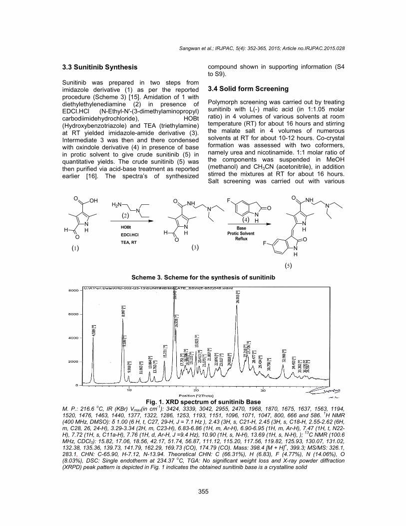
Fig. 1M. P.: 216.6
oC, IR (KBr) Vmax(in cm
-1): 3424, 3339, 3042, 2955, 2470, 1968, 1870, 1675, 1637, 1563, 1194,
1520, 1476, 1463, 1440, 1377, 1322, 1286, 1253, 1193, 1151, 1096, 1071, 1047, 800, 666 and 586. (400 MHz, DMSO): δ 1.00 (6 H, t, C27, 29m, C28, 26, 24-H), 3.29-3.34 (2H, m, C23H), 7.72 (1H, s, C11a-H), 7.76 (1H, d, ArMHz, CDCl3): 15.82, 17.06, 18.56, 42.17, 51.74, 56.87, 111.12, 115.20, 117.56, 119.82, 125.93, 130.07, 131.02, 132.38, 135.36, 139.73, 141.79, 162.29, 169.73 (CO), 174.79 (CO). 283.1, CHN: C-65.90, H-7.12, N-13.94.(8.03%), DSC: Single endotherm at 234.37(XRPD) peak pattern is depicted in Fig. 1
Sangwan et al.; IRJPAC, 5(4): 352-365, 2015; Article no.IRJPAC.2015.0
355
Sunitinib was prepared in two steps from imidazole derivative (1) as per the reported
Amidation of 1 with diethylethylenediamine (2) in presence of
dimethylaminopropyl) ), HOBt
) and TEA (triethylamine) amide derivative (3).
then and there condensed with oxindole derivative (4) in presence of base in protic solvent to give crude sunitinib (5) in quantitative yields. The crude sunitinib (5) was
base treatment as reported of synthesized
compound shown in supporting information (S4 to S9).
3.4 Solid form Screening
Polymorph screening was carried out by treating sunitinib with L(-) malic acid (in 1:1.05 molar ratio) in 4 volumes of various solvents at room temperature (RT) for about 16 hours and stirring the malate salt in 4 volumes of numerous solvents at RT for about 10-12 hours. Coformation was assessed with two coformernamely urea and nicotinamide. 1:1 molar ratio of the components was suspended in MeOH (methanol) and CH3CN (acetonitrile), in addition stirred the mixtures at RT for about 16 hours. Salt screening was carried out with various
NH
O NH
O
H
N
BaseProtic Solvent
Reflux
NH
O
NH
FO
NH
F
O
(3)
(4)
(5)
Scheme 3. Scheme for the synthesis of sunitinib
1. XRD spectrum of sunitinib Base 1): 3424, 3339, 3042, 2955, 2470, 1968, 1870, 1675, 1637, 1563, 1194,
1520, 1476, 1463, 1440, 1377, 1322, 1286, 1253, 1193, 1151, 1096, 1071, 1047, 800, 666 and 586. C27, 29-H, J = 7.1 Hz ), 2.43 (3H, s, C21-H, 2.45 (3H, s, C18-H,
(2H, m, C23-H), 6.83-6.86 (1H, m, Ar-H), 6.90-6.95 (1H, m, Ar-H), 7.47 (1H, t, N22H), 7.76 (1H, d, Ar-H, J =9.4 Hz), 10.90 (1H, s, N-H), 13.69 (1H, s, N-H), );
: 15.82, 17.06, 18.56, 42.17, 51.74, 56.87, 111.12, 115.20, 117.56, 119.82, 125.93, 130.07, 131.02, 132.38, 135.36, 139.73, 141.79, 162.29, 169.73 (CO), 174.79 (CO). Mass: 398.4 [M + H]
+, 399.3; MS/MS: 326.1,
13.94. Theoretical CHN: C (66.31%), H (6.83), F (4.77%), N (14.06%), O (8.03%), DSC: Single endotherm at 234.37
oC, TGA: No significant weight loss and X-ray powder diffraction
(XRPD) peak pattern is depicted in Fig. 1 indicates the obtained sunitinib base is a crystalline solid
, 2015; Article no.IRJPAC.2015.028
compound shown in supporting information (S4
Polymorph screening was carried out by treating malic acid (in 1:1.05 molar
ratio) in 4 volumes of various solvents at room temperature (RT) for about 16 hours and stirring the malate salt in 4 volumes of numerous
12 hours. Co-crystal formation was assessed with two coformers, namely urea and nicotinamide. 1:1 molar ratio of the components was suspended in MeOH
CN (acetonitrile), in addition stirred the mixtures at RT for about 16 hours. Salt screening was carried out with various
NH
NHN
O
): 3424, 3339, 3042, 2955, 2470, 1968, 1870, 1675, 1637, 1563, 1194, 1520, 1476, 1463, 1440, 1377, 1322, 1286, 1253, 1193, 1151, 1096, 1071, 1047, 800, 666 and 586.
1H NMR
H, 2.55-2.62 (6H, H), 7.47 (1H, t, N22-
); 13
C NMR (100.6 : 15.82, 17.06, 18.56, 42.17, 51.74, 56.87, 111.12, 115.20, 117.56, 119.82, 125.93, 130.07, 131.02,
, 399.3; MS/MS: 326.1, Theoretical CHN: C (66.31%), H (6.83), F (4.77%), N (14.06%), O
ray powder diffraction solid

organic acids such as adipic acid, glutaric acid, nicotinic acid, 4-hydroxybenzic acidsaccharin. Mixtures of sunitinib and salt(in 1:1.05 molar ratio) were stirred in 10volumes of MeOH (methanol) or acetonitrile at RT for about 12-16 hours and the resulting solutions either clear or suspensions cooled down to 0-5 ºC and stirred at 0about 1 hour before filtration. In all the above cases the solids were separated from the solvethrough vacuum filtration and dried them at 45-50 ºC under vacuum for about 10scheme 4 to 8.
3.4.1 Sunitinib Glutarate
5.0 g of Sunitinib base was suspended in 75 ml of methanol at RT under N2. 1.82 g of glutaric acid (1.1 meq) was added to the mixture at RT. The mixture was stirred at RT for about 16 hours and then cooled down to 0-5 oC and stirred at this temperature for 1 hour. Red colored solids
NH
O NHN
NH
FO + OH
O
Sunitinib Base
Glutaric Acid
Scheme 4. Scheme for the synthesis of
Fig. 2. XRD spectrum of sunitinib% Yield = 88, M. P. = 204.2
oC, IR (KBr) V
1326, 1194, 1179, 697, 666, and 588. 1
(2H, p, C34-H, J = 7.44 Hz), 2.23 (4H, t, C33, 35(6H, m, C24, 26, 28-H), 3.27-3.32 (2H, C23
7.44 (1H, t, N22-H, J = 5.52, 5.56 Hz), 7.71 (1H, s, C11aH),13.68 (1H, s, N1-H), DSC: Single endotherm at 202.38
Sangwan et al.; IRJPAC, 5(4): 352-365, 2015; Article no.IRJPAC.2015.0
356
adipic acid, glutaric acid, hydroxybenzic acid and
. Mixtures of sunitinib and salt-former (in 1:1.05 molar ratio) were stirred in 10-15
or acetonitrile at 16 hours and the resulting
solutions either clear or suspensions were further C and stirred at 0-5 ºC for
about 1 hour before filtration. In all the above cases the solids were separated from the solvent through vacuum filtration and dried them at
for about 10-15 hours,
5.0 g of Sunitinib base was suspended in 75 ml . 1.82 g of glutaric
added to the mixture at RT. The mixture was stirred at RT for about 16 hours
C and stirred at this temperature for 1 hour. Red colored solids
were collected by vacuum filtration and washed the material with 25 ml of methanol. Dproduct under vacuum at 50
oC temperature for
12 to 14 h. The crystalline Form I of glutarate salt of sunitinib (scheme 4) was obtained and characterized by X-ray powder diffraction (XRPD) peaks pattern substantially as depicted in Fig. 2. 3.4.2 Sunitinib Adipate
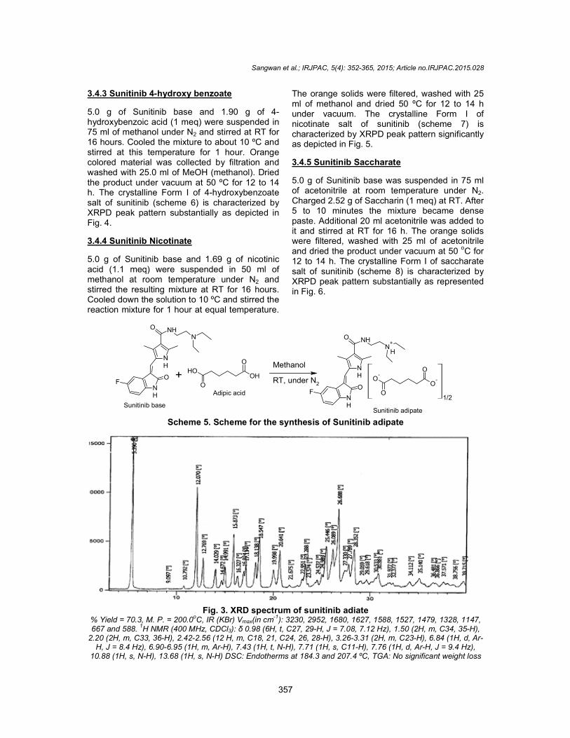
5.0 g of Sunitinib base and 2.0 g of adipic acid (1.1 meq) were suspended in 75 ml of methanol at RT under N2. The resulting mixture was stirred for 5 hours at RT. The red colored solids were collected by vacuum filtration and washed wit50 ml of methanol at RT. Dried the product under vacuum at 50 oC for 12 to 14 h. Form I of adipate salt of sunitinib (scheme 5) was characterized by XRPD peak pattern substantially as shown in Fig. 3.
NH
O NHN
+
NH
FO
O-
O
Sunitinib glutarate
OH
O
Glutaric Acid
Methanol
RT under N2
Scheme for the synthesis of sunitinib glutarate
XRD spectrum of sunitinib glutarate C, IR (KBr) Vmax(in cm
-1): 3427, 3189, 3047, 2987, 2203, 1679, 1628, 1572, 1525,
1H NMR (400 MHz, CDCl3): δ 0.98 (6 H, t, C29, 27-H, J = 7.08, 7.12
(4H, t, C33, 35-H, J = 7.04 Hz), 2.43 (6H, d, C18, 21-H, J = 7.8 Hz), 2.503.32 (2H, C23-H, m), 6.85 (1H, d, Ar-H, J = 4.6 Hz), 6.94 (1H, d, Ar-
H, J = 5.52, 5.56 Hz), 7.71 (1H, s, C11a-H), 7.74 (1H, d, Ar-H, J = 9.4 Hz), 10.88DSC: Single endotherm at 202.38
oC, TGA: No significant weight loss
, 2015; Article no.IRJPAC.2015.028
were collected by vacuum filtration and washed the material with 25 ml of methanol. Dried the
C temperature for The crystalline Form I of glutarate salt
of sunitinib (scheme 4) was obtained and ray powder diffraction
(XRPD) peaks pattern substantially as depicted
5.0 g of Sunitinib base and 2.0 g of adipic acid (1.1 meq) were suspended in 75 ml of methanol
. The resulting mixture was stirred for 5 hours at RT. The red colored solids were collected by vacuum filtration and washed with 50 ml of methanol at RT. Dried the product under
The crystalline Form I of adipate salt of sunitinib (scheme 5) was characterized by XRPD peak pattern
+
H
OH
O
Sunitinib glutarate
): 3427, 3189, 3047, 2987, 2203, 1679, 1628, 1572, 1525, H, J = 7.08, 7.12), 1.69
H, J = 7.8 Hz), 2.50-2.60 -H, J = 8.48 Hz),
H, J = 9.4 Hz), 10.88 (1H, s, N13-TGA: No significant weight loss
3.4.3 Sunitinib 4-hydroxy benzoate
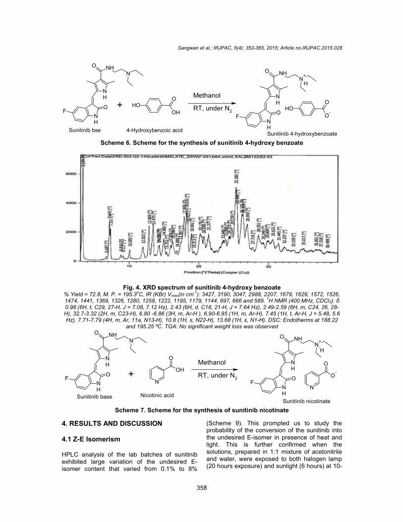
5.0 g of Sunitinib base and 1.90 g of 4hydroxybenzoic acid (1 meq) were suspended in 75 ml of methanol under N2 and stirred at RT for 16 hours. Cooled the mixture to about 10stirred at this temperature for 1 hour. Orange colored material was collected by filtration and washed with 25.0 ml of MeOH (methanol). Dried the product under vacuum at 50 ºC for 12 to 14 h. The crystalline Form I of 4-hydroxybenzoate salt of sunitinib (scheme 6) is characterized by XRPD peak pattern substantially as depicted in Fig. 4.
3.4.4 Sunitinib Nicotinate
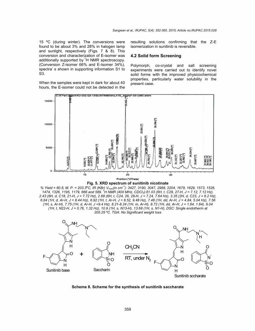
5.0 g of Sunitinib base and 1.69 g of nicotinic acid (1.1 meq) were suspended in 50 ml of methanol at room temperature under Nstirred the resulting mixture at RT for 16 hours. Cooled down the solution to 10 ºC and stirred the reaction mixture for 1 hour at equal temperature.
NH
O NHN
NH
FO
OH
O
Sunitinib base
+
Adipic acid
Scheme 5. Scheme for the synthesis of Sunitinib
Fig. 3% Yield = 70.3, M. P. = 200.0oC, IR (KBr) V667 and 588. 1H NMR (400 MHz, CDCl
2.20 (2H, m, C33, 36-H), 2.42-2.56 (12 H, m, C18, 21, C24, 26, 28H, J = 8.4 Hz), 6.90-6.95 (1H, m, Ar-H), 7.43 (1H, t, N
10.88 (1H, s, N-H), 13.68 (1H, s, N-H) DSC: Endotherms at 184.3 an
Sangwan et al.; IRJPAC, 5(4): 352-365, 2015; Article no.IRJPAC.2015.0
357
hydroxy benzoate
5.0 g of Sunitinib base and 1.90 g of 4-hydroxybenzoic acid (1 meq) were suspended in
and stirred at RT for 16 hours. Cooled the mixture to about 10 ºC and stirred at this temperature for 1 hour. Orange colored material was collected by filtration and washed with 25.0 ml of MeOH (methanol). Dried
C for 12 to 14 hydroxybenzoate
salt of sunitinib (scheme 6) is characterized by XRPD peak pattern substantially as depicted in
5.0 g of Sunitinib base and 1.69 g of nicotinic (1.1 meq) were suspended in 50 ml of
methanol at room temperature under N2 and stirred the resulting mixture at RT for 16 hours.
C and stirred the reaction mixture for 1 hour at equal temperature.
The orange solids were filtered, washed with 25 ml of methanol and dried 50 ºC for 12 to 14 h under vacuum. The crystalline Form I of nicotinate salt of sunitinib (scheme 7) is characterized by XRPD peak pattern significantly as depicted in Fig. 5.
3.4.5 Sunitinib Saccharate
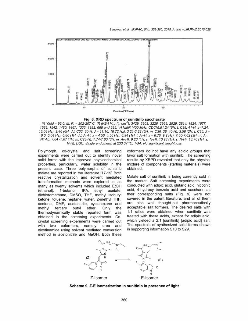
5.0 g of Sunitinib base was suspended in 75 ml of acetonitrile at room temperature under NCharged 2.52 g of Saccharin (1 meq) at RT. After 5 to 10 minutes the mixture became dense paste. Additional 20 ml acetonitrile was added to it and stirred at RT for 16 h. The orange solids were filtered, washed with 25 ml of acetonitrile and dried the product under vacuum at 50 12 to 14 h. The crystalline Form I of saccharate salt of sunitinib (scheme 8) is characterized by XRPD peak pattern substantially as in Fig. 6.
OH
ONH
O NHN
+
NH
FO
H
O
O-
O
Sunitinib adipate
Adipic acid
Methanol
RT, under N2
Scheme for the synthesis of Sunitinib adipate
3. XRD spectrum of sunitinib adiate C, IR (KBr) Vmax(in cm-1): 3230, 2952, 1680, 1627, 1588, 1527, 1479, 1328, 1147,
H NMR (400 MHz, CDCl3): δ 0.98 (6H, t, C27, 29-H, J = 7.08, 7.12 Hz), 1.50 (2H, m, C34, 35(12 H, m, C18, 21, C24, 26, 28-H), 3.26-3.31 (2H, m, C23-H), 6.84 (1H, d, Ar
H), 7.43 (1H, t, N-H), 7.71 (1H, s, C11-H), 7.76 (1H, d, Ar-H, J = 9.4 Hz), DSC: Endotherms at 184.3 and 207.4 ºC, TGA: No significant weight loss
, 2015; Article no.IRJPAC.2015.028
filtered, washed with 25 C for 12 to 14 h
The crystalline Form I of nicotinate salt of sunitinib (scheme 7) is characterized by XRPD peak pattern significantly
5.0 g of Sunitinib base was suspended in 75 ml of acetonitrile at room temperature under N2. Charged 2.52 g of Saccharin (1 meq) at RT. After 5 to 10 minutes the mixture became dense paste. Additional 20 ml acetonitrile was added to
r 16 h. The orange solids were filtered, washed with 25 ml of acetonitrile and dried the product under vacuum at 50
oC for
The crystalline Form I of saccharate salt of sunitinib (scheme 8) is characterized by XRPD peak pattern substantially as represented
O-
O
1/2
): 3230, 2952, 1680, 1627, 1588, 1527, 1479, 1328, 1147, (2H, m, C34, 35-H),
H), 6.84 (1H, d, Ar-H, J = 9.4 Hz),
TGA: No significant weight loss
Sangwan et al.; IRJPAC, 5(4): 352-365, 2015; Article no.IRJPAC.2015.028
358
NH
O NHN
NH
FO OH
O
OH
Sunitinib bse
NH
O NHN
+
NH
FO
H
OHO
O-
Sunitinib 4-hydroxybenzoate
+
4-Hydroxybenzoic acid
Methanol
RT, under N2
Scheme 6. Scheme for the synthesis of sunitinib 4-hydroxy benzoate
Fig. 4. XRD spectrum of sunitinib 4-hydroxy benzoate % Yield = 72.8, M. P. = 195.3
oC, IR (KBr) Vmax(in cm
-1): 3427, 3190, 3047, 2988, 2207, 1679, 1629, 1572, 1526,
1474, 1441, 1369, 1326, 1280, 1258, 1222, 1195, 1179, 1144, 697, 666 and 589. 1H NMR (400 MHz, CDCl3): δ
0.98 (6H, t, C29, 27-H, J = 7.08, 7.12 Hz), 2.43 (6H, d, C18, 21-H, J = 7.64 Hz), 2.49-2.59 (6H, m, C24, 26, 28-H), 32.7-3.32 (2H, m, C23-H), 6.80 -6.86 (3H, m, Ar-H ), 6.90-6.95 (1H, m, Ar-H), 7.45 (1H, t, Ar-H, J = 5.48, 5.6 Hz), 7.71-7.79 (4H, m, Ar, 11a, N13-H), 10.8 (1H, s, N22-H), 13.68 (1H, s, N1-H). DSC: Endotherms at 188.22
and 195.25 ºC. TGA: No significant weight loss was observed
NH
O NHN
NH
FO
N
O
OH
Sunitinib base
NH
O NHN
+
NH
FO
H
N
O
O-
Sunitinib nicotinate
+
Nicotinic acid
Methanol
RT, under N2
Scheme 7. Scheme for the synthesis of sunitinib nicotinate
4. RESULTS AND DISCUSSION 4.1 Z-E Isomerism HPLC analysis of the lab batches of sunitinib exhibited large variation of the undesired E-isomer content that varied from 0.1% to 8%
(Scheme 9). This prompted us to study the probability of the conversion of the sunitinib into the undesired E-isomer in presence of heat and light. This is further confirmed when the solutions, prepared in 1:1 mixture of acetonitrile and water, were exposed to both halogen lamp (20 hours exposure) and sunlight (6 hours) at 10-
Sangwan et al.; IRJPAC, 5(4): 352-365, 2015; Article no.IRJPAC.2015.028
359
15 ºC (during winter). The conversions were found to be about 3% and 28% in halogen lamp and sunlight, respectively (Figs. 7 & 8). This conversion and characterization of E-isomer was additionally supported by 1H NMR spectroscopy. (Conversion Z-isomer 66% and E-Isomer 34%), spectra’ s shown in supporting information S1 to S3. When the samples were kept in dark for about 40 hours, the E-isomer could not be detected in the
resulting solutions confirming that the Z-E isomerization in sunitinib is reversible.
4.2 Solid form Screening Polymorph, co-crystal and salt screening experiments were carried out to identify novel solid forms with the improved physicochemical properties, particularly water solubility in the present case.
Fig. 5. XRD spectrum of sunitinib nicotinate
% Yield = 60.8, M. P. = 203.3ºC, IR (KBr) Vmax(in cm-1): 3427, 3190, 3047, 2988, 2204, 1679, 1629, 1573, 1526, 1474, 1326, 1195, 1179, 666 and 589. 1H NMR (400 MHz, CDCl3):δ1.03 (6H, t, C29, 27-H, J = 7.12, 7.12 Hz),
2.43 (6H, d, C18, 21-H, J = 7.72 Hz), 2.68 (6H, t, C24, 26, 28-H, J = 7.24, 7.64 Hz), 3.35 (2H, d, C23, J = 6.2 Hz), 6.84 (1H, d, Ar-H, J = 8.44 Hz), 6.92 (1H, t, Ar-H, J = 8.52, 9.48 Hz), 7.48 (1H, dd, Ar-H, J = 4.84, 5.04 Hz), 7.56
(1H, s, Ar-H), 7.75 (1H, d, Ar-H, J =9.4 Hz), 8.21-8.24 (1H, m, Ar-H), 8.72 (1H, dd, Ar-H, J = 1.64, 1.64), 9.04 (1H, t, N22-H, J = 0.76, 1.32 Hz), 10.9 (1H, s, N13-H), 13.68 (1H, s, N1-H), DSC: Single endotherm at
205.25 ºC. TGA: No Significant weight loss
NH
O NHN
NH
FO NH
S
OO
O
Sunitinib base
NH
O NHN
+
NH
FO
H
N-
S
OO
O
Sunitinib sccharate
Saccharin
+CH3CN
RT, under N2
Scheme 8. Scheme for the synthesis of sunitinib saccharate
Sangwan et al.; IRJPAC, 5(4): 352-365, 2015; Article no.IRJPAC.2015.028
360
Fig. 6. XRD spectrum of sunitinib saccharate % Yield = 92.0, M. P. = 202-207
oC, IR (KBr) Vmax(in cm
-1): 3429, 3303, 3226, 2969, 2929, 2814, 1824, 1677,
1589, 1542, 1480, 1467, 1333, 1192, 668 and 585. 1H NMR (400 MHz, CDCl3):δ1.24 (6H, t, C39, 41-H, J=7.24,
13.04 Hz), 2.46 (6H, dd, C33, 30-H, J = 11.16, 18.72 Hz), 3.21-3.22 (6H, m, C36, 38, 40-H), 3.58 (2H, t, C35, J = 6.0, 6.04 Hz), 6.86 (1H, dd, Ar-H, J = 4.56, 4.56 Hz), 6.94 (1H, t, Ar-H, J = 8.76, 9.2 Hz), 7.56-7.62 (3H, m, Ar, N1-H), 7.64 -7.67 (1H, m, C23-H), 7.74-7.80 (3H, m, Ar-H), 9.23 (1H, s, N-H), 10.93 (1H, s, N-H), 13.76 (1H, s,
N-H), DSC: Single endotherm at 233.07 ºC. TGA: No significant weight loss
Polymorph, co-crystal and salt screening experiments were carried out to identify novel solid forms with the improved physicochemical properties, particularly, water solubility in the present case. Three polymorphs of sunitinib malate are reported in the literature.[17-19] Both reactive crystallization and solvent mediated transformation methods were explored in as many as twenty solvents which included EtOH (ethanol), 1-butanol, IPA, ethyl acetate, dichloromethane, DMSO, THF, methyl isobutyl ketone, toluene, heptane, water, 2-methyl THF, acetone, DMF, acetonitrile, cyclohexane and methyl tertiary butyl ether. Only the thermodynamically stable reported form was obtained in the screening experiments. Co-crystal screening experiments were carried out with two coformers, namely, urea and nicotinamide using solvent mediated conversion method in acetonitrile and MeOH. Both these
coformers do not have any acidic groups that favor salt formation with sunitinb. The screening results by XRPD revealed that only the physical mixture of components (starting materials) were obtained.
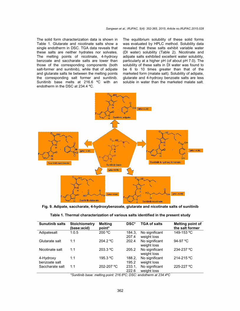
Malate salt of sunitinib is being currently sold in the market. Salt screening experiments were conducted with adipic acid, glutaric acid, nicotinic acid, 4-hydroxy benzoic acid and saccharin as their corresponding salts (Fig. 9) were not covered in the patent literature, and all of them are also well thought-out pharmaceutically acceptable salt formers. The desired salts with 1:1 ratios were obtained when sunitinib was treated with these acids, except for adipic acid, which yielded a 2:1 [sunitinib] [adipic acid] salt. The spectra’s of synthesized solid forms shown in supporting information S10 to S29.
NH
O NHN
NH
FO
NH
NH
NHO
N
O
F
(Z)(E)
Z-Isomer E-Isomer
Scheme 9. Z-E Isomerization in sunitinib in presence of light
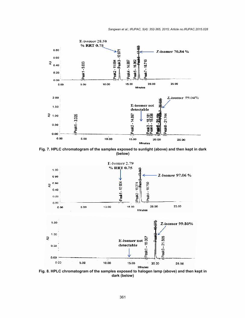
Fig. 7. HPLC chromatogram of the samples exposed to sunlight (above
Fig. 8. HPLC chromatogram of the samples exposed to halogen lamp (above) and then kept in
Sangwan et al.; IRJPAC, 5(4): 352-365, 2015; Article no.IRJPAC.2015.0
361
7. HPLC chromatogram of the samples exposed to sunlight (above) and then kept in dark(below)
8. HPLC chromatogram of the samples exposed to halogen lamp (above) and then kept in dark (below)
, 2015; Article no.IRJPAC.2015.028
) and then kept in dark
8. HPLC chromatogram of the samples exposed to halogen lamp (above) and then kept in
The solid form characterization data is shown in Table 1. Glutarate and nicotinate salts show a single endotherm in DSC. TGA data reveals that these salts are neither hydrates nor solvates. The melting points of nicotinate, benzoate and saccharate salts are lower than those of the corresponding components (both salt-former and sunitinib), while that of adipate and glutarate salts lie between the melting points the corresponding salt former and sunitinib. Sunitinib base melts at 216.6 endotherm in the DSC at 234.4 ºC.
Fig. 9. Adipate, saccharate, 4-hydroxybenzoate, glutarate and nicotinate salts of sunitinib
Table 1. Thermal characterization of various salts
Sunutinib salts Stoichiometry(base:acid)
Adipatesalt 1:0.5
Glutarate salt 1:1
Nicotinate salt 1:1
4-Hydroxy benzoate salt
1:1
Saccharate salt 1:1
*Sunitinib base: melting point: 216.6
Sangwan et al.; IRJPAC, 5(4): 352-365, 2015; Article no.IRJPAC.2015.0
362
The solid form characterization data is shown in Table 1. Glutarate and nicotinate salts show a single endotherm in DSC. TGA data reveals that these salts are neither hydrates nor solvates. The melting points of nicotinate, 4-hydroxy benzoate and saccharate salts are lower than those of the corresponding components (both
former and sunitinib), while that of adipate and glutarate salts lie between the melting points the corresponding salt former and sunitinib.
ºC with an
The equilibrium solubility of these solid forms was evaluated by HPLC method. Solubility data revealed that these salts exhibit variable water (DI water) solubility (Table 2). Nicotinate and adipate salts exhibited excellent water solubility, particularly at a higher pH (of about pH 7.0). The solubility of these salts in DI water was found to be 6 to 10 times greater than that of the marketed form (malate salt). Solubility oglutarate and 4-hydroxy benzoate salts are less soluble in water than the marketed malate salt.
hydroxybenzoate, glutarate and nicotinate salts of sunitinib
Table 1. Thermal characterization of various salts identified in the present study
Melting point*
DSC* TGA of salts Melting point of the salt former
200 ºC 184.3, 207.4
No significant weight loss
149
204.2 ºC 202.4 No significant weight loss
94-97
203.3 ºC 205.2 No significant weight loss
234
195.3 ºC 188.2, 195.2
No significant weight loss
214
202-207 ºC 233.1, 222.6
No significant weight loss
225
*Sunitinib base: melting point: 216.6ºC; DSC: endotherm at 234.4ºC
, 2015; Article no.IRJPAC.2015.028
The equilibrium solubility of these solid forms was evaluated by HPLC method. Solubility data revealed that these salts exhibit variable water
Nicotinate and exhibited excellent water solubility,
particularly at a higher pH (of about pH 7.0). The DI water was found to
be 6 to 10 times greater than that of the marketed form (malate salt). Solubility of adipate,
hydroxy benzoate salts are less soluble in water than the marketed malate salt.
hydroxybenzoate, glutarate and nicotinate salts of sunitinib
identified in the present study
Melting point of the salt former 149-153 ºC
97 ºC
234-237 ºC
214-215 ºC
225-227 ºC
Sangwan et al.; IRJPAC, 5(4): 352-365, 2015; Article no.IRJPAC.2015.028
363
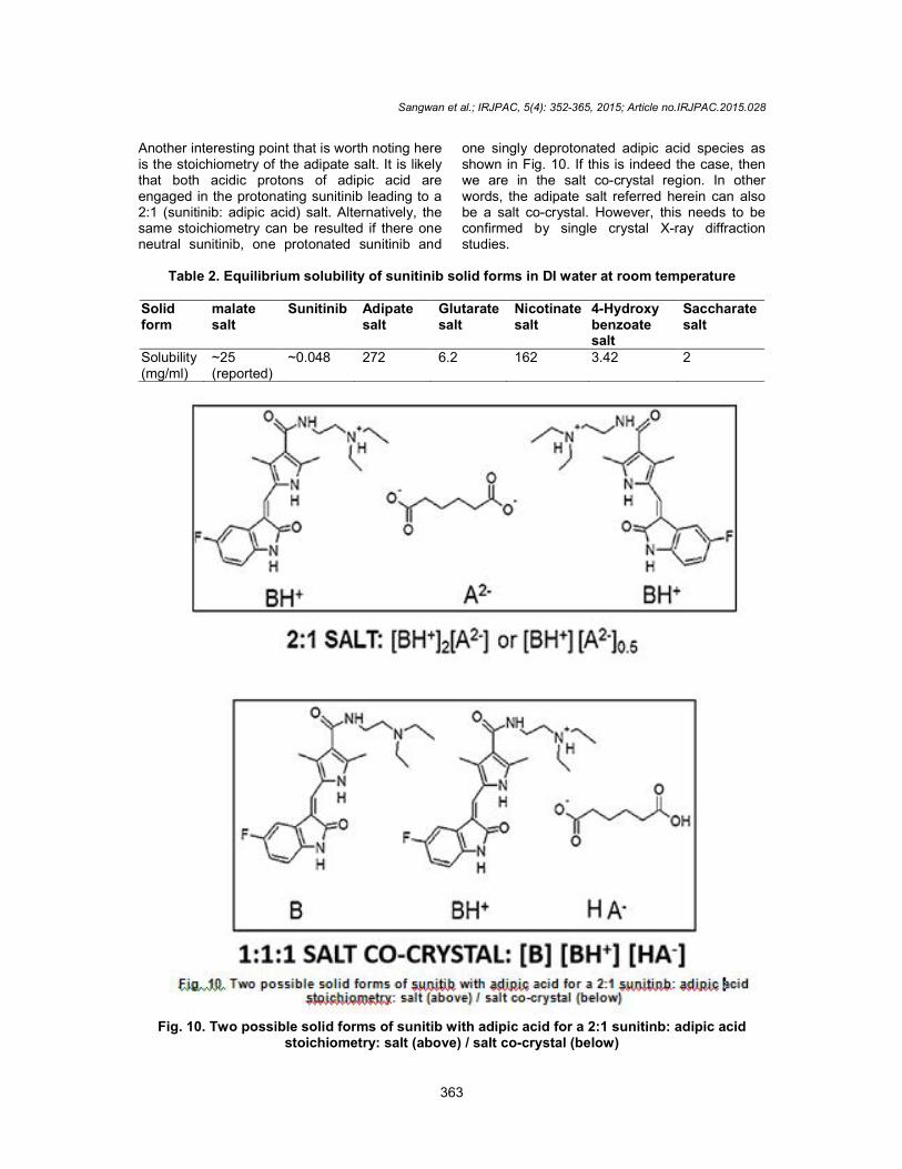
Another interesting point that is worth noting here is the stoichiometry of the adipate salt. It is likely that both acidic protons of adipic acid are engaged in the protonating sunitinib leading to a 2:1 (sunitinib: adipic acid) salt. Alternatively, the same stoichiometry can be resulted if there one neutral sunitinib, one protonated sunitinib and
one singly deprotonated adipic acid species as shown in Fig. 10. If this is indeed the case, then we are in the salt co-crystal region. In other words, the adipate salt referred herein can also be a salt co-crystal. However, this needs to be confirmed by single crystal X-ray diffraction studies.
Table 2. Equilibrium solubility of sunitinib solid forms in DI water at room temperature
Solid form
malate salt
Sunitinib Adipate salt
Glutarate salt
Nicotinate salt
4-Hydroxy benzoate salt
Saccharate salt
Solubility (mg/ml)
~25 (reported)
~0.048 272 6.2 162 3.42 2
Fig. 10. Two possible solid forms of sunitib with adipic acid for a 2:1 sunitinb: adipic acid
stoichiometry: salt (above) / salt co-crystal (below)
Sangwan et al.; IRJPAC, 5(4): 352-365, 2015; Article no.IRJPAC.2015.028
364
5. CONCLUSION Sunitinib base as well as its malate salts are poorly soluble compounds, primarily in water. Consequently novel solid forms of sunitinib were prepared using pharmaceutically acceptable solid form conformers with improved physicochemical properties like solubility. Thermodynamically stable crystalline solid forms of sunitinib adipate, glutarate, nicotinate, 4-hydroxybenzoate and saccharate were prepared by reactive crystallization process, out of which Nicotinate and Adipate salts exhibited excellent water solubility, at a pH of about pH 7.0 in deionized water (DI). The solubility of these salts in DI water was found to be 6 to 10 times greater than that of the marketed form (malate salt). Z-E isomerization in sunitinib was also investigated in presence of light and heat, likewise additionally confirmed by 1H-NMR. Importantly, these results suggest that care should be taken while handling the analytical solutions of this API during HPLC testing for the most part, protection from light and heat for obtaining consistent and accurate data.
COMPETING INTERESTS Authors have declared that no competing interests exist.
REFERENCES 1. Berge SM, Bighley LD, Monkhouse DC,
Pharmaceutical salts, J Pharm. Sci. 1977;66:1-19.
2. (a) Shan N, Zaworotko MJ, Blagden N. The role of cocrystals in pharmaceutical science, Drug Discovery Today. 2008;13:440-446. (b) Blagden N, Matas MD, Gavan PT, York P. Crystal engineering of active pharmaceutical ingredients to improve solubility and dissolution rates, Advanced Drug Delivery Rev. 2007;59:617-630. (c) Schultheiss N, Newman A. Pharmaceutical Cocrystals and their Physicochemical Properties, Cryst. Growth Des. 2009;9:2950-2967. (d) Chen J, Sarma B, Evans JMB, Myerson AS. Pharmaceutical crystallization, Cryst. Growth Des. 2011;11:887-897. (e) Eddleston MD, Jones W. Formation of tubular crystals of pharmaceutical compounds, Cryst. Growth Des. 2010;10: 365-370.
(f) David SE, Timmins P, Conway BR. Impact of the counterion on the solubility and physicochemical properties of salts of carboxylic acid drugs, Drug Dev. Ind. Pharm. 2012;3893-103.
3. Byrn SR, Pfeiffer RR, Stowell JG. Solid-
State Chemistry of Drugs, 2nd
edition, SSCI, Inc., West Lafayette, Indiana; 1999.
4. Bernstein J, Polymorphism in Molecular Crystals; Clarendon Press: Oxford, U.K.; 2002.
5. Hilfiker R, Blatter F, Raumer. Von MV, Relevance of Solid-state properties for pharmaceutical products in polymorphism in the pharmaceutical industry; R. Hilfiker, Eds.; Wiley-VCH: Weinheim; 2006.
6. (a) Allesø M, Berg FVD, Cornett C, Jørgensen FS, Halling-Sørensen B, Diego HLD, Hovgaard L, Aaltonen J, Rantanen J. Solvent diversity in polymorph screening. J. Pharm. Sci. 2008;97:2145-2159. (b) Characterization of piroxicam crystal
modifications, Vrecer F, Vrbinc M, Meden A. Int. J Pharm. 2003;256:3-15. (c) Ranitidine Hydrochloride,Trifkovic M, Rohani S, Mirmehrabi M, Polymorphic generation through solvent selection. Org. Process Res. Dev. 2007;11:138-143. (d) Miller JM, Collman BM, Greene LR, Grant DJW, Blackburn AC, Identifying the stable polymorph early in the drug discovery -Development process, Pharm. Dev. Technol. 2005;10:291-297.
7. Mejías VL, Kampf JW, Matzger AJJ. Structural Investigation of a Pentamorph. Am. Chem. Soc. 2009;131:4554-4555.
8. (a) Lee EH, Byrn SR, Carvajal MT, Additive-induced metastable single crystal of mefenamic acid. Pharm. Res. 2006;23:2375-2380. (b) He X, Stowell JG, Morris KR, Pfeiffer RR, Li H, Stahly GP, Byrn SR. Stabilization of a metastable polymorph of 4-methyl-2-nitroacetamide by isomorphic additive,
Cryst. Growth Des. 2001;1305-312. (c) García OP, Rasmuson AC. influence of additives on nucleation of vanillin: experiments and introductory molecular
simulations, cryst. Growth Des. 2004;4: 1025-1037.
9. Kollmannsberger C, Soulieres D, Wong R, Scalera A, Gaspo R, Bjarnason G. Sunitinib therapy for metastatic renal cell carcinoma: recommendations for
management of side effects. Review, Can. Urol. Assoc. J. 2007;1:41-54.

Sangwan et al.; IRJPAC, 5(4): 352-365, 2015; Article no.IRJPAC.2015.028
365
10. Motzer RJ, Hutson TE, Tomczak P, et al. Sunitinib versus interferon alfa in metastatic renal-cell carcinoma. N Engl J Med. 2007;356:115-124.
11. Bisht S, Feldmann G, Brossart P. Pharmacokinetics and pharmacodynamics of sunitinib for the treatment of advanced pancreatic neuroendocrine tumors. Drug Metab. Toxicol. 2013;9:777-788.
12. Selic L. Novel salts of sunitinib, US20110263671 A1; 2011.
13. Sanwal SS, Kumar SMD, Sathyanarayana S. Process for the preparation of crystalline form ii of l-malic acid salt of sunitinib, WO2011061613 A1; 2011.
14. Sistla A, Shenoy N, Reversible Z-E. Isomerism and Pharmaceutical Implications for SU5416, Drug Dev. Ind. Pharm. 2005;31:1001-1007.
15. Tang PC, Miller TA, Sun XLi, L, Wei CC, Shrazian S, Liang C, Vojkovsky T, Nematalla AS, Hawley M. Anticancer agents, US 6573293 B2; 2003.
16. Bigatti E, Canavesi A, Macdonald PL, Scarpitta F. Process for preparing sunitinib and salts thereof, Patent Application No. 0247767; 2009.
17. Gore VG, Patkarv L, Hublikar MG, Mande H, Pokharkar KS, Bansode P. Novel pyrrole derivatives; 2011. WO2011004200 A1.
18. Polymorphs of sunitinib malate, Reddy BP, Reddy KR, Reddy RR, Reddy DM, Rao TS, US8329740 B2; 2012.
19. Salts of sunitinib, Kumar SMD, Prasad M, Sanwal SS, Sathyanarayana S, Thaper RK, AU2010296849; 2010.
_________________________________________________________________________________ © 2015 Sangwan et al.; This is an Open Access article distributed under the terms of the Creative Commons Attribution License (http://creativecommons.org/licenses/by/4.0), which permits unrestricted use, distribution, and reproduction in any medium, provided the original work is properly cited.
Peer-review history: The peer review history for this paper can be accessed here:
http://www.sciencedomain.org/review-history.php?iid=809&id=7&aid=7436