novel dissection approach of equine back muscles: new ... · extrinsic, which are attached to the...
TRANSCRIPT

RESEARCH ARTICLE
Novel dissection approach of equine back muscles:
new advances in anatomy and topography - and
comparison to present literature.
Rikke Mark Schultz1, DVM, Vibeke Sødring Elbrønd2, DVM, Ph.D.
Author’s affiliations:
1. Equine Practice, Karlebovej 22, DK- 2980 Kokkedal.
2. Dept. of Animal and Veterinary Sciences, Vet. Faculty, SUND, KU, Denmark
Published: November 19, 2018 Citation: Elbrønd V. et al. (2018). Novel dissection approach of equine back muscles: new advances in anatomy and topography - and comparison to present literature. Science Publishing Group Journal 1(2). Corresponding Author: Vibeke Elbrønd Dept. of Animal and Veterinary Sciences, Vet. Faculty, SUND, KU, Denmark E-mail: [email protected] Keywords: back muscles, topography, m. iliocostalis, m. longissimus dorsi, m. spinalis
Abstract
Knowledge of the anatomy and topography of the equine back are
essential for a correct diagnosis and treatment as well as
communication among therapists, especially since different authors
have not always agreed upon the anatomical topography of the
epaxial back muscles. In this study, we performed a novel 3-D
dissection procedure that focused on maintaining the integrity of the
myofascial role in muscle topography. A total of 17 horses were
carefully dissected, recorded and videotaped. The results revealed
some interesting points. 1) The iliocostalis muscle (IL) was found to be
clearly distinct from the longissimus dorsi muscle (LD) and positioned
ventral to the lateral edge of LD. 2) Two distinct variations in the
origin of the IL, i) from the Bogorozky tendon and the ventral
epimysium of m. longissimus dorsi (LD) at the caudo-lateral region at
L1 to L5, and ii) from the lumbar myofascia lateral to the lumbar
transverse processes at the level of L2 to L4 have been found. 3) A
fold in the LD from the thoracolumbar junction to the cervicothoracic
junction was identified. It is concluded that: i) the IL muscle is the
smallest of the 3 muscles in the erector spinae group with variations
in the origo; ii) the LD fold may play a stabilizing role and the lateral
section of the LD may act bilaterally in extension and unilaterally in
lateral flexion; iii) the m. spinalis (SP), incorrectly labelled in previous
studies, may be related to extension only in the cervicothoracic
region, whilst the LD may be primarily responsible for extension in
the thoracolumbar region.
Copyright:
© 2018 Science Publishing Group
This open access article is distributed
under the terms of the Creative
Commons Attribution Non-
Commercial License.

Novel dissection approach of equine back muscles: new advances in anatomy and topography - and comparison to present literature.
Science Publishing Group Copyright 2018 www.spg.ltd
2
Introduction
Knowledge of the anatomy and topography of the equine back is essential for optimal examination and treatment of back problems in horses. The literature comprises of divergent information about the anatomy of muscles, especially m. iliocostalis (IL), which is positioned and outlined markedly differently by a number of authors [1-11].
Two major muscle groups are located in the equine back and based on their position in relation to the vertebrae, to the segmental innervation of the spinal nerves dorsal versus ventral, they are grouped into the epaxial and hypaxial muscles [5, 7, 12, 13]. The epaxial spinal muscles are arranged above the axis of the vertebral transverse processes and are subdivided into the extrinsic, which are attached to the appendicular skeleton, and the intrinsic, attached to the spine. The intrinsic muscles are further subdivided into three parallel systems: the iliocostalis, the longissimus and the transversospinal [6, 12, 13]. The transversospinal muscles are located close to the vertebrae and comprise the m. multifidi (M), m. rotatores, m. interspinales and m. semispinales in the cervical region [14]. These muscles control the local vertebral movements, stabilize the spine [13, 14] and in horses and humans they are suggested to be involved in proprioception [13, 14]. The errector spinae group comprises the iliocostal and longissimus system [6, 13] and consists of: m.longissimus pars thoracis and lumborum, which in general is referred to as m.longissimus dorsi (LD) and m.iliocostalis pars thoracis and lumborum (IL) and m. spinalis (SP) [6, 13].
When bilaterally contracted, the epaxial back muscle group performs an extension of the back, and when unilaterally contracted a lateral flexion [7, 9, 13]. The epaxial muscles also balance the back and prevent hyperflexion [4].
The m. longissimus dorsi is a segmented muscle with several areas of origin: the cranial edge of the ilium wing; the sacrum and procc. mammillare and procc. spinosus of the lumbar and thoracic vertebrae [6]. This longest muscle of the body is positioned lateral to m. multifidi on the transverse lumbar processes and the arcus costarum. It inserts on to the transverse processes of the lower cervical vertebrae C4 to C7 [6]. Due to the full span of LD from the pelvis to the 4th cervical vertebrae, static concentric contraction of this muscle provides static extension of the lower cervical, the thoraco-lumbar vertebrae and the pelvis [7, 13, 15]. The gluteus medius supports lateral and medial rotation during extension and flexion of the pelvis. The origo of this muscle is located to the dorsal aspect of the fascia of LD [6] at the level of L2-L3, on the fascia gluteus, the ilium, the sacrum and the lig. sacroischiadicus and the insertion distal to the trochanter tertius on the lateral side of the femur [7, 9, 12]. In static contraction it also hinders a normal motion of the pelvis and can be seen as swellings in the lumbar region. From a clinical point of view, a problem in the lumbar region often involves a lower cervical component and vice versa due to the insertion of LD.
M. iliocostalis is a segmented, thin and fibrous muscle lateral to LD [7, 9, 12]. According to [6, 7], IL is subdivided into a lumbar, a thoracic and a cervical part. All parts have multiple small muscle bellies transforming into flat long tendons with insertions on the costae. Each muscle belly spans over multiple vertebral segments. The lumbar part has an origo on crista iliaca, proc. transversus of the lumbar vertebrae and the fascia sheet dividing LD and IL, also called the “Bogorozky

Novel dissection approach of equine back muscles: new advances in anatomy and topography - and comparison to present literature.
Science Publishing Group Copyright 2018 www.spg.ltd
3
tendon” (fig. 1B illustrates the latter) is also described as a fusion of the lumbar part of LD and IL. The origo of thoracic part is located to the lumbar segments. The multiple insertions of the IL muscle which are flat and tendinous, are on the caudal surface of the 1st to the 15th rib and the more profound insertions on the cranial edge on the 4th to the 18th ribs as well as on the transverse processes of the 7th cervical vertebrae [6]. The function of the IL muscle is to stabilize the thorax and lumbar vertebral column and it supports in expiration the main respiratory muscle, the diaphragm [6].
Fig. 1 A, B, C. Fig. 1A shows the left side of a dissected equine back from T6 till L4. Most dorsal is the fleshy part of SP to the left and the tendinous part to the right. Below is the vertical medial part of LD (LD medial) followed by the lateral horizontal part (LD lateral). Further below is IL with its fleshy origo in the lumbar region and the characteristic tendinous insertions on the ribs. The IL and LD lat., are separated by a line of adipose and areolar tissue in cranial direction (arrowheads). At the level of L2-3 a tendinous/fascia origo of the IL is present (arrow) and seen at a higher magnification in fig. 1C (arrow). Fig. 1B presents the muscular origo of the IL and the position where the Bogorozky tendon is located (asterisk). The figures 1B and 1C represent the main variations in the origo of the LD.

Novel dissection approach of equine back muscles: new advances in anatomy and topography - and comparison to present literature.
Science Publishing Group Copyright 2018 www.spg.ltd
4
The origo of m. spinalis is according to [6, 7] located to the procc. spinosi of the six lumbar and six last thoracic vertebrae (T13-18). From here the muscle passes on in a cranio-horizontal direction towards the first thoracic vertebrae and inserts on the spinous processes of the lower cervical vertebrae (C7 to C3). The first part of the muscle is tendinous, and the fleshy part starts at T13-T14 [7]. Except for a description of the equine SP detaching from the medial side of the LD [12], there is no mention of a precise origin of this muscle, rather only a specific mention of a division into a thoracic and cervical part. The function of SP is to extend the cranial thoracic area in conjunction with the lower cervical vertebrae. SP acts in an antagonistic fashion to the hypaxial muscles of the neck (m.longus colli and capitis) [6]. In the thoracic region, the SP is overlaid by m. trapezius pars thoracica, which is much thinner than the SP (see fig. 2A, left side). The SP may be one of the muscles likely affected in a negative way to poor saddle fit, and the atrophy of this muscle may be a cause of the exposure of the withers, which can be observed as obvious and sharp.
The aim of this study has therefore been to utilize dissections to more precisely describe the anatomy and topography of the equine back from a clinical, anatomical and in vivo perspective.
Material and methods
Animals, ethics and euthanization: Seventeen horses of different breeds (warmblood (danish, dutch, swedish), Icelandic, welsh, trotter, pony, arabian, shetland pony), age (1-27 years) and sex (9 m / 8 f) were collected over a period of two years (2016-2017). The number of horses was reached at, as being those horses donated within the set time frame for this study at University of Copenhagen, KU, as well as giving a comprehensive overview of similarity to the authors. The horses used in this study were donated to the University for euthanization for a variety of reasons, including; chronic lameness, lack of ability of the owner to care for the horse and advanced age of the horse. The horses were euthanized by stunning using a captured bolt pistol applied to the cranium at the point of the proximal midline of the frontal bones so as to reach the cerebrum, and subsequently bled in accordance with the Code of Ethics of the World Medical Association (Declaration of Helsinki).
Dissections and transverse sections: Fifteen of the seventeen horses were dissected, but two of the seventeen horses were eviscerated, frozen and cut into segmental slices. A novel approach to the dissection procedure focused on the maintenance of the full body expansion of the myofascia so that the interactions and connections between the muscles and the fascia relations could be more clearly interpreted. The specific procedures for this approach to the dissection was gained from the work on equine myofascia kinetic lines [16]. This focuses on the long lines of functionality of myofascia throughout the full body. When anatomical observations were counter to previous documentation, care was taken to rigorously confirm the presence of these differences in additional dissection specimens. The transverse sections of the 2 horses were found to be of considerable assistance for comparison with the anatomical topography of the dissections.

Novel dissection approach of equine back muscles: new advances in anatomy and topography - and comparison to present literature.
Science Publishing Group Copyright 2018 www.spg.ltd
5
Dissection procedure: After euthanization horses were either positioned upside down, hanging from their hindlimbs, or were placed side-wards recumbent on a table. The horses were skinned and the superficial fascia with the m. cutaneous trunci was retracted to one side and subsequently removed. Hereafter the thoracolumbar fascia was opened in the dorsal midplane and carefully dissected in the cranioventral direction removing also the m. trapezius, the m. latissimus dorsi and the front limb and shoulder including the girdle muscles except for the m. serratus ventralis and dorsalis. Thereafter, the serratus muscles were dissected carefully to present the underlying epaxial extrinsic back muscles and fascia (LD, SP, IL) as presented in fig. 1A. The thoracic part of the SP was dissected from the fleshy part in a cranial direction to study the insertion of this muscle at the lower cervical vertebrae and in a caudal direction to determine more closely the fibrous part and the origo as well as the attachments to the dorsal fascia sheet of the LD. The LD was subsequently dissected in its full length. When dissecting the lumbar part of LD, the gluteus medius and the fascia connected to the muscle were removed from the dorsal cavity of LD, so as to better assess the topography of this region. The shape of the LD in the different regions/segments (see fig. 1A) as well as its 3-D interaction with the SP and fascia was noted and compared to the transverse sections. The IL was isolated by lifting and dissecting the LD in a dorsal direction up to the origo of IL. The origo of this muscle was carefully noted and once again the morphology and topography were compared to the transverse sections.
Imaging: Throughout the anatomy and topography analyses, images and video sequences were made for all three of the major muscles (LD, SP and IL) and for the different fascia connections and relations. Images were recorded using a Canon GX1- powershot and a Ricoh GX 200 camera. The images were processed using Adobe Photoshop CS4 extended version.
Results
The longissimus muscle (LD), the lumbar section: The dissections confirmed the previously described anatomy of the LD. In the cross sections variations of the LD shape was observed. Close to the origo, in the lumbar region, the muscle was “L”- shaped (fig. 2B, left side) with a medial vertical and lateral horizontal part. The medial vertical part was covered with a thick epimysium which was not the case for the fleshy lateral horizontal section. In the thoracic region it was oval shaped (figs. 2A, left side, and 3A, B) and markedly flat and thin close to the insertion at the lower cervical vertebrae. In the lumbar region, at the level from L2-L3, the m. gluteus medius overlaid the LD and filled up the open space of the L-shape (fig. 2B, left side, white markings).
The dissections revealed a previously undiscussed folding of the LD muscle. From its L-shape in the lumbar region (fig. 2B, left side, white markings) the medial vertical and the lateral horizontal parts approach one another and transform into a fold at the thoracolumbar transmission (fig. 2A, 3A and B) to near the insertion on the cervical vertebrae. On the transverse sections this fold presents as an uneven line of intermuscular tendinous fascia septum (fig. 2A, white arrowhead) with contact to the thoracolumbar fascia. The dissections confirmed that at the inner edges of the fold (the uneven line) were the thick epimysium. Furthermore, in between the two edges of the fold, areolar fascia tissue and interstitial fluid was present.

Novel dissection approach of equine back muscles: new advances in anatomy and topography - and comparison to present literature.
Science Publishing Group Copyright 2018 www.spg.ltd
6
Fig. 2A and B represent transverse sections of the equine segments T9 (fig. 2A) and L3 (fig. 2B). The white markings in fig. 2A and B, on the left side, represent the results from this study. The black markings on the right in fig. 2A are exactly as presented in [4] (fig. 2), and in fig. 2B, as presented by [2] (fig. 1c).
Fig. 2A identifies T, SP, LD, M, IL, SVT. At the left side LD presents ventral to SP. LD is covered by a thick epimysial and tendinous fascia sheet (white arrow). Note the clear segmentation of the LD (white arrowhead). The IL with a small cross-sectional area, are present ventral to the lateral edge of LD. Fig. 2B: At the level of L3, the L-shaped LD has a large cross-sectional area and the IL is no longer present. The GM covers the lateral part of LD.
Compare the differences in the labelling between fig. 2A and 2B, left side our results and right side results from respectively [4] and [2].
Novel dissection approach of equine back muscles: new advances in anatomy and topography - and comparison to present literature.
Science Publishing Group Copyright 2018 www.spg.ltd
7
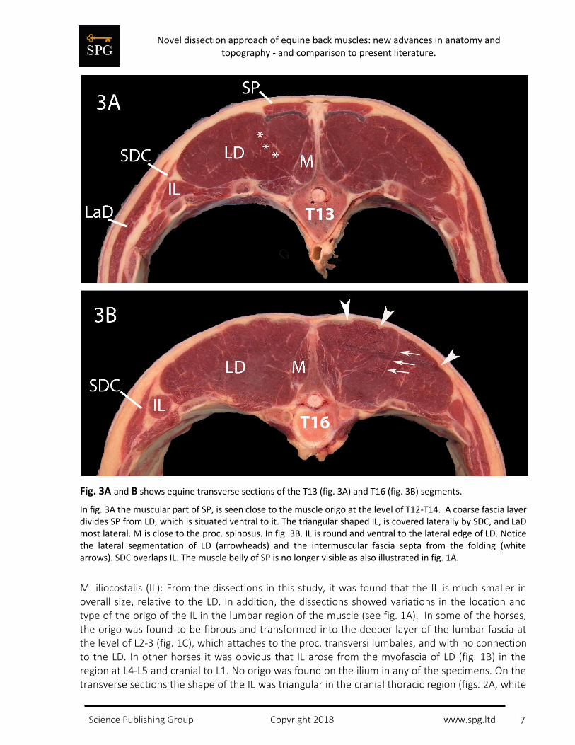
Fig. 3A and B shows equine transverse sections of the T13 (fig. 3A) and T16 (fig. 3B) segments.
In fig. 3A the muscular part of SP, is seen close to the muscle origo at the level of T12-T14. A coarse fascia layer divides SP from LD, which is situated ventral to it. The triangular shaped IL, is covered laterally by SDC, and LaD most lateral. M is close to the proc. spinosus. In fig. 3B. IL is round and ventral to the lateral edge of LD. Notice the lateral segmentation of LD (arrowheads) and the intermuscular fascia septa from the folding (white arrows). SDC overlaps IL. The muscle belly of SP is no longer visible as also illustrated in fig. 1A.
M. iliocostalis (IL): From the dissections in this study, it was found that the IL is much smaller in overall size, relative to the LD. In addition, the dissections showed variations in the location and type of the origo of the IL in the lumbar region of the muscle (see fig. 1A). In some of the horses, the origo was found to be fibrous and transformed into the deeper layer of the lumbar fascia at the level of L2-3 (fig. 1C), which attaches to the proc. transversi lumbales, and with no connection to the LD. In other horses it was obvious that IL arose from the myofascia of LD (fig. 1B) in the region at L4-L5 and cranial to L1. No origo was found on the ilium in any of the specimens. On the transverse sections the shape of the IL was triangular in the cranial thoracic region (figs. 2A, white

Novel dissection approach of equine back muscles: new advances in anatomy and topography - and comparison to present literature.
Science Publishing Group Copyright 2018 www.spg.ltd
8
arrows and 3A, white asterisk) to round (lower thoracic region, fig. 3B, IL) and had a width of approx. 2 centimetres. It was situated ventral to the lateral edge of LD. The multiple tendon insertions provide the muscle with its unique appearance (fig. 1A). The thoracic part of the IL was covered by m. serratus dorsalis as presented in fig.3A, left side.
M. spinalis (SP): This study showed from both longitudinal dissections and cross sections (fig. 1A, 2A and 3A, left side, white markings) that the fleshy part of SP had its origin on the profound layer of the fascia thoracolumbalis and the tendinous part of LD from T12 to T14 as well as the spinous processes. M. spinalis was situated dorsal to the LD, proceeded in a cranial direction and inserted dorsally with a tendinous connection to the transmission of the elastic funiculi nuchalis and the supraspinous ligament at T6-8 and with a tendinous connection to the spinous processes at C5, 6 and 7.
Discussion
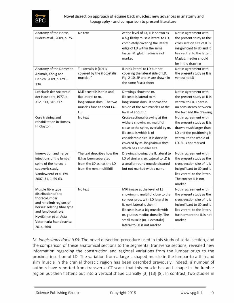
In the present study of the equine back muscles, several anatomical and topographical similarities to the literature were found, but new discoveries, as well as important differences and divergences, were also observed and presented in table 1.
Table 1. Comparison of the text and illustrations of the equine epaxial muscles in anatomical textbooks, clinical textbooks and scientific articles/papers to illustrate the disagreement of the placement, outline and positioning of the different back muscles.
Publication Text Pictures/illustrations Comparison of previous
data with results of the
present study
Equine Back Pathology,
2009, Kidd, p. 21. Ed.:
Henson.
m. iliocostalis = m.
longissimus costarum.
“..most lateral of the large
group of epaxial muscles..”
and “..originates from the
deep layer of the
lumbodorsal fascia back to
L3-L4..”
No illustrations In agreement with the
present study
Clinical Anatomy of the
Horse, 2005. Clayton et
al. p. 33
No text Picture of foal with correctly
marked m. iliocostalis
In agreement with the
present study
Diagnosis and
Management of
Lameness in the Horse,
2003, Ross & Dyson, p.
510.
“..the iliocostalis muscles
are small. Caudally, these
muscles fuse to form the
erector spinae muscles..”
Picture: Transverse section of
the back at the level of L3, area
lateral to m.multifidii is named
“erector spinae muscles”
Not in agreement with
the present study as LD
and IL are the erector
spinae muscles and IL
does not become part of
LD
Physical therapy and
Massage for the Horse,
1996, Denoix & Pailloux,
p.33, 36, 58.
“..Iliocostalis ..” on the
outside of the group
(erector spinae group)
Drawings show the m.
iliocostalis lateral to m.
longissimus dorsi
Not in agreement with
the present study as IL is
ventral to LD
Novel dissection approach of equine back muscles: new advances in anatomy and topography - and comparison to present literature.
Science Publishing Group Copyright 2018 www.spg.ltd
9
Anatomy of the Horse,
Budras et al., 2009, p. 75.
No text At the level of L3, IL is shown as
a big fleshy muscle lateral to LD,
completely covering the lateral
edge of LD within the same
fascia. M. glut. medius is not
marked
Not in agreement with
the present study as the
cross section size of IL is
insignificant to LD and it
lies ventral to the latter.
M.glut. medius should
be in the drawing
Anatomy of the Domestic
Animals, König and
Liebich, 2009, p.129 –
134.
“..Laterally it (LD) is covered by the iliocostalis muscle..”
IL runs lateral to LD but not covering the lateral side of LD. Fig. 2-10. SP and M are drawn in
the same fascia sheet
Not in agreement with the present study as IL is ventral to LD
Lehrbuch der Anatomie
der Haustiere,1977, p.
312, 313, 316-317.
M.iliocostalis is thin and
flat lateral to m.
longissimus dorsi. The two
muscles fuse at about L4-
L5.
Drawings show the m.
iliocostalis lateral to m.
longissimus dorsi. It shows the
fusion of the two muscles at the
level of about L1
Not in agreement with
the present study as IL is
ventral to LD. There is
no consistency between
the text and the drawing
Core training and rehabilitation in Horses. H. Clayton,
No text Cross-sectional drawing at the
withers showing m. multifidi
close to the spine, overlaid by m.
iliocostalis which is of
considerable size. It is dorsally
covered by m. longissimus dorsi
which has a smaller size
Not in agreement with
the present study as IL is
drawn much larger than
LD and the positioning is
ventral to the whole of
LD. SL is not marked
Innervation and nerve
injections of the lumbar
spine of the horse: a
cadaveric study.
Vandeweerd et al. EVJ
2007, 31, 1, 59-63.
The text describes how the
IL has been separated
from the LD as has the LD
from the mm. multifidii
Drawing showing the IL lateral to
LD of similar size. Lateral to LD is
a smaller round muscle pictured
but not marked with a name
Not in agreement with
the present study as the
cross-section size of IL is
insignificant to LD and it
lies ventral to the latter.
The correct IL is not
marked
Muscle fibre type distribution of the thoracolumbar and hindlimb regions of horses: relating fibre type
and functional role.
Hyytiäinen et al. Acta
Veterinaria Scandinavica
2014, 56:8
No text MRI image at the level of L3
showing m. multifidi close to the
spinous proc. with LD lateral to
it, next lateral is the m.
iliocostalis as a big muscle with
m. gluteus medius dorsally. The
small muscle (m. iliocostalis)
lateral to LD is not marked
Not in agreement with
the present study as the
cross-section size of IL is
insignificant to LD and it
lies ventral to the latter,
furthermore the IL is not
marked
M. longissimus dorsi (LD): The novel dissection procedure used in this study of serial section, and the comparison of these anatomical sections to the segmental transverse sections, revealed new information regarding the construction and regional variations from the lumbar origo to the proximal insertion of LD. The variation from a large L-shaped muscle in the lumbar to a thin and slim muscle in the cranial thoracic region has been described previously. Indeed, a number of authors have reported from transverse CT-scans that this muscle has an L shape in the lumbar region but then flattens out into a vertical shape cranially [3] [13] [8]. In contrast, two studies in

Novel dissection approach of equine back muscles: new advances in anatomy and topography - and comparison to present literature.
Science Publishing Group Copyright 2018 www.spg.ltd
10
the L3/L4 region [1] and [5], found the LD to appear as a more rectangular to oval shape. In addition, they incorrectly indicated that IL was positioned lateral to the LD. The muscle marked as IL [1, 5] was 50% and 25%, respectively, of the cross sectional area, much larger than the IL found in this study (fig. 2B). M. gluteus medius does not appear in any of the figures. This muscle is important as it overlies the LD, fills out the “hollow” space in the LD [10] and has one of the origos on the LD fascia [5]. Another study [2] utilized a MRI scan of the lumbar region and in the labelling of the muscles at the L3 level there are some inconsistencies. The m. gluteus medius is in the correct position as well as the medial vertical part of the L-shaped LD but the lateral horizontal part of LD is named as IL. There seems to be a very small cross section to IL on the image, which is not marked.
Due to the unique topography of the LD found in other studies, and refined in this study, where the medial vertical section of the LD is covered with tight fascia and the fleshy lateral horizontal section is uncovered, the evidence that the two parts of the LD have different functions is compelling. This finding is supported by another study[3], which showed that the muscle fascicle orientation is latero-medial in the medial part closest to the spinous processes, and dorso-ventral in the lateral part along the whole spine except from T2 and L6. These findings suggest that the medial part plays a stabilizing role and the lateral part gives rise to the extending/lateral flexing function of the muscle.
To further support this theory, a sEMG study was performed on the LD activity [17] which showed segmental differences in the activity of the LD in relation to the gait cycle at the walk and trot. They found that the more cranial segments showed more unilateral activity and suggested that the LD has a more prominent role in lateral flexion in this region.
Including the observations of the presence of areolar tissue within the intermuscular septae in the thoracic part and thereby a possibility to slide and glide independently [18], the overall LD composition and our findings help to support the anatomical and possible functional variation in the two parts of the LD. The location of the IL was also evaluated in this study and based on the findings we conclude that IL is situated ventral to the lateral edge of LD. In addition, we observed two distinct and individual variations in the origo; i) from the Bogorozky tendon and the ventral epimysium of LD at the caudo-lateral part at the level of L1 to L5 or ii) from the deep lumbar fascia lateral to the lumbar transverse processes at the level of L2 to L4. This observation differs from [6] [7] which included the aforementioned origos but also indicated the crista iliaca as a primary origo which we did not find in this study. However, our findings agree with [6] and [7] with regard to the variation of the origos. Our findings could very well be explained as a result of the way the dissections have been performed. The novel approach in the dissections was to include preservation of the fascia to understand the muscles, the functionality, the 3-dimensional interactions and biomechanics of the region. The fascia and their complex organisation in the body improves the understanding of how the topography, organisation and functionality of the region is arranged [16]. The agonistic function of the IL and LD can produce an extension of the back and a simultaneous expiratory posture of the thorax. From this we propose that horses with a static extended back and weak abdominal muscles (concentric contraction of erector spinae) may experience inspiratory problems, thereby adding yet another factor to the condition typified

Novel dissection approach of equine back muscles: new advances in anatomy and topography - and comparison to present literature.
Science Publishing Group Copyright 2018 www.spg.ltd
11
as poor performance and stress. In previous studies [1, 2, 4, 5], the positioning of the IL is considered to be incorrect and is confused with the LD, based on the findings of this study.
In the aforementioned references the lateral part of the muscle is indicated as being IL. It is possible that the interfacial septa from the folding in the LD caused a misinterpretation of the separation between the LD and IL which is typically seen in carnivores [7, 12]. Studies of serial transverse sections of the back as performed in this study show the distinction between the muscles. The mislabelling of the LD and IL is important to mention, as the size of the correct IL muscle is significantly smaller and the IL is with no exception the smallest of the three muscles in the erector spinae group.
M. spinalis (SP) has the shortest muscle belly of the epaxial muscles with a span from the level of T12- T14 and in a cranial direction to the lower cervical vertebrae [6, 7]. The muscle is situated dorsal to LD on the myofascia of its thoracic part. In an anatomical reference [4] the spinalis muscle is mistakenly labelled as the LD (fig. 2A) and is also wrongly described in the text, likewise the LD is marked in the place of SP. The incorrect labelling of the muscles could result in a misunderstanding of the topography and anatomy in this region which could lead to an incorrect biomechanical evaluation of the muscle group mechanics. The SP is generally thought to be related to extension only in the cervicothoracic region, and the LD is primarily responsible for extension in the thoracolumbar region.
Conclusion
This study shows some novel differences in the anatomy and topography of the erector spinae muscles as compared to previous publications. For example, IL has previously been confused with LD, that distinct variations in the origin of the IL have been found, that a fold in the LD from the thoracolumbar junction to the cervicothoracic junction exists, and that SP has previously been identified as LD. It is concluded that the IL is the smallest of the three muscles in the erector spinae group, that the LD medial part may serve a stabilizing role whilst the lateral part of the LD may act bilaterally in extension and unilaterally in lateral flexion and finally, that the SP may be related to extension only in the cervicothoracic region, whilst the LD may be primarily responsible for extension in the thoracolumbar region. The identified errors in the literature can quite easily give rise to misunderstandings with respect to local medical treatments in connection with back treatment or incorrect interpretations of ultrasound or MR images. As a precise and well formulated knowledge of anatomy is the basic language in intra-veterinary communication, it is essential that this information is detailed and presented correctly. The authors emphasize the need for future studies to understand the functional implications of these findings, as well as the use of correct topography and anatomy in future publications.
Acknowledgements
The authors would like to thank Vibe Bøgelund Hansen and Jan Lykke Jensen for their excellent technical assistance, Dr. Adrian P. Harrison, a Cambridge University graduated for proofreading and not least the owners of the horses for their generous donation.

Novel dissection approach of equine back muscles: new advances in anatomy and topography - and comparison to present literature.
Science Publishing Group Copyright 2018 www.spg.ltd
12
References
1. Vandeweerd, J.M., et al., Innervation and nerve injections of the lumbar spine of the horse: a cadaveric study. Equine Vet J, 2007. 39(1): p. 59-63.
2. Hyytiäinen, H.K., et al., Muscle fibre type distribution of the thoracolumbar and hindlimb regions of horses: relating fibre type and functional role. Acta Veterinaria Scandinavica, 2014. 56(1): p. 8.
3. Ritruechai, P., R. Weller, and J.M. Wakeling, Regionalisation of the muscle fascicle architecture in the equine longissimus dorsi muscle. Equine Vet J, 2008. 40(3): p. 246-51.
4. Clayton, H.M., Core Training and Rehabilitation in Horses. Vet Clin North Am Equine Pract, 2016. 32(1): p. 49-71.
5. Budras, K.-D., Sack, W.O., Röck, S.,, Anatomy of the horse,. Fifth ed. 2009, Hannover, Germany: Schlütersche Verlagsgesellschaft GmbH & Co K.G.,. 195.
6. König, H.E., Liebich, H.G., , Veterinary Anatomy of Domestic Mammals. fourth ed. 2009, Stuttgart, Deutschland: Schattauer.
7. Nickel, R., Schummer, A., Seiferle, E., Bewegungsapparat, Lehrbuch der Anatomie der Haustiere I. fifth ed. 1977, Berlin und Hamburg, Deutschland: Verlag Paul Parey.
8. Denoix, J.-M. and S.J. Dyson, Chapter 54 - Thoracolumbar Spine, in Diagnosis and Management of Lameness in the Horse. 2003, W.B. Saunders: Saint Louis. p. 509-521.
9. Denoix J.-M., P.J.-P., Physical therapy and massage for the horse. Fourth ed. 2009, UK: Manson Publishing Ltd.
10. Ehrle, A., et al., Structure and Innervation of the Equine Supraspinous and Interspinous Ligaments. Anat Histol Embryol, 2017. 46(3): p. 223-231.
11. A., K.J., Equine Back Pathology: Diagnosis and Treatment. 2009, UK: Wiley-Blackwell.
12. Schaller, O., Illustrated Veterinary Anatomical Nomenclature. 2 ed, ed. O. Schaller. 2007, Stuttgart: Enke Verlag.
13. Haussler, K.K., Osseous spinal pathology. Vet Clin North Am Equine Pract, 1999. 15(1): p. 103-12, vii.
14. Stubbs, N.C., et al., Functional anatomy of the caudal thoracolumbar and lumbosacral spine in the horse. Equine Vet J, 2006. 36.
15. Groesel, M., et al., A preliminary model study of the equine back including activity of longissimus dorsi muscle. Equine Vet J Suppl, 2010(38): p. 401-6.
16. Elbrond, V.S. and R.M. Schultz, Myofascia - the unexplored tissue: Myofascial kinetic lines in horses, a model for describing locomotion using comparative dissection studies derived from human lines. . Medical Research Archives, 2015. 4(3): p. 22.
Novel dissection approach of equine back muscles: new advances in anatomy and topography - and comparison to present literature.
Science Publishing Group Copyright 2018 www.spg.ltd
13
17. Wakeling, J.M., Ritruechai, P., Dalton, S., Nankervis, K., Segmental variation in the activity and function of the equine longissimus dorsi muscle during walk and trot. Equine and Comparative Exercise Physiology, 2007. 4(2): p. 95-103.
18. Stecco, C., et al., Hyaluronan within fascia in the etiology of myofascial pain. Surg Radiol Anat, 2011. 33(10): p. 891-6.
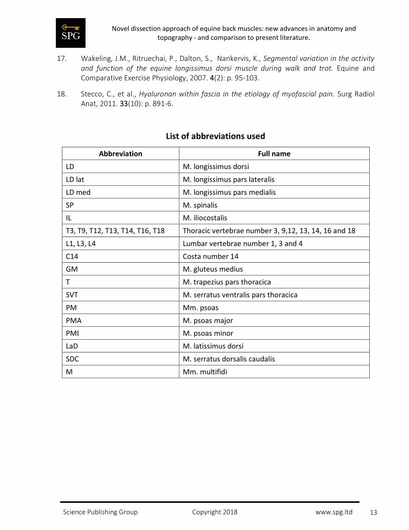
List of abbreviations used
Abbreviation Full name
LD M. longissimus dorsi
LD lat M. longissimus pars lateralis
LD med M. longissimus pars medialis
SP M. spinalis
IL M. iliocostalis
T3, T9, T12, T13, T14, T16, T18 Thoracic vertebrae number 3, 9,12, 13, 14, 16 and 18
L1, L3, L4 Lumbar vertebrae number 1, 3 and 4
C14 Costa number 14
GM M. gluteus medius
T M. trapezius pars thoracica
SVT M. serratus ventralis pars thoracica
PM Mm. psoas
PMA M. psoas major
PMI M. psoas minor
LaD M. latissimus dorsi
SDC M. serratus dorsalis caudalis
M Mm. multifidi