neurons communicate with other cells at synapsesgandha.weebly.com/.../chapter_34.3_synapses.pdf ·...
TRANSCRIPT
N E U R O N S C O M M U N I C AT E W I T H
O T H E R C E L L S AT S Y N A P S E S
3 4 . 3
NEURONS COMMUNICATE WITH OTHER CELLS AT SYNAPSES
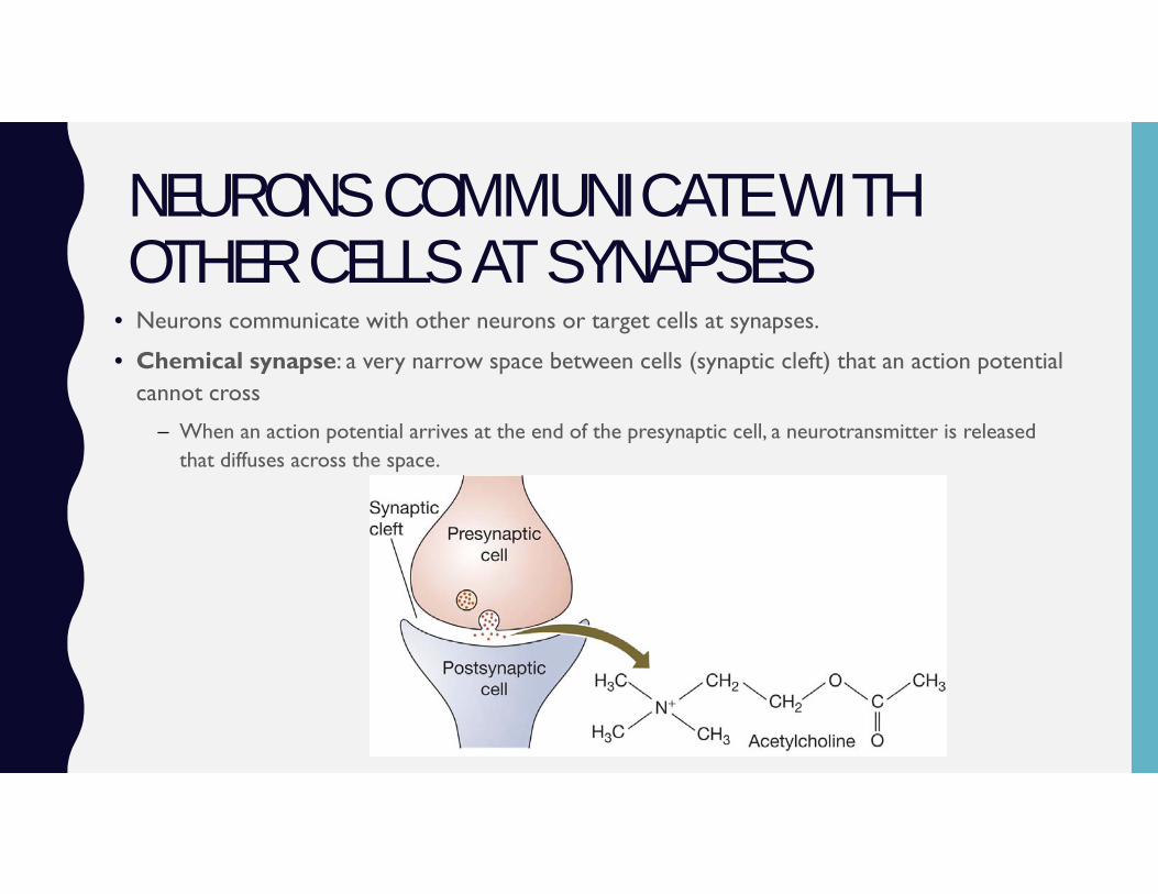
• Neurons communicate with other neurons or target cells at synapses.
• Chemical synapse: a very narrow space between cells (synaptic cleft) that an action potential cannot cross
– When an action potential arrives at the end of the presynaptic cell, a neurotransmitter is released that diffuses across the space.
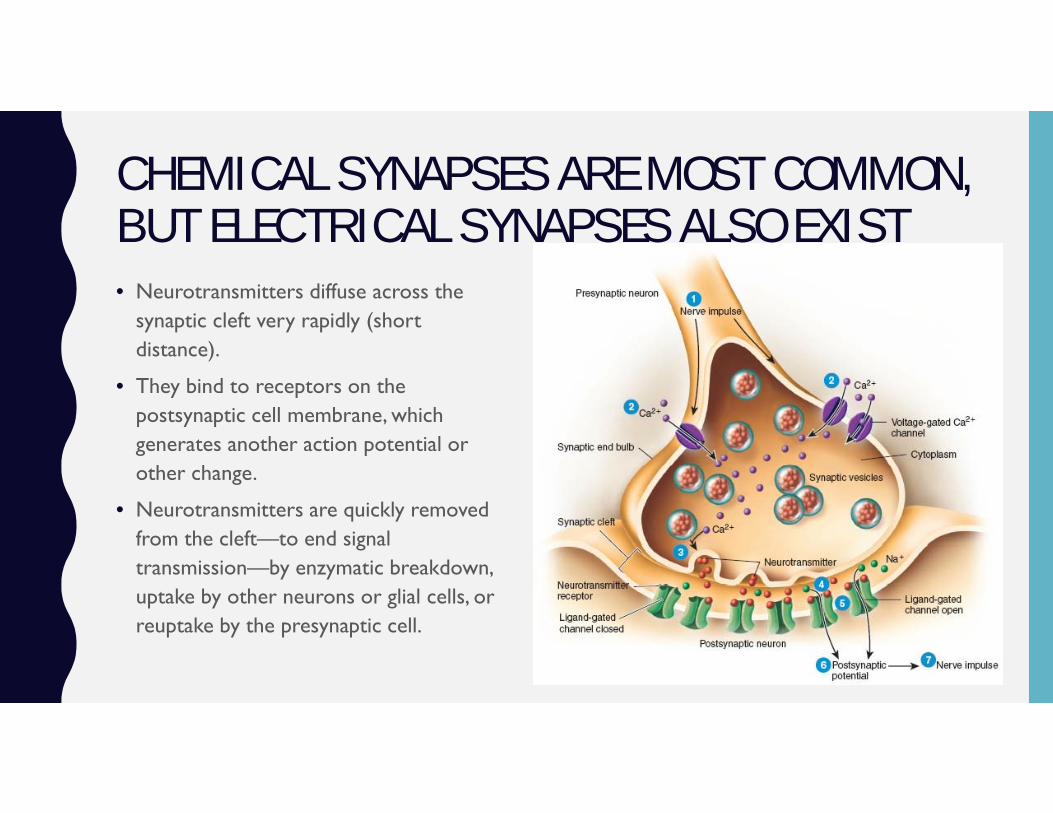
CHEMICAL SYNAPSES ARE MOST COMMON, BUT ELECTRICAL SYNAPSES ALSO EXIST• Neurotransmitters diffuse across the
synaptic cleft very rapidly (short distance).
• They bind to receptors on the postsynaptic cell membrane, which generates another action potential or other change.
• Neurotransmitters are quickly removed from the cleft—to end signal transmission—by enzymatic breakdown, uptake by other neurons or glial cells, or reuptake by the presynaptic cell.
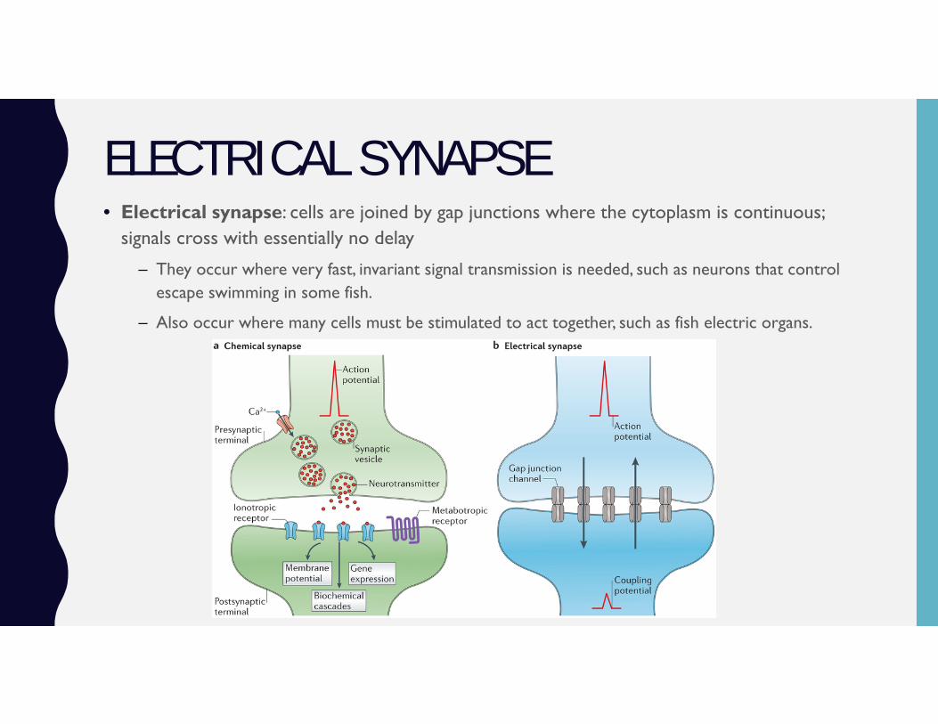
ELECTRICAL SYNAPSE • Electrical synapse: cells are joined by gap junctions where the cytoplasm is continuous;
signals cross with essentially no delay
– They occur where very fast, invariant signal transmission is needed, such as neurons that control escape swimming in some fish.
– Also occur where many cells must be stimulated to act together, such as fish electric organs.
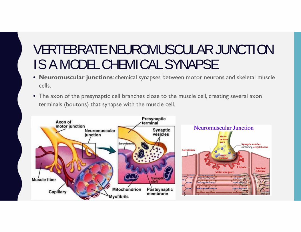
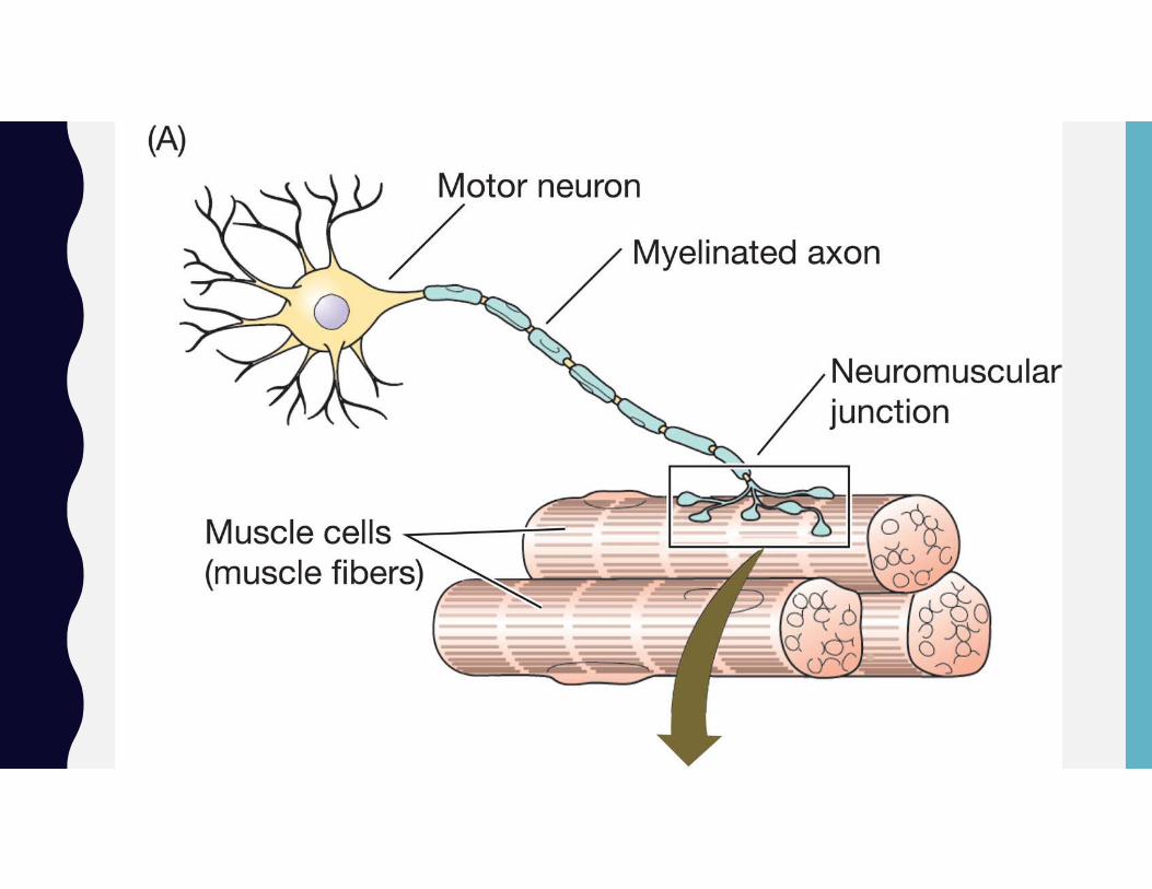
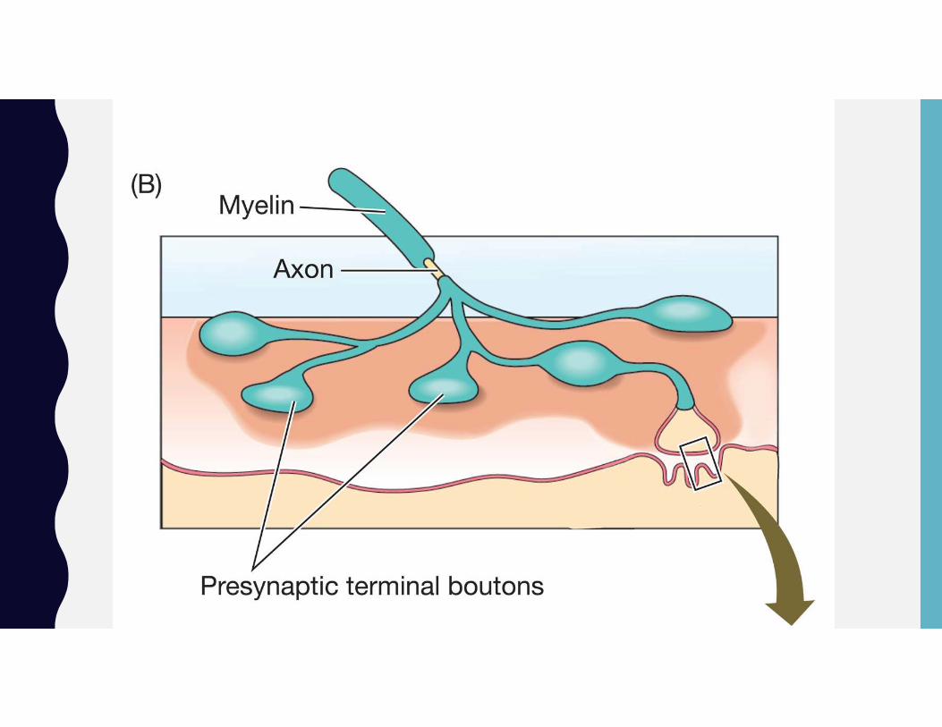
VERTEBRATE NEUROMUSCULAR JUNCTION IS A MODEL CHEMICAL SYNAPSE • Neuromuscular junctions: chemical synapses between motor neurons and skeletal muscle
cells.
• The axon of the presynaptic cell branches close to the muscle cell, creating several axon terminals (boutons) that synapse with the muscle cell.
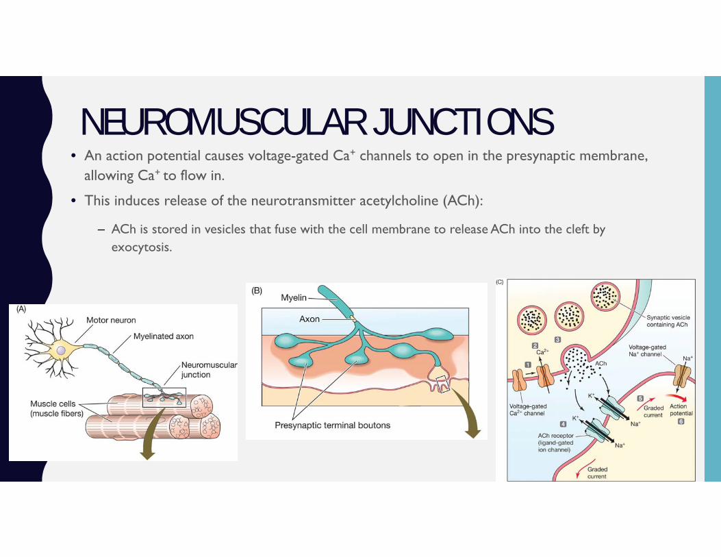
NEUROMUSCULAR JUNCTIONS• An action potential causes voltage-gated Ca+ channels to open in the presynaptic membrane,
allowing Ca+ to flow in.
• This induces release of the neurotransmitter acetylcholine (ACh):
– ACh is stored in vesicles that fuse with the cell membrane to release ACh into the cleft by exocytosis.

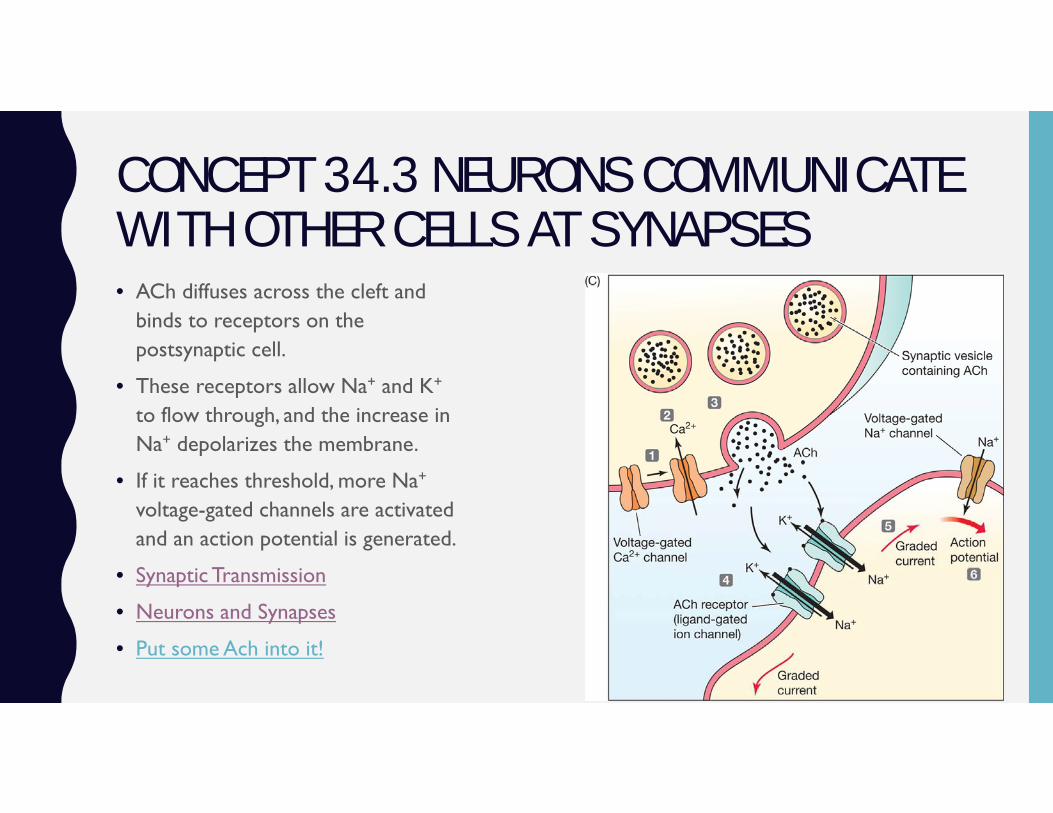
CONCEPT 34.3 NEURONS COMMUNICATE WITH OTHER CELLS AT SYNAPSES• ACh diffuses across the cleft and
binds to receptors on the postsynaptic cell.
• These receptors allow Na+ and K+
to flow through, and the increase in Na+ depolarizes the membrane.
• If it reaches threshold, more Na+
voltage-gated channels are activated and an action potential is generated.
• Synaptic Transmission
• Neurons and Synapses
• Put some Ach into it!
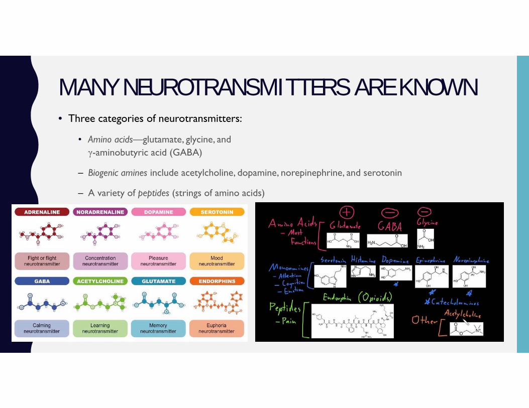
MANY NEUROTRANSMITTERS ARE KNOWN• Three categories of neurotransmitters:
• Amino acids—glutamate, glycine, andγ-aminobutyric acid (GABA)
– Biogenic amines include acetylcholine, dopamine, norepinephrine, and serotonin
– A variety of peptides (strings of amino acids)
MANY NEUROTRANSMITTERS ARE KNOWN
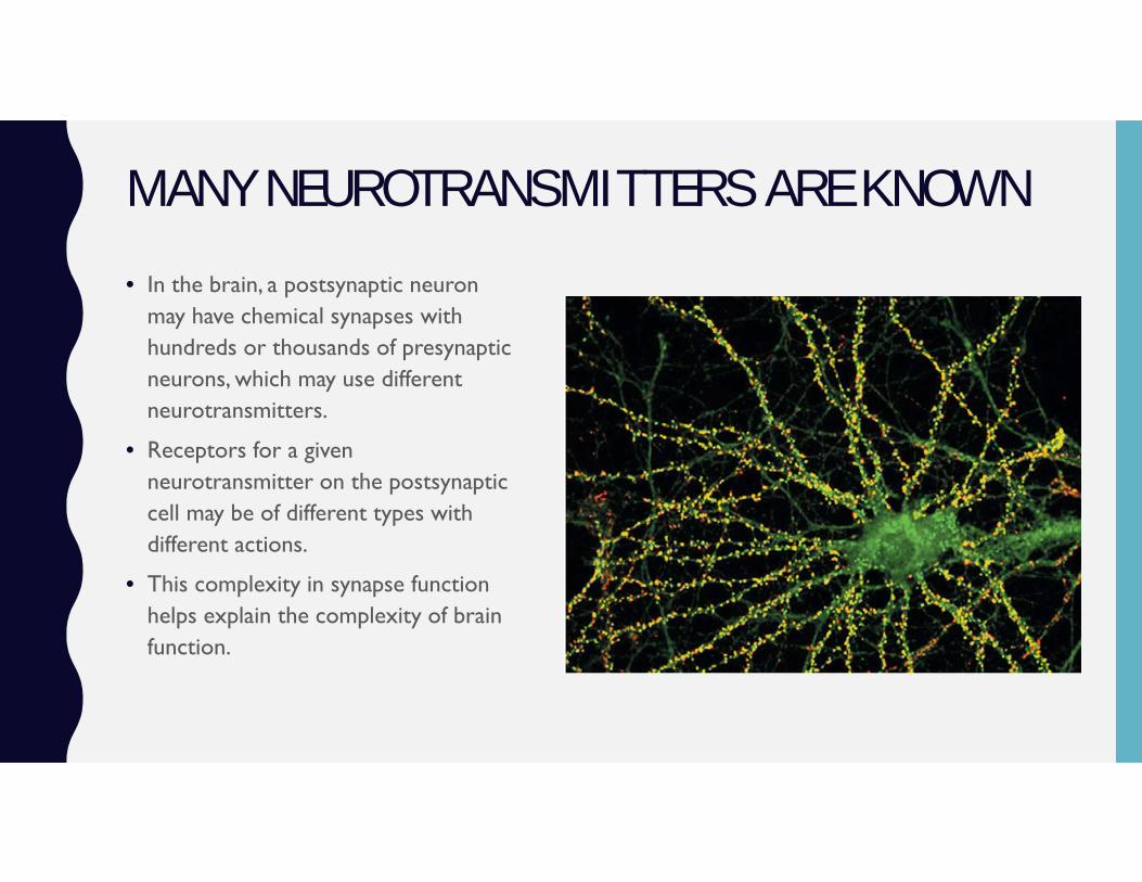
• In the brain, a postsynaptic neuron may have chemical synapses with hundreds or thousands of presynaptic neurons, which may use different neurotransmitters.
• Receptors for a given neurotransmitter on the postsynaptic cell may be of different types with different actions.
• This complexity in synapse function helps explain the complexity of brain function.
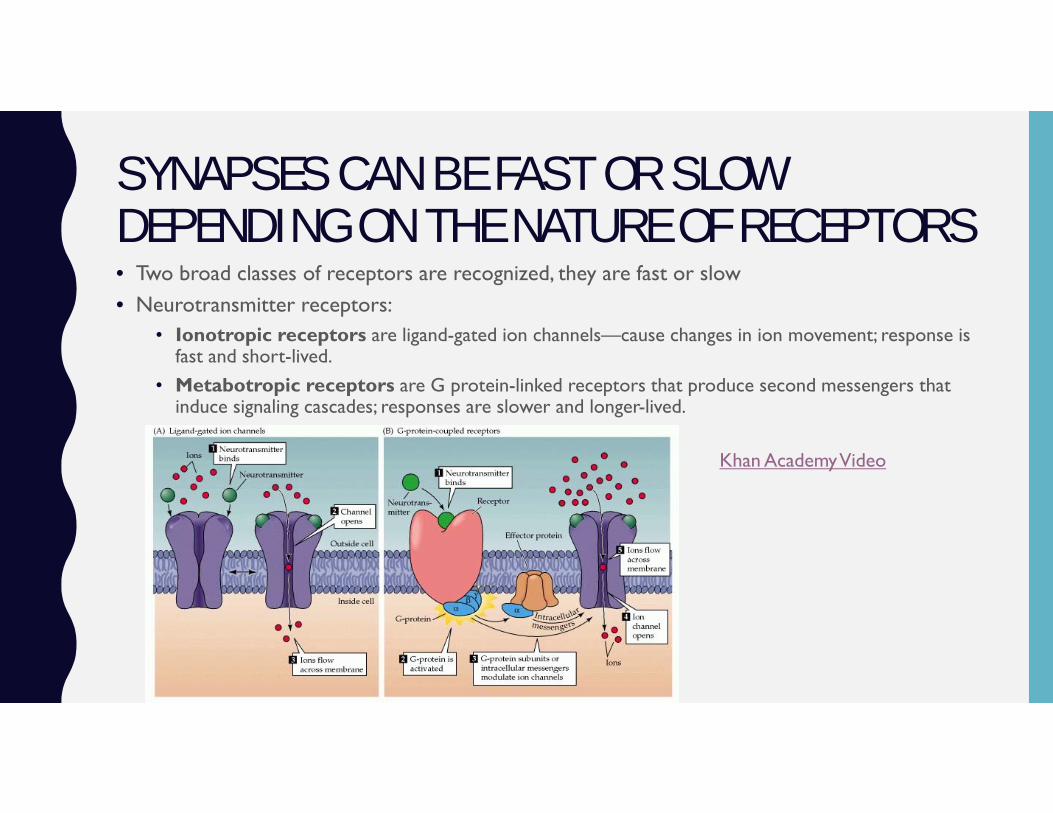
SYNAPSES CAN BE FAST OR SLOW DEPENDING ON THE NATURE OF RECEPTORS• Two broad classes of receptors are recognized, they are fast or slow• Neurotransmitter receptors:
• Ionotropic receptors are ligand-gated ion channels—cause changes in ion movement; response is fast and short-lived.
• Metabotropic receptors are G protein-linked receptors that produce second messengers that induce signaling cascades; responses are slower and longer-lived.
Khan Academy Video

FAST SYNAPSES PRODUCE POSTSYNAPTIC POTENTIALS THAT SUM TO DETERMINE ACTION POTENTIAL PRODUCTION
• Excitatory synapses produce graded membrane depolarizations called excitatory postsynaptic potentials (EPSPs); shift membrane potential towards threshold.
• Inhibitory synapses shift membrane potential away from threshold; produce graded membrane hyperpolarizations called inhibitory postsynaptic potentials (IPSPs).
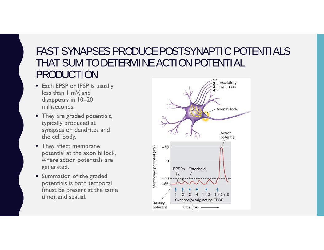
FAST SYNAPSES PRODUCE POSTSYNAPTIC POTENTIALS THAT SUM TO DETERMINE ACTION POTENTIAL PRODUCTION• Each EPSP or IPSP is usually
less than 1 mV, and disappears in 10–20 milliseconds.
• They are graded potentials, typically produced at synapses on dendrites and the cell body.
• They affect membrane potential at the axon hillock, where action potentials are generated.
• Summation of the graded potentials is both temporal (must be present at the same time), and spatial.
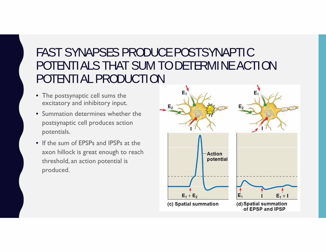
FAST SYNAPSES PRODUCE POSTSYNAPTIC POTENTIALS THAT SUM TO DETERMINE ACTION POTENTIAL PRODUCTION• The postsynaptic cell sums the
excitatory and inhibitory input.
• Summation determines whether the postsynaptic cell produces action potentials.
• If the sum of EPSPs and IPSPs at the axon hillock is great enough to reach threshold, an action potential is produced.
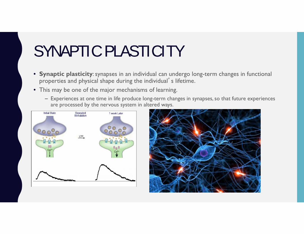
SYNAPTIC PLASTICITY • Synaptic plasticity: synapses in an individual can undergo long-term changes in functional
properties and physical shape during the individual’s lifetime.• This may be one of the major mechanisms of learning.
– Experiences at one time in life produce long-term changes in synapses, so that future experiences are processed by the nervous system in altered ways.
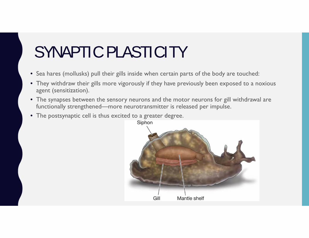
SYNAPTIC PLASTICITY • Sea hares (mollusks) pull their gills inside when certain parts of the body are touched:
• They withdraw their gills more vigorously if they have previously been exposed to a noxious agent (sensitization).
• The synapses between the sensory neurons and the motor neurons for gill withdrawal are functionally strengthened—more neurotransmitter is released per impulse.
• The postsynaptic cell is thus excited to a greater degree.
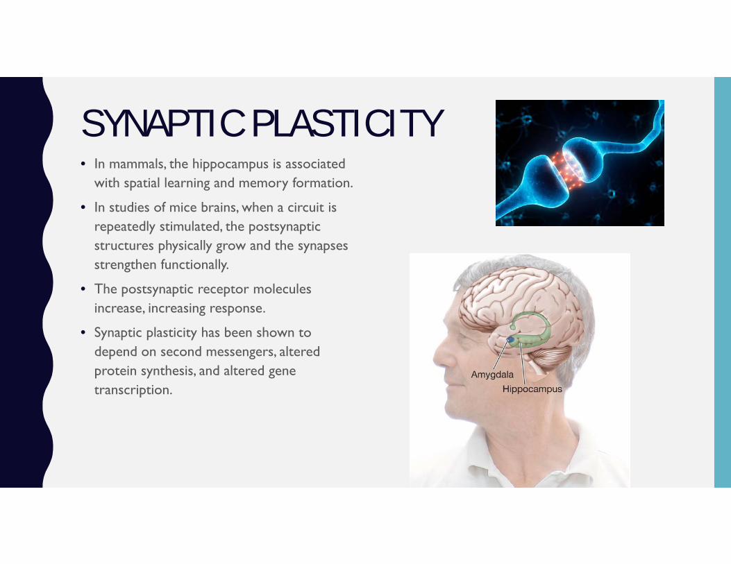
SYNAPTIC PLASTICITY • In mammals, the hippocampus is associated
with spatial learning and memory formation.
• In studies of mice brains, when a circuit is repeatedly stimulated, the postsynaptic structures physically grow and the synapses strengthen functionally.
• The postsynaptic receptor molecules increase, increasing response.
• Synaptic plasticity has been shown to depend on second messengers, altered protein synthesis, and altered gene transcription.
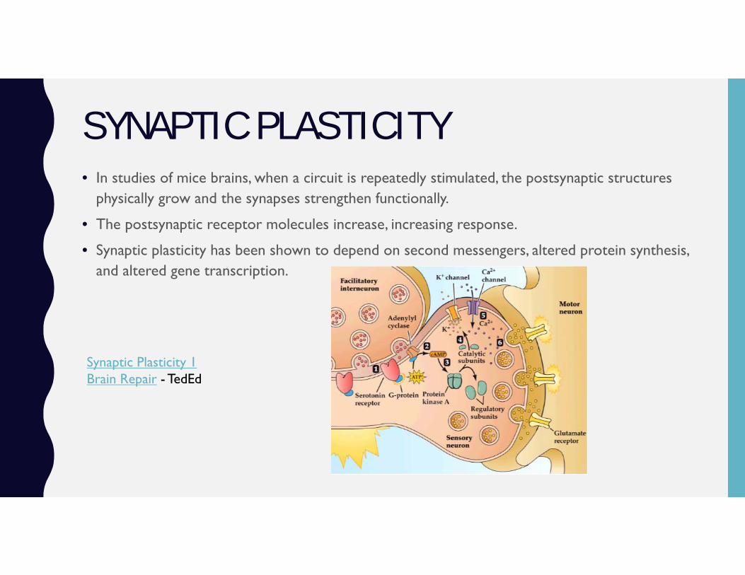
SYNAPTIC PLASTICITY • In studies of mice brains, when a circuit is repeatedly stimulated, the postsynaptic structures
physically grow and the synapses strengthen functionally.
• The postsynaptic receptor molecules increase, increasing response.
• Synaptic plasticity has been shown to depend on second messengers, altered protein synthesis, and altered gene transcription.
Synaptic Plasticity 1Brain Repair - TedEd